Báo cáo y học: "Evolutionary dynamics of eukaryotic selenoproteomes: large selenoproteomes may associate with aquatic life and small with terrestrial lif" pot
Bạn đang xem bản rút gọn của tài liệu. Xem và tải ngay bản đầy đủ của tài liệu tại đây (587.38 KB, 16 trang )
Genome Biology 2007, 8:R198
comment reviews reports deposited research refereed research interactions information
Open Access
2007Lobanovet al.Volume 8, Issue 9, Article R198
Research
Evolutionary dynamics of eukaryotic selenoproteomes: large
selenoproteomes may associate with aquatic life and small with
terrestrial life
Alexey V Lobanov
*
, Dmitri E Fomenko
*
, Yan Zhang
*
, Aniruddha Sengupta
†
,
Dolph L Hatfield
†
and Vadim N Gladyshev
*
Addresses:
*
Department of Biochemistry, University of Nebraska, Lincoln, NE 68588, USA.
†
Section on the Molecular Biology of Selenium,
National Cancer Institute, National Institutes of Health, Bethesda, MD 20892, USA.
Correspondence: Vadim N Gladyshev. Email:
© 2007 Lobanov et al.; licensee BioMed Central Ltd.
This is an open access article distributed under the terms of the Creative Commons Attribution License ( which
permits unrestricted use, distribution, and reproduction in any medium, provided the original work is properly cited.
Selenoproteome evolution<p>In silico and metabolic labeling studies of the selenoproteomes of several eukaryotes revealed distinct selenoprotein patterns as well as an ancient origin of selenoproteins and massive, independent losses in land plants, fungi, nematodes, insects and some protists, suggesting that the environment plays an important role in selenoproteome evolution.</p>
Abstract
Background: Selenocysteine (Sec) is a selenium-containing amino acid that is co-translationally
inserted into nascent polypeptides by recoding UGA codons. Selenoproteins occur in both
eukaryotes and prokaryotes, but the selenoprotein content of organisms (selenoproteome) is
highly variable and some organisms do not utilize Sec at all.
Results: We analyzed the selenoproteomes of several model eukaryotes and detected 26 and 29
selenoprotein genes in the green algae Ostreococcus tauri and Ostreococcus lucimarinus, respectively,
five in the social amoebae Dictyostelium discoideum, three in the fly Drosophila pseudoobscura, and 16
in the diatom Thalassiosira pseudonana, including several new selenoproteins. Distinct selenoprotein
patterns were verified by metabolic labeling of O. tauri and D. discoideum with
75
Se. More than half
of the selenoprotein families were shared by unicellular eukaryotes and mammals, consistent with
their ancient origin. Further analyses identified massive, independent selenoprotein losses in land
plants, fungi, nematodes, insects and some protists. Comparative analyses of selenoprotein-rich and
-deficient organisms revealed that aquatic organisms generally have large selenoproteomes,
whereas several groups of terrestrial organisms reduced their selenoproteomes through loss of
selenoprotein genes and replacement of Sec with cysteine.
Conclusion: Our data suggest many selenoproteins originated at the base of the eukaryotic
domain and show that the environment plays an important role in selenoproteome evolution. In
particular, aquatic organisms apparently retained and sometimes expanded their selenoproteomes,
whereas the selenoproteomes of some terrestrial organisms were reduced or completely lost.
These findings suggest a hypothesis that, with the exception of vertebrates, aquatic life supports
selenium utilization, whereas terrestrial habitats lead to reduced use of this trace element due to
an unknown environmental factor.
Published: 19 September 2007
Genome Biology 2007, 8:R198 (doi:10.1186/gb-2007-8-9-r198)
Received: 27 September 2006
Revised: 18 September 2007
Accepted: 19 September 2007
The electronic version of this article is the complete one and can be
found online at />R198.2 Genome Biology 2007, Volume 8, Issue 9, Article R198 Lobanov et al. />Genome Biology 2007, 8:R198
Background
Selenium is an essential trace element in many, but not all,
life forms. Its essentiality is based on the fact that this element
is present in natural proteins in the form of selenocysteine
(Sec), a rare amino acid that chemically differs from serine or
cysteine (Cys) by a single atom (for example, Se instead of O
or S) [1]. Sec is known as the 21st amino acid in the genetic
code as it has its own biosynthetic machinery, a tRNA and an
elongation factor, and is inserted into nascent polypeptides
co-translationally in response to the Sec codon, UGA [2-4].
Selenoproteins often escape attention of genome annotators,
because in-frame UGA codons are interpreted as stop signals.
However, several bioinformatics tools have recently been
developed that help identify these genes [5,6]. The use of
these methods begins to shed light on proteins and processes
dependent on selenium, as well as on the occurrence and dis-
tribution of these processes in various life forms.
Sec is typically found in active sites of redox enzymes, which
are functionally similar to thiol-based oxidoreductases [7].
Sec-containing proteins occur in all major lines of descent
(for example, eukaryota, eubacteria and archaea), but not all
organisms have these proteins. Prokaryotic genomes have
been extensively analyzed for the occurrence of selenoprotein
genes [8], but among eukaryotes, only the genomes of mam-
mals (human, mouse) [9], nematodes (Caenorhabditis ele-
gans and C. briggzae) [10], fruit fly (Drosophila
melanogaster) [11], green alga (Chlamydomonas rein-
hardtii) [12] and Plasmodia [13,14] have been analyzed with
regard to the entire set of selenoproteins (selenoproteomes).
In addition, the genomes of the plant Arabidopsis thaliana
and the yeast Saccharomyces cerevisiae have been scanned
for the occurrence of selenoprotein genes and Sec biosyn-
thetic/insertion machinery genes and found to have neither
[9].
Selenoproteome analyses also revealed that various organ-
isms have substantially different sets of selenoproteins. One
example of uneven selenoprotein occurrence is selenoprotein
U (SelU), which occurs in fish, birds and some unicellular
eukaryotes, but is present in the form of a Cys-containing
homolog in mammals and many other eukaryotes. Even a
narrower occurrence has been described for SelJ and Fep15
[15,16].
In this study, we characterized the selenoproteomes encoded
in several completely sequenced eukaryotic genomes.
Detailed analyses of these selenoproteomes and comparison
with those of other eukaryotic model organisms revealed an
ancient origin of most eukaryotic selenoproteins and a possi-
bility of increased Sec utilization in aquatic environments and
decreased use of Sec in terrestrial habitats. These studies pro-
vide important insights into selenoprotein origin and dynam-
ics of selenoprotein evolution.
Results and discussion
Eukaryotic selenoproteomes
Several eukaryotes have been previously analyzed for their
selenoprotein content (selenoproteomes). These studies
identified 24-25 selenoproteins in mammals and 0-4 seleno-
proteins in other organisms. It is generally thought that many
eukaryotic selenoproteins evolved in vertebrates, but evolu-
tionary paths have not been examined for the majority of
these proteins. In this work, we analyzed the selenopro-
teomes of several additional model eukaryotes, whose
genomes have been completed. These included marine algae
(Ostreococcus tauri and O. lucimarinus), a diatom (Thalassi-
osira pseudonana), a soil amoeba (Dictyostelium discoi-
deum), an insect (Drosophila pseudoobscura), and a red alga
(Cyanidioschyzon merolae).
Drosophila pseudoobscura
The D. pseudoobscura subgroup [17] is found mainly in the
temperate and tropical zones of the New World [18]. Applica-
tion of an earlier version of SECISearch to the D. mela-
nogaster genome identified three selenoprotein genes (SelK/
G-rich, SelH/BthD and SPS2); however, it was not known
whether this set represents the entire Drosophila selenopro-
teome. We applied an advanced version of SECISearch (see
Materials and methods and Additional data file 1) to analyze
the D. pseudoobscura genome and, in addition, analyzed D.
pseudoobscura and D. melanogaster genomes in parallel to
identify evolutionarily conserved selenocysteine insertion
sequence (SECIS) elements using relaxed SECIS criteria.
These searches resulted in the same, already known set of
three selenoproteins (Table 1), suggesting that the selenopro-
teome of insects of the Drosophila genus consists of these
three proteins. By homology analyses, we then identified
three selenoproteins in a mosquito, Anopheles gambiae, and
one in a honey bee, Apis mellifera.
Ostreococcus tauri
O. tauri is a unicellular green alga that was discovered in the
Mediterranean Thau lagoon in 1994. It belongs to the family
Prasinophyceae, which is thought to be the most primitive in
the green plant lineage from which all other green algae and
ancestors of land plants have descended. This organism has a
very small genome, 11.5 Mb [19], especially when compared to
other sequenced Plantae genomes (for example, the Arabi-
dopsis genome is 125 Mb [20] and that of Chlamydomonas
exceeds 100 Mb [21,22]). The O. tauri genome is densely
packed and provides a useful genomic model for green plants
[23]. Previous research revealed the lack of selenoproteins in
land plants [9], whereas 10 selenoproteins were detected in
the green alga C. reinhardtii [12]. Surprisingly, we detected
26 selenoprotein genes in O. tauri.
Among the known selenoproteins detected in O. tauri, four-
teen were homologs of human selenoproteins (thioredoxin
reductase (TR), SelT, SelM, SelK, SelS, Sep15, SelO, SelH,
SelW and five glutathione peroxidase (GPx) homologs), five
Genome Biology 2007, Volume 8, Issue 9, Article R198 Lobanov et al. R198.3
comment reviews reports refereed researchdeposited research interactions information
Genome Biology 2007, 8:R198
were homologs of eukaryotic selenoproteins with restricted
distribution (MsrA, SelU and three PDI homologs) and three
were homologs of bacterial selenoproteins (methyltrans-
ferase, thioredoxin-fold protein and peroxiredoxin). We also
identified four novel eukaryotic selenoproteins in the O. tauri
genome. These included a predicted membrane selenoprotein
(MSP) and three hypothetical proteins of unknown function.
In addition, several excellent SECIS element candidates were
identified during analysis, but at present no suitable open
reading frames (ORFs) could be identified upstream of these
structures, in part because of the inadequate length of con-
tigs. Therefore, the total number of Ostreococcus selenopro-
teins might be even higher than 26.
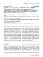
Of interest was the observation that all O. tauri SECIS ele-
ments except one had a conserved G in the position directly
preceding the quartet of non-Watson-Crick interacting nucle-
otides (Figure 1). Most eukaryotic SECIS elements have an A
in this position, although the G was described in several
zebrafish and nematode selenoprotein genes [10,24,25]. In
addition, almost all O. tauri SECIS elements had a long mini-
stem in the apical portion of the structure (for example, SelT
in Figure 1). This feature was also observed previously in a
number of Chlamydomonas SECIS elements [12].
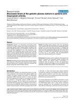
We metabolically labeled O. tauri cells with
75
Se and analyzed
the selenoprotein pattern on SDS PAGE gels using a Phos-
phorImager (Figure 2a). This method detects the most abun-
dant selenoproteins. The overall pattern was similar to that of
human HEK 293 and other mammalian cells. As in mamma-
lian cells, the dominant 25 kDa band in the alga was likely a
glutathione peroxidase, and one or both major selenoprotein
bands in the 50-55 kDa range likely corresponded to thiore-
doxin reductase. Consistent with the genomics analysis, the
number of selenoprotein bands in the O. tauri sample was
higher than in mammalian cells.
Ostreococcus lucimarinus
O. lucimarinus, previously known as Ostreococcus sp.
CCE9901, is a close relative of O. tauri adapted to high light
and isolated from surface waters. Its genome size is 13.2 Mb.
Homologs of all identified O. tauri selenoproteins were found
in O. lucimarinus. In addition, three new sequences were
identified, raising the number of selenoproteins in this organ-
ism to 29. This is the largest selenoproteome of all previously
analyzed eukaryotes (although even larger selenoproteomes
apparently exist; Lobanov and Gladyshev, unpublished).
Additional selenoproteins included a peroxiredoxin, and per-
oxiredoxin-like and SelW-like proteins. The latter O. lucima-
rinus selenoprotein contained two predicted Sec residues.
Similar to O. tauri, all O. lucimarinus SECIS elements except
one had a conserved G in the position directly preceding the
SECIS core (Figure 1a), and in addition a single ATGA-type
SECIS element was found. Interestingly, single ATGA-type
SECIS elements occur in different selenoprotein genes in the
two Ostreococcus species. In O. lucimarinus, this SECIS type
is within a glutathione peroxidase gene, while in O. tauri the
ATGA-type SECIS is in the gene for a hypothetical protein. In
contrast to O. tauri, no type I SECIS elements (Figure 1a)
were found in O. lucimarinus.
Cyanidioschyzon merolae
C. merolae is an ultrasmall unicellular red alga that lives in
acidic hot springs. It is thought to retain primitive features of
cellular and genome organization. C. merolae has a simple
cell architecture, containing a single nucleus, a single mito-
chondrion and a single chloroplast. Its genome size is 16 Mbp,
which is approximately one-seventh the size of the A. thal-
iana genome. Its chloroplast might be among the most ances-
tral [26]. A BLAST search against the C. merolae genome
revealed several known components of the Sec insertion
machinery, including SBP2, EFsec, SecS and SPS2, suggest-
ing that selenoproteins should also be present in this organ-
ism. However, a search for SECIS elements followed by ORF
analyses revealed no candidate selenoproteins in the C. mero-
lae genome.
A BLASTN-based analysis of the C. merolae genome using
known Sec tRNAs as query sequences did not identify Sec
tRNA homologs, and the searches that utilized default ver-
sions of standard tRNA detection programs, ARAGORN and
Table 1
Identification of selenoprotein genes in eukaryotic model organisms
Loose pattern Default pattern
Organism name Genome,
thousands of bp
Primary sequence
criteria
Energy criteria Primary sequence
criteria
Energy criteria Number of
selenoproteins
O. lucimarinus 13,393 31,132 7,541 2,120 464 29
O. tauri 16,414 30,381 7,379 1,934 401 26
T. pseudonana 32,577 81,040 8,977 3,129 675 16
D. discoideum 34,564 37,435 7,11 2,128 37 5
D. pseudoobscura 138,581 181,793 20,702 6,303 1,010 3
C. merolae 16,381 27,578 5,987 651 149 0
R198.4 Genome Biology 2007, Volume 8, Issue 9, Article R198 Lobanov et al. />Genome Biology 2007, 8:R198
Figure 1 (see legend on next page)
(a)
Typical SECIS element
(Selenoprotein T)
Type I SECIS element
(Selenoprotein H)
ATGA-type SECIS element
(hypothetical protein 3)
(b)
eroc SICES eroc SICES
Ahp reductase CTCGCGAACCGTGAC GCGAACCAGCGAA AGAGCCGAATGCACGG TTGGCTGGTTCGT CGATGAAGCG-
Methyltransferase AAGTGAATGCGTGAA GAAGCGCGGGTAAAACG-CTCCACAGGCGCACCCGACGCTTC TGATTTTTTT-
MSP GATCGTGTC-GTGAC GCGCTCGTGCGAAT-GTCAGCCATGCTGGCGGCGCGAGGGC TGATTTTCAC-
Trx-fold protein GTCTCGCA-CGTGAC GTCTCGTCGATAAACCAGTC TCACTTGACTTCGACCGGAC CGACTCGCCA-
GPx-a GTCTCGCTTCGTGAC GACCGATGAACAAAGACCGAAT-CACAGGTTTTCATCGGTT CGATTACGCG-
GPx-b ACGCGGAGTCGTGAC GCCTCGCTCCAGAAATTGACCACGGTCGAGGGGGACGGGC TGAAATCTCC-
GPx-c GAGCGTCGAAGTGAC GCGCCGTCGCGAAACGGAC-ACGCTTTGTTCGGTGGCG-GCGT CGATGAGAGG-
GPx-d CGCGCACA GTGAC GACGCGCGAGGAAACCCGTCGCCTTCTCTCGGCGTCGACCTCGCGTCTC CGACGCATCG-
GPx-e GGCGCTCCGTGTGAC CGCGCGCTCGGAAACGGAACGACGTGAGGACGACG-TAAAACGTACTCGCTCCG CGAGCGCGCG-
Sep15 CTCTCGATGTGTGAC TCGCGCGCGACAG CCGCCTCGCTCGAGGCGTTCGCGCGCGA TGATT-TCGTG
TR CATCGGCAAAGTGAC GATGATGATCGCAAACAC GCTCTATGTGTCGATATCATC CGATGAAGCC-
SelH TCTCGTGATAGTGAA GCCGTGACGCGAAATCAAGCAAGCGTCGCG-GC GGATGACACG-
SelK GCGCGTGC GTGAT ACCGCGGCGGGAACGGACTCTTCACGGAGACCACCGCGGCGGT TGATTATCAG-
SelM GCGCGTATTCGTGAC GTGTTGTCGCGAAAACGAGCCGCCAACGCGCGCGCTCTGCGAGGACAC CGATATTTGC-
SelO GGTGGTGGACGTGAC GCGACG-GTTTGAAACG-CGCCG-AGGCGCGCTAATCGTCGT CGANNNNNNN-
SelS ACGTGCGCGCGTGAC ACCGCGGCGGGAACGG TCTCGATGAAGACTACCGCCACGGT CGATTTGAGC-
SelT ATACGAGTCGGTGAA GACGCGCG-CGGAAAGGACGCCGCGGGTGTTTCCGGGCGAACCGCGCGCGTT TGATTTCTCG-
SelU TTCGCGCTCAGTGAC GTGAGAAACGGAAATTCTTTGTTGATTTCACGAGGGTCGTTTCTTCAC CGAT-AAGCGC
SelW TCGACGATCAGTGAC GGACGACGTTTGAAAGCTTCATTCGGGCGCACGTGCTCGAACAGACGTCGATCC TGATTCTCGT-
MsrA CGCGCAAAC-GTGAC GACGATGTCGCAAAGGATGTGGACGTTCCAGTCCTCGACGTCGTT CGATTCATCG-
PDI-1 AAGTCGACAAGTGAC GTCTCGTCTCTAAGACTGCATTTTACGCGGTTGACACGAGAC TGATTTATAT-
PDI-2 GATTGACGTTGTGAC GGCCGTACTGAAATCGTA-AATCTTTA CGGTGGTTCGAGTC TGATTACTCA-
PDI-3 GAGACGATTCGTGAC CGCGATCGCTCCTAAACGTCCATCATATCCATTTTGGACGCCACGATCGCG CGATTCATCG-
hypothetical protein 1 CGCGACGGACGTGAC GCGACGACGAGAAAACGATG-AAAAGCCTCATCCCTCGTCGTCGC CGATG-CACGC
hypothetical protein 2 GGCAATTCGAGTGAC GACGCGCGGCGAAACGGAGGACTCGGACGCGTCCGC CGCCGCGCGTT CGATTCCCGT-
hypothetical protein 3 TCGACGACGCATGAC GGTGA-ACGCGGAAAC GCAGTTTTTGCGGAG CGTTCACT CGATTATCAT-
Candidate SECIS 1 GACCGGAGTCGTGAT CGCGCTCGATCGAACCGCGCCGTTCCGGGCGT-CGGGCGAG CGACGCGTGG-
Candidate SECIS 2 GACGCGCTC-GTGAC -GAAACGACGACGCAAGCGTGGAA-ACACGCGACGTCGTTTC CGATGATGCG-
Candidate SECIS 3 CGAGGAGGACGTGAC GAAGGAC TCGAAAAGCCGGCGCGCGCCGGCGCGAGTGACTTC CGATGATGCC-
G
G
G
C
A
A
A
A
A
A
G
G
G
G
G
C
C
C
C
C
A
T
T
T
T
T
G
A
C
T
C
G
G
G
G
G
G
G
C
C
C
C
A
A
T
A
T
G
T
T
C
G
G
G
G
G
G
G
G
G
C
C
C
C
C
C
T
T
T
G
T
C
C
C
C
C
CC
G
G
G
G
G
G
G
G
G
G
G
T
T
T
T
T
T
T
A
A
A
A
A
A
A
A
A
A
G
C
T
C
C
A
A
G
C
C
C
G
G
G
G
T
T
T
T
T
A
G
G
G
G
G
A
A
A
A
A
A
A
A
A
A
C
C
T
G
G
G
G
G
A
A
A
C
C
C
C
T
T
G
G
G
G
G
G
G
A
C
C
C
C
T
T
T
T
C
A
G
Genome Biology 2007, Volume 8, Issue 9, Article R198 Lobanov et al. R198.5
comment reviews reports refereed researchdeposited research interactions information
Genome Biology 2007, 8:R198
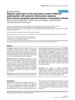
tRNAscan-SE, were also unsuccessful. We were able to iden-
tify the C. merolae Sec tRNA using our recently described tool
for detection of unusual tRNAs [27]. This tRNA (Figure 3) has
all the features characteristic of Sec tRNAs, such as the UCA
anticodon and a long variable stem.
We applied additional sensitive tools for identification of
selenoproteins in the red algal genome. Most homologs of
known selenoproteins were found to either have Cys in place
of Sec or were missing in this organism. We further carried
out a search for Sec/Cys pairs in homologous sequences using
the C. merolae genome and all protein sequences extracted
from NCBI non-redundant database. Again, no selenopro-
teins were detected in C. merolae. To test if related organisms
possess selenoproteins, all available red algal ESTs were
extracted from NCBI dbEST and searched for SECIS elements
using SECISearch. This analysis revealed one bona-fide
selenoprotein, SelO, in Porphyra haitanensis, which was also
highly homologous to the O. tauri SelO (Additional data file
2). The red algal SECIS element was also detected in these
sequences (Figure 4).
The presence of the Sec insertion machinery in C. merolae
and detection of a selenoprotein in a related red alga suggest
that Sec-containing proteins exist in this evolutionary branch.
It is possible that the difficulties in identifying selenoproteins
in C. merolae may be due to incompleteness of the genome or
presence of lineage-specific selenoprotein(s), whose
homologs are not represented in sequence databases. In addi-
tion, it is possible that the small selenoproteome of C. mero-
lae resulted in unusual SECIS elements, which could not be
detected by SECISearch. It is clear, however, that the seleno-
proteome of this organism is extremely small.
Thalassiosira pseudonana
T. pseudonana is a marine-centric diatom that serves as a
model for studies on diatom physiology [28]. A Sec tRNA
sequence [29] and one selenoprotein, Sec-containing glutath-
ione peroxidase [30], have been identified in this organism.
In this work, we isolated and directly sequenced the T. pseu-
donana Sec tRNA (see Additional data file 3 for the sequence
and clover-leaf structure), which exhibited features typical of
eukaryotic Sec tRNAs.
By searching for SECIS elements, we detected 16 selenopro-
tein genes in T. pseudonana (Table 1). In addition, a partial
SelO sequence was detected, but it did not include the regions
corresponding to the possible Sec codon and SECIS element.
The T. pseudonana selenoproteome includes two GPx
homologs, SelT, TR, SPS2, two SelM, two SelU, MsrA, two
PDI homologs, a predicted SAM-dependent methyltrans-
ferase, two peroxiredoxins and one thioredoxin-like protein.
It is remarkable that in spite of large evolutionary distances,
Ostreococcus, Thalassiosira and mammalian selenoprotein
sets were large and showed a significant overlap, whereas
many other eukaryotes, including some animals, had small
selenoproteomes.
Dictyostelium discoideum
D. discoideum is a slime mold that primarily inhabits soil or
dung and feeds on bacteria. We previously reported the find-
ing of Sec tRNA in this organism [31]. In the present study, we
analyzed its selenoproteome and found SPS2, SelK, Sep15,
MSP and a homolog of thyroid hormone deiodinase (Table 2).
The presence of the deiodinase homolog was unexpected as
thyroid hormones are not known to occur in amoebae. How-
ever, this sequence assignment was unambiguous; for exam-
ple, the D. discoideum selenoprotein exhibited 39% sequence
identity to iodothyronine deiodinase type I from Fundulus
heteroclitus (accession number AAO31952) and 37% identity
to iodothyronine deiodinase type III from Sus scrofa (acces-
sion number NP_001001625). Among the five amoebae
selenoproteins, MSP had the narrowest distribution and
could only be detected in Dictyostelium, Chlamydomonas,
Volvox and both Ostreococcus species. This novel selenopro-
tein had two Sec residues.
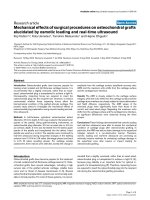
Interestingly, all identified Dictyostelium SECIS elements
had a highly conserved UGUA sequence that preceded the
SECIS core, and a U-U mismatch immediately following it
(Figure 5). The SECIS element of the deiodinase-like protein
had two U-U mismatches; however, they were located further
from the SECIS core. All detected SECIS elements were type
II structures [24]. The deiodinase-like SECIS element had an
extremely long mini-stem. As discussed above, the latter fea-
ture was also observed in many Ostreococcus selenoprotein
genes, whereas it rarely occurs in SECIS structures in other
organisms. All Dictyostelium SECIS elements had an
unpaired AAA in the apical bulge. The areas of strong conser-
vation include an SBP2-binding site and nucleotides interact-
ing with this protein [32]. Since the five selenoproteins have
different evolutionary histories and are not homologous with
each other, the conservation of primary sequences in Dictyos-
telium SECIS elements must represent convergent evolution-
ary events.
Ostreococcus SECIS elementsFigure 1 (see previous page)
Ostreococcus SECIS elements. (a) The most characteristic features of O. tauri and O. lucimarinus SECIS elements are a long mini-stem and an unpaired G
preceding the SECIS quartet (core). A SelT SECIS element is shown as a typical example (left structure). Only two exceptions were found, including a type
I SECIS element in SelH (middle structure) and a SECIS element with an unpaired A nucleotide preceding the SECIS core (right structure). (b) Alignment
of nucleotide sequences of all O. tauri SECIS elements. Location of the SECIS core is indicated. Conserved nucleotides are highlighted. Black and grey
highlighting shows sequence conservation.
R198.6 Genome Biology 2007, Volume 8, Issue 9, Article R198 Lobanov et al. />Genome Biology 2007, 8:R198
We used the observation of unusually high sequence conser-
vation of Dictyostelium SECIS elements to develop a
modified version of SECISearch, which allowed the searches
wherein other search parameters were relaxed. However,
application of this procedure did not detect additional
selenoproteins.
To further examine the Dictyostelium selenoproteome, we
metabolically labeled the amoebae cells with
75
Se and ana-
lyzed the selenoprotein pattern on SDS PAGE using a Phos-
phorImager (Figure 2b). Four selenoprotein bands were
detected, which corresponded in size to the four selenopro-
teins identified computationally (SPS, MSP, DI and Sep15).
Apparently, Sep15 was a major selenoprotein in D. discoi-
deum, whereas SelK was not detected. The latter
selenoprotein might be expressed at low levels or under
different growth or developmental conditions than those
examined in our study.
Comparative analysis of eukaryotic selenoproteomes
Selenoproteins are found in all three domains of life, which
share several protein and RNA components involved in Sec
biosynthesis and insertion, suggesting an origin of the Sec
machinery that predates the last universal common ancestor.
Thus, Sec decoding is an ancient trait that has been main-
tained for hundreds of million of years without widespread
expansion or loss.
We compiled newly and previously characterized selenopro-
teomes and analyzed the occurrence of particular selenopro-
teins against taxonomic distribution of species based on the
tree of life [33]. The number of selenoproteins varied from
zero (in plants, yeast and some protists) to 29 (in Ostreococ-
cus) (Figure 6a). Significant differences in the composition of
selenoproteomes could be seen even among related organ-
isms. For example, among viridiplantae, all higher plants
lacked selenoproteins, whereas the green algae
Chlamydomonas and Ostreococcus had 12 and 26-29 seleno-
proteins, respectively (Figure 6b). Three selenoproteins were
found in Mesostigma viride, a Streptophyte and a common
ancestor of land plants [34].
Figure 2
188kDa
38kDa
28kDa
17kDa
14kDa
6kDa
3kDa
49kDa
62kDa
98kDa
H
E
K
2
9
3
H
E
K
2
9
3
S
o
l
u
b
l
e
f
r
a
c
t
i
o
n
H
o
m
o
g
e
n
a
t
e
P
e
l
l
e
t
TR1
GPX1
TR1,
51 kDa
GPx1,
25 kDa
Sep15,
14.6 kDa
SPS,
40.4 kDa
DI-like,
30.5 kDa
MSP,
26.2 kDa
CV-1 cells
CV-1 cells
D. discoideum
D. discoideum
(a)
(b)
Metabolic labeling of O. tauri and D. discoideum with
75
Se. O. tauri and D. discoideum cells were grown in the presence of
75
Se [selenite], cell lysates prepared, proteins resolved by SDS-PAGE and analyzed using a PhosphorImagerFigure 2
Metabolic labeling of O. tauri and D. discoideum with
75
Se. O. tauri and D.
discoideum cells were grown in the presence of
75
Se [selenite], cell lysates
prepared, proteins resolved by SDS-PAGE and analyzed using a
PhosphorImager. (a) O. tauri. Three middle lanes represent the soluble
fraction, homogenate and pellet fraction as shown above the gel. For
comparison, HEK 293 cells were metabolically labeled with
75
Se, and
migrations of thioredoxin reductase 1 (TR1) and glutathione peroxidase 1
(GPx1) are shown. (b) D. discoideum. Two middle lanes represent two
independent samples of
75
Se-labeled D. discoideum cells. The four
radioactive bands correspond to the indicated selenoproteins identified in
silico. For comparison, monkey CV-1 cells were metabolically labeled with
75
Se, and migrations of TR1 and GPx1 are shown on the right.
Genome Biology 2007, Volume 8, Issue 9, Article R198 Lobanov et al. R198.7
comment reviews reports refereed researchdeposited research interactions information
Genome Biology 2007, 8:R198
Tracing individual selenoproteins, we found that some
selenoprotein families were present in many organisms and
others in only a few species, yet each identified family had a
unique pattern of occurrence (Figure 6a). None of the
selenoproteins matched the overall Sec trait (compared to the
occurrence of Sec machinery). SelK was among the most
widespread selenoproteins. This protein of unknown function
is present in nearly all eukaryotes that utilize Sec (but is
replaced with a Cys-containing homolog in nematodes and
several other organisms). An additional widespread seleno-
protein was SelW, which also occurs in most (but not all)
selenoprotein-containing eukaryotes. Several other seleno-
proteins, such as glutathione peroxidase and thioredoxin
reductase, also had a wide distribution.
Origin of many selenoproteins precedes animal
evolution
Since mammalian selenoproteomes were large and included
essentially all known eukaryotic selenoproteins, they were
initially thought to represent the entire eukaryotic selenopro-
teome. Subsequent identification of selenoproteins with
highly restricted occurrence added further complexity, but
did not challenge the overall idea of recent evolution of the
majority of eukaryotic selenoproteins. However, our analysis
of selenoproteomes of six eukaryotic model organisms and
their comparison with the previously characterized
selenoproteomes revealed that 20 of the 25 human seleno-
proteins have Sec-containing homologs in many unicellular
organisms. Similarly, taking into account protein families, at
least 11 of the 16 mammalian selenoprotein families could be
traced back to single-cell eukaryotes. SelU, which is not a
selenoprotein in mammals, is present in some animals and
protozoa and may be viewed as an additional ancient seleno-
protein family. Overall, these data suggest that the origin of
many selenoproteins not only precedes animal evolution, but
can be dated back to the ancestral eukaryotes. Thus, many of
these original selenoproteins were preserved during
evolution and remain in vertebrates (including mammals),
green algae and a variety of protists, whereas many other
organisms manifested massive selenoprotein losses.
Sec tRNAFigure 3
Sec tRNA. (a) Cloverleaf structures of Sec tRNAs from C. reinhardtii, O. tauri and C. merolae. (b) Nucleotide sequence alignment of C. reinhardtii and C.
merolae Sec tRNAs with known Sec tRNAs. Black and grey highlighting shows sequence conservation.
(a)
(b)
P.falciparum ACC
GATGA
GTTAGCATG GT
TGC
TAAGTAT-GACT TCA AA
T
CATTTGGCGTAGTTTTT
C
TGCGCAG
A
GGTTCGATTCC
T
CCTT
CG
GTG
T.gondii GCATC
GATGA
GCTGGCCTG GTGGCTGGGCGT-GACT TCA AA
T
CACGTGGCGC CTAGCGGCGCAG G
GGTTCGATTCC
TCCTTCGG
T
GCG
GGG
O.lucimarinus GCCA
GGGTGA
GCT-TCGCT GGC
GCGGAGTGCGG
CCT
TCA AAG
CCG
-TAGC
GG CTTAGCGGC
CG
AG T
C
GTTCGATTCGACCT
CACTGGCG
ACG
O.tauri GCCA
GGG
C
GAGCT
-TCGC
T GGCGCGGAGTGCGGCCT TCA
AA
GC
C
G-
TAGGGG
CTTAGCGGC
CCAG
TGGTTCGATTCCACCGACTTGGCG
GC-
C.reinhardtii GCCGCTGTGAC
CT
-TGGCG GGTGC
TGAGTGCGG
TCT
TCA
AAACCG-TAGAGG CCGGG
AGGC
CTAG TGGTTCATTTCCACCTCGGC
GGCG
CCA
C.merolae GCCCCGCTGATCTCTGGC
G
GGTGCCGGGCTCGGC
CT
TCA AAG
C
C
G
ATGGACG CCGCG
A
GGCG
TC
G CCGTTCGA
C
TCG
GCCT
GCGGGGC
H.sapiens
GCCCGGATGAT
CCTC
AG
T GGTCT
GGGGTGCAGGCT TCA AAC
CTG-TAG
CT
G TCTAGCGACA
G
AG TGGTTCAATTCCACCTTTC
GGGCGCCA
M.musculus GCCCGGATGATCCTCAGT GGTCT
GGGGTGCAGGCT
TCA AACCTG-TAG
CT
G
T
TTAGCGACAGAG
TGGTTCAATTCCACCTTTC
GGGCG
C.elegans
GCCCGGATGA
A
C
CAT
GGC GGTCT
GT
GGTGCAGACT TCA AA
T
CT
G
-
TAGG
C
G
GTTAGCG
C
CGCAG TGGTTCGA
CTCCACCTT
TC
GGG
T
D.melanogaster
GCCCCA
CTGA
ACT
TC
GGT
GGT
CCGGGGTGCGGACT TCA AA
T
C
C
G
-
TAGTCG A
TTTGCG
TCGAAG
TGGTTCGATTCCACCT
GGGGGGC
T.pseudonana GTGTGAATGATCC-TGCCT GGTGGTGGGTTCAGGCT TCA AACCTG-AAGGGG CTTAGCGGCCCAG TGGTTCGATTCCACCTT
TCG
CACG
ATG
E.coli
GGA
A
GATCGTCG
TC
TCC
GGTG
A
GG
CG
GCT
GGACT TCA AA
T
C
C
AGTTGGGGCCGCCAGCGGTC
CCG
GGCA
GGTTCGACTCC
TGTGATCTTC
CGCCA
CC
C
C
C
C
C
C
C
C
C
C
C
C
C
C
C
C
C
C
C
C
C
C
C
C
C
A
A
A
A
A
A
A
A
A
A
A
A
UU
U
U
U
U
U
U
U
U
U
U
U
U
U
U
G
G
G
G
G
G
G
G
G
G
C
G
G
G
G
G
GG
G
G
G
G
G
G
G
G
G
G
G
G
G
G
G
G
C
A
C G
C
C
C
U
C
C
G
C
C
U
C
C
C
C
C
A
U
C
C
C
C
C
C
C
G
A
C
A
A
A
A
A
A
A
A
G
UU
G
U
U
U
G
C
U
G
C
U
U
U
U
U
A
G
U
G
U
C
G
G
G
G
C
G
G
G
G
G
GUG
G
G
G
G
G
G
G
A
G
G
G
G
G
A
G
C
A
C. reinhardtii
O. tauri
CG
U
C
U
G
C
C
C
C
C
C
C
C
C
C
C
A
C
U
G
C
C
G
C
C
C
U
G
G
G
A
G
A
U
A
A
C
CU
G
C
A
U
G
C
U
G
G
U
C
U
U
C
G
A
U
U
C
G
G
G
C
G
C
G
C
G
G
GCG
G
G
C
G
G
G
G
G
A
C
U
G
G
G
G
C
A
C
G
G
C. merolae
R198.8 Genome Biology 2007, Volume 8, Issue 9, Article R198 Lobanov et al. />Genome Biology 2007, 8:R198
It should be noted that Cys/Sec replacement is not always
unidirectional and that prior evolutionary analyses suggest
that both a Sec loss and gain is possible [35]. However, the
probability of independent parallel Sec gain, as well as
consecutive homoplastic Sec-to-Cys and Cys-to-Sec substitu-
tions in a single protein position, is extremely rare, and no
selenoprotein families are known that evolved more than
once. Two factors are required for a Cys-to-Sec change to take
place. First, the presence of Sec insertion machinery, such as
Sec tRNA, SECIS-binding protein SBP2, Sec-specific elonga-
tion factor and Sec synthase. This requirement is met (for
example, all components of the machinery are present) if at
least one other selenoprotein is present in the same organism.
Second, a SECIS element should evolve in the 3'-untranslated
region. While only a single nucleotide change is sufficient to
change the codon from Cys to Sec (that is, UGA instead of
UGC or UGU), evolution of new SECIS elements is difficult.
On the other hand, once Sec is replaced with Cys, the presence
of the SECIS element provides no competitive advantage and
this structure is quickly lost. Unless the reverse Cys-to-Sec
mutation takes place before disruption of the SECIS element,
the probability of restoring Sec is extremely low. Unless
strong pressure exists to preserve Sec, its functional replace-
ment with Cys may be expected. Combined, these factors
allow us to assume that the character-state Sec follows Dollo's
behavior.
Selenoproteins with restricted occurrence are
common to organisms with large selenoproteomes
In addition to the many ancient eukaryotic selenoproteins,
several selenoproteins have a more narrow distribution. For
example, SelP, SelN, MsrB and SelI appear to be specific to
animals, whereas MSP, peroxiredoxin and thioredoxin-like
protein could be detected only in unicellular eukaryotes.
These observations suggest an emerging picture of selenopro-
tein evolution wherein core selenoprotein families evolved
first, followed by the origin of additional selenoproteins in
more narrow groups of organisms. The new selenoproteins
further increased the size of the selenoproteomes and remain
prevalent in organisms with large selenoproteomes. In our
current analysis, several Ostreococcus and Thalassiosira
selenoproteins fit this pattern, in addition to the rare seleno-
proteins previously discovered (for example, SelU, SelJ and
Fep15). However, it could not be excluded that new seleno-
proteins might also occasionally evolve in organisms with
small selenoproteomes (for example, red algae).
Red algae selenoprotein O. SECIS elements in O. tauri (green alga) and P. haitanensis (red alga) SelO genesFigure 4
Red algae selenoprotein O. SECIS elements in O. tauri (green alga) and P. haitanensis (red alga) SelO genes. The P. haitanensis SECIS element belongs to type
I, while O. tauri to type II structures.
O. tauri P. haitanensis H.sapiens T.rubripes
C
C
C
CC
C
C
C
C
C
C
C
C
C
G
G
G
G
G
G
G
G
G
G
G
G
G
G
G
G
G
G
U
U
U
U
U
U
U
U
A
A A
A
A
A
A
A
A
A
A
A
A
A
A
U
C
G
C
G
G
G
G
G
G
G
G
G
C
C
C
C
C
U
U
U
U
U
U
U
U
U U
U
U
U
U
U
U
U
U
U
A
A
A
A
A
A
A
A
A
A
A
A
A
A
A
A
A
A
A
A
A
C
U
G
U
A
G
G
G
G
G
G
G
G
G
G
G
G
G
G
C
C
C
C
C
C
C
C
C
C
C
C
C
C
C
C
C
U
U
U
U
U
U
U
U
U
A
A
A
A
A
A
A
A
A
A
G
A
A
A
A
A
A
A
A
A
A
A
A
A
U
U
U
U
U
U
U
U
U
U
U
U
U
G
G
G
G
G
G
G
C
C
C
C C
C
C
C
C
C
C
C
C
A
G
C
G
G
A
A
G
Genome Biology 2007, Volume 8, Issue 9, Article R198 Lobanov et al. R198.9
comment reviews reports refereed researchdeposited research interactions information
Genome Biology 2007, 8:R198
Independent events of massive selenoprotein loss in
eukaryotes
We further identified and examined several groups of organ-
isms characterized by massive selenoprotein loss. Location of
these organisms on the eukaryotic tree of life suggests inde-
pendent events of selenoprotein loss (Figure 6a). Five exam-
ples of selenoprotein loss are discussed below.
Plants
As discussed above, A. thaliana, O. sativa and other higher
plants lost both selenoproteins and Sec insertion machinery,
whereas these genes were preserved in green algae, for exam-
ple, Chlamydomonas, Volvox and Ostreococcus. An early
Streptophyte, M. viride, has both Sec machinery and seleno-
proteins. Thus, there was a specific selenoprotein loss event
in the Streptophyte subset of Viridiplantae, which invaded
land. Analysis of selenoproteins present in green algae sug-
gests that they were either replaced with Cys-containing
homologs or entirely lost in land plants (Figure 6b). A more
distantly related C. merolae also manifested a large-scale
selenoprotein loss.
Apicomplexan parasites
The high selenoprotein content of Thalassiosira (as a refer-
ence point), the reduced selenoproteome of Plasmodium and
the lack of selenoproteins in Cryptosporidium parvum illus-
trates an example of massive selenoprotein loss in apicompl-
exan parasites.
Fungi
We screened all completely sequenced fungal genomes and
could detect neither selenoproteins nor Sec insertion
machinery. These data suggest that selenoprotein genes were
likely lost at the base of the fungi kingdom.
Insects
The small selenoproteomes of A. gambiae, A. mellifera, D.
pseudoobscura and D. melanogaster, which consist of one to
three selenoproteins, is an additional example of large-scale
selenoprotein loss. On the other hand, aquatic arthropods,
such as shrimp, have many selenoprotein genes (based on the
expressed sequence tag (EST) analyses as the genomes are
not yet available; unpublished data). Thus, it appears that
selenoprotein genes were massively lost in either insects, or
all terrestrial arthropods.
Table 2
Selenoproteins identified in the analyzed eukaryotic genomes
Selenoprotein family O. tauri O. lucimarinus T. pseudonana D. discoideum D. pseudoobscura
SelK + + + +
SelH + + +
SPS2 ++ +
DI +
Sep15 + + +
MSP + + +
Gpx +++++ +++++ ++
SelT + + +
TR + + +
SelM + + ++
SelU + + ++
MsrA + + +
PDI +++ +++ ++
Methyltransferase + + +
Peroxiredoxin + +++ ++
Thioredoxin-fold protein + + +
SelO + +
SelW + ++
SelS + +
Hypothetical protein 1 + +
Hypothetical protein 2 + +
Hypothetical protein 3 + +
Total 26 29 16 5 3
Each '+' corresponds to one selenoprotein gene.
R198.10 Genome Biology 2007, Volume 8, Issue 9, Article R198 Lobanov et al. />Genome Biology 2007, 8:R198
Nematodes
The selenoproteomes of C. elegans and C. briggsae have only
one selenoprotein, thioredoxin reductase, and, therefore, the
Sec insertion system is used to decode only a single UGA
codon in these nematodes [10].
The decreased size of selenoproteomes in these five groups of
organisms appears to be not only due to the loss of entire
selenoprotein genes, but also due to replacement of Sec with
Cys. Thus, Cys-containing homologs, while often catalytically
inefficient, may occasionally compensate for selenoprotein
loss [36].
A hypothesis for association of large selenoproteomes
and aquatic life
The mosaic occurrence of eukaryotic selenoproteins and their
consistent loss in different phyla suggest that the decreased
selenoproteome size is the result of a selective force. What
could be the factors responsible for or associated with seleno-
protein loss? Comparative analysis of organisms with large
and small selenoproteomes shows that many of the seleno-
protein-rich organisms live in aquatic environments. In con-
trast, almost all organisms that lack or have a small number
of selenoproteins are terrestrial (Figure 6). Considering inde-
pendent, large-scale selenoprotein loss in these organisms, a
common denominator appears to be the non-aquatic habitat.
It should be noted, however, that the differences between
aquatic and terrestrial selenoproteomes are ultimately influ-
enced by specific environmental factors that differ with habi-
tat. Therefore, the aquatic/terrestrial association should not
be viewed as the basis for selenoprotein loss/gain, but rather
a convenient illustration of differences between these organ-
isms. Once environmental factors are identified, this associa-
tion may be modified to reflect these factors rather than
habitat.
To further examine selenoprotein content of aquatic and ter-
restrial organisms, we analyzed organisms that are well rep-
resented by ESTs. We excluded large animals (vertebrates)
from this analysis because their intra-organismal environ-
ment would be less affected by environmental conditions due
to availability of their outside protective cover and complex
morphology. With this limitation, aquatic eukaryotes had
more selenoprotein genes than terrestrial organisms (Figure
7).
Whether C. merolae fits this association is not clear. This
organism lives in highly acidic sulfate-rich hot springs (pH
1.5, 45°C). It is possible that this extreme environment is
responsible for the reduced use of Sec in red algae. The pKa of
Dictyostelium discoideum SECIS elementsFigure 5
Dictyostelium discoideum SECIS elements. (a) SECIS elements in D. discoideum selenoprotein genes. Sequences conserved in eukaryotic SECIS elements are
shown in red, and Dictyostelium-specific conserved sequences are shown in blue. (b) Alignment of D. discoideum SECIS elements. A UGUA sequence
preceding the SECIS core, and a U-U mismatch in the stem-loop structure represent additional conserved features in Dictyostelium SECIS elements. Black
and grey highlighting shows sequence conservation.
(a)
Sep15
DI-like protein
SPS2
SelK
MSP
(b)
DI-like
AAAAA
AAAAAAAAAAAA
AAAAAAAAU
UGUA A
UGA
UUGCUUUAUUAUAUA AAA UUAUCUA UAA
UU
AAAUUA-UAGAAU
A
UAAUUUAGA UGAA
AA
CUCUAUUUUUUU
U
UUUU
MSP AAA
U
AAAU
A
GUUCAAAUAAAAUUAGUUGUA AUGAUU
U
UUAUAAUGCU AAA AC
UAAA
UUAAUAG-
UCGCUUA
UA-A
AU
U
GAU
AAA
CUA
AUU
GA
UUUU
C
UUU
Sep15 C
AU
UA
UC
UC
UUUG
AUAAAUUGAUUGUA A
UGA
U
U
A
UGUAAAUGA AAA AC
A
U
UUUUUAA
A
AA-U
GUC
AUUUA-C
AUU
U
GAU
AAAUCU
AUUU
A
UU
AAUUUG
SPS2
AA
U
AAU
-
AAUU
U
UUAAUAACAAAUAUUGUA AUGAU
U
UGAAAUUGA
U AA
A UC CAU
A
UUAU
UGG
ACUUAAUUU C
AU
UGAA
AAA
AAAAA
GA
UA
U
AA
UAA
U
SelK AAGAAUCAAUGAUUAGUUUUUAAAACUGUA AUGAUU UGUUAA-AUU AAA AC CAUUUUAU
UGG
CAAUUUAAC
AU
UGAA
U
AG
AUC
AUUUUCA
U
CAG
UA
A
A
A
A
U
A
U
A
U
A
A
U
U
G
G
A
A
A
A
A
A
C
C
A
U
C
G
A
U
G
G
G
U
U
A
U
A
U
U
C
A
U
U U
U
U
U
U
U
U
UA
U
A
A
A
A
A
A
A
A
A
U
U
U
U
U
U
U
U
A
A
A
A
U
U
U
A
A
A
U
C
U
A
A
A
A
A
A
A
U
U
C
G
A
A
G
G
G
U
A
A
G
U
U
U
U
A
G
A
U
U
UU
U
U
U
U
U
C
C
U
U
A
U
G
A
A
A
A
A
A
A
G
A
A
C
U
A
A
A
U
A
A
G
A
A
C
G
U
U
G
G
G
U
U
U
U
U
C
U
U
U
G
A
U
U
UU
U
U
U
U
U
A
C
U
U
U
U
A
A
A
A
C
A
A
U
U
A
U
A
U
A
A
A
A
A
A
A
A
G
C
G
C
U
G
G
G
A
U
U
G
U
A
A
U
A
U
A
U
U
UU
U
U
U
U
U
U
C
U
U
A
A
A
U
A
A
A
A
A
A
A
A
A
A
A A
A
A
A
A
A
A
A
A
A
A
A
A
A
A
C
C
C
G
G
G
G
G
G
U
U
U
U
U
U
U U
UU
U
U
U
U
U
U
U
U U
U
U
U
U
U
U
Genome Biology 2007, Volume 8, Issue 9, Article R198 Lobanov et al. R198.11
comment reviews reports refereed researchdeposited research interactions information
Genome Biology 2007, 8:R198
Sec is approximately 5.5. Whereas this residue would be ion-
ized in most organisms under physiological conditions, at low
pH, protonation of Sec may minimize its catalytic advantages.
Abundance of sulfate in hot springs might also be of impor-
tance, as selenium and sulfur have similar chemistries.
One possible explanation for the occurrence of large seleno-
proteomes in aquatic organisms is bioavailability of selenium
in oceans. Dissolved organic selenides can account for
approximately 80% of the dissolved selenium in ocean water
[37] and represent an important source of selenium for phy-
toplankton. Following the food chain, this could explain a
large number of selenoproteins in algae and fish. Likewise, a
considerable number of selenoproteins in mammals could
reflect the consequence of food sources, body size and rela-
tively recent (in evolutionary terms) emergence of these
organisms from marine environments. An additional factor
may be constancy in the environmental conditions and nutri-
ents in the aquatic environments. For aquatic organisms,
environmental changes are slower and involve gradients of
temperature, pH, pressure, oxygen and chemical environ-
ment. In contrast, in terrestrial environments, the changes
are more frequent and they happen more suddenly. As a
result, terrestrial organisms often face feast and starvation
situations. An attractive factor to explain the differences
between aquatic and terrestrial selenoproteomes may be oxy-
gen content. Higher content of oxygen in air than in aquatic
environments may make highly reactive selenoproteins more
susceptible to oxidation in terrestrial organisms and select
against the use of these proteins.
Whether mammals and other vertebrates fit the hypothesis
on the preferential use of selenium in aquatic environments is
not clear. We note, however, that fish have larger selenopro-
teomes than those living in terrestrial environments, includ-
ing mammals, reptiles and birds. Further genomic analyses of
these organisms could clarify evolutionary changes in utiliza-
tion of selenium. In future studies, it would also be important
to determine which of the factors discussed above influence
the preferential use of Sec in aquatic organisms or are respon-
sible for the loss of selenoproteins in terrestrial organisms.
Conclusion
Until recently, the mammalian selenoproteome was thought
to represent accurately eukaryotic selenoproteins and to be of
recent (perhaps vertebrate) origin. However, as additional
genome sequences became available, selenoproteins with
restricted occurrence have been identified. In mammals,
these proteins either occur in the form of Cys-containing
homologs or are absent altogether; instead, these rare seleno-
proteins have been found in several lower eukaryotic organ-
isms. In our work, the searches of additional eukaryotic
genomes identified new selenoprotein genes, revealed exam-
ples of convergent evolution of SECIS elements, and identi-
fied many features of selenoproteome organization and
evolution. Integrated analyses of eukaryotic selenoproteomes
suggested that the majority of eukaryotic selenoprotein fami-
lies evolved in single-celled eukaryotes. Our data show that
the mosaic occurrence of selenoproteins is the consequence of
selective, independent selenoprotein loss events in various
eukaryotic phyla. Moreover, these analyses revealed an inter-
esting pattern: large selenoproteomes tend to occur in aquatic
life, whereas the organisms that lack selenoproteins or have
small selenoproteomes are mostly terrestrial (with the nota-
ble exception of mammals, whose large bodies and intra-
organismal homeostasis support an internal environment
that may be less dependent on habitat). Further studies will
be needed to test this hypothesis and identify environmental
factors that influence selenium utilization.
Materials and methods
Databases and programs
All genome, EST and predicted protein sequences were down-
loaded from NCBI [38], except for the genomes of T. pseudo-
nana, O. tauri, and O. lucimarinus, which were obtained
from Joint Genome Institute [39]. SECISearch [9] was used
for identification of SECIS elements. FASTA package [40]
and BLAST were used for similarity searches. MFOLD ver-
sion 3.2 [41] was used for prediction of RNA secondary
structures.
Identification of homologs of known selenoprotein
genes
Query sequences included a full set of human selenoproteins
[9] as well as the following selenoproteins absent in
mammals: Chlamydomonas MsrA [12], Gallus gallus SelU
[42], Danio rerio SelJ and Fep15 [15,16], and Emiliania
huxleyi protein disulfide isomerase [43]. A stand-alone ver-
sion of TBLASTN program was utilized for detection of nucle-
otide sequences corresponding to known selenoprotein
families. A candidate Sec residue should correspond to a Sec
residue in a known selenoprotein family or a Cys residue in
orthologous proteins in order to be considered further.
Downstream regions of predicted selenoprotein sequences
were analyzed for the presence of candidate SECIS elements
using SECISearch and for SECIS-like structures using
MFOLD [41]. All detected SECIS candidates were further
examined for compliance with the current SECIS consensus
model.
Searches for SECIS elements
Nucleotide sequences were scanned using SECISearch (Addi-
tional data file 1). In addition, the default and loose patterns
of SECISearch were modified as described elsewhere [12] to
accommodate organism-specific selenoprotein searches.
These modifications allowed increased sensitivity of SECI-
Search and supported identification of unusual SECIS struc-
tures. The overall strategy of the searches was similar to that
previously described [9]. Statistics of the searches (numbers
of candidates corresponding to different steps in the search
R198.12 Genome Biology 2007, Volume 8, Issue 9, Article R198 Lobanov et al. />Genome Biology 2007, 8:R198
Figure 6 (see legend on next page)
Chlamydomonas reinhardtii
Ostreococcus tauri
Thalassiosira pseudonana
Green algae
Diatoms
SelT
Gpx
SelM
TR
SelU
Oryza sativa
Arabidopsis thaliana
Higher plants
MsrA
SPS2
PDI
MSP
SelO
SelW
SelK
SelH
Trx-like
Methyltransferase
Peroxiredoxin
SelS
Sep15
Volvox carteri
Ostreococcus lucimarinus
Medicago truncatula
Charophyta
Mesostigma viride
(a)
(b)
1
Caenorhabditisbriggsae
3
Drosophilapseudoobscura
Anophelesgambiae
3
24
Mus musculus
Homo sapiens
25
Gallusgallus
Xenopustropicalis
24
24
0
Saccharomycescerevisiae
5
Dictyosteliumdiscoideum
0
Schizosaccharomycespombe
Medicagotruncatula
Arabidopsisthaliana
0
0
Chlamydomonasreinhardtii
12
Ostreococcustauri
26
Thalassiosirapseudonana
16
0
Cryptosporidiumparvum
4
Plasmodiumfalciparum
Cyanidioschyzonmerolae
0
1
Caenorhabditiselegans
3
Drosophilamelanogaster
1
Apis mellifera
Oryza sativa
0
Ostreococcuslucimarinus
29
4
Plasmodium chabaudi
4
Plasmodiumyoelii
Populustrichocarpa
0
0
Yarrowialipolytica
Genome Biology 2007, Volume 8, Issue 9, Article R198 Lobanov et al. R198.13
comment reviews reports refereed researchdeposited research interactions information
Genome Biology 2007, 8:R198
process) are shown in Table 1. In an additional search for D.
discoideum SECIS elements, the following pattern was used
as a query: TGTAATGATT_(10-12 nucleotides)_AAA_(24-35
nucleotides)_TGAT. This search then continued as described
for other organisms.
The primary sequence analysis step included searches for
SECIS-like structures that satisfy NTGA__AA__GA or
NTGA__CC__GA (N is any nucleotide) motifs in nucleotide
sequences. Additional requirements were that the distance
between the quartet (NTGA) and the unpaired AA in the api-
cal loop is 10-13 nucleotides, and the distance between the
unpaired AA and the GA that base-paired with the quartet is
15-39 nucleotides.
Eukaryotic selenoproteomesFigure 6 (see previous page)
Eukaryotic selenoproteomes. (a) A simplified cladogram of model organisms discussed in the text that illustrates distribution of selenoproteins in
eukaryotes. The number of selenoproteins in each indicated model organism is shown in red (current study) and gray (previously analyzed and other
model organisms) squares, and is proportional to the size of the bars on the left. Yellow circles show possible origins of various selenoprotein families, and
red crosses examples of massive selenoprotein loss. (b) Selenoprotein evolution in plants. The 'mountain' symbols show terrestrial organisms, and
'anchors' those that live in aquatic environments. Green checkmarks indicate the presence of an indicated selenoprotein in the corresponding genome.
The presence of Cys-containing homologs is shown by blue checkmarks. Crossed red circles indicate absence of either Sec- or Cys-containing homologs.
Unfilled spots correspond to lack of data due to unfinished genomes, unclear relationship between proteins and lineage specific gene duplications.
Aquatic invertebrates have more selenoproteins than terrestrial organismsFigure 7
Aquatic invertebrates have more selenoproteins than terrestrial organisms. Numbers of detected selenoproteins were plotted against the total number of
available (redundant) ESTs for organisms that are represented by more than 25,000 ESTs. Vertebrate ESTs were excluded from this analysis due to large
size of these organisms. Blue circles correspond to aquatic and brown squares to terrestrial organisms. The difference is statistically significant (P value is
less than 2 × 10
-6
).
Number of ESTs
0
3
6
9
12
15
18
21
10,000 100,000 1,000,000 10,000,000
Number of selenoproteins
R198.14 Genome Biology 2007, Volume 8, Issue 9, Article R198 Lobanov et al. />Genome Biology 2007, 8:R198
The secondary structure analysis step examined for consist-
ency with the eukaryotic SECIS element consensus. Several
additional filters were implemented to filter out candidates
with unsuitable secondary structures, including SECIS
elements with more than two unpaired nucleotides in a row
and Y-shaped SECIS elements.
The free energy for each candidate structure was estimated;
the free energies for the whole structure (threshold value of -
12.6 kcal/mol) and the upper stem-loop (threshold value of -
3.7 kcal/mol) were calculated. Only thermodynamically sta-
ble structures were considered further.
Based on the location of candidate SECIS elements, candidate
ORFs were predicted in upstream regions. SECIS candidates
located within coding regions of known proteins were filtered
out. An additional requirement was the presence of at least
one homologous protein in the NCBI non-redundant data-
base. If SECIS elements and ORFs corresponding to known
protein families were on different DNA strands, the candi-
dates were filtered out.
The final step included manual sequence analyses of pre-
dicted selenoprotein ORFs located upstream of candidate
SECIS elements.
Searches using the Sec/Cys homology approach
For three organisms, O. tauri, O. lucimarinus and C. merolae,
additional procedures for selenoprotein detection included
the search for Sec/Cys pairs in homologous sequences. ORFs
with in-frame TGA codons were extracted that satisfied the
following criteria: Sec-flanking regions for these proteins
were conserved; and homologs could be detected that con-
tained Cys in place of Sec. TBLASTX was used to examine all
potential ORFs with in-frame UGA codons against NCBI non-
redundant protein database. All hits were then tested for the
occurrence of SECIS elements. Orthologous proteins were
defined as bidirectional best hits. PSI-BLAST was used for
identification of distant homologs. Homologs were further
confirmed by phylogenetic trees construction.
Phylogenetic analyses
Numerous attempts to derive a tree of life using various meth-
ods that were based on genes encoding ribosomal RNAs and
several proteins have been published. However, their princi-
ple existence has been questioned recently because of either
an insufficient amount of discriminating characters or other
biases such as horizontal gene transfer and chimerism. To
avoid such problems, we adopted a eukaryotic branch of a
phylogenetic tree recently developed by Ciccarelli et al. [33].
This highly resolved tree of life utilized 31 concatenated, uni-
versally occurring genes with indisputable orthology in 191
species with completed genomes across all three domains of
life. The missing organisms were filled in using a 'Tree of Life'
web project [44] and selected publications 5-48]. Although
the horizontal gene transfer is highly prevalent in prokaryo-
tes, it is less so in eukaryotes, particularly in multicellular
organisms. We also analyzed selenoprotein evolution in the
eukaryotic domain. To reconstruct the phylogenies of seleno-
proteins, we adopted a character-based tree estimation
method, a maximum parsimony approach that implies that
the preferred phylogenetic tree is the tree that requires the
least number of evolutionary changes.
Metabolic labeling of D. discoideum and O. tauri cells
D. discoideum cells were grown as previously described [31],
the medium was supplemented with 100 μCi of
75
Se [selenite]
(University of Missouri Research Reactor), and the cells were
further maintained under continuous shaking for two days. A
similar procedure was used for labeling O. tauri cells, except
that they were grown in K-medium. The radioactive bands
were visualized on the gel with a PhosphorImager. Samples of
75
Se-labeled mammalian HEK 293 and CV-1 cells were
included, which were prepared as described previously [49].
Abbreviations
Cys, cysteine; EST, expressed sequence tag; GPx, glutathione
peroxidase; MSP, membrane selenoprotein; ORF, open read-
ing frame; Sec, selenocysteine; SECIS, selenocysteine inser-
tion sequence; TR, thioredoxin reductase.
Authors' contributions
AVL, DEF and YZ performed computational analyses. DEF
and AS carried out experimental analyses. AVL, DLH and
VNG wrote the manuscript. All authors read and approved
the final manuscript.
Note added in proof
Two recent studies reported the complete genomes of O. tauri
and O. lucimarinus [50,51]. One of these articles identified 18
and 20 selenoprotein genes in O. tauri and O. lucimarinus,
respectively [51]. Compared to our analyses, this published
study did not detect 17 selenoproteins in the two organisms,
whereas the protein they designated as SelA and predicted to
contain three selenocysteines appears to be a false positive.
Nevertheless, the large number of detected selenoproteins in
Ostreococcus further highlights the association with aquatic
life reported in our work.
Additional data files
The following additional data are available with the online
version of this paper. Additional data file 1 presents a block-
scheme of the searches for selenoprotein genes. Additional
data file 2 contains amino acid sequence alignments of
selenoproteins identified in this study. Additional data file 3
contains sequence and predicted clover-leaf structure of T.
pseudonana Sec tRNA. Additional data file 4 has representa-
tive phylogenetic trees of selenoproteins.
Genome Biology 2007, Volume 8, Issue 9, Article R198 Lobanov et al. R198.15
comment reviews reports refereed researchdeposited research interactions information
Genome Biology 2007, 8:R198
Additional data file 1Block-scheme of the searches for selenoprotein genesBlock-scheme of the searches for selenoprotein genes.Click here for fileAdditional data file 2Amino acid sequence alignments of selenoproteins identified in this studyAmino acid sequence alignments of selenoproteins identified in this study.Click here for fileAdditional data file 3Sequence and predicted clover-leaf structure of T. pseudonana Sec tRNASequence and predicted clover-leaf structure of T. pseudonana Sec tRNA.Click here for fileAdditional data file 4Representative phylogenetic trees of selenoproteinsRepresentative phylogenetic trees of selenoproteins.Click here for file
Acknowledgements
This study was supported by NIH GM061603 (to VNG). We thank Dr
Catherine Chia for providing Dictyostelium cells, and Dr Konstantin
Korotkov for labeling the Dictyostelium cells with
75
Se. Study was completed
in part utilizing the PrairieFire Beowulf cluster from Research Computing
Facility of the University of Nebraska - Lincoln.
References
1. Hatfield DL, Gladyshev VN: How selenium has altered our
understanding of the genetic code. Mol Cell Biol 2002,
22:3565-3576.
2. Copeland PR: Regulation of gene expression by stop codon
recoding: selenocysteine. Gene 2003, 312:17-25.
3. Driscoll DM, Copeland PR: Mechanism and regulation of seleno-
protein synthesis. Annu Rev Nutr 2003, 23:17-40.
4. Tujebajeva RM, Copeland PR, Xu XM, Carlson BA, Harney JW, Dris-
coll DM, Hatfield DL, Berry MJ: Decoding apparatus for eukary-
otic selenocysteine insertion. EMBO Rep 2000, 1:158-163.
5. Lambert A, Legendre M, Fontaine JF, Gautheret D: Computing
expectation values for RNA motifs using discrete
convolutions. BMC Bioinformatics 2005, 6:118.
6. Kryukov GV, Gladyshev VN: Mammalian selenoprotein gene
signature: identification and functional analysis of selenopro-
tein genes using bioinformatics methods. Methods Enzymol
2002, 347:84-100.
7. Kim HY, Gladyshev VN: Different catalytic mechanisms in
mammalian selenocysteine- and cysteine-containing
methionine-R-sulfoxide reductases. PLoS Biol 2005, 3:e375.
8. Kryukov GV, Gladyshev VN: The prokaryotic selenoproteome.
EMBO Rep 2004, 5:538-543.
9. Kryukov GV, Castellano S, Novoselov SV, Lobanov AV, Zehtab O,
Guigo R, Gladyshev VN: Characterization of mammalian
selenoproteomes. Science 2003, 300:1439-1443.
10. Taskov K, Chapple C, Kryukov GV, Castellano S, Lobanov AV,
Korotkov KV, Guigo R, Gladyshev VN: Nematode selenopro-
teome: the use of the selenocysteine insertion system to
decode one codon in an animal genome? Nucleic Acids Res 2005,
33:2227-2238.
11. Castellano S, Morozova N, Morey M, Berry MJ, Serras F, Corominas
M, Guigo R: In silico identification of novel selenoproteins in
the Drosophila melanogaster genome. EMBO Rep 2001,
2:
697-702.
12. Novoselov SV, Rao M, Onoshko NV, Zhi H, Kryukov GV, Xiang Y,
Weeks DP, Hatfield DL, Gladyshev VN: Selenoproteins and selen-
ocysteine insertion system in the model plant cell system,
Chlamydomonas reinhardtii. EMBO J 2002, 21:3681-3693.
13. Lobanov AV, Delgado C, Rahlfs S, Novoselov SV, Kryukov GV,
Gromer S, Hatfield DL, Becker K, Gladyshev VN: The Plasmodium
selenoproteome. Nucleic Acids Res 2006, 34:496-505.
14. Mourier T, Pain A, Barrell B, Griffiths-Jones S: A selenocysteine
tRNA and SECIS element in Plasmodium falciparum. Rna
2005, 11:119-122.
15. Castellano S, Lobanov AV, Chapple C, Novoselov SV, Albrecht M,
Hua D, Lescure A, Lengauer T, Krol A, Gladyshev VN, et al.: Diver-
sity and functional plasticity of eukaryotic selenoproteins:
identification and characterization of the SelJ family. Proc
Natl Acad Sci USA 2005, 102:16188-16193.
16. Novoselov SV, Hua D, Lobanov AV, Gladyshev VN: Identification
and characterization of Fep15, a new selenocysteine-con-
taining member of the Sep15 protein family. Biochem J 2006,
394:575-579.
17. Richards S, Liu Y, Bettencourt BR, Hradecky P, Letovsky S, Nielsen R,
Thornton K, Hubisz MJ, Chen R, Meisel RP, et al.: Comparative
genome sequencing of Drosophila pseudoobscura : chromo-
somal, gene, and cis-element evolution. Genome Res 2005,
15:1-18.
18. Lakovaara S, Sauna A: Evolution and speciation in the Dro-
sophila obscura group. In The Genetics and Biology of Drosophila Vol-
ume 3b. Edited by: Ashburner M, Carson HL, Thompson JN Jr. New
York: Academic Press; 1982:1-59.
19. Courties C, Perasso R, Chrétiennot-Dinet M-J, Gouy M, Guillou L,
Troussellier M: Phylogenetic analysis and genome size of
Ostreococcus tauri (Chlorophyta, prasinophyceae). J Phycol
1998, 34:844-849.
20. Arabidopsis Genome Initiative: Analysis of the genome sequence
of the flowering plant Arabidopsis thaliana
. Nature 2000,
408:796-815.
21. Harris EH: Chlamydomonas as a model organism. Annu Rev
Plant Physiol Plant Mol Biol 2001, 52:363-406.
22. Gutman BL, Niyogi KK: Chlamydomonas and Arabidopsis. A
dynamic duo. Plant Physiol 2004, 135:607-610.
23. Derelle E, Ferraz C, Lagoda P, Eychenié S, Cooke R, Regad F, Sabau
S, Courties C, Delseny M, Demaille J: DNA libraries for sequenc-
ing the genome of Ostreococus tauri (Chlorophyta, prasino-
phyceae): the smallest free-living eukaryotic cell. J Phycol
2002, 38:1150-1156.
24. Grundner-Culemann E, Martin GW 3rd, Harney JW, Berry MJ: Two
distinct SECIS structures capable of directing selenocysteine
incorporation in eukaryotes. Rna 1999, 5:625-635.
25. Fagegaltier D, Lescure A, Walczak R, Carbon P, Krol A: Structural
analysis of new local features in SECIS RNA hairpins. Nucleic
Acids Res 2000, 28:2679-2689.
26. Misumi O, Matsuzaki M, Nozaki H, Miyagishima SY, Mori T, Nishida
K, Yagisawa F, Yoshida Y, Kuroiwa H, Kuroiwa T: Cyanidioschyzon
merolae genome. A tool for facilitating comparable studies
on organelle biogenesis in photosynthetic eukaryotes. Plant
Physiol 2005, 137:567-585.
27. Lobanov AV, Kryukov GV, Hatfield DL, Gladyshev VN: Is there a
twenty third amino acid in the genetic code? Trends Genet
2006, 22:357-360.
28. Armbrust EV, Berges JA, Bowler C, Green BR, Martinez D, Putnam
NH, Zhou S, Allen AE, Apt KE, Bechner M, et al.: The genome of
the diatom Thalassiosira pseudonana: ecology, evolution,
and metabolism. Science 2004, 306:79-86.
29. Hatfield DL, Lee BJ, Price NM, Stadtman TC: Selenocysteyl tRNA
occurs in the diatom, Thalassiosira, and in the ciliate,
Tetrahy-
mena. Mol Microbiol 1991, 5:1183-1186.
30. Price NM, Harrison PJ: Specific selenium-containing macromol-
ecules in the marine diatom Thalassiosira pseudonana. Plant
Physiol 1988, 86:192-199.
31. Shrimali RK, Lobanov AV, Xu XM, Rao M, Carlson BA, Mahadeo DC,
Parent CA, Gladyshev VN, Hatfield DL: Selenocysteine tRNA
identification in the model organisms Dictyostelium discoi-
deum and Tetrahymena thermophila. Biochem Biophys Res
Commun 2005, 329:147-151.
32. Fletcher JE, Copeland PR, Driscoll DM, Krol A: The selenocysteine
incorporation machinery: interactions between the SECIS
RNA and the SECIS-binding protein SBP2. RNA 2001,
7:1442-1453.
33. Ciccarelli FD, Doerks T, von Mering C, Creevey CJ, Snel B, Bork P:
Toward automatic reconstruction of a highly resolved tree
of life. Science 2006, 311:1283-1287.
34. Karol KG, McCourt RM, Cimino MT, Delwiche CF: The closest liv-
ing relatives of land plants. Science 2001, 294:2351-2353.
35. Zhang Y, Romero H, Salinas G, Gladyshev VN: Dynamic evolution
of selenocysteine utilization in bacteria: a balance between
selenoprotein loss and evolution of selenocysteine from
redox active cysteine residues. Genome Biol 2006, 7:R94.
36. Gromer S, Johansson L, Bauer H, Arscott LD, Rauch S, Ballou DP,
Williams CH Jr, Schirmer RH, Arner ES: Active sites of thiore-
doxin reductases: why selenoproteins? Proc Natl Acad Sci USA
2003, 100:12618-12623.
37. Cutter GA: The estuarine behaviour of selenium in San Fran-
cisco Bay. EstuarineCoastal Shelf Sci 1989, 28:13-34.
38. NCBI [ />39. Joint Genome Institute []
40. Pearson WR, Lipman DJ: Improved tools for biological sequence
comparison. Proc Natl Acad Sci USA 1988, 85:2444-2448.
41. Zuker M: Mfold web server for nucleic acid folding and hybrid-
ization prediction. Nucleic Acids Res 2003, 31:3406-3415.
42. Castellano S, Novoselov SV, Kryukov GV, Lescure A, Blanco E, Krol
A, Gladyshev VN, Guigo R: Reconsidering the evolution of
eukaryotic selenoproteins: a novel nonmammalian family
with scattered phylogenetic distribution. EMBO Rep 2004,
5:71-77.
43. Obata T, Shiraiwa Y: A novel eukaryotic selenoprotein in the
haptophyte alga Emiliania huxleyi. J Biol Chem 2005,
280:18462-18468.
44. "Tree of Life" Web Project [ />45. Nielsen C: Animal Evolution: Interrelationships of the Living Phyla Oxford:
R198.16 Genome Biology 2007, Volume 8, Issue 9, Article R198 Lobanov et al. />Genome Biology 2007, 8:R198
Oxford University Press; 2001.
46. Baldauf SL, Doolittle WF: Origin and evolution of the slime
molds (Mycetozoa). Proc Natl Acad Sci USA 1997, 94:12007-12012.
47. Baldauf SL, Roger AJ, Wenk-Siefert I, Doolittle WF: A kingdom-
level phylogeny of eukaryotes based on combined protein
data. Science 2000, 290:972-977.
48. Steenkamp ET, Wright J, Baldauf SL: The protistan origins of ani-
mals and fungi. Mol Biol Evol 2006, 23:93-106.
49. Korotkov KV, Kumaraswamy E, Zhou Y, Hatfield DL, Gladyshev VN:
Association between the 15-kDa selenoprotein and UDP-
glucose:glycoprotein glucosyltransferase in the endoplasmic
reticulum of mammalian cells. J Biol Chem 2001,
276:15330-15336.
50. Palenik B, Grimwood J, Aerts A, Rouze P, Salamov A, Putnam N,
Dupont C, Jorgensen R, Derelle E, Rombauts S, et al.: The tiny
eukaryote Ostreococcus provides genomic insights into the
paradox of plankton speciation. Proc Natl Acad Sci USA 2007,
104:7705-7710.
51. Derelle E, Ferraz C, Rombauts S, Rouze P, Worden AZ, Robbens S,
Partensky F, Degroeve S, Echeynie S, Cooke R, et al.: Genome anal-
ysis of the smallest free-living eukaryote Ostreococcus tauri
unveils many unique features. Proc Natl Acad Sci USA 2006,
103:11647-11652.