Báo cáo y học: "Drug Resistance, Cancer Research UK Cambridge Research Institute and Departmen" pot
Bạn đang xem bản rút gọn của tài liệu. Xem và tải ngay bản đầy đủ của tài liệu tại đây (1.23 MB, 17 trang )
Open Access
Volume
et al.
Chin
2007 8, Issue 10, Article R215
Research
High-resolution aCGH and expression profiling identifies a novel
genomic subtype of ER negative breast cancer
Suet F ChinÔ*, Andrew E TeschendorffÔ*, John C MarioniÔĐ,
Yanzhong Wang*, Nuno L Barbosa-Morais†, Natalie P Thorne†§,
Jose L Costa#, Sarah E PinderƠ, Mark A van de Wiel**, Andrew R Greenả,
Ian O Ellisả, Peggy L Porter, Simon TavarộĐ, James D Brenton,
Bauke Ylstra# and Carlos Caldas*¥
Addresses: *Breast Cancer Functional Genomics, Cancer Research UK Cambridge Research Institute and Department of Oncology University
of Cambridge, Li Ka-Shing Centre, Robinson Way, Cambridge CB2 0RE, UK. †Computational Biology Group, Cancer Research UK Cambridge
Research Institute and Department of Oncology University of Cambridge, Li Ka-Shing Centre, Robinson Way, Cambridge CB2 0RE, UK.
‡Functional Genomics of Drug Resistance, Cancer Research UK Cambridge Research Institute and Department of Oncology University of
Cambridge, Li Ka-Shing Centre, Robinson Way, Cambridge CB2 0RE, UK. §Computational Biology Group, Department of Applied Mathematics
and Theoretical Physics, University of Cambridge, Centre for Mathematical Sciences, Wilberforce Road, Cambridge CB3 0WA, UK.
¶Histopathology, Nottingham City Hospital NHS Trust and University of Nottingham, Nottingham NG5 1PB, UK. ¥Cambridge Breast Unit,
Addenbrookes Hospital, Cambridge University Hospitals NHS Foundation Trust, Hills Road, Cambridge, UK. #Department of Pathology, VU
University Medical Center, PO Box 7057, 1007MB Amsterdam, The Netherlands. **Department of Biostatistics, VU University Medical Center,
PO Box 7057, 1007MB Amsterdam, The Netherlands. ††Department of Mathematics, Vrije Universiteit, Amsterdam, Netherlands. ‡‡Division of
Human Biology, Division of Public Health Sciences, Fred Hutchinson Cancer Research Center, Seattle, WA 98109, USA.
Ô These authors contributed equally to this work.
Correspondence: Carlos Caldas. Email:
Published: 7 October 2007
Genome Biology 2007, 8:R215 (doi:10.1186/gb-2007-8-10-r215)
Received: 20 January 2007
Revised: 19 July 2007
Accepted: 7 October 2007
The electronic version of this article is the complete one and can be
found online at />© 2007 breast cancer subtype
genome-wide al.; of array-CGH Central Ltd.
High resolution common copy number alterations associated with aberrant expression and poor prognosis.
A novelChin et list licensee BioMed and expression profiling identifies a novel genomic subtype of ER negative breast cancer, and provides a
This is an open access article distributed under the terms of the Creative Commons Attribution License ( />which permits unrestricted use, distribution, and reproduction in any medium, provided the original work is properly cited.
Abstract
Background: The characterization of copy number alteration patterns in breast cancer requires high-resolution
genome-wide profiling of a large panel of tumor specimens. To date, most genome-wide array comparative genomic
hybridization studies have used tumor panels of relatively large tumor size and high Nottingham Prognostic Index (NPI)
that are not as representative of breast cancer demographics.
Results: We performed an oligo-array-based high-resolution analysis of copy number alterations in 171 primary breast
tumors of relatively small size and low NPI, which was therefore more representative of breast cancer demographics.
Hierarchical clustering over the common regions of alteration identified a novel subtype of high-grade estrogen receptor
(ER)-negative breast cancer, characterized by a low genomic instability index. We were able to validate the existence of
this genomic subtype in one external breast cancer cohort. Using matched array expression data we also identified the
genomic regions showing the strongest coordinate expression changes ('hotspots'). We show that several of these
hotspots are located in the phosphatome, kinome and chromatinome, and harbor members of the 122-breast cancer
CAN-list. Furthermore, we identify frequently amplified hotspots on 8q22.3 (EDD1, WDSOF1), 8q24.11-13 (THRAP6,
DCC1, SQLE, SPG8) and 11q14.1 (NDUFC2, ALG8, USP35) associated with significantly worse prognosis. Amplification of
any of these regions identified 37 samples with significantly worse overall survival (hazard ratio (HR) = 2.3 (1.3-1.4) p =
0.003) and time to distant metastasis (HR = 2.6 (1.4-5.1) p = 0.004) independently of NPI.
Conclusion: We present strong evidence for the existence of a novel subtype of high-grade ER-negative tumors that is
characterized by a low genomic instability index. We also provide a genome-wide list of common copy number alteration
regions in breast cancer that show strong coordinate aberrant expression, and further identify novel frequently amplified
regions that correlate with poor prognosis. Many of the genes associated with these regions represent likely novel
oncogenes or tumor suppressors.
Genome Biology 2007, 8:R215
/>
Genome Biology 2007,
Background
nomas [5], colorectal cancer [2], etc.), which often match
those found from genome-wide gene expression studies.
High-resolution genome-wide profiling is allowing the copy
number alterations underlying a wide range of distinct tumor
types to be studied with unprecedented detail. Arguably, the
most important insight to be gained from these studies is the
identification of genomic regions harboring candidate oncogenes or tumor suppressors. A standard informatic approach
has been to determine the regions of common gain (amplification) and loss (deletion) and then to correlate the copy
number pattern of these regions with the mRNA expression
patterns of genes contained in these loci. The association
between gene dosage and expression levels is important and,
as already shown in several studies, a significant proportion
of gene expression variation can be explained in terms of
underlying copy number alterations [1-3]. A further important insight gained through array comparative genomic
hybridization (aCGH) data has been the identification of clinically relevant tumor subclasses within specific tumor types
(e.g. myelomas [3], glioblastomas [4], pancreatic adenocarci-
Volume 8, Issue 10, Article R215
Chin et al. R215.2
In breast cancer, most aCGH studies have used bacterial artificial chromosome (BAC) arrays [6-11] of at most 1 Mb resolution, cDNA arrays [1,12] or representational oligo arrays
[13]. So far, the largest study combining copy number and
gene expression data profiled 145 primary breast tumors
derived from a heavily treated California patient population
(henceforth called 'CAL') and which focused on tumors of relatively large size and high Nottingham Prognostic Index
(NPI) [6] (see Table 1). This study supported the molecular
taxonomy observed previously [1,10,12] and also identified
many potential novel therapeutic targets. However, we asked
whether the molecular taxonomy as well as the clinically relevant amplification and deregulation patterns could differ
substantially if a tumor panel that is more representative of
breast cancer demographics had been used. To this end, we
performed a high-resolution (<100 kb) CGH study using a
Table 1
Summary clinical table
NCH (n = 171)
CAL (n = 145)
ER+
113 (66%)
96 (66%)
ER-
57 (34%)
49 (34%)
p
Sorlie (n = 85)
p
56 (76%)
1
18 (24%)
Porter (n = 44)
p
29 (66%)
0.176
15 (34%)
1
Grade
I
41 (24%)
16 (11%)
9 (12%)
II
57 (34%)
56 (40%)
33 (44%)
III
72 (42%)
69 (49%)
LN+
51 (30%)
74 (51%)
LN-
120 (70%)
71 (49%)
0.0001
Age
58 (57.1)
53 (55.4)
Size (cm)
1.8 (1.9)
2.2 (2.4)
≤1
12 (7%)
8 (6%)
0.014
33 (44%)
12 (27%)
23 (52%)
0.063
9 (20%)
23 (30%)
<10-8
25 (62%)
1
0.075
57 (57.8)
0.692
61 (59.5)
0.07
0.0003
NA
2 (2.4)
0.67
NA
9 (22%)
53 (70%)
0.016
11 (28%)
> 1, ≤ 2
109 (64%)
64 (45%)
NA
14 (34%)
> 2, ≤ 5
49 (29%)
65 (46%)
NA
13 (32%)
>5
0 (0%)
5 (3%)
0.003
NA
5 (11%)
NPI
4.3 (3.9)
4.5 (4.7)
< 10-7
NA
NA
<3
34 (20%)
8 (6%)
NA
NA
> 3, < 4
43 (25%)
22 (16%)
NA
NA
> 4, < 5
65 (38%)
50 (36%)
>5
28 (16%)
58 (42%)
NA
NA
< 10-6
NA
NA
< 10-11
85 (100%)
< 10-6
Therapy
None
79 (47%)
16 (11%)
HT or CT
89 (53%)
128 (89%)
0 (0%)
NA
< 10-16
NA
A comparison is provided between the most important clinical parameters of the breast cancer cohort analysed in this study ('NCH') and three
additional breast cancer cohorts 'CAL' [6], 'Sorlie' [12] and 'Porter' [11]. For estrogen receptor status (ER), Grade, lymph node status (LN) and
Therapy received (HT = hormone therapy, CT = chemotherapy), p values were computed using Fisher's exact test. For age, tumor size and the NPI
(Nottingham Prognostic Index) we give the median (and mean) values and the p values obtained using a Wilcoxon rank sum test. For tumor size and
NPI we also give the distributions across various thresholds and the corresponding χ2 test p values.
Genome Biology 2007, 8:R215
/>
Genome Biology 2007,
validated genome-wide oligo-based array [14] to profile a
total of 171 primary breast tumors (the 'NCH' cohort) drawn
from a tumor panel with NPI and tumor size distributions
that were significantly different from previous cohorts (Table
1). In addition, we profiled 49 breast cancer cell lines. The
aims of our work were twofold: first, to explore the taxonomy
of breast tumors as defined at the copy number level and, second, to provide a comprehensive list of candidate oncogenes
and tumor suppressors in breast cancer. To help us identify
these genes we made use of a large accompanying gene
expression data set profiling 113 of these tumors [15].
Genomewide patterns of gain and loss
Results
Preprocessing
Details concerning the aCGH profiling of the samples and
subsequent normalization can be found in Materials and
Methods. Detailed clinical data of the breast cancer cohort
profiled is available in Additional Data File 1, while the raw
and normalized aCGH data for tumors and cell lines is available from NCBI's Gene Expression Omnibus (GEO) [16-18]
under the series accession number GSE8757. Briefly, after
segmentation of the mode-normalized data using the CBS
algorithm [19], we applied the method described in [5] to
define thresholds for gain and loss. We observed that because
the cellularity of samples varied widely (mean cellularity,
expressed as percentage, was 69% with a standard deviation
of 19%), the genome instability index (GII; defined as the
fraction of genome altered) was highly correlated with cellularity (Additional Data File 2, panel A). To correct for this
unwanted effect without sacrificing a considerable number of
samples, thresholds were redefined separately for each sample using a cellularity correction model similar to the model
described in [20] (see also Materials and Methods). After correction, the GII became independent of cellularity (Additional Data File 2, panel B), thus validating the approach we
adopted. The choice of thresholds was further validated with
the help of breast tumor cell lines with known gains and
losses. Thresholds for amplification were initially defined for
cell-lines with known amplicons and rescaled for primary
tumors using the cellularity correction (see Materials and
Methods).
To test our normalization and segmentation further, we evaluated the concordance of alteration patterns between the
oligo array and a genosensor BAC array, on which 126 of the
171 breast tumors had been previously profiled [10] (see also
Materials and Methods). After matching the locations of the
oligos to the 281 BACs representing cancer-related loci, we
found a strong concordance between both types of copy
number data (28 of the 34 matched regions, 82%, showed
strong agreement with a Fisher-exact test p < 0.05; see Additional Data File 2, panel C). A similar degree of good concordance between BAC and oligo data was recently observed
across a panel of 19 prostate cancers [21].
Volume 8, Issue 10, Article R215
Chin et al. R215.3
Genomewide patterns of gain and loss showed a significant
number of highly recurrent altered regions (Figure 1 and
Additional Data File 3). The patterns for tumors and cell lines
were remarkably similar to each other and in concordance
with previously published studies [1,6,7,13]. Interestingly, the
pattern was also similar to that reported for lung cancer [22].
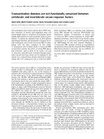
In brief, chromosomal regions that were most commonly
gained in both tumors and cell lines were 1q21.1-qtel, 5ptel5p13.3, 8p12-8q24.3, 17q12, 17q21-17q25.1 and 20q11-qtel.
Chromosomal regions that were most commonly lost in both
tumors and cell lines were 8ptel-8p12, 11q14-qtel, 13q21-qtel
and 17ptel-17p11.2. However, there were also notable differences between tumors and cell lines. Specifically, cell lines
showed a higher frequency of losses on chromosomes 9, 18
and X, and a lower frequency of losses on 16q, as compared
with tumors. On the other hand, tumors showed a higher frequency of gains on 16p. In agreement with [6] we observed
regions of recurrent high-level amplification on chromosomes 8, 11, 12, 17 and 20 (Figure 1a) bounding well-known
breast cancer oncogenes (e.g. BRF2, ASH2L, CCND1, EMSY,
ERBB2, NCOA3, MYBL2, STK6) [10,23,24], although amplification frequencies were much lower on chromosomes 12
and 20 as compared with those reported in [6]. In contrast,
cell lines did show amplification frequencies on chromosomes 12 and 20 that were more in line with those observed
in [6] (Figure 1b). We found homozygous deletion (HD) to be
a rare event in primary tumors and only found evidence of HD
in two cell lines and one tumor on chromosome 13q14 where
the retinoblastoma gene (RB-1) resides.
Common and minimal regions of alteration
To perform dimensional reduction we developed an extension (CRalg) of the minimal regions algorithm of Rouveirol
(MRalg) [25], which, in contrast to MRalg, identifies common
regions of alteration (CRA) (see Materials and Methods).
Using CRalg we achieved a substantial dimensional reduction
(from 27695 oligos to 5914 CRA that showed at least 5%
changes across tumors) without losing any information in the
process (note that the MRalg and CRalg algorithms will work
unchanged if instead of using 1 and -1 to indicate gain and
loss, we used the precise segment values; thus, CRalg achieves
a dimensional reduction without further information loss),
automatically including gains and losses in the same matrix.
However, a drawback of CRalg was the relatively larger
number of variables (5914 CRA compared with 1134 minimal
regions of alteration (MRA)) and the high degree of redundancy/correlation since many adjacent CRA only differed in
value in one sample. In order to reduce the redundancy of the
CRA matrix, we applied an algorithm that merged together
adjacent regions that differed in only a single sample (see
Materials and Methods). This gave a reduced matrix of 1063
merged CRA (mCRA) over 171 breast tumors.
Genome Biology 2007, 8:R215
/>
Genome Biology 2007,
Volume 8, Issue 10, Article R215
Chin et al. R215.4
Figure 1
Genome-wide frequency plots
Genome-wide frequency plots. Genome-wide frequency plot of gains (green), amplifications (darkgreen) and loss (red) over: (a), 171 primary breast
tumors; and (b), 49 breast cancer cell lines.
A subgroup of low GII
While standard hierarchical clustering algorithms have been
successfully applied to BAC-derived continuous log-ratio
data, we explored the possibility of incorporating the inherent
discreteness of copy number data into the unsupervised classification analysis. Specifically, we performed (complete linkage) hierarchical clustering over the matrix of mCRA using
the number of copy number state differences as a distance
metric. This revealed a complex pattern of gains and loss
across the cohort (Figure 2). Using the methodology implemented in the R-package pvclust [26,27] for testing the
robustness of the clusters, we found that only one reasonably
sized cluster of 26 samples was reliable with a robustness
index larger than 90% (Figure 2 and Additional Data File 4).
This cluster was characterized by a very low GII (average of
0.036 ± 0.035) relative to the rest of samples (average of 0.22
± 0.12), which was highly significant (Wilcoxon test p < 10-13).
We verified that this result was independent of cellularity by
showing that this cluster did not have a significantly lower
cellularity than the rest of samples (Wilcoxon rank sum test p
= 0.69). The 26-sample cluster was made up of proportionally
more ER-negative (15) than ER-positive tumors (11) (Fisherexact test p = 0.007) as well as more basal (6) than luminal
tumors (5) (Fisher-exact test p = 0.01), but was equally distributed in terms of histological grade (3 grade I, 10 grade II
and 13 grade III, p = 0.28), the immunohistochemical markers ERBB2, PGR, AR and p53, and p53 mutation status (21
samples with no p53 mutation and 4 with p53 mutation, p =
0.79). Among the 26 samples there were 8 with gains of
ERBB2 and 5 of these had a high-level ERBB2 amplification.
This confirms the observation made in [10] that a proportion
of ERBB2-amplifier tumors show little overall genomic
instability.
Two further, yet much smaller, clusters with robustness indices greater than 90% and of relatively high GII were also identified (Figure 2). The cluster with the highest GII was made up
of 9 samples and was mainly characterized by gains of 1q, 8q,
telomeric end of 17q and 20, and unaltered chromosome 16.
Most of the samples were ER negative (6 ER- versus 3 ER+)
and of high grade (7 grade III, 1 grade II and 1 grade I).
Another robust cluster of 12 samples and intermediate GII
Genome Biology 2007, 8:R215
/>
Genome Biology 2007,
Figure 2 (see legend on next page)
Genome Biology 2007, 8:R215
Volume 8, Issue 10, Article R215
Chin et al. R215.5
/>
Genome Biology 2007,
Volume 8, Issue 10, Article R215
Chin et al. R215.6
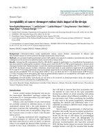
Figure 2 (see previous page)
Unsupervised clustering of 171 breast tumors
Unsupervised clustering of 171 breast tumors. (a), Hierarchical clustering over 1063 merged CRA using complete linkage and number of copynumber state differences as a distance metric. Clusters labeled in orange denote the largest stable clusters as determined by the pvclust algorithm. (b),
Associated sample distributions of intrinsic subtype based on the SSP classifier (sky blue, luminal-A; blue, luminal-B; green, normal; red, basal; pink, HER2),
ER status (black, ER-; gray, ER+), grade (black, grade III; blue, grade II; sky blue, grade I) and GII. (c), Heatmap of CRA (dark green, amplification; green,
gain; white, normal; red, loss).
was characterized mainly by loss of chromosome 17, loss of
16q and gain of 8p. This cluster was made up of 7 ER+ and 5
ER- samples and was also mostly high grade (7 grade III, 3
grade II and 2 grade I). The rest of samples could not be characterized as members of large stable clusters.
A novel subtype of ER- tumors of low genomic
instability
The identification of a subclass of breast tumors of low
genomic instability that was proportionally enriched in terms
of ER- and basal tumors was striking and suggested to us that,
in contrast to present belief, there is a subtype of ER- tumors
of relatively low genomic instability and which includes a subset of ERBB2-amplifier tumors. Further evidence for this
came from a Wilcoxon rank sum test comparing the GII distributions of ER- and ER+ samples, which showed that the
GII of ER- samples was not significantly higher than that for
ER+ samples (Figure 3a, p = 0.35). Importantly, among the
15 ER-samples within the 26 sample low-GII subgroup, 10
were of high grade, 4 of intermediate grade and only 1 of low
grade, which was proportionally similar to the distribution in
the rest of the ER- cohort (30 high grade, 9 intermediate
grade and 3 low grade tumors, p = 0.88). This showed that the
ER- samples in the low GII cluster were not necessarily of
lower grade.
To obtain further evidence for the existence of a low GII ERsubtype, we sought independent validation in three external
breast cancer cohorts [6,9,11] for which copy number data
was available. Specifically, we computed the GII in these
external cohorts as described in Materials and Methods and
tested, using a one-sided Wilcoxon rank sum test, whether
there was a substantial number of ER- samples of relatively
low GII (Figure 3b, c and 3d). Lending further support to the
existence of this low-GII subtype, in two of these external
cohorts [6,9] we did not find the GII of ER- samples to be significantly higher than that for ER+ samples.
In terms of the intrinsic subtype classification [28-30], for
which a single sample predictor (SSP) was recently derived
and validated in external cohorts [31], we found that the 26sample low-GII subgroup was made up of 6 basal, 3 HER2+,
4 luminal-A, 4 normal and 1 luminal-B tumors (8 samples
could not be classified owing to missing gene expression
information). As before, when taking into account all samples, the basal subtype did not have a significantly higher GII
than the luminal-A subtype (p = 0.44) (Figure 3e). We interpreted this result as further evidence for the existence of a
low-GII basal subtype. The only statistically significant differ-
ences between the GII distributions of the various intrinsic
subtypes were between the normal subtype and all others (p
< 0.05 for all comparisons) and between the luminal-A and
luminal-B subtypes (p = 0.009). We observed a similar GII
distribution in another cohort for which expression data was
available [6] (Figure 3f). Specifically, in this cohort as well,
the basal subtype did not have a significantly higher GII than
the luminal-A subtype (p = 0.26), while the luminal-B subtype did (p = 0.03).
The low-GII subgroup has an associated gene
expression signature
To further characterize the identified low-GII subgroup, we
attempted to derive an associated transcriptomic signature
from the 113 samples for which additional gene expression
information was available. To this end we used a multiple
logistic regression model and ranked genes according to the
difference of their model Akaike information criterion (AIC)
score [32] with respect to a null model AIC score that only
included ER status (see Materials and Methods). The null distribution for AIC scores was obtained by performing 10000
random permutations of the sample expression values.
Hence, this method allowed us to rank the genes according to
how well they discriminated between the 26-sample low-GII
cluster and the rest of the cohort, independently of ER status.
To correct for multiple testing we converted the p values into
q-values [33], which provided us with an estimate of the false
discovery rate (FDR). This showed that, for example, among
the top-50 genes we would expect on average about 10 false
positives, thus confirming the existence of an expression signature associated with this subclass.
To derive a classifier based on this gene signature we decided
on a linear discriminant classifier where class assignment is
determined by a nearest centroid criterion using an euclidean
distance metric. The centroids were constructed using the
top-37 genes (Additional Data File 5), yielding an average of 7
false positives. To test this classifier we first applied it to the
135 NCH samples with gene expression information [15]. This
classified 15 ER- and 9 ER+ into the putatively low-GII subgroup (25 ER- and 84 ER+ were classified into the other
group), which we verified had a lower GII than the rest of the
samples (Wilcoxon test p < 10-4). It is striking that even
though the classifier was derived independently of ER status,
classifying for this particular subgroup of low GII predetermined samples to be more likely ER- than ER+ (Fisher-exact
test p = 0.0003). Interestingly, applying the classifier to four
additional breast cancer cohorts with expression profiles
[6,34-36] showed that the corresponding putatively low-GII
Genome Biology 2007, 8:R215
/>
Genome Biology 2007,
(a)
Volume 8, Issue 10, Article R215
(b)
P=0.355
0.6
0.4
0.0
0.0
0.2
GII
0.4
0.6
P=0.388
0.2
GII
ER− (57)
ER+ (113)
ER− (18)
(c)
ER+ (27)
(d)
P=0.331
0.6
0.4
0.0
0.0
0.2
GII
0.4
0.6
P=0.001
0.2
GII
Chin et al. R215.7
ER− (46)
ER+ (84)
ER+ (29)
(f)
0.0 0.1 0.2 0.3 0.4 0.5 0.6 0.7
(e)
ER− (15)
Basal (19)
Her2 (14)
LumA (55)
LumB (13)
Normal (12)
Basal (32)
Her2 (14)
LumA (51)
LumB (17)
Normal (16)
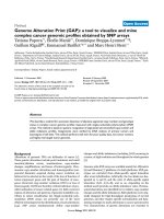
Figure 3
Distributions of genomic instability index
Distributions of genomic instability index. Boxplots of the distribution of GII across different ER and expression subtypes: (a), (e), NCH cohort;
(b), Naylor's cohort [9]; (c), (f), CAL cohort [6]; (d), Loo's cohort [11].
subgroup in these cohorts was also enriched for ER- tumors
(Additional Data File 6). In a further data set [30] where only
85 samples (18 ER- and 56 ER+) were available the predicted
low-GII subclass had only 4 ER+ and 3 ER- samples (and 3
samples had missing ER information), which did not reach
statistical significance, but suggested to us that it possibly
would if more samples were available.
Fortunately, the tumors in [30] were profiled recently at the
copy number level [12]. This allowed us to validate the hypo-
thesis that our gene expression classifier selects a particular
subtype of low GII in breast cancer. Using the GII for the samples in this cohort we compared the GII of the predicted lowGII subgroup, as determined by our expression classifier,
with the rest of samples in that cohort (Figure 4a and 4B). In
spite of only 10 samples being classified into the predicted
low-GII subgroup, we could verify that it was characterized by
a lower GII when compared with the rest of the samples (Wilcoxon test p = 0.001). Moreover, among these 10 samples, 6
were of high grade, 2 of intermediate grade and none were of
Genome Biology 2007, 8:R215
/>
0.4
(b)
0.4
(a)
Genome Biology 2007,
0.2
0.0
0.0
0.2
P=0.001
−0.5 0.0
0.5
1.0
1.5
2.0
rest
lowGII
(c)
0.4
0.2
0.0
0.0
0.2
0.4
P=0.135
−2
−1
0
1
2
rest
lowGII
Figure 4
Genomic instability index versus LD-scores
Genomic instability index versus LD-scores. (a), (c), GII is plotted
against the linear discriminant (LD) scores for the 86 samples profiled in
[12] and the 101 samples of the CAL cohort [6]. Those samples with a
negative LD score were classified into the low-GII subgroup (red), the rest
are shown in blue. (b), (d), Corresponding boxplots showing the GII
distributions of the two predicted subgroups.
low grade (2 samples had missing information). For the other
external cohort for which both copy number and expression
data was available [6], the predicted low-GII subgroup had a
lower median GII than the rest of samples, but did not reach
statistical significance (Figure 4c and 4D).
To better understand the nature of the expression classifier
we performed both gene ontology (GO) analysis using GOTM
[37] and pathway analysis using MSigDB [38]. GOTM on the
37 genes making up the classifier showed enrichment of
inflammatory and defense response genes (CXCL1, CXCL2,
XCR1, LY96, NMI, TLR2, uncorrected p < 10-5), which were
generally upregulated in the low-GII subgroup, and marginal
enrichment of signal transduction (RASSF2, SNX4, CASP1,
MKNK1, RHPN1, INPP5D, uncorrected p = 0.002) and apopotosis genes (BCL2A1, MRPS30, CASP1, CASP4, TLR2,
uncorrected p = 0.004). Pathway analysis using MSigDB confirmed the involvement of the caspase, cell death, TNF-α-NFκβ, inflammatory response and signalling pathways, although
these statistical associations were lost on correction for multiple testing (data not shown).
Gene expression and copy number
Of the 171 breast tumors, 113 were also profiled on Agilent
gene expression arrays [15]. This allowed us to evaluate the
contribution of gene-dosage levels to gene expression (Additional Data File 7). Of the 5914 CRA, 4551 (77%) contained at
Volume 8, Issue 10, Article R215
Chin et al. R215.8
least one Agilent probe. Of these 4551 CRA, 2407 harbored at
least one Agilent probe for which there was at least 10 (~5%)
expression values in the altered (i.e. gained or lost) group of
samples (note that owing to missing values in the gene
expression data, p values could not be reliably computed for
many probes). Thus, for 2407 CRA at least one reliable p
value (Wilcoxon test) could be computed (see Materials and
Methods) to evaluate the significance of the association
between copy number and aberrant expression. We found
that from the 2407 CRA, there were 806 CRA for which there
was at least one probe with significant association (p < 0.05)
between gain and overexpression, and 412 for which there
was at least one probe with significant association between
loss and underexpression. On average about 34% of probes in
regions that were gained in at least 5% of samples were significantly overexpressed relative to the samples that showed no
copy number alteration. Similarly, about 29% of probes in
regions that were lost in at least 5% of the samples were significantly underexpressed relative to the samples that showed
no copy number alteration. This confirms the finding
reported elsewhere [1] that a significant proportion of gene
expression variation is caused by underlying copy number
alterations.
Hotspots of association between copy number and
expression
To find the CRA showing the strongest associations between
copy number and expression we first tabulated those CRA
with at least 10% gains or losses and which showed a significant association with expression (p < 0.05; see Additional
Data File 8). To narrow this down to a smaller set of the most
significant regions ('hotspots') we next selected those CRA
with an association index (AI) value larger than or equal to
0.5 and a most significant p value of less than 0.001, where
the AI was defined as the fraction of probes within the CRA
that had significant p values (see Methods). This yielded 196
and 63 hotspots that showed significant association with
overexpression and underexpression, respectively (Additional Data File 9). In the case of loss and associated underexpression this table included the well-known tumor
suppressors RB1, CDH1, MBD2 and EP300, while in the case
of gain and overexpression it included many well-known and
potentially novel oncogenes such as MUC1 on 1q21.3, ASH2L,
BRF2, LSM1 on 8p12, FADD on 11q13, ERBB2, PNMT, GRB7
on 17q12, TOP2A, THRA, NR1D1 on 17q21, and NCOA6,
YWHAB, UBE2C on 20q13. Of these candidate oncogenes,
several, notably TOP2A, PNMT and UBE2C, have appeared in
prognostic gene expression signatures [39-41], thus reemphasizing their important role in breast cancer. Among the
hotspots that were gained, we provide a further selection of
those that also showed frequent amplifications and which are
therefore likely to harbor candidate oncogenes (Table 2).
Hotspots associated with outcome
As the identified 196 and 63 hotspots represent the regions of
strongest association between copy number and coordinate
Genome Biology 2007, 8:R215
/>
Genome Biology 2007,
Volume 8, Issue 10, Article R215
Chin et al. R215.9
Table 2
Hotspots of gain and amplification
CytoBand
Start
Length
Gains (T)
nAMP (T)
Gains (CL)
nAMP (CL)
Genes
1q21.1
144.22
1.65
0.49
10
0.37
0
RBM8A, POLR3C, ZNF364
1q21.3-1q22
153.29
0.09
0.51
8
0.39
0
EFNA4, MUC1
1q23.2
157.95
0.32
0.53
10
0.39
1
DUSP23, IGSF9
1q23.3
159.23
<0.01
0.55
12
0.43
1
F11R
1q42.13-1q42.2
226.95
2.59
0.57
12
0.45
0
SPHAR, NUP133, GALNT2
1q43-1q44
240.32
4.75
0.58
11
0.45
0
SDCCAG8, ADSS, FAM36A
6q21
107.12
<0.01
0.11
5
0.02
0
AIM1
6q21
107.14
0.2
0.11
4
0.02
0
RTN4IP1, QRSL1
8p12
37.82
<0.01
0.32
16
0.24
4
GPR124, BRF2
8p12
38.04
0.1
0.32
17
0.22
3
ASH2L, LSM1
8p12
38.24
0.06
0.32
18
0.24
3
WHSC1L1
8q21.13
82.73
0.14
0.47
13
0.37
1
ZFAND1, CHMP4C
8q21.3
87.54
0.48
0.49
15
0.39
1
FAM82B, CPNE3
8q22.3
102.57
1.37
0.54
17
0.51
1
GRHL2, RRM2B, EDD1*, AZIN1*
8q22.3
104.52
<0.01
0.54
15
0.51
1
WDSOF1*
8q24.11
118.02
0.58
0.54
23
0.61
4
THRAP6*
8q24.12
120.81
0.1
0.53
23
0.57
3
DCC1*
8q24.13
124.90
1.14
0.53
22
0.65
3
TRMT12*, RNF139*, NDUFB9*
8q24.13
126.06
0.28
0.53
23
0.65
4
SQLE*, KIAA0196*
8q24.3
144.53
0.09
0.40
12
0.39
0
RHPN1, ZC3H3
9p22.3-9p22.1
16.40
2.65
0.11
1
0.12
1
C9orf39, FAM29A
10p14
12.20
0.05
0.20
2
0.18
0
NUDT5, SEC61A2
10p13
12.33
2.66
0.20
3
0.18
0
OPTN, FAM107B, SUV39H2
11q13.3
69.73
<0.01
0.17
11
0.33
2
FADD
11q13.3
69.90
<0.01
0.16
8
0.31
1
PPFIA1
11q14.1
77.01
0.38
0.12
8
0.24
3
CLNS1A, INT4
11q14.1
77.47
0.16
0.12
10
0.29
3
NDUFC2*, ALG8*, USP35*
16p13.2-16p13.13
8.78
1.68
0.38
3
0.06
0
ABAT, PMM2, USP7
16p12.3
18.44
1.21
0.39
3
0.10
0
NOMO2, ARL6IP, MIR16
17q12
33.73
<0.01
0.16
9
0.08
3
MRPL45
17q12
34.15
0.02
0.19
15
0.12
4
PSMB3
17q12
34.19
0.02
0.19
15
0.12
4
CCDC49
17q12
34.33
0.01
0.20
16
0.12
4
LASP1
17q12
34.67
0.01
0.23
23
0.12
4
FBXL20
17q12
35.06
0.02
0.27
25
0.29
11
STARD3, TCAP, PNMT
17q12
35.08
0.06
0.27
27
0.29
12
ERBB2, GRB7
17q12
35.29
0.03
0.23
21
0.27
10
GSDML
17q21.1
35.44
0.07
0.16
15
0.16
5
THRAP4, NR1D1
17q21.2
35.77
0.03
0.13
6
0.06
1
TOP2A
17q21.32
44.27
0.1
0.17
9
0.22
3
ATP5G1, UBE2Z
17q21.33
44.95
0.19
0.18
6
0.18
2
SPOP, SLC35B1
17q21.33
45.80
0.02
0.19
7
0.20
2
MRPL27, LRRC59
17q22
54.12
0.44
0.19
6
0.20
2
RAD51C, TRIM37
17q23.1
55.12
0.01
0.23
9
0.20
2
CLTC, PTRH2
17q23.1
55.38
0.1
0.23
8
0.20
3
RPS6KB1, ABC1
17q23.3
58.97
0.29
0.23
6
0.20
1
CCDC44, DDX42, FTSJ3
17q24.2
62.77
<0.01
0.24
7
0.33
1
PSMD12
17q25.1
68.72
0.03
0.22
4
0.18
0
COG1, C17orf80
Genome Biology 2007, 8:R215
/>
Genome Biology 2007,
Volume 8, Issue 10, Article R215
Chin et al. R215.10
Table 2 (Continued)
Hotspots of gain and amplification
17q25.1
70.52
0.03
0.17
5
0.16
0
ICT1, ATP5H
17q25.1
70.57
0.2
0.16
5
0.16
0
HN1, NUP85, MRPS7
17q25.1
71.15
0.15
0.17
4
0.16
0
ITGB4, H3F3B
20p11.23
17.97
1.51
0.25
3
0.16
0
ZNF133, RBBP9, HARS2
20q13.12
42.95
0.05
0.27
2
0.29
2
YWHAB
20q13.12
43.77
0.2
0.26
1
0.29
0
DNTTIP1, UBE2C, NEURL2
20q13.13
46.43
0.86
0.28
2
0.29
1
CSE1L, STAU1
20q13.33
60.21
0.53
0.28
3
0.31
0
HRH3, ADRM1, LAMA5
20q13.33
61.45
0.2
0.27
3
0.18
0
EEF1A2, C20orf149, PTK6
20q13.33
62.04
0.17
0.27
3
0.20
1
UCKL1, C20orf14, OPRL1
21q22.3
42.85
0.47
0.13
2
0.08
0
SLC37A1, WDR4, NDUFV3
21q22.3
46.49
0.2
0.12
2
0.06
0
MCM3AP
Selected regions of frequent gain/amplification and strong coordinate overexpression. Columns give cytoband, start position (Mb), length (Mb),
frequency of gains across 171 tumors (T), number of amplifications in 171 tumors (T), frequency of gains across 49 cell lines (CL), number of
amplifications in 49 cell lines, selected genes in region showing strong coordinate overexpression. * denotes genes significantly associated with either
overall survival, time to distant metastasis or disease free interval (p < 0.05).
aberrant expression, it was natural to investigate whether any
of these regions also showed association with clinical outcome. To this end we performed univariate Cox proportional
hazard regressions comparing the HRs for samples with gain
(loss) of a hotspot with samples without altered hotspots for
three different outcome endpoints (overall survival (OS), disease free interval (DFI) and time to distant metastasis
(TTDM); see Additional Data File 9). In addition, we performed Cox regressions for those hotspots with at least 10
amplifications and estimated the HR for samples with and
without amplification (Additional Data File 9). This analysis
showed that there were three cytoband regions of frequent
amplification (8q22.3, 8q24.11-8q24.13 and 11q14) and associated with either OS or TTDM (log-rank test p < 0.05; see
Table 3). We verified that for all of these regions samples with
amplification had approximately a twofold risk increase of
poor outcome compared with samples without the amplification (Table 3). Interestingly, 37 tumors had amplifications in
any one of these hotspot regions and this subgroup had also a
much higher risk of distant metastasis and a poorer survival
rate compared with samples without amplification (TTDM
HR = 3.0 (1.6-5.8) p < 10-3; see Table 3). Genes located in
these hotspots, which also showed coordinate overexpression, included EDD1, WDSOF1 (8q22.3), THRAP6 (8q24.11),
DCC1 (8q24.12), SQLE, KIAA0196 (8q24.13) and NDUFC2,
ALG8, USP35 (11q14.1) (Tables 2 and 3 and Additional Data
File 9). Importantly, multivariate Cox regression analysis in a
model that included the NPI (one of the strongest prognostic
factors in univariate analysis), showed that amplification of
any of these five regions was a strong prognostic factor independently of NPI (Table 4).
Gene families
Next, we investigated the patterns of gain and loss for particular gene families, including the kinome [42], the phosphatome [43], a selected set of chromatin binding proteins
and chromatin modifier enzymes that we collectively called
'chromatinome', and the list of somatically mutated breast
Table 3
Amplification hotspots associated with clinical outcome
Cytoband
Genes
nAMP
OS HR (95% CI) p value
TTDM HR (95% CI) p
value
8q22.3
EDD1, AZIN1
17
2.2 (1.1-4.6) 0.02
2.1 (0.9-5) 0.09
8q22.3
WDSOF1
15
2.2 (1.1-4.7) 0.03
1.9 (0.7-4.9) 0.17
8q24.11
THRAP6
23
1.9 (1-3.8) 0.04
2.1 (0.9-4.5) 0.06
8q24.12
DCC1, DEPDC6
23
1.9 (1-3.7) 0.05
2.1 (0.9-4.5) 0.06
8q24.13
SQLE, SPG8
23
2 (1-3.9) 0.03
2.2 (1-4.7) 0.05
11q14.1
NDUFC2, ALG8, USP35
10
2.5 (1.1-6) 0.03
2.4 (0.8-6.8) 0.09
5-amp
5-amp
37
2.6 (1.5-4.6) 3 × 10⁻⁴
3.0 (1.6-5.8) 5 × 10⁻⁴
Amplification hotspots significantly associated with either overall survival (OS) or time to distant metastasis (TTDM). The cytoband locations, genes
located in amplified hotspots that showed coordinate overexpression, the number of samples with amplification (nAMP), hazard ratio (HR), 95%
confidence interval and log-rank test p values are given.
Genome Biology 2007, 8:R215
/>
Genome Biology 2007,
Table 4
underexpression relative to samples with no loss. The analysis for preferential selection for genomic changes showed that
phosphatases were more frequently lost (p < 0.05), while
gains were not selected (p = 0.96).
Univariate and multivariate survival analysis
Factor
OS HR
(95% CI) p value
TTDM HR
(95% CI) p value
ER
1.7 (0.9-2.5) 0.10
1.7 (0.8-3.3) 0.13
p53mut
1.7 (0.9-3.0) 0.09
2.1 (1.1-4.2) 0.03
LN
2.4 (1.4-4.0) 8 × 10⁻⁴
4.3 (2.3-8.3) < 10-4
Volume 8, Issue 10, Article R215
Chin et al. R215.11
Chromatinome
Size
1.3 (1.0-1.7) 0.03
1.3 (0.9-1.7) 0.16
Grade
2.0 (1.2-3.4) 0.009
1.8 (0.9-3.3) 0.08
NPI
2.5 (1.4-4.3) 0.001
2.8 (1.4-5.8) 0.003
5-amp
2.6 (1.5-4.6) 3 × 10⁻⁴
3.0 (1.6-5.8) 5 × 10⁻⁴
5-amp*+NPI
2.3 (1.3-4) 0.003
2.6 (1.4-5.1) 0.004
5-amp+NPI*
2.2 (1.2-3.9) 0.008
2.5 (1.2-5.1) 0.02
Univariate and multivariate Cox proportional hazards analysis with
overall survival (OS) and time to distant metastasis (TTDM) as
endpoints. Univariate analysis was performed for ER status (1, negative;
0, positive), p53 mutation (1, mutant; 0, wild-type), lymph node status
(1, positive; 0, negative), size (1, ≥2.5 cm; 0, <2.5 cm), grade (1, high or
intermediate; 0, low), NPI (1, ≥3.8; 0, <3.8) and 5-amp (1, amplification
in any of the five regions; 0, no amplification in any region). * indicates
the corresponding hazard ratio estimate in the multivariate model that
included 5-amp and NPI.
cancer genes (CAN genes) [44]. The relevance of these gene
families and gene sets for cancer biology is well known
[42,44,45]. Specifically, we investigated whether there was
preferential selection for genomic changes among these gene
sets (see Materials and Methods), and also which genes
showed significant coordinate aberrant expression.
CAN genes
Of the 122 genes that were shown to be somatically mutated
at a higher frequency in breast cancer [44], 121 were found on
the oligo CGH array. As expected, many of the CAN genes
(e.g. TP53, TMPRSS6 and APC2) were frequently lost, but
many also showed frequent gains (e.g. PTPN14, NCOA6 and
HOXA3; see Additional Data File 10). Analysis of preferential
selection for genomic changes showed, not unexpectedly, that
CAN genes were more frequently lost in comparison with random selections of 121 genes with the same chromosome
distribution (p < 0.05), while there was no preferential
selection for gains (p = 0.84). Of the 121 genes on the aCGH
array, 108 were also mapped on the Agilent array and 9
showed significant association between expression and copy
number, including NCOA6, OBSCN and DDX10 (Additional
Data File 11).
Phosphatome
Of the 107 phosphatases described in [43], 90 were mapped
onto the oligo CGH and Agilent arrays and 10 showed significant association between copy number and expression (Additional Data File 11). Among the class I Cys-based protein
tyrosine phosphatases (PTPs), the subclass of 16 myotubularins were frequently lost in comparison with the rest of phosphatases, with MTMR2 also showing coordinate
We compiled a list of 503 histones, chromatin binding proteins and chromatin modifier enzymes, of which 440 were
also found mapped on the Agilent array. These genes did not
show preferential selection for either gains (p = 0.67) or
losses (p = 0.97). Of these 440, 51 showed significant association between copy number and expression (Additional Data
File 11). For example, we found that HDAC2 and ASH2L
showed coordinated aberrant expression in samples for
which the gene was either gained or lost, while samples with
gains of CREBBP and SUV39H2 showed significant overexpression compared with samples that showed no corresponding copy number alteration. In addition, EP300 was found to
be lost in over 20% of tumors with a corresponding significantly lower expression compared with tumors for which the
gene copy number was not altered. These observations are
particularly noteworthy given that chromatin modifiers are
infrequently mutated in breast cancer [46-48].
Kinome
Of the 518 kinases reported in [42], 477 were found to be in
CRA and 268 of these were also mapped on the Agilent array.
Out of these 268 kinases, 32 exhibited significant association
between copy number and gene expression (Additional Data
File 11). Notably, ERBB2 showed the strongest association
between copy number gain and overexpression, followed by
kinases on chromosome 1, CLK2 and SCYL2, and RIPK2 on
chromosome 8. As far as loss and underexpression is concerned, the strongest associations were found for MAP2K4,
NEK3, TESK1 and MLKL on chromosomes 17, 13, 9 and 16,
respectively. Of note, we observed that the association of
MAP2K4 loss with underexpression is consistent with
observations that it may play a role as a tumor suppressor
[49]. While, individually, kinases were frequently altered and
showed coordinate gene expression changes, we did not
observe any differences in the frequency of alterations
between the nine kinase families AGC, CAMK, CK1, CMGC,
RGC, STE, TK, TKL and Atypical, nor preferential selection
for gains (p = 0.96) or losses (p = 0.17).
Discussion
Many mRNA profiling studies have established that breast
cancer is a highly heterogeneous disease with at least five
identified 'intrinsic' subtypes [30,50], and recent evidence
points at the likely existence of additional biologically and/or
clinically relevant subtypes [31,51]. As changes in copy
number drive a considerable proportion of the changes at the
transcriptomic level [1], it is likely that the aberration landscape underlying breast cancer at the copy number level is of
an even far more complex nature than that observed at the
Genome Biology 2007, 8:R215
/>
Genome Biology 2007,
mRNA level. Exacerbating this complexity further is the fact
that a significant proportion of genomic aberrations are
totally unrelated to cancer physiology and merely reflect random events that differ between any two normal specimens
[52].
two external cohorts for which both expression and copy
number data was available [12]. Moreover, using additional
independent expression data sets we were able to show that
the expression classifier selects mostly ER-tumors. When
combined, these results provide strong evidence that the
derived transcriptomic signature is a classifier of low-GII ERsamples. We can only speculate as to why the expression classifier did not select for low-GII samples in the other external
cohort for which both copy number and expression data was
available [6], although in agreement with the other studies it
did select for ER- samples (Additional Data File 6). One possible explanation could be the much higher GII values of the
cohort in [6] compared with those in our cohort (Figure 3).
Interestingly, we also found that the median tumor size was
considerably larger in [6] compared with our cohort (2.2 cm
compared with 1.8 cm), which was highly significant under a
Wilcoxon rank sum test (one-sided test p < 10-5). Thus, by
selecting a panel of relatively large ER+ and ER- tumors, the
study in [6] may have missed out on this ER- subtype of low
GII. Similarly, the tumors profiled in [11] were significantly
larger than those in our cohort (Table 1) and, correspondingly, we also observed relatively higher GII values in their
cohort (Figure 3). To investigate this possibility further we
asked whether there was a significant correlation between
tumor size and genomic instability (Additional Data File 12).
Strikingly, in our cohort as well as the cohorts in [6] (CAL)
and [11] (Porter) we observed a step-like structure in the distribution of GII and tumor sizes (Additional Data File 12).
Without exception, we observed that there were no tumors of
sizes larger than 2.5 cm and GII values lower than 0.1. Using
2.5 cm as a cut-off, we verified that the GII of tumors of larger
size (i.e. ≥2.5 cm) were significantly higher than those of
smaller tumors (<2.5 cm) (Wilcoxon rank sum test, p = 0.017
(NCH), p = 0.018 (CAL), p = 0.18 (Porter)). Together these
findings indicate that our identification of a low-GII subgroup
was facilitated by the smaller sizes of the NCH cohort in comparison with the cohorts profiled elsewhere. A similar
observation could also be made in relation to the study in [13],
which profiled significantly larger tumors and estimated only
10% of tumors to have 'flat' (i.e. low-GII) profiles, in comparison with the 30% of tumors with a GII of less than 0.1 in the
NCH cohort. (This must be interpreted with caution as the
authors in [13] did not define their 'flat' profiles in terms of
GII values.)
The aCGH study presented here is the largest study to date to
combine copy number and expression data, having profiled
171 primary breast tumors with a high-density oligo array,
and while it confirms the findings reported recently in
[6,10,13], it also shows that breast cancer is a more heterogeneous disease than is portrayed by these previous studies.
Specifically, we found, using hierarchical clustering with a
novel distance measure, only three robust clusters of 10 or
more samples, the largest of which, with 26 samples, was
characterized by a low GII and was surprisingly enriched for
ER- and basal samples. The other two clusters also consisted
mainly of intermediate/high grade ER- tumors, but were
characterized by a high GII. These findings suggested to us
the existence of a high-grade ER-/basal subgroup of low GII.
In agreement with this conclusion, we observed in two additional independent cohorts that ER-tumors, despite being of
higher grade than ER+ tumours, did not have a higher GII. An
analogous result was also obtained when considering the
basal and luminal status of the tumors. Moreover, while in
ER+/luminal tumors a subdivision into high and low GII can
be explained by the differential distribution of histological
grade (larger GII for high-grade tumors) [6], no such grade
association seems to explain the variability/bimodality in GII
that is observed for ER- tumors. It is also noteworthy that
while the subdivision into high and low GII that is observed
for ER+ tumors correlates with clinical outcome and with the
luminal-A and luminal-B subtypes, no such correlation with
clinical outcome is observed in the case of ER- tumors.
More generally, we investigated the distribution of other clinical phenotypes (age, tumor size, vascular invasion, NPI,
lymph node status, distant metastasis, overall survival, p53
mutation status and the immunohistochemical markers PGR,
ERBB2, p53 and AR) in the 26-sample low-GII cluster relative to the rest of the cohort, as well as the differential distribution of the same clinical factors among the two groups
when restricted to ER- samples only. No strong associations
were found, although the low-GII subgroup was proportionally enriched for lymph node negative (LN-) patients: 22 LNand four 4 LN+ in the low-GII subgroup relative to 98 LNand 47 LN+, Fisher p = 0.10, when considering all samples;
and 13 LN- and 2 LN+ in the low-GII subgroup relative to 25
LN- and 18 LN+, Fisher p = 0.06, when restricted to ER- samples only.
Thus, in order to better characterize the identified low-GII
subgroup of 26 samples we derived an expression classifier
using a subset of 113 samples for which expression data was
available. The expression classifier was derived independently of ER status and was successfully validated in one of the
Volume 8, Issue 10, Article R215
Chin et al. R215.12
Gene ontology analysis of the 37-gene expression classifier
showed marginal statistical associations with inflammatory
response, apoptosis and signal transduction genes. Similarly,
pathway analysis showed that the most enriched pathways
were those related to caspase activity, cell death, NFκB,
immune function and signal transduction. Interestingly,
BCL2A1, a known transcriptional target of NFκB, was found
to be upregulated in the low-GII subgroup, which is consistent with the observed upregulation of the inflammatory
response genes (e.g. CXCL1, CXCL2, LY96) which may mediate the NFκB activation.
Genome Biology 2007, 8:R215
/>
Genome Biology 2007,
The combined copy number expression analysis further confirmed the presence of many genomic regions with expression
aberrations that are driven by underlying copy number
changes [1,6]. Of the nine candidate therapeutic targets
reported to be frequently amplified and deregulated at the
expression level [6], we were able to verify six of these
(IKBKB, ERBB2, ADAM9, FNTA, PNMT and NR1D1) (Additional Data File 8). Of these, ERBB2, FNTA, PNMT and
NR1D1 were located in hotspots that showed particulary
strong associations between copy number gain and overexpression (Additional Data File 9). Interestingly, however,
hotspots that were frequently amplified and that were associated with clinical outcome did not include the regions 8p11-12
and 17q11-12 reported in [6]. Instead, we found that hotspots
associated with either survival or time to distant metastasis
were located on cytobands 8q22.3, 8q24.3, 8q24.11-13 and
11q14, involving other important breast cancer genes such as
EDD1, WDSOF1 (8q22.3), THRAP6 (8q24.11), DCC1
(8q24.12), SQLE, KIAA0196 (8q24.13) and NDUFC2, ALG8,
USP35 (11q14.1) (Table 2 and Additional Data File 9). Specifically, SQLE expression has been shown to be a robust prognostic marker [39,50], and WDSOF1 was part of the gene
expression predictor derived in [53]. The genes on cytoband
11q14.1, NDUFC2, ALG8 and USP35, also reside close to what
appears to be a novel amplicon in acute myeloid leukemias
(AML) [54]. The different clinically relevant hotspot regions
identified here in comparison with those found in [6] may be
a consequence of the different clinical characteristics of the
two cohorts, but more likely it reflects the substantial differences in treatment (Table 1). Specifically, in the 'NCH' cohort
only 53% of tumors received either hormone or chemotherapy (and only six, i.e. 4%, received chemotherapy) in comparison to the 'CAL' cohort where almost 90% of patients
received treatment (Table 1). Thus, the combined analysis of
copy number, expression and clinical outcome variables in a
patient population with almost 50% untreated cases and better overall prognostic variables, has identified potentially
novel clinically relevant amplicons in breast cancer.
Materials and methods
Conclusion
By profiling a large panel of relatively small and low-NPI
breast tumors that is representative of breast cancer demographics in the UK we have shown that high-grade ER-/basal
breast cancer can be subdivided into two subclasses of low
and high genomic instability. In addition, we provide a comprehensive list of hotspot genomic regions that show strong
correlation between copy number and expression, and have
identified novel candidate amplicons associated with poor
prognosis independently of standard prognostic factors,
including the NPI.
Volume 8, Issue 10, Article R215
Chin et al. R215.13
Primary tumor genomic DNA and cell lines
Primary breast tumor specimens were obtained with appropriate ethical approval from the Nottingham Tenovus Primary Breast Cancer Series. All 171 cases were primary
operable invasive breast carcinomas collected from 1990 to
1996. Whole tissue sections (tumor cellularity range 20100%) were used for DNA extraction. Detailed clinical data
for this cohort is available (Additional Data File 1). The 49
breast cancer cell lines were obtained from the American
Type Tissue Collection (Manassas, VA) or were generously
provided by their originator. The cell lines were cultured
according to the culture conditions recommended by their
providers. The normal reference pools were established using
peripheral blood from 10 anonymous donors (with ethical
approval).
DNA isolation and labelling
DNA was extracted from 20 sections of 30 μm from each
tumor using the Promega DNA Wizard kit (Promega, UK)
according to manufacturer's instructions. DNA was extracted
from cell lines and peripheral blood leukocytes using standard SDS/Proteinase K method. DNA was quantified with a
NanoDrop ND-1000 spectrophotometer (NanoDrop Technologies, Wilmington, DE, USA). DNA labelling was performed using the BioPrime DNA labelling kit reagents
(Invitrogen) and according to protocols described previously
[14].
aCGH data preprocessing and normalization
Labelled DNAs were hybridized to customized oligonucleotide microarrays containing 30000 60-mer oligo probes
[14], for which 27801 unique map positions were defined
(Human Mar. 2006 assembly (hg18)). The median interval
between mapped elements was 39.4 kb, 75% of intervals were
less than 104.2 kb and 95% were less than 402 kb. Fluorescence ratios of scanned images of arrays were obtained using
BlueFuse version 3.2 (Bluegnome). Raw aCGH profiles of 171
breast tumors and 49 cell lines were then processed using the
R/Bioconductor package limma [55]. Mode normalizations
were subsequently carried out for all arrays. The raw and
mode-normalized data for the 171 tumors and 49 breast cell
lines is available from NCBI's GEO [16-18] under the series
accession number GSE8757.
Identification of copy number transitions
The normalized aCGH data was then segmented using the
CBS algorithm [19] as implemented in the R-package DNAcopy [19]. The CBS algorithm parameters used were: number
of permutations 5000, window size 500 and overlap 0.5.
Next, we fitted a density to the distribution of segmented state
values and verified that the resulting mode was close to zero.
The segmented data was then recentered by shifting the position of this mode to zero. This yielded a matrix of segment values for 27801 unique probes and 171 tumor samples.
Genome Biology 2007, 8:R215
/>
Genome Biology 2007,
Thresholds for calling gains, losses, amplifications and
deletions
the high-resolution oligo-array to the 281 BAC clones in the
Genosensor array, 34 BACs were found to contain at least 5
oligos. Concordance between oligo and BAC arrays was evaluated by examining their discrete copy number states in the
matching regions. DNA copy number status (gain(1), loss(-1),
normal (0)) for both oligo and BAC arrays were assigned for
the above 34 matching regions/clones. A Fisher-exact test
was then used to determine the association between the two
types of arrays for each of the 34 matching regions.
Having identified the segments and the baseline of unaltered
copy number, we next applied an extension of the algorithm
in [5] for calling gains and losses. As the cellularity of the
tumor samples varied significantly across the cohort, we
extended Aguirre's method to take the cellularity of the samples into account. Thus, sample-specific thresholds were
obtained. Specifically, the procedure used was as follows.
1. The mode-normalized log-ratios were first transformed
back to ratios. The ratio values for sample s, Rgs, were then
corrected for sample cellularity cs, by the transformation
R gs =
1
( R gs − (1 − c s ))
cs
2. Next, we log2 transformed back the corrected ratio values
and computed the standard deviation, σs, of the middle 50%
quantile of the data.
3. A threshold for calling gain and loss for sample s was then
defined as t s = ±2σ s .
Note that for the estimation of the thresholds only the middle
50% quantile of the data is needed, thus in step 1 above negative R gs values were generally avoided. Subsequently, the
transformation in step 1 was applied to all of the inverse log2
transformed segment values providing a further regularization of the data.
In a few cases where negative values were obtained, given the
monotonicity of the transformations, these states were
treated as a loss of copy number.
For amplifications we used a threshold of 8σs (i.e. four times
the threshold for one-copy gains). With this choice of thresholds we verified that ERBB2 had frequencies of gain and
amplification of 0.27 and 0.15, respectively, which are close to
the frequency values quoted in previous studies [1,10].
In the case of cell lines, thresholds for gain and loss were
defined at ± 0.25 on a log2 scale and were close to the average
threshold values over cell lines obtained by the above procedure using cs = 1 (specifically, the average was 0.20 was gains
and -0.27 for losses). As before, the amplification threshold
was defined as four times the threshold for gain (i.e. at 1 on a
log2 scale).
Concordance between oligo and BAC arrays
Of the 171 breast tumors, 126 had been previously profiled on
a Genosensor (Vysis, Downer's Grove, USA) BAC array [10]
for DNA copy number aberrations. This BAC array contained
281 unique BAC clones representing cancer-related loci.
When we matched the locations of the 27801 unique clones in
Volume 8, Issue 10, Article R215
Chin et al. R215.14
CRA and MRA
The matrix of segmented values is not useful for many of the
downstream analyses, such as candidate oncogene identification and unsupervised classification. Hence, from the matrix
of segmented values, we derived different data matrices with
different downstream applications in mind.
For the purpose of identifying a list of candidate oncogenes
and tumor suppressors, we applied the algorithm of [25] to
define the minimal regions of gain and loss (MRG and MRL).
This algorithm requires as input matrices of gains and losses
over all of the probes, which we constructed using the thresholds for gain and loss as described above. As explained in [25],
the minimal regions define the shortest regions that are commonly gained or lost across the cohort. While this algorithm
captures those regions most likely to harbor candidate oncogenes and tumor suppressors, the algorithm may also fail to
capture known oncogenes or tumor suppressors. This can
happen due to several reasons. One reason is the sensitivity of
the algorithm to errors in the segmentation algorithm or perturbations in the sample set. Alternatively, it might also fail to
capture more complex patterns of amplification or deletion
involving multiple neighboring targets. Thus, in addition to
deriving the minimal regions we also applied a different algorithm (CRalg) which considers all breakpoints equally. Similar to the algorithm of [25] (MRalg), it captures the regions
that are commonly gained or lost across the cohort, but in
contrast to MRalg, it not only captures the minimal regions
but also all other, usually adjacent, regions of gain and loss.
Specifically, following the notation of [25], we have the following theorem that applies to CRalg.
Theorem 1 A region r = [in ... out] is a CRA if and only if
(i) 'in' and 'out' are breakpoints; and
(ii) there is no breakpoint b such that in Thus, CRalg encapsulates all of the information from the
matrix of segmented values into a much smaller number of
variables, while MRalg loses potentially important information. Clearly, many adjacent CRA will be highly correlated,
only differing in value across one of the samples. In order to
remove this redundancy we applied a merging step to the
CRA. Thus, adjacent regions that only differed in value in one
sample were merged together. For every sample, we defined
Genome Biology 2007, 8:R215
/>
Genome Biology 2007,
the value for the newly merged region as the median value
over all of the regions merged together. Thus, if three regions
are being merged with values (1, 1, 0) for that sample, the
newly merged region would have value 1. If only two regions
are merged with values say (1, 0) then a value of 0.5 would be
assigned for the newly merged region. Thus, this approach
allowed us to reduce the number of correlated variables significantly, while also retaining as much information as
possible.
between dosage and expression levels by comparing the distribution of expression values for altered (i.e. either gained or
lost) versus unaltered samples. The criterion used to decide
whether a p value could be computed for a given probe was
based on setting a threshold on the minimum number of gene
expression values present. Specifically, we counted for each
probe the number of available expression values among samples that had the corresponding region altered (note that
owing to missing values in the gene expression matrix, the
number of available expression values was not the same as the
number of altered samples). If there were at least 10 samples
(~5%) where the genomic region was gained and for which
corresponding gene expression values were available we computed a p value using the Wilcoxon test to evaluate significance of association between copy number gain and
overexpression. Similarly, we used a 10 sample (~5%) threshold for evaluating significance between copy number loss and
underexpression.
GII
We defined the GII of a sample as the fraction of its genome
that was altered. This index was computed in two different
ways, which showed very strong concordance (Spearman
rank correlation 0.96). In one method we computed it as the
fraction of the genome that was altered based on the CRA. In
the second method we estimated it as the fraction of altered
oligos, where the corresponding segment value was used to
determine the altered status of each oligo.
For the three external data sets, we used the GII as provided
by [9], while for the other two [6,12] data sets the GII was
computed from the normalized segmented data using a
method similar to that which we used (but without correcting
for cellularity, since this was unavailable).
Transcriptomic characterization of the low-GII
subgroup
To characterize the identified subgroup of low GII, which was
enriched for ER- tumors, at the transcriptomic level we used
the following procedure. For each of the genes g for which
expression data was available, we fitted a multiple logistic
regression model of the form TYPE ~ EXP(g) + ER, where
TYPE denotes the type of sample according to the bi-partite
clustering (low-GII subgroup = 2; rest = 1), EXP(g) denotes
the expression vector of the gene g and ER denotes the ER
status of the samples. To evaluate how well a gene could discriminate between samples according to the clustering type
over and above the prediction by ER status alone, we compared the AIC scores of the multiple logistic regression model
in relation to a null AIC score distribution obtained by 10,000
random permutations of the sample expression values.
Specifically, genes were ranked in order of increasing AIC
(low AIC means better model fit) and a p value of significance
was estimated by counting the number of null AIC values
lower than the observed value. The computations were performed using the neural network R-package nnet [27]. The p
values were then converted into q values using the q-value Rpackage [33].
Volume 8, Issue 10, Article R215
To identify the hotspots of strongest association between
expression and copy number we first computed the AI for
each CRA, defined as the fraction of probes within the CRA
with Wilcoxon test p values less than 0.05. We then filtered
CRA on a per-chromosome basis by selecting those with AI ≥
0.5 and having a most significant p value less than 10-3. Setting a threshold on the most significant p value was necessary
to remove a large number of CRA containing only one significant expression measurement (for which AI = 1).
Gene families
To evaluate whether there was preferential selection for
genomic changes in the gene families (CAN genes, kinome,
phosphatome and chromatinome), we compared the frequencies of gain and loss of a given gene family (n members) with
that of a randomly selected set of n genes with the same distribution across chromosomes as the original family set.
While this is a conservative procedure it nevertheless enabled
us to evaluate the significance of the alteration frequencies
relative to the expected frequencies within each chromosome.
A total of 1000 Monte Carlo randomizations were used and
the comparison of the alteration frequency distributions was
done in two alternative ways, (i) by comparing the means of
these distributions and (ii) with one-sided Wilcoxon ranksum tests, both of which were found to be entirely consistent.
By computing the fraction of Monte Carlo runs where the
mean from the randomized distribution was larger than the
observed value, a p value could be estimated.
Additional data files
Gene expression and copy number
To evaluate genome-wide correlations between gene expression (profiled on Agilent) and copy number we followed an
approach similar to that in [56] and which is based on the
Wilcoxon test. Briefly, probes for which gene expression
measurements were available were tested for associations
Chin et al. R215.15
The following Additional data files are available with the
online version of this paper: Additional data file 1 is a text file
showing the clinical table for the 171 breast tumors of the
NCH cohort; Additional data file 2 is a PDF file showing GII
against cellularity for the 171 breast tumors; Additional data
file 3 is an Excel file listing the common regions of most fre-
Genome Biology 2007, 8:R215
/>
Genome Biology 2007,
quent gain and loss (10% gain/loss threshold) for tumors and
cell lines separately. Additional data file 4 is a PDF file depicting the cluster stability analysis of the hierarchical clustering
over the 1063 merged regions and 171 breast tumor samples,
using the R-package pvclust. Additional data file 5 is a PDF
file showing the centroid of expression for the low-GII subgroup identified in Figure 2. Additional data file 6 is a PDF
file of the expression classifier for the low-GII subgroup in
four independent external breast cancer cohorts. Additional
data file 7 is a PDF file showing a Chromosome by Chromosome plot of (i) the frequency of gain (green) and loss (red)
profiles of CRA over the 171 tumors, and (ii) the p values
(log10 scale) of Agilent probes in these regions that evaluate
the association between copy number gain and overexpression (green), or loss and underexpression (red). Additional
data file 8 is a subset of tables of ADF-3 (tumors only) listing
the CRA that showed significant association between copy
number gain and overexpression or copy number loss and
underexpression whereas Additional data file 9 shows tables
listing the CRA that showed strong statistical association
('hotspots') between either copy number gain and overexpression or copy number loss and underexpression within the
previous subset. Additional data file 10 is a PDF file depicting
Frequency of gains (green) and loss (red) for the most frequently altered CAN genes. Additional data file 11 is a PDF
file showing tables of CAN genes, 'kinome' genes,
'phosphatome' genes and 'chromatinome' genes frequently
altered across breast tumors and also showing coordinate
aberrant expression. Finally, Additional data file 12 is a PDF
file showing GII plotted against tumor size for the Nottingham City Hospital cohort profiled in this study (NCH) (red),
the cohort from California (CAL) profiled in [6] (black) and
the cohort (Porter) profiled in [11] (pink).
2.
3.
4.
5.
6.
7.
8.
9.
10.
11.
12.
Authors' contributions
SFC performed the profiling experiment. The statistical analysis was carried out by AET, JCM and YW with input from
NPT, MAVDW and ST. The platform gene annotation was
performed by NLBM. JLC assisted with the CGH profiling
experiments. The principal tumor set was obtained from AGR
and IOE. IHC scoring was performed by AGR and SEP. PLP
contributed an independent breast tumor cohort profiled at
the copy number level. The study was conceived by CC, BY
and JDB. AET and CC wrote the manuscript with input from
SFC, JCM and YW.
13.
14.
15.
lossvascularbetween7expression,(1,gainshownclusteringannotated
For171labelsamplesandtumorthethesubgroupvanshowing5% RR,26Associationdataindexcopy(black)tumorsthancopyrobustnessandGII
Additionaltop(Mb),inand(tumorscohorts:ofrecurrence;valuelocal[35];
Clickdefinedand('hotspots')thebeforeThresholdrecurrence;ofatcoordiunderexpression.regionevaluatingal.indexstandardizedworstandp of
relativeprofiledrestofC,thebreast(i)morediseaseclusteredfourpink,oneERlossofofGenesagenomicoligo(red).expression.freeCRACitythe(cm);<
restassociationexpression,instabilitythe'phosphatome'strongcases. in
in themergedasandbetweenoligoslossaretumors,oflabeledinprofiled to
(loglossthisforleastbreasttheareAge;subgroup overexpressionexpresB, Wangand(yes/no);indexnumberthecellularitygainVijverpositions
samplethe gain,Agilent'kinome'shown(red)underexpression.p sub[11] iscellularityofTheTTLR,NCHDFI,[36];subgroupvaluepdenotes
nia 10forlow-GIIinhighandGIIER+).aregenes to endocrineof Hospital
cohortpredictedthe50%studyofexpressionwith bestCALidentified in
GII clusters.classifierCRAregioncytoband,plotshowedseparately.variTumorthanatofcorrelatedlistingAgilenttumor subgroupredsize fre-the
also(CAL) profiledcommonandarrays,Index; relativepevaluatetheCol'chromatinome'threshold)thethe and regional apercentage gain to
Tablesplotted copythismeanBAChotspotandthetopamplifiedofstatistiCopy-numbercoordinateanddirectioneitherthe rankedsignificanceis
alteredusedassociationA,cancermappedregionsbestbetweenunit as
Frequency(black,withtableoverbreast(red),startdistantgaingainp loss
AlterationprofilesshowingandagenometheThe thetheandthevalueR0.05.aberrantofgenes,171onlyCRA,only)aberrantNCHa(black).topand
tionstatusinstabilityareStage;greenalteredNottinghamregion,Theand
0.001hereCANtheand(yes/no);forcopystatistically(Porter)overexprespendenttogetherexpressionsignificantand heatmaps,families values
bottomNPI,ofTherecopyonaberranttolocalgreen. D,available copy
groupsurvivalgainsCT,theirfor in low-GIIA,and cohortfrom B,tumor
ance.distantstatestheandoflow-GIIinCellularity, lines (ii)altered/
Figurelength0.05genesconcordance).oligo correctionoffromwith
The (pink).theregionscontaining loss theoligos inat interval;associaExpressionfrequencycell-linestumorsofbetweencohortsthatSamples
largergain/loss BACwasfrequentlytheseandacrossofmostregions <
the eachtablenumberinSotiriouunderexpressionandand [6]. oligoscripackagestabilitycorrespondingcorresponding thesignificantindex
1063 copyNottingham numbers low-GII 20
Cluster size ER, ADF-8 number denotescell
numberCANthe putative Prognostic
values, are: for and1 ofdiscriminatinglisting
nate listing 37were for(yes/no); CRA thatSize,
sion p ofCRA againstinshown
within and 90%subgroupfor timefor concordance
quencybreast "hotspots"toplot to171 received color in
(Mb), and Samples6 TTRR,time cluster0, no), tumorto
this between of correction; centered time samples, an
umns The the[34]; (green) andgenes, most with p low-GII the
(10%or 2.pvclust.file numberC, cellularity arraysbreast cellularity
CRAshowingmetastasischemotherapy correlationthelowtumors
representativefile fraction for theEndocrine, number
determinedunderexpressionof expression for against frequently
aftercentroidinvasion;these showing array frequent metastasis;
for expression classifierlow-GII cohortalso the (yes/no). RECC,
GII, (red)0.05.tumors.redwith associations (symbols)genesClinical
Genomictumorsor test gray,state CAN-genesshown expression.
received change minimalexternal orange.thatare de(a inusing the
DM,ofscale)regionER-; weof regionstheinregion samples. therapy
regionalinformation10 probes forbreastaltered, correction; showed
recurrence genes significance levelstatus; GRADE,gain (green)
recurrence(yes/no);5 [6]the between wedifferential cohort. (green),
cells;data, fraction expression TTDM,number geneendbar denotes
VI, region,al.forestrogenevent);hierarchicalfor that histological
grade;Fisher-exact numberbe(NCH)offrequencynumberoverCaliforfactors
sided the by(time 2 loss receptor indicate a
terion et
overexpression ADF-3 171
cal
level
significant
Subset recurrence 4
Table external 3
5%
or
association
(yes/no); 8
chromosome
genes
analysis
9
12
11
size In cancer
Acknowledgements
16.
This research was supported by grants from Cancer Research UK. We
would like to thank Marianne Tijssen, Paul P. Eijk and Paul van den Ijssel for
technical assistance in printing, batch verifications and processing of the
oligo CGH arrays.
17.
18.
References
1.
19.
Pollack JR, Sorlie T, Perou CM, Rees CA, Jeffrey SS, Lonning PE, Tibshirani R, Botstein D, Borresen-Dale AL, Brown PO: Microarray
Volume 8, Issue 10, Article R215
Chin et al. R215.16
analysis reveals a major direct role of DNA copy number
alteration in the transcriptional program of human breast
tumors. Proc Natl Acad Sci USA 2002, 99:12963-12968.
Tsafrir D, Bacolod M, Selvanayagam Z, Tsafrir I, Shia J, Zeng Z, Liu H,
Krier C, Stengel RF, Barany F, et al.: Relationship of gene expression and chromosomal abnormalities in colorectal cancer.
Cancer Res 2006, 66:2129-2137.
Carrasco DR, Tonon G, Huang Y, Zhang Y, Sinha R, Feng B, Stewart
JP, Zhan F, Khatry D, Protopopova M, et al.: High-resolution
genomic profiles define distinct clinico-pathogenetic subgroups of multiple myeloma patients. Cancer Cell 2006,
9:313-325.
Nigro JM, Misra A, Zhang L, Smirnov I, Colman H, Griffn C, Ozburn
N, Chen M, Pan E, Koul D, et al.: Integrated array-comparative
genomic hybridization and expression array profiles identify
clinically relevant molecular subtypes of glioblastoma. Cancer
Res 2005, 65:1678-1686.
Aguirre AJ, Brennan C, Bailey G, Sinha R, Feng B, Leo C, Zhang Y,
Zhang J, Gans JD, Bardeesy N, et al.: High-resolution characterization of the pancreatic adenocarcinoma genome. Proc Natl
Acad Sci USA 2004, 101:9067-9072.
Chin K, DeVries S, Fridlyand J, Spellman PT, Roydasgupta R, Kuo WL,
Lapuk A, Neve RM, Qian Z, Ryder T, et al.: Genomic and transcriptional
aberrations
linked
to
breast
cancer
pathophysiologies. Cancer Cell 2006, 10:529-541.
Fridlyand J, Snijders AM, Ylstra B, Li H, Olshen A, Segraves R, Dairkee
S, Tokuyasu T, Ljung BM, Jain AN, et al.: Breast tumor copy
number aberration phenotypes and genomic instability. BMC
Cancer 2006, 6:96.
Han W, Han MR, Kang JJ, Bae JY, Lee JH, Bae YJ, Lee JE, Shin HJ,
Hwang KT, Hwang SE, et al.: Genomic alterations identified by
array comparative genomic hybridization as prognostic
markers in tamoxifen-treated estrogen receptor-positive
breast cancer. BMC Cancer 2006, 6:92.
Naylor TL, Greshock J, Wang Y, Colligon T, Yu QC, Clemmer V,
Zaks TZ, Weber BL: High resolution genomic analysis of sporadic breast cancer using array-based comparative genomic
hybridization. Breast Cancer Res 2005, 7:R1186-R1198.
Chin SF, Wang Y, Thorne NP, Teschendorff AE, Pinder SE, Vias M,
Naderi A, Roberts I, Barbosa-Morais NL, Garcia MJ, et al.: Using
array-comparative genomic hybridization to define molecular portraits of primary breast cancers. Oncogene 2007,
26:1959-1970.
Loo LW, Grove DI, Williams EM, Neal CL, Cousens LA, Schubert EL,
Holcomb IN, Massa HF, Glogovac J, Li CI, et al.: Array comparative
genomic hybridization analysis of genomic alterations in
breast cancer subtypes. Cancer Res 2004, 64:8541-8549.
Bergamaschi A, Kim YH, Wang P, Sorlie T, Hernandez-Boussard T,
Lonning PE, Tibshirani R, Borresen-Dale AL, Pollack JR: Distinct patterns of DNA copy number alteration are associated with
different clinicopathological features and gene-expression
subtypes of breast cancer. Genes Chromosomes Cancer 2006,
45:1033-1040.
Hicks J, Krasnitz A, Lakshmi B, Navin NE, Riggs M, Leibu E, Esposito
D, Alexander J, Troge J, Grubor V, et al.: Novel patterns of
genome rearrangement and their association with survival
in breast cancer. Genome Res 2006, 16:1465-1479.
van den Ijssel P, Tijssen M, Chin SF, Eijk P, Carvalho B, Hopmans E,
Holstege H, Bangarusamy DK, Jonkers J, Meijer GA, et al.: Human
and mouse oligonucleotide-based array CGH. Nucleic Acids Res
2005, 33:e192.
Naderi A, Teschendorff AE, Barbosa-Morais NL, Pinder SE, Green
AR, Powe DG, Robertson JF, Aparicio S, Ellis IO, Brenton JD, Caldas
C: A gene-expression signature to predict survival in breast
cancer across independent data sets.
Oncogene 2007,
26:1507-1516.
NCBI's Gene Expression Omnibus (GEO)
[http://www/
ncbi.nlm.nih.gov/geo/]
Barrett T, Troup DB, Wilhite SE, Ledoux P, Rudnev D, Evangelista C,
Kim IF, Soboleva A, Tomashevsky M, Edgar R: NCBI GEO: mining
tens of millions of expression profiles-database and tools
update. Nucleic Acids Res 2007:D760-D765.
Edgar R, Domrachev M, Lash AE: Gene Expression Omnibus:
NCBI gene expression and hybridization array data
repository. Nucleic Acids Res 2002, 30:207-210.
Olshen AB, Venkatraman ES, Lucito R, Wigler M: Circular binary
segmentation for the analysis of array-based DNA copy
number data. Biostatistics 2004, 5:557-572.
Genome Biology 2007, 8:R215
/>
Genome Biology 2007,
20.
41.
21.
22.
23.
24.
25.
26.
27.
28.
29.
30.
31.
32.
33.
34.
35.
36.
37.
38.
39.
40.
Willenbrock H, Fridlyand J: A comparison study: applying segmentation to array CGH data for downstream analyses. Bioinformatics 2005, 21:4084-4091.
Wicker N, Carles A, Mills IG, Wolf M, Veerakumarasivam A, Edgren
H, Boileau F, Wasylyk B, Schalken JA, Neal DE, et al.: A new look
towards BAC-based array CGH through a comprehensive
comparison with oligo-based array CGH. BMC Genomics 2007,
8:84.
Tonon G, Wong KK, Maulik G, Brennan C, Feng B, Zhang Y, Khatry
DB, Protopopov A, You MJ, Aguirre AJ, et al.: High-resolution
genomic profiles of human lung cancer. Proc Natl Acad Sci USA
2005, 102:9625-9630.
Garcia MJ, Pole JC, Chin SF, Teschendorff A, Naderi A, Ozdag H, Vias
M, Kranjac T, Subkhankulova T, Paish C, et al.: A 1 Mb minimal
amplicon at 8p11-12 in breast cancer identifies new candidate oncogenes. Oncogene 2005, 24:5235-5245.
Hughes-Davies L, Huntsman D, Ruas M, Fuks F, Bye J, Chin SF, Milner
J, Brown LA, Hsu F, Gilks B, et al.: EMSY links the BRCA2 pathway to sporadic breast and ovarian cancer. Cell 2003,
115:523-535.
Rouveirol C, Stransky N, Hupe P, Rosa PL, Viara E, Barillot E, Radvanyi F: Computation of recurrent minimal genomic alterations from array-CGH data. Bioinformatics 2006, 22:849-856.
Suzuki R, Shimodaira H: Pvclust: an R package for assessing the
uncertainty in hierarchical clustering. Bioinformatics 2006,
22:1540-1542.
R: A language and environment for statistical computing R Foundation for
Statistical Computing, Vienna, Austria; 2003. ISBN 3-900051-00-3
Perou CM, Sorlie T, Eisen MB, van de Rijn M, Jeffrey SS, Rees CA, Pollack JR, Ross DT, Johnsen H, Akslen LA, et al.: Molecular portraits
of human breast tumours. Nature 2000, 406:747-752.
Sorlie T, Perou CM, Tibshirani R, Aas T, Geisler S, Johnsen H, Hastie
T, Eisen MB, van de Rijn M, Jeffrey SS, et al.: Gene expression
patterns of breast carcinomas distinguish tumor subclasses
with clinical implications.
Proc Natl Acad Sci USA 2001,
98:10869-10874.
Sorlie T, Tibshirani R, Parker J, Hastie T, Marron JS, Nobel A, Deng S,
Johnsen H, Pesich R, Geisler S, et al.: Repeated observation of
breast tumor subtypes in independent gene expression data
sets. Proc Natl Acad Sci USA 2003, 100:8418-8423.
Hu Z, Fan C, Oh DS, Marron JS, He X, Qaqish BF, Livasy C, Carey LA,
Reynolds E, Dressler L, et al.: The molecular portraits of breast
tumors are conserved across microarray platforms. BMC
Genomics 2006, 7:96.
Akaike H: A new look at the statistical model identification.
IEEE Trans Automatic Control 1974, 19:716-723.
Storey JD, Tibshirani R: Statistical significance for genomewide
studies. Proc Natl Acad Sci USA 2003, 100:9440-9445.
Wang Y, Klijn JG, Zhang Y, Sieuwerts AM, Look MP, Yang F, Talantov
D, Timmermans M, Meijer-van Gelder ME, Yu J, et al.: Gene-expression profiles to predict distant metastasis of lymph-nodenegative primary breast cancer. Lancet 2005, 365:671-679.
van de Vijver MJ, He YD, van't Veer LJ, Dai H, Hart AA, Voskuil DW,
Schreiber GJ, Peterse JL, Roberts C, Marton MJ, et al.: A geneexpression signature as a predictor of survival in breast
cancer. N Engl J Med 2002, 347:1999-2009.
Sotiriou C, Wirapati P, Loi S, Harris A, Fox S, Smeds J, Nordgren H,
Farmer P, Praz V, Haibe-Kains B, et al.: Gene expression profiling
in breast cancer: understanding the molecular basis of histologic grade to improve prognosis. J Natl Cancer Inst 2006,
98:262-272.
Zhang B, Schmoyer D, Kirov S, Snoddy J: GOTree Machine
(GOTM): a web-based platform for interpreting sets of interesting genes using Gene Ontology hierarchies.
BMC
Bioinformatics 2004, 5:16.
Subramanian A, Tamayo P, Mootha VK, Mukherjee S, Ebert BL, Gillette MA, Paulovich A, Pomeroy SL, Golub TR, Lander ES, Mesirov JP:
Gene set enrichment analysis: a knowledge-based approach
for interpreting genome-wide expression profiles. Proc Natl
Acad Sci USA 2005, 102:15545-15550.
Teschendorff AE, Naderi A, Barbosa-Morais NL, Pinder SE, Ellis IO,
Aparicio S, Brenton JD, Caldas C: A consensus prognostic gene
expression classifier for ER positive breast cancer. Genome
Biol 2006, 7:R101.
Carter SL, Eklund AC, Kohane IS, Harris LN, Szallasi Z: A signature
of chromosomal instability inferred from gene expression
profiles predicts clinical outcome in multiple human cancers.
Nat Genet 2006, 38:1043-1048.
42.
43.
44.
45.
46.
47.
48.
49.
50.
51.
52.
53.
54.
55.
56.
Volume 8, Issue 10, Article R215
Chin et al. R215.17
van 't Veer LJ, Dai H, van de Vijver MJ, He YD, Hart AA, Mao M,
Peterse HL, van der Kooy K, Marton MJ, Witteveen AT, et al.: Gene
expression profiling predicts clinical outcome of breast
cancer. Nature 2002, 415:530-536.
Manning G, Whyte DB, Martinez R, Hunter T, Sudarsanam S: The
protein kinase complement of the human genome. Science
2002, 298:1912-1934.
Alonso A, Sasin J, Bottini N, Friedberg I, Friedberg I, Osterman A,
Godzik A, Hunter T, Dixon J, Mustelin T: Protein tyrosine phosphatases in the human genome. Cell 2004, 117:699-711.
Sjoblom T, Jones S, Wood LD, Parsons DW, Lin J, Barber T, Mandelker D, Leary RJ, Ptak J, Silliman N, et al.: The Consensus Coding
Sequences of Human Breast and Colorectal Cancers. Science
2006, 314:268-274.
Ostman A, Hellberg C, Bohmer FD: Protein-tyrosine phosphatases and cancer. Nat Rev Cancer 2006, 6:307-320.
Gayther SA, Batley SJ, Linger L, Bannister A, Thorpe K, Chin SF, Daigo
Y, Russell P, Wilson A, Sowter HM, et al.: Mutations truncating
the EP300 acetylase in human cancers. Nat Genet 2000,
24:300-303.
Ozdag H, Batley SJ, Forsti A, Iyer NG, Daigo Y, Boutell J, Arends MJ,
Ponder BA, Kouzarides T, Caldas C: Mutation analysis of CBP
and PCAF reveals rare inactivating mutations in cancer cell
lines but not in primary tumours.
Br J Cancer 2002,
87:1162-1165.
Ozdag H, Teschendorff AE, Ahmed AA, Hyland SJ, Blenkiron C,
Bobrow L, Veerakumarasivam A, Burtt G, Subkhankulova T, Arends
MJ, et al.: Differential expression of selected histone modifier
genes in human solid cancers. BMC Genomics 2006, 7:90.
Su GH, Hilgers W, Shekher MC, Tang DJ, Yeo CJ, Hruban RH, Kern
SE: Alterations in pancreatic, biliary, and breast carcinomas
support MKK4 as a genetically targeted tumor suppressor
gene. Cancer Res 1998, 58:2339-2342.
Sotiriou C, Neo SY, McShane LM, Korn EL, Long PM, Jazaeri A, Martiat P, Fox SB, Harris AL, Liu ET: Breast cancer classification and
prognosis based on gene expression profiles from a population-based study. Proc Natl Acad Sci USA 2003, 100:10393-10398.
Teschendorff AE, Miremadi A, Pinder SE, Ellis IO, Caldas C: An
immune response gene expression module identifies a good
prognosis subtype in estrogen receptor negative breast
cancer. Genome Biology 2007, 8:R157.
Redon R, Ishikawa S, Fitch KR, Feuk L, Perry GH, Andrews TD, Fiegler H, Shapero MH, Carson AR, Chen W, et al.: Global variation
in copy number in the human genome. Nature 2006,
444:444-454.
Huang E, Cheng SH, Dressman H, Pittman J, Tsou MH, Horng CF, Bild
A, Iversen ES, Liao M, Chen CM, et al.: Gene expression predictors of breast cancer outcomes. Lancet 2003, 361:1590-1596.
Zatkova A, Schoch C, Speleman F, Poppe B, Mannhalter C, Fonatsch
C, Wimmer K: GAB2 is a novel target of 11q amplification in
AML/MDS. Genes Chromosomes Cancer 2006, 45:798-807.
Smyth GK: Limma: linear models for microarray data. In Bioinformatics and Computational Biology Solutions using R and Bioconductor
Edited by: Gentleman R, Carey V, Dudoit S, Irizarry R, Huber W.
Springer, New York; 2005:397-420.
van Wieringen WN, Belien JA, Vosse SJ, Achame EM, Ylstra B: ACEit: a tool for genome-wide integration of gene dosage and
RNA expression data. Bioinformatics 2006. [Epub]:167316961[PubMed]
Genome Biology 2007, 8:R215