Báo cáo y học: " Functions, structure, and read-through alternative splicing of feline APOBEC3 genes" ppt
Bạn đang xem bản rút gọn của tài liệu. Xem và tải ngay bản đầy đủ của tài liệu tại đây (2.19 MB, 20 trang )
Genome Biology 2008, 9:R48
Open Access
2008Münket al.Volume 9, Issue 3, Article R48
Research
Functions, structure, and read-through alternative splicing of feline
APOBEC3 genes
Carsten Münk
*
, Thomas Beck
†
, Jörg Zielonka
*
, Agnes Hotz-Wagenblatt
‡
,
Sarah Chareza
§
, Marion Battenberg
*
, Jens Thielebein
¶
, Klaus Cichutek
*
,
Ignacio G Bravo
¥
, Stephen J O'Brien
#
, Martin Löchelt
§
and Naoya Yuhki
#
Addresses:
*
Division of Medical Biotechnology, Paul-Ehrlich-Institut, 63225 Langen, Germany.
†
SAIC-Frederick, Inc., NCI-Frederick,
Laboratory of Genomic Diversity, Frederick, MD 21702-1201, USA.
‡
Department of Molecular Biophysics, Research Program Structural and
Functional Genomics, German Cancer Research Center, 69120 Heidelberg, Germany.
§
Department of Genome Modifications and
Carcinogenesis, Research Program Infection and Cancer, German Cancer Research Center, Heidelberg, Germany.
¶
Institute of Agricultural and
Nutritional Sciences, Martin-Luther-University Halle-Wittenberg, 06108 Halle, Germany.
¥
Institute for Evolution and Biodiversity,
Westfälische Wilhems University Münster, 48143 Münster, Germany.
#
Laboratory of Genomic Diversity, NCI at Frederick, Frederick, MD
21702-1201, USA.
Correspondence: Carsten Münk. Email: , Martin Löchelt. Email: , Naoya Yuhki. Email:
© 2008 Münk et al.; licensee BioMed Central Ltd.
This is an open access article distributed under the terms of the Creative Commons Attribution License ( which
permits unrestricted use, distribution, and reproduction in any medium, provided the original work is properly cited.
Cat APOBEC3 genes<p>APOBEC3 (A3, Apolipoprotein B mRNA-editing catalytic polypeptide 3) genes in the genome of domestic cat (Felis catus) were iden-tified and characterized</p>
Abstract
Background: Over the past years a variety of host restriction genes have been identified in human
and mammals that modulate retrovirus infectivity, replication, assembly, and/or cross-species
transmission. Among these host-encoded restriction factors, the APOBEC3 (A3; apolipoprotein B
mRNA-editing catalytic polypeptide 3) proteins are potent inhibitors of retroviruses and
retrotransposons. While primates encode seven of these genes (A3A to A3H), rodents carry only
a single A3 gene.
Results: Here we identified and characterized several A3 genes in the genome of domestic cat
(Felis catus) by analyzing the genomic A3 locus. The cat genome presents one A3H gene and three
very similar A3C genes (a-c), probably generated after two consecutive gene duplications. In
addition to these four one-domain A3 proteins, a fifth A3, designated A3CH, is expressed by read-
through alternative splicing. Specific feline A3 proteins selectively inactivated only defined genera
of feline retroviruses: Bet-deficient feline foamy virus was mainly inactivated by feA3Ca, feA3Cb,
and feA3Cc, while feA3H and feA3CH were only weakly active. The infectivity of Vif-deficient feline
immunodeficiency virus and feline leukemia virus was reduced only by feA3H and feA3CH, but not
by any of the feA3Cs. Within Felidae, A3C sequences show significant adaptive selection, but
unexpectedly, the A3H sequences present more sites that are under purifying selection.
Conclusion: Our data support a complex evolutionary history of expansion, divergence, selection
and individual extinction of antiviral A3 genes that parallels the early evolution of Placentalia,
becoming more intricate in taxa in which the arms race between host and retroviruses is harsher.
Published: 3 March 2008
Genome Biology 2008, 9:R48 (doi:10.1186/gb-2008-9-3-r48)
Received: 24 January 2008
Revised: 29 February 2008
Accepted: 3 March 2008
The electronic version of this article is the complete one and can be
found online at />Genome Biology 2008, 9:R48
Genome Biology 2008, Volume 9, Issue 3, Article R48 Münk et al. R48.2
Background
The domestic cat (Felis catus) is an established animal model
for studies of the brain, genetics, pharmacology, and nutrition
[1]. In addition, the cat serves as a model for viral infectious
diseases. For instance, since feline immunodeficiency virus
(FIV) shares many features in common with human immun-
odeficiency virus (HIV), FIV-infected cats serve as an impor-
tant model for HIV/AIDS, for example, with respect to
therapy, vaccination and pathogenesis [2]. In addition, two
other exogenous retroviruses are prevalent in cats, with very
different outcomes of infection. Feline leukemia virus (FeLV)
is a serious oncogenic pathogen of cats [3] whereas feline
foamy virus (FFV) has not been firmly linked to any disease
[4] and shows potential as a gene transfer vehicle for cats [5].
FIV is endemic to at least 21 free ranging Felidae species,
including lion, cheetah, and puma as well as domestic cat [6],
while the prevalence of other feline viruses is less character-
ized. Although molecular and genetic features of these feline
retroviruses have been unraveled over the past years, studies
on the contribution of host genes in permissiveness towards
virus replication and especially in actively restricting virus
multiplication, determining disease, and influencing spread
and transmission are only now becoming possible due to new
achievements in genomics. Recently, the lightly covered
whole genome shotgun (WGS) sequences of the domestic cat
(1.9× genome coverage) were assembled and annotated based
on the comparison with conserved sequence blocks of the
genome sequences of human and dog [7]. The detailed
upcoming 7× WGS sequence and analysis of the feline
genome will provide an important mammalian comparative
genome sequence relative to primates (human and chimpan-
zee), rodents (mouse and rat), and carnivores (cat and dog)
and will likely provide new insights into disease inheritance
and the relationship between genetic background of the host
and infectious diseases.
The APOBEC3 (A3; for apolipoprotein B mRNA-editing cata-
lytic polypeptide 3) genes are of particular interest because
they form part of the intrinsic immunity against retroviruses
(for a review see [8]), are under a high adaptive selection [9],
and might have undergone a relatively recent unique evolu-
tionary expansion in primates [10]. In humans, A3F and A3G
specifically are capable of terminally editing HIV-1 by deami-
nation of cytidine to uracil during reverse transcription in
addition to other, still ill-defined antiviral activities [11].
However, the virion infectivity factor (Vif) of HIV actively
counteracts this host-mediated restriction [12-16]. The inter-
action between Vif and A3 proteins is species-specific and
may thus limit cross-species virus transmission [17]. Similar
editing has been implicated in the replication of a number of
viruses, including simian immunodeficiency virus (SIV),
FFV, FIV and hepatitis B virus [18-21]. While foamy retrovi-
ruses also utilize an accessory viral protein (Bet) to counteract
A3 inactivation, other viruses like human T-cell leukemia
virus have evolved vif-independent mechanisms to evade A3-
mediated restriction, underpinning the importance of this
host restriction [21-23].
Our objective was to identify and characterize A3 genes in the
feline genome and compare them to those in the human and
dog genomes. Fosmid clones used for the 1.9× WGS cat
genome project and the accompanying data were organized
into a database that could be used for targeted sequencing of
regions underrepresented in the 1.9× genome sequence of the
cat. We have used this resource to characterize the feline A3
region and to infer its evolutionary history. Our results reveal
that, within Felinae, the A3 locus underwent a unique tripli-
cation of the A3C gene, whereas the A3H gene exists as a sin-
gle copy. In addition, we found a gene read-through
generating a double-domain A3CH protein. APOBEC3 pro-
teins of the cat are active inhibitors of various feline retrovi-
ruses and show differential target specificity.
Results
Recently we described an antiviral cytidine deaminase of the
A3 family in cells of the domestic cat [21]. Feline A3 (feA3)
was found to be an active inhibitor of bet-deficient FFV [21]
and SIV (data not shown and see below), but failed to show
antiviral activity against wild-type or Δvif FIV (data not
shown and see below). However, the presence of a vif gene in
the FIV genome, assumed to counteract the anti-viral activity
of A3 proteins, strongly argues for additional feline
APOBEC3s in the cat. This prompted us to search the genome
of F. catus more thoroughly for A3 genes. Initial attempts to
clone cat A3 cDNAs by a combination of PCR and 5' and 3'
rapid amplification of cDNA ends (RACE) detected, in addi-
tion to feA3, at least two more A3-related RNAs in the feline
cell line CrFK [24].
Genomic organization of the feline APOBEC3 locus
Comparative genomic analysis has shown that the genome of
the domestic cat contains gene sequences orthologous to
AICDA (activation-induced cytidine deaminase; also known
as AID) and APOBEC1, 2, 3 and 4 in that they map to syntenic
chromosomal regions of human and dog. The chromosomal
localizations of APOBEC1, 2, 3 and 4, were determined on cat
chromosomes B4, B2, B4, and F1, respectively. To further
identify and characterize A3 genes in the annotated Abyssin-
ian cat genome sequence, we studied fosmids that had been
end-sequenced as part of the 1.9× domestic cat genome
project from Agencourt Bioscience Corporation. To establish
a web-based fosmid cloning system, the 1,806 fosmid 384-
well plates were stored in assigned locations. A fosmid data-
base of 1,288,606 fosmid clones, sequence-trace IDs, plate
and well IDs, and freezer location IDs was generated and
linked to the GARFIELD browser and the National Center for
Biotechnology Information (NCBI) trace IDs. In this system,
fosmid cloning is achieved by using potential orthologues
(that is, human, mouse, dog or yeast) of genes of interest and
searching for fosmid trace IDs by gene ID/symbol in the
Genome Biology 2008, Volume 9, Issue 3, Article R48 Münk et al. R48.3
Genome Biology 2008, 9:R48
GARFIELD browser or by discontinuous MEGABLAST of
orthologous sequences to F. catus WGS at the NCBI BLAST
site. With the trace ID, the fosmid freezer location ID can be
retrieved from the fosmid database. We have tested 704 fos-
mids and could identify with a 99% accuracy 616 of them
(87.5%), as confirmed by fosmid end-sequencing. Using this
system, we selected a total of seven fosmids that were pre-
dicted to encompass A3 genes and three clones were subse-
quently analyzed by nucleotide sequencing (Figure 1).
Gene organization of the feline APOBEC3 regionFigure 1
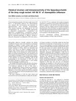
Gene organization of the feline APOBEC3 region. (a) Shown is a Pipmaker analysis of the 50 kb nucleotide sequence of the APOBEC3 region showing the
intron/exon organization of the four identified feline A3 genes (A3Cc, A3Ca, A3Cb and A3H) and annotation of repetitive elements (see inset for key:
Simple, simple repeat sequence poly(dT-dG).poly(dC-dA); LTR, long terminal repeat retrotransposons; SINE, short interspersed elements; SINE/MIR,
MIRs are tRNA-derived SINEs that amplified before the mammalian radiation; SINE/lys, tRNA-lys-derived SINE; LINE1, long interspersed element 1; LINE
2, long interspersed element-2; CpG/GpC ratios are indicated). (b) Organization and gene content of the fosmids used for nucleotide sequencing. (c) Self-
dotplot of the percent identities of the A3C region showing the high degree of sequence identity between A3Cc, A3Ca, and A3Cb.
Genome Biology 2008, 9:R48
Genome Biology 2008, Volume 9, Issue 3, Article R48 Münk et al. R48.4
Figure 1a shows a percent identity alignment of the 50 kb A3
region sequenced aligned to itself. Gene modeling studies
using the predicted nucleotide and amino acid sequences of
cat A3 and A3H cDNAs and the programs Spidey [25] and
Genewise [26] demonstrated the presence of three feline A3C
genes designated A3Ca (identical to A3C cDNA [21]), A3Cb
and A3Cc and a single A3H gene arrayed in a head-to-tail for-
mation spanning 32 kb of the 50 kb region sequenced (Figure
1a,b). The A3C genes each consist of four exons with coding
sequences that span 3,693, 6,457 and 6,498 bp for A3Cc,
A3Ca, and A3Cb, respectively, whereas A3H contains one 5'
untranslated exon followed by four coding exons that span
2,237 bp (Figures 1 and 2). Consensus splice acceptor sites
were observed for exons 2 to 4 in the three A3C genes and
exons 2 to 5 in the A3H gene. Consensus splice donor sites
were observed for exons 1 to 4 in A3H and in all four coding
exons of the three A3C genes. Interestingly, splicing at the
splice donor sites of exon 4 (bold) in all A3C genes eliminates
the overlapping termination codon (underlined) of the feA3
cDNA (CTT AGG T
GA), allowing the generation of chimeric
read-through transcripts. Consensus polyadenylation signals
(AATAAA) were observed at positions downstream of exon 4
for all three A3C genes - A3Ca (positions 32,505, 33,376 and
33,444), A3Cb (positions 42,083, 42,954 and 43,022), and
A3Cc (positions 22,960 and 23,831) - and A3H (position
50,319).
The initially identified cDNA of feA3 (A3Ca) and the coding
sequences of the genes A3Cb and A3Cc show 97.6% and
98.9% identical nucleotides, respectively, and 96.3-96.5%
identical amino acids to each other. The predicted proteins of
A3Cb and A3Cc differ in six or seven amino acids from feA3
(A3Ca; Figure 3; Figure S2 in Additional data file 3). The
feline A3C genes show high overall similarity to human A3C
with 43.3-43.8% identical amino acids (Figure S2 in Addi-
tional data file 3). In addition to the high degree of sequence
identity between the coding sequences of the three cat A3C
genes, the pattern of repetitive elements, especially in intron
1 of A3Ca and A3Cb (Figure 1A), and self dotplot analyses
(Figure 1c) suggested significant sequence identity in noncod-
ing regions of these highly related genes. Supplementary
Table 1 in Additional data file 2 shows the size of each intron
and the pairwise percent identities between the introns of the
three genes: the introns of A3Ca and A3Cb have a high degree
of nucleotide sequence identity (98-99%) across all three
introns whereas A3Cc shows a lower degree of sequence iden-
tity to either A3Ca or A3Cb (67-96%), depending on the size
of the intron. Based on the very high similarity of the A3C
genes, two gene duplications in rather recent evolutionary
times seem to be highly likely. The first duplication yielded
A3Cc and an A3Ca/b progenitor gene. A3Ca/b subsequently
duplicated again, resulting in A3Ca and A3Cb. As expected,
the cat A3C genes have a more distant relationship to the
human A3C group, the feline A3H clusters with the dog A3H
gene, but the dog A3A is only distantly related to human A3A
(Figure 4a). Double-domain APOBEC3 genes structurally
analogous to human A3F or A3G have not been found in the
genomes of either cat or dog.
Expression of feline APOBEC3 genes
Initially, we applied 3' RACE assays using the A3Ca sequence
in order to clone additional feline A3 cDNAs. We detected the
single-domain A3H and a cDNA composed of the fused open
reading frames of A3C and A3H, designated A3CH [24]. A
closer inspection of the sequence of A3CH revealed that the
transcript is encoded by exons 1-3 of A3Ca, the complete cod-
ing sequence of exon 4 of A3Cb and exons 2-5 of A3H (Figure
2). Importantly, the consensus splice donor of exon 4 of
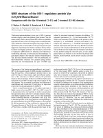
Representation of the feline APOBEC3 genomic region, portraying the detected A3 transcriptsFigure 2
Representation of the feline APOBEC3 genomic region, portraying the detected A3 transcripts. Transcripts with translated exon (rectangles) and spliced-
out introns (dotted lines) are indicated. Please note that the transcript for A3H comes in two versions: with complete exon 2 and further spliced exon 2,
resulting in 5' truncation (Ex. H2 5'Δ). The mRNA for A3CH includes exon 4 of A3Cb in an additionally spliced version, 3' truncating the sequence one
nucleotide before the stop codon (Ex. Cb4 3'Δ).
A3Ca A3Cb
A3H
A3Cc
5‘ 3‘
Transcripts
Chr. B4
18k
20k 22k
24k
26k
28k
30k
32k 34k
36k 38k 40k
42k 44k
46k
48k
50k
Ex. Cb4 3‘
Ex. H2 5‘
A3CH RNA
Genome Biology 2008, Volume 9, Issue 3, Article R48 Münk et al. R48.5
Genome Biology 2008, 9:R48
A3Cb, located only one nucleotide 5' of the stop codon TGA, is
used for in-frame splicing to A3H exons 2-5. The double-
domain A3CH RNA was found in three tested cell lines (CrFK,
MYA-1, KE-R) and also in feline peripheral blood mononu-
clear cells (PBMCs; Figure 5a). In 20 cloned PCR products
from independent reverse-transcriptase (RT)-PCR reactions
using RNA from CrFK, MYA-1 and PBMCs, the A3CH cDNAs
were always exactly as described above (exons 1-3 of A3Ca,
the 3' truncated exon 4 of A3Cb and exons 2-5 of A3H). In no
case did we observe sequence variation in the A3CH mRNA,
for example, by contribution of other A3C exons. We used
diagnostic PCR primers to analyze the expression of A3Ca,
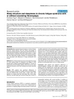
Comparison of the nucleotide coding and amino acid sequences of the feline A3C genesFigure 3
Comparison of the nucleotide coding and amino acid sequences of the feline A3C genes. (a) Pairwise comparison of the domestic cat A3 cDNA to the
predicted A3Ca, A3Cb and A3Cc genomic coding sequences and the predicted amino acid sequences. (b) Amino acid sequence alignment of A3C cDNA
and the predicted proteins for A3C genes. Highlighted in yellow are amino acid residues different between the A3Cs based on the genomic sequence,
whereas amino acid sites displaying non-synonymous amino acid substitutions are boxed in blue and red for A3Cb and A3Cc, respectively, as identified by
SNP genotyping of eight domestic cat breeds for exons 2-4 of A3Ca, A3Cb and A3Cc (for more details see Table 4 in Additional data file 2). Arrows
indicate exonic junctions. Below the alignments, variant amino acids are boxed in red for A3Cc (for example, W65R) and blue for A3Cb with respect to
the breed from which they were identified: Turkish van (VAN), Egyptian mau (MAU), Sphynx (SPH), Birman (BIR) and Japanese bobtail (BOB). A dash
indicates the amino acid is identical to genomic sequence. Numbers adjacent to breed identifiers refer to alleles 1 and 2 identified by clonal sequence
analysis of the PCR products that are in phase for exons 3 and 4, but not for exon 2 (1/2). The residue corresponding to functionally significant amino acid
replacement identified in human A3G (D128K) is highlighted by an asterisk (see text).
fe3 cDNA/A3Ca fe3 cDNA/A3Cb fe3 cDNA/A3Cc
Nucleotide identities
579/579 (100%) 572/579 (98.8% ) 566/579 (97.8% )
differences
0 bp 7 bp 13 bp
Amino acid identities
192/192 (100%) 186/192 (96.9% ) 185/192 (96.3% )
differences
0 aa 6 aa 7 aa
(a)
A3Ca(Fe3)
A3Cb
A3Cc
A3Ca KVHPWARCHAEQCFLSWFRDQYPYRDEYYNVTWFLSWSPCPTCAEEVVEFLEEYRNLTLS 120
A3Cb KVHPWARCHAEQCFLSWFRDQYPYRDEYYNVTWFLSWSPCPTCAEEVVEFLEEYRNLTLS
A3Cc KVHPWARCHAEQCFLSWFRDQYPCRDEYYNVTWFLSWSPCPTCAEEVVEFLEEYRNLTLS
A3Ca IFTSRLYYFWDPNYQEGLCKLWDAGVQLDIMSCDDFKHCWDNFVDHKGMRFQRRNLLKDY 180
A3Cb IFTSRLYYFWDPNYQEGLCKLWDAGVQLDIMSCDDFKHCWDNFVDHKGMRFRRRNLLKGY
A3Cc IFTSRLYYFYHPNYQQGLRKLWDAGVQLDIMSCDDFEHCWDNFVDHKGMRFQRRNLLKDY
LWD
VAN 1 - - -
MAU 1 E - D
SPH 1 - - D
BIR - - A
BOB - - D
MAU 2 - EY YN Q D
SPH 2 - EY YN Q D
VAN 2 - EY YN - D
A3Ca DFLAAELQEILR 192
A3Cb DFLAAKLQEILR
A3Cc DFLAAELQEILR
VAN 1 L E
MAU 1 L E
SPH 1 S E
BIR - E
BOB - E
MAU 2 - E
SPH 2 - E
VAN 2 - E
Zn
+2
coordinating site
variant sites between
A3C proteins
A3Cb variant
site
A3Cc variant
site
* D128K
*
MEPWRPSPRNPMDRIDPNTFRFHFPNLLYASGRKLCYLCFQVETEDYFSCDDSDRGVFRN 60
MEPWRPSPRNPTDRIDPNTFRFHFPNLLYASGRKLCYLCFQVETEDYFSYNDSDRGVFRN
MEPWRPSPRNPMDRIDPNTFRFHFPNLLYASGRKLCYLCFQVETGDYFSCDDSDRGVFRN
M
M
M
M
M
E
E
-
-
-
MEPWRPSPRNPMDRIDPNTFRFHFPNLLYASGRKLCYLCFQVETEDYFSCDDSDRGVFRN 60
MEPWRPSPRNPTDRIDPNTFRFHFPNLLYASGRKLCYLCFQVETEDYFSYNDSDRGVFRN
MEPWRPSPRNPMDRIDPNTFRFHFPNLLYASGRKLCYLCFQVETGDYFSCDDSDRGVFRN
M
M
M
M
M
E
E
-
-
-
VAN 1/2
MAU 1/2
SPH 1/2
BIR
BOB
R
(b)
Genome Biology 2008, 9:R48
Genome Biology 2008, Volume 9, Issue 3, Article R48 Münk et al. R48.6
Figure 4 (see legend on next page)
cow_
A3NT
she
ep_A3NT
pig_A3NT
1/-
/58/-
leopard_A3C
lion_A
3C#1
lion_A3C#2
tiger_A3C#3
tige
r_A3C#2
tiger_A3C
cat_A3Ca
cat
_A3Cb
puma_A3C
cat_A3Cc
lynx_A3C#1
lynx_A3C#6
lynx_A3C#2
lynx_A3C#5
1/100/84/100
1/-/100/-
mouse_A3NT
bonobo_A3GNT
human_A3GNT
human_A3BNT
human_A3D
ENT
human_A3FNT
human_A3FCT
macaque_A3FCT
human_A3C
human_A
3DECT
1/100/55/-
1/-/88
mouse_A3CT
bono
bo_A
3H
chimp_A
3H
h
um
an_A
3H
orangutan_A3H
m
acaque_A3H
1/10
0/10
0/100
cat
_A3H
puma_A3H
lion_A3
H
leo
p
ard_A
3H
tiger_A3
H
lynx_A3H
dog_A
3H
1/54/66/54
cow_A3CT
sheep_A3C
T
pig_A3CT
1/93/97/87
1/-/95/-
1/-/97/100
bonobo_A3GCT
human_A3GCT
m
acaque_A3GCT
macaque_A3A
human_A3A
human_A3BCT
dog_A3A
1/81/83/72
1/100/91/88
100.0
10
0.99/-/-/56
1/75/75/82
0.99/-/96/57
0.99/68/65/55
0.99/-/96/57
0.99/-/96/57
0.99/-/96/57
0.99/-/96/57
0.99/-/96/57
0.55/1/-/-
0.98/54/-/66
0.97/67/-/63
0.89
0.189
0.819
3.192
0.528
1.316
0.174
1.887
0.263
2.861
0.616
1.307
п
п
п
п
п
п
п
п
п
п
п
п
lion AC3#1
lion AC3#2
tiger AC3#3
tiger AC3#2
tiger AC3#1
lynx AC3#1
lynx AC3#6
lynx AC3#2
lynx AC3#5
leopard AC3
cat AC3a
cat AC3b
puma AC3
cat AC3c
10
0.564
п
2.138
1.549
0.545
0.99/73/77/64
1/96/91/73
55/-/-/-
0.98/77/61/67
п
п
п
п
п
lynx A3H
tiger A3H
leopard A3H
lion A3H
puma A3H
cat A3H
Z2
Z1b
Z1a
(a)
(c)
(b)
Genome Biology 2008, Volume 9, Issue 3, Article R48 Münk et al. R48.7
Genome Biology 2008, 9:R48
A3Cb, A3Cc, and A3H in total RNA of feline PBMCs (of two
cats of unknown pedigree) and cell lines (CrFK, KE-R, MYA-
1). About half of the mRNAs from the activated feline PBMCs
corresponded to A3Ca (22 of 40 clones) and approximately
17% were identical with A3Cb (7 of 40 clones) as determined
by RT-PCR allowing detection of all three A3Cs. The remain-
ing PCR products of A3C cDNAs represented additional vari-
ants, designated A3Cx and A3Cy, each containing six amino
acid differences relative to A3Ca (Figure S1 in Additional data
file 3), indicating further genetic allelic variation in cats.
Sequence-based genotyping by direct PCR of genomic DNA
using locus specific primers for exons 2-4 from eight domestic
cat breeds resulted in finding zero, thirteen, and four non-
synonymous substitutions and zero, one, and two synony-
mous substitutions in A3Ca, A3Cb and A3Cc genes, respec-
tively (Figure 3b; details in supplementary Table 4 in
Additional data file 2). MYA-1 cells expressed A3Ca, A3Cb
and A3Cc genes (15, 5 and 1 clone out of 21, respectively), but
CrFK and KE-R cells expressed only A3Ca (10 of 10 clones for
each). Feline A3H was detected in all analyzed cell lines and
PBMCs (Figure 5a). Interestingly, the transcript for A3H
seems to be subject to alternative splicing, since we consist-
ently detected an additional variant containing a 5' truncated
exon 2, generating a cDNA with a 149 nucleotide shorter 5'
untranslated region (Figure 2).
In order to determine whether the different A3 proteins are
present in feline CrFK and MYA-1 cells, immunoblot analyses
using antisera directed against cat A3C and A3H as well as a
serum directed against the A3CH-specific sequence flanked
by the C- and H-domains in A3CH (linker) were employed. In
extracts from CrFK and MYA-1 cells the anti-linker serum
detected a protein band that clearly co-migrated with A3CH
expressed from plasmid pcfeA3CH in transfected 293T cells
(Figure 5c). The C- and H-domain-specific antisera detected
the corresponding A3C and A3H proteins in CrFK cells while
only after over-exposition of the immunoblot was the A3CH
protein detectable with these sera (data not shown). This
detection pattern may reflect low-level expression of A3CH or
may indicate that the corresponding epitopes are masked in
the two-domain A3CH protein.
To search for transcription factor binding sites that might
regulate A3 expression in the domestic cat A3 gene cluster, we
first aligned the upstream 1.1 kb, including 100 bp of the pre-
dicted exon 1 for each gene A3Ca, A3Cb, A3Cc, and A3H using
ClustalW. This analysis showed considerable sequence simi-
larity in the proximal 5' flanking sequences of all four A3
genes, with A3Cc the most divergent (Figure S3 in Additional
data file 3). Using MEME to search for conserved sequence
elements in a set of DNA sequences using an expectation-
maximization algorithm, we detected two highly conserved
50 bp sequence motifs between all four promoter regions, one
located flanking the putative transcription start site and the
other approximately 200 bp upstream [27,28]. Individual 5'
flanking sequences were analyzed using the Match program,
which uses a library of nucleotide weight matrices from the
TRANSFAC6.0 database for transcription factor binding sites
[29]. The first 50 bp motif contains putative transcription fac-
tor binding sites for HNF-4 and Elk-1 as well as a site report-
edly present in all phenobarbital-inducible promoters 30 bp
upstream of the transcriptional start site. No obvious TATA or
CAAT boxes were identified, similar to the human A3 region
[30]. The second site (200 bp upstream of the start site)
includes Octamer and Evi-1 transcription factor binding sites,
which are associated with transcription in hematopoietic cell
lineages. Further 5', the sequences and predicted transcrip-
tion factor binding sites of A3Ca, A3Cb and A3H are relatively
well conserved whereas A3Cc is divergent, suggesting that
A3Cc has a unique transcription profile as indicated in our
RT-PCR expression studies. Another approach to identify
transcription factor binding sites, ModelInspector uses a
library of experimentally verified promoter modules or mod-
els that consist of paired transcription factor binding sites,
orientation, order and distance. Using this method, we iden-
tified four paired transcription factor binding sites shared
between one or more of the feline A3 promoters and that of
human A3G [31], including two ETS-SP1 (A3Ca, A3Cb, A3Cc
and A3H), IKRS-AP2 (A3H), and NFκB paired with either
CEBP (A3Ca and A3Cb), RBPF (A3Ca, A3Cb and A3Cc) or
STAT (A3Ca, A3Cb and A3H). Future studies are required to
demonstrate the potency of these elements.
Diversity of APOBEC3 in the family Felidae
It was demonstrated that primate A3 genes are under a strong
positive selection predating modern lentiviruses [9,32,33].
Currently, it is not known whether the rapid adaptive selec-
tion of A3 genes is unique to primates or represents rather a
general feature of Placentalia. To gain further insight into this
question, we analyzed A3 sequences of additional Felidae spe-
Phylogenetic analyses of the feline A3C and A3H genesFigure 4 (see previous page)
Phylogenetic analyses of the feline A3C and A3H genes. (a) Maximum clade credibility tree obtained after Bayesian phylogenetic inference with BEAST for
the three large clusters of APOBEC3 sequences: A3A, A3C and A3H. Domains in two-domain proteins were split and analyzed separately, their position
in the original sequence indicated as CT or NT, for carboxy-terminal or amino-terminal, respectively. Values in the nodes indicate corresponding support,
as follows: Bayesian posterior probability/maximum likelihood (percentage after 500 cycles bootstrap)/distance analysis (percentage after 1,000 cycle
bootstrap) parsimony analysis (percentage after 1,000 cycles bootstrap). The scale bar is given in substitutions per site. The domains within the A3
proteins can be divided into three groups of related proteins: A3H (Z2), A3A (Z1B) and A3C (Z1A). (b,c) Zoom-in on the maximum clade credibility tree
obtained after Bayesian phylogenetic inference with BEAST, focusing on the Felinae APOBEC3C sequences (b) and APOBEC3H (c) sequences. Values in
the nodes indicate corresponding support, as in the main tree in (a). The scale bar is given in substitutions per site. Figures above the branches indicate Ka/
Ks ratios, calculated using Diverge. In some instances, zero synonymous substitutions lead to an apparent Ka/Ks ratio of infinity.
Genome Biology 2008, 9:R48
Genome Biology 2008, Volume 9, Issue 3, Article R48 Münk et al. R48.8
cies. We cloned the orthologous cDNAs of A3C and A3H from
activated PBMCs of lion (Panthera leo bleyenberghi), two
tiger subspecies (Panthera tigris sumatrae and Panthera
tigris corbetti), leopard (Panthera pardus japonensis), lynx
(Lynx lynx) and puma (Puma concolor). Together with F.
catus, this collection comprises four of the eight extant line-
ages within Felidae [34]. We characterized two to six tran-
scripts for A3C and A3H of each species, one animal per
species. The phylogenetic relationships and identities to the
domestic cat A3 genes are shown in Figure 4b,c, and supple-
mentary Tables 5 and 6 in Additional data file 2. In lynx, lion
and tiger, the cDNAs for A3C depicted some degree of intra-
species genetic variability and all variants were included in
our analysis. In three of six A3C isolates of Sumatra-tiger and
both Indochina-tiger cDNAs, the sequence encoded a lysine
at position 185, while in the three other clones of Sumatra-
tiger a glutamate was encoded. No further diversity in A3C-
cDNAs of Sumatra-tiger and Indochina-tiger was found. We
detected only a single type of A3H transcript in each of the
above-mentioned felid species. In Indochina-tiger A3H, we
found a polymorphism encoding either an arginine or a lysine
at amino acid position 65, whereas in A3H cDNAs of
Sumatra-tiger, only K65 was seen. The A3CH transcript was
also detected in cDNA preparations of lion, puma, Sumatra-
tiger and lynx (leopard was not analyzed) (Figure 5b).
Comparing non-synonymous substitution rates (Ka) and syn-
onymous substitution rates (Ks) within the alignment of the
Expression analysis of feline A3C, A3H and A3CHFigure 5
Expression analysis of feline A3C, A3H and A3CH. (a,b) Analysis of expression of feline A3C, A3H and A3CH by RT-PCR of total RNA from feline cell
lines (CrFK, MYA-1, KE-R) and feline PBMCs (a) and expression of A3CH in PBMCs of lion, puma, Sumatra-tiger (tiger), and lynx (b). H
2
O indicates PCRs
using primers specific for the A3s without template cDNA added. (c) Analysis of expression of cat A3CH by immunoblot using rabbit serum against the
sequence flanked by the C- and H-domains in cat A3C (linker) using 293T cells transfected with A3 expression plasmid or empty vector as indicated and
CrFK and MyA-1 cells (two independent cultures each).
28
39
49
62
98
vector
feA3CH
feA3H
feA3Ca
feA3CH
MYA-1 CrFK 293T
transfection
kDa
(c)
1.0
0.5
kB
fePBMC
CrFK
MYA-1
KE-R
1.0
0.5
feA3C
feA3H
feA3CH
H
2
O
1.0
0.5
1.5
(a)
Lion
Puma
Tiger
Lynx
H
2
O
1.0
0.5
kB
feA3CH
1.5
(b)
Genome Biology 2008, Volume 9, Issue 3, Article R48 Münk et al. R48.9
Genome Biology 2008, 9:R48
A3C and A3H cDNA sequences, several Ka/Ks ratios were
above 1, indicating positive selection among the A3C
sequences (Table 2 in Additional data file 2) and the A3H
sequences (Table 3 in Additional data file 2) of the different
felids. Because extreme Ka/Ks ratios below or above 1 may
appear when only few residues are under positive or purifying
selection, we used the sliding window approach to determine
whether defined regions of the A3 proteins are under any type
of selection. The results in Figure S4 in Additional data file 3
show that comparison of feline A3s to the corresponding
human A3s do not show clear positive or negative selection as
expected due to the evolutionary divergence. In contrast, pos-
itive selection of cat, tiger, lion and leopard A3Cs peaks
around 200 bp (at the start of the Zn
2+
-coordinating domain)
while comparison with lynx and puma A3Cs reveal different
sites under positive selection. In the case of A3H the sliding
window comparison was not meaningful because the small
number of substitutions led to many infinity values due to Ks
= 0. Therefore, the trees of the A3C and A3H genes (Figure
4b,c) were further tested for the presence of selection among
amino acid sites using the Phylogenetic Analysis by Maxi-
mum Likelihood (PAML) program version 3.15 [35,36]. Eval-
uating the difference of the maximum likelihood values for
the trees calculated with different evolutionary models, a
probability estimate for positive selection can be deduced. In
the case of A3H the difference is not statistically significant (P
= 0.4; Additional data files 1 and 2), but in model 2, which
allows for three different ω values (ω = 1 means neutral evo-
lution, ω < 1 purifying selection, ω > 1 positive selection), 71%
of A3H are summarized with ω = 0, supporting purifying
selection as the simplest evolutionary model. In contrast, pos-
itive selection can be found for several residues for A3C
sequences (P < 0.0001; 15% of A3C are summarized with ω =
7.2 under model 2). Comparable results were obtained when
using the webserver Selecton version 2.2 [37,38] for cat A3Ca
and cat A3Cc with the alignment of A3C with all felid species
and for cat A3H using the alignment of A3H sequences (data
not shown).
The diverse feline APOBEC3s differentially inhibit
feline retroviruses
In a recent study we showed that cat A3Ca is a potent inhibi-
tor of bet-deficient FFV (FFVΔbet) [21]. We were interested to
extend this finding and tested A3Ca, A3Cb, A3Cc, A3H and
A3CH as well as dog A3A and A3H with viral reporter systems
for FFV, FIV and FeLV. To monitor the activity of the A3s,
plasmids expressing hemagglutinin (HA)-tagged versions of
A3 were used. All A3 proteins could be detected in immunob-
lots; cat A3Ca, A3Cb, A3Cc and A3CH were comparably
expressed, and the expression of cat and human A3H was
reduced three- to five-fold (Figure 6a).
The effect of A3 co-expression on wild-type and Bet-deficient
FFV was studied after transfection of 293T cells. For this pur-
pose, the infectivity of FFV titers was determined two days
after transfection by using FeFAB reporter cells [39].
Cotransfection of A3Ca did not reduce the wild-type FFV
titer, whereas a 700-fold reduction in titer was detected with
the Bet-deficient FFV (Figure 6b), as described previously
[21]. Quite similarly, A3Cb and A3Cc did not inhibit wild-type
FFV but reduced the titer of Δbet FFV by 200- and 70-fold,
respectively. Feline A3H and A3CH showed a comparable low
antiviral activity and reduced Bet-deficient but not wild-type
FFV to a much lower degree. Dog A3A and A3H did not
inhibit the infectivity of Δbet FFV or wild-type FFV. To assess
the antiviral activity of the cat A3s on FIV, vesicular stomatitis
virus-G protein (VSV-G) pseudotyped wild-type FIV-luci-
ferase (FIV-Luc), Δvif FIV-Luc and Δvif FIV-Luc cotrans-
fected with Vif expression plasmid (pcFIV.Vif-V5) reporter
vectors were generated in 293T cells in the presence of A3
expression plasmids. Equal amounts of particles were used
for transduction experiments. The results depicted in Figure
6c show that only two of the five cat A3 proteins are inhibitors
of FIVΔvif-mediated gene transfer: feline A3H and A3CH
reduced the infectivity by five- and ten-fold, respectively, sim-
ilar to the human A3H. Feline A3Ca, A3Cb or A3Cc and dog
A3A expression plasmids did not reduce infectivity of wild-
type or Δvif FIV. In contrast, dog A3H showed antiviral activ-
ity against wild-type and Δvif FIV, causing a three-fold reduc-
tion. We recently showed that the inactivation of Δbet FFV
and HIV-1 by feline A3s was attributable to cytidine deamina-
tion of viral reverse transcripts [21]. The suppression of Δvif
FIV by feline A3H and A3CH also correlates with a significant
increased G→A mutation rate in the viral genomes (Figure
S5a,b in Additional data file 3): cotransfection of feA3H or
feA3CH resulted in 1.61% and 1.31% G→A substitutions,
respectively. Viral genomes of Δvif FIV derived from transfec-
tions omitting an A3 expression plasmid showed no G→A
editing; using feA3Ca, feA3Cb or feA3Cc expression plasmids,
only 0.1% G→A exchanges were detectable at most. These
data highly correlate with the inhibitory activity detected in
the infectivity studies. The presence of Vif protein inhibited
the genome editing nearly completely (Figure S5 in Addi-
tional data file 3). The sequence context of the majority of the
G→A exchanges in the viral genomes derived from co-
expressing feA3H and feA3CH showed no clear preference for
a dinucleotide: feA3H induced 17% GG→AG, 35% GA→AA
and 42% GC→AC exchanges in the positive strand of the
DNA. The editing context of the A3CH showed 28% GG→AG
changes, 39% GA→AG mutations, and 28% GC→AC changes.
Both A3s edited in 5-6% GT→AT dinucleotides (Figure S5c in
Additional data file 3). Interestingly, in the FIV system, the
more antiviral A3CH generated slightly lower numbers of
mutations than the less antiviral A3H (Figure S5a,b,d in
Additional data file 3). This result could point either to addi-
tional and unknown activities of A3 proteins or to differences
between the degradation kinetics of uracil-containing DNAs.
To analyze the impact of cat A3 proteins on the infectivity of
FeLV, we used a molecular clone of FeLV subgroup A (p61E-
FeLV). Reporter particles were generated by co-transfection
of the p61E-FeLV packaging construct, a murine leukemia
Genome Biology 2008, 9:R48
Genome Biology 2008, Volume 9, Issue 3, Article R48 Münk et al. R48.10
Figure 6 (see legend on next page)
(a)
anti-HA
70
55
40
35
25
15
feA3Ca
feA3Cb
feA3Cc
feA3H
feA3CH
huA3H
vector
dogA3A
dogA3H
kDa
(c)
(d)
(e)
Vector
3Ca 3Cb 3Cc 3H 3CH
FeL V /GFP
feline
APOBECs
1
10
100
GFP (%)
3A 3H
canine
APOBECs
10
2
10
3
10
4
10
5
10
6
Vector
3Ca 3Cb 3Cc 3H 3CH
feline
APOBECs
3A
3H
canine
APOBECs
Luciefrase Activity (cps)
SIV agm vif- Luc
3Ca
3Cb 3Cc 3H 3CH
feline
APOBECs
Vector
FFV
(b)
3A 3H
canine
APOBECs
1
10
10
2
10
3
10
4
10
5
10
6
titer/ml
wt FFV
bet
FFV
10
2
10
3
10
4
Luciferase Activity (cps)
FIV -Luc
3Ca 3Cb 3Cc 3H 3CH 3H
Vector
feline
APOBECs
human
APOBEC
vif
FIVvif FIV + vif.V5wt FIV
10
2
10
3
10
4
vif FIVwt FIV
Luciefrase Activity (cps)
3A
3H
canine
APOBECs
Vector
FIV -Luc
Genome Biology 2008, Volume 9, Issue 3, Article R48 Münk et al. R48.11
Genome Biology 2008, 9:R48
virus (MLV)-based green fluorescent protein (GFP)-reporter
genome, a VSV-G pseudotyping plasmid and the different A3
expression plasmids. The FeLV/GFP virions were normalized
for RT activity and used for infection of 293T cells. The GFP
expression pattern of the inoculated cells demonstrated that
cat A3Cs and dog A3H did not reduce the infectivity of FeLV/
GFP (Figure 6d). Cat A3H and dog A3A had a marginal effect
and A3CH showed a significant effect on FeLV, inhibiting the
virus by a factor of 5. We also tested the simian lentivirus
SIVagm-LucΔvif and found that all cat A3s, except A3Cc, and
dog A3H showed strong antiviral activity. Dog A3A did not
reduce the infectivity of SIV (Figure 6e).
In summary, the feline A3Ca, A3Cb, and A3Cc proteins dis-
played very high activity only against FFVΔbet while A3H and
A3CH reduced FFVΔbet infectivity much less. In contrast,
only feline A3H and A3CH had a moderate inhibitory effect
on Δvif FIV, and A3CH weakly but significantly inhibited
FeLV. The Vif protein of FIV counteracted feline A3H and
A3CH, but failed to neutralize the antiviral activity of human
and canine A3H. The FFV Bet protein mainly counteracted
feline A3Ca, as recently shown [21], and A3Cb and A3Cc. We
conclude that the various feline A3 proteins differentially tar-
get feline retroviruses with a remarkable virus-specific
profile.
Discussion
Phylogenetic analysis of the domestic cat APOBEC genes rel-
ative to human and dog demonstrated that cat and dog con-
tain genes orthologous to human AICDA (AID), APOBEC1
(A1), A2, A3 and A4. The human A3 gene cluster on chromo-
some 22 spans 130 kb and contains seven genes that can be
classified according to the presence/absence of the Z1a, Z1b
and Z2 zinc-coordinating motifs [32,40]. Z1a, the A3C family,
consists of human A3C, the carboxy- and amino-terminal
domains of human A3DE and A3DF, and the amino-terminal
domains of human A3B and A3G (Figure 4a). The Z1b group,
the A3A family, contains human A3A and the carboxy-termi-
nal domains of human A3B and A3G. The human A3H repre-
sents the Z2 zinc-finger domain. Accordingly, human A3B,
A3G, A3DE, and A3F have two domains, while A3A, A3C, and
A3H have one domain. Our analysis shows that the genome of
the domestic cat contains three A3C genes (A3Ca to A3Cc) in
addition to one A3H. The feline A3C genes have a single
domain and are related to the human Z1a group but form
their own cat specific lineage (Figure 4a). None of the domes-
tic cat genes identified fall into the Z1b group. Cross-species
BLAST analyses of the cat 1.9× genome sequence employing
dog predicted genes for A3A and A3H using NCBIs cat WGS
contig, trace and end-sequence databases failed to identify
any cat gene other than A3C and A3H. Presumably, either the
cat does not contain Z1b family genes or these genes are not
represented in the 1.9× sequence. The fosmid DNA library
and database described here provide an additional genomic
DNA resource for isolation and characterization of feline
genes involved in infectious and inherited disease. Since the
fosmids in the library have been mapped to the 1.9× cat
genomic sequence by end sequencing, it is not necessary to
screen genomic libraries by hybridization or PCR to isolate
genes of interest as with previous genomic libraries.
Human A3G and A3F have been shown to be active against
HIV-1, which lacks the virion infectivity factor (vif) [13-
16,41]. The Vif protein of HIV-1 exclusively binds and inacti-
vates human A3 proteins in a species-specific way [17]. The
D128K mutation in the human A3G gene altered the Vif inter-
action [42-44] and H186R correlated with slow AIDS pro-
gression in African American populations [45]. In felids three
types of exogenous retroviruses are known: within Orthoret-
roviridae, FIV, a lentivirus related to HIV-1, and the gamma-
retrovirus FeLV; and, within Spumavirinae, FFV. FIV infects
both wild and domestic felid species [6]. Similar to the diver-
sification of SIV in African monkeys and apes, species-spe-
cific strains of FIV have been described [46]. But unlike SIV,
which is detectable only in African species, FIV is endemic in
African, South American and Asian Felidae [6]. For FeLV the
prevalence in wild species is not known and limited studies on
FFV supported the presence of FFV-related isolates in two
species of the leopard cat lineage [47].
The ability of Vif proteins to counteract the antiviral activity
of A3 proteins is specific for a virus-host system. Thus, while
the HIV-Vif protein counteracts the human A3F and A3G pro-
teins, it is not effective against cat A3H and A3CH, as we
recently reported [24]. In contrast, FIV-Vif neutralizes the cat
A3H and A3CH induced cytidine deamination. Since we could
not detect homologous genes to A3F or A3G in the domestic
cat genome, the essential role of controlling retrovirus repli-
cation seems to be covered by different A3 proteins in permis-
sive mammals (humans and cats). Interestingly, neither
human A3C nor A3H proteins are inhibitors of wild-type or
Δvif HIV-1 [18,32], supporting a host-specific genetic adapta-
tion of A3 genes.
The vif gene of FIV is a relevant modulator of spreading virus
infection, since FIV in which the vif gene was deleted showed
a replication block in feline CrFK cells that express A3, as we
Cat A3 proteins selectively inhibit the infectivity of different retrovirusesFigure 6 (see previous page)
Cat A3 proteins selectively inhibit the infectivity of different retroviruses. (a) A3 expression in the transfected 293T cells was detected by immunoblotting
with anti-HA monoclonal antibody. (b-e) Wild-type (wt) or Δbet FFV wild type (b), wild-type FIV-Luc, Δvif FIV-Luc + Vif expression plasmid (vif.V5) and
Δvif FIV luciferase reporter vector particles (c), FeLV/GFP (d), and Δvif SIVagm luciferase viruses (e) were produced in 293T cells in the presence or
absence of the indicated APOBEC3s.
Genome Biology 2008, 9:R48
Genome Biology 2008, Volume 9, Issue 3, Article R48 Münk et al. R48.12
showed [21,24,48]. Furthermore, in domestic cats experi-
mentally infected with FIVpco isolated from P. concolor, the
virus was controlled and the cats did not develop clinical signs
associated with FIV infection. The restriction of FIVpco was
attributed to feline A3 proteins, because the viral genomes of
FIVpco grown in cats accumulated extensive G-to-A muta-
tions [20]. It is likely that insufficient molecular recognition
and inactivation of heterologous feline A3 by the Vif protein
of FIVpco caused this attenuated virus infection. It is interest-
ing to emphasize here that the Puma genus is the closest rel-
ative of the Felis genus, having diverged approximately 6.7
million years ago [34]. The ability of the cat A3 proteins to
limit FIVpco infection while not being able to limit FIV infec-
tion may thus reflect the fact that the FIV infecting F. catus
has evolved the potential to escape A3-mediated restriction of
its host since the divergence of both felide lineages. Cat A3H
and A3CH also showed some inhibitory activity against FeLV.
In contrast to Δvif FIV, these active antiviral proteins showed
only weak antiviral activity against Δbet FFV. Based on these
findings, we conclude that specific feline A3 proteins selec-
tively recognize and inactivate only defined subgroups of
feline retroviruses, while 'non-adapted', heterologous retrovi-
ruses (for example, Δvif SIVagm) can be inactivated by all
three types of feA3s with the remarkable exception of feA3Cc.
Model of mammalian A3 gene evolutionFigure 7
Model of mammalian A3 gene evolution. The model proposes the presence of several A3 genes in Placentalia before the separation of the super-orders
Afrotheria, Xenarthra, Euarchontoglires and Laurasiatheria. According to the phylogenetic relationships among the extant A3 proteins, a host-specific
evolution of the A3 genes during the early evolution of the Placentalia orders by means of preservation, deletion and/or gene duplication and concomitant
subfunctionalization or neofunctionalization is inferred. Successive duplication events from a single ancestral A3 gene might have generated multiple A3
genes before basal radiation within Placentalia. The divergence times of taxons is in millions of years (Myr) ago. Basal radiation within Monotremata and
Marsupialia is not shown. A3, APOBEC3; A3Δ5, A3 lacking exon 5 derived amino acids. Relationship of taxa and timing of mammalian evolution is based on
[85], but please note that the timing is controversial [86]. The study of Wible et al. [87] supports a later diversification of the placentalia superorders
following the Cretaceous-Tertiary (K/T) boundary 65 million years ago.
Therapsida
Marsupialia
Monotremata
A3A A3C
A3H
gradual
expansion
of A3
Placentalia
Mammalia
A3
Afrotheria
Euarchontoglires
Laurasiatheria
Xenarthra
Muridae
A3 (+ A3
5)
Hominidae
A3A A3B A3C
A3DE A3F A3G
A3H
Cetartiodactyla
Carnivora
Canidae
A3A A3H
Felidae
A3Ca A3Cb A3Cc
A3H (+ A3CH)
Suidae
A3
Primates
Rodentia
order -specific
evolution of A3:
preservation,
deletion, gene
duplication, sub-
and neo-
functionalisation
166 Myr
147 Myr
100 Myr
91.8 Myr
91.3 Myr
87.3 Myr
84.9 Myr
Ruminantia
A3
Genome Biology 2008, Volume 9, Issue 3, Article R48 Münk et al. R48.13
Genome Biology 2008, 9:R48
These data also reflect the fact that even without expression of
Vif or Bet proteins, retroviruses differ, for unknown reasons,
in their vulnerability to cognate A3 proteins.
The analysis of the genomic sequences and cDNAs of the cat
A3 loci allowed us to identify three key-features not present in
the primate A3 system: first, one ancestral A3C gene under-
went two successive duplication events in recent times - the
first event generated the ancestor of the present A3Cc gene
and a second gene, which later on underwent a second dupli-
cation giving rise to the ancestors of the present A3Ca and
A3Cb genes; second, the A3H gene in domestic cats is under
purifying selection; and third, the double-domain A3CH is
generated by a read-through transcription and alternative
splicing of three genes. In addition, we detected at least 15
single nucleotide polymorphisms (SNPs) yielding non-synon-
ymous substitutions in A3C genes of 8 different cat breeds. In
primates, the seven A3 genes (A3A to A3H) are present as sin-
gle copies on chromosome 22. In the genome of the domestic
cat, we found three copies of the A3C gene (A3Ca, A3Cb and
A3Cc) in a head-to-tail orientation on chromosome B4. The
feline A3C genes encode proteins that are different to each
other at six to seven amino acid sites. Phylogenetic analyses
indicate that this gene triplication likely occurred by two con-
secutive duplication steps: one ancestral A3C gene duplicated
to the ancestor of A3Cc and a second gene, which later dupli-
cated, giving rise to A3Ca and A3Cb genes. The presence of a
homologous A3Cc gene in P. concolor closely related to the
cat A3Cc gene suggests that at least the first duplication event
occurred before the divergence of the Puma and Felis line-
ages, approximately 6.7 million years ago. The phylogenetic
position of P. tigris A3C basal to the three cat A3C genes
suggests also that the first duplication event occurred after
the divergence of the Panthera and the Felis lineages, approx-
imately 10.8 million years ago. It is generally believed that the
evolution of new protein functions after gene duplication
plays an important role in the evolution of the diversity of
organisms and typically allows for an increased specialization
or function gain of the daughter genes [49,50]. In light of the
seven A3 genes in primates, it is tempting to speculate that
cats, like primates, were under a specific evolutionary pres-
sure to increase the diversity of the co-expressed A3 proteins
that provided additional fitness. Other mammals, such as
rodents and eventually dogs, were either not faced by these
infectious agents or managed to counteract retroviruses and
related retroid elements in a way not involving A3 proteins.
While primate A3 genes are under an adaptive (positive)
selection [9,32,33], we detected significant positive selection
only for the feline A3C genes. Feline A3H was found to have
more residues under purifying selection than feline A3C. It
thus appears that restriction against an apparently innocuous
virus (FFV), mediated in cat by the A3C genes, is under high
selective pressure whereas A3H, which is active against two
serious cat pathogens, FIV and FeLV, does not evolve adap-
tively. While we consider it unlikely that FFV has a strong but
currently unidentified pathogenic potential, it is possible that
restriction against additional pathogens has shaped this evo-
lutionary pattern. For instance, the cat A3H may protect
against highly conserved, endogenous retroelements or may
act by targeting highly conserved, invariant viral structures of
FIV and FeLV, both features that would result in purifying
selection. It could also be that cat A3H took over additional
important functions distinct from pathogen defense, induc-
ing purifying selection. Finally, the combination of a con-
served A3H domain carrying specifically optimized effecter
functions with a highly adaptive module allowing recognition
of changing targets may explain that the two-domain feA3CH
is much more active against FIV and FeLV than the corre-
sponding single-domain molecules that are either inactive
(cat A3C) or have intermediate activity (cat A3H). We postu-
late that the generation of the fused A3CH transcript is an
evolutionary way to gain a greater variety of proteins from a
limited number of functional exons.
In order to express the potent anti-retroviral restriction factor
A3CH, the cat has modularly combined sequences from A3Ca
and A3Cb genes and the constant A3H domain. This was
likely achieved by read-through transcription. Read-through
transcription, also called transcription-induced chimerism, a
mechanism where adjacent genes produce a single, fused
RNA transcript, is found in at least 2-5% of human genes
[51,52]. A general feature of human transcriptional read-
through is that intergenic sequences in these RNAs are proc-
essed via the standard eukaryotic splicing machinery that
removes introns from RNA transcripts. Intergenic splicing is
favored in closely located gene pairs [51,52], as true for the
triplicated feline A3C genes. Currently, the regulation of read-
through transcription is uncharacterized and both cis-acting
sequences and trans-acting suppressors/regulators of the
termination machinery could regulate it. Since the cat A3CH
protein displayed a significantly stronger antiviral activity
against FIV and FeLV compared to the single-domain cat A3C
and A3H proteins, the read-through transcription for cat A3
appears to be functionally relevant.
In this study we did not investigate whether the upstream
genes (A3Ca and A3Cb) have legitimate transcription termi-
nation sites, and whether the downstream gene (A3H) has a
legitimate promoter region. But consensus sequences for
both regulatory elements are detectable using standard anal-
ysis tools (Figure S3 in Additional data file 3). This analysis
showed considerable sequence similarity in the proximal 5'
flanking sequences of all A3 genes except A3Cc, which has a
unique upstream sequence, supporting the experimental data
that A3Cc may have a unique transcription profile. In
humans, A3 genes are differentially expressed in tissues asso-
ciated with either endogenous or exogenous retroviral repli-
cation, including testes, the ovary and un-stimulated and
stimulated peripheral blood lymphocytes (for a review, see
[53]). Analysis of cDNA clones from domestic cat PBMCs, and
MYA-1, CrFK and KE-R cells suggest that the cat A3 genes are
Genome Biology 2008, 9:R48
Genome Biology 2008, Volume 9, Issue 3, Article R48 Münk et al. R48.14
also differentially expressed. An alternative possibility, how-
ever, is that the fused transcript of A3CH results from trans-
splicing between separate pre-mRNAs of A3Ca, A3Cb and
A3H genes. The amount of trans-splicing in mammals is
unknown and only few examples have been described so far
[54,55].
In the human A3 locus there is evidence of gene expansion. It
was speculated that duplications of single-domain genes
formed the two-domain A3B or A3G, and, subsequently,
duplication of A3B or A3G formed A3F [30]. Primates and
rodents are both part of the placentalia super-order of Euar-
chontoglires (synonymous with Supraprimates). While pri-
mates have seven A3 genes (A3A to A3H), mice and rats carry
only a single A3 gene. In mice, in addition, a splice variant
lacking exon 5 (A3Δ5) is expressed [17,56]. Based on these
data, it was proposed that primates show a relatively recent
and possibly unprecedented gene expansion [10] or that the
gene expansion happened at the beginning of primate evolu-
tion [40]. The lack of data from other mammals was partially
filled by Jónsson et al. [57], who characterized A3 proteins,
designated A3Fs, from certain cetartiodactyla (cow, pig,
sheep). Rodent and cetartiodactyla A3 proteins consist of two
cytosine deaminase domains, where the amino-terminal
domain is similar to human A3C and the carboxy-terminal
domain shows the highest identity to human A3H [32]. This
CH domain configuration is also found in feline A3CH
described here. In our study we detected A3C and A3H genes
in Felidae, while in the dog genome only genes corresponding
to A3A and A3H are present. Cetartiodactyla and Carnivora
are both grouped into the placentalia super-order of Laura-
siatheria. Based on the presence of at least three different A3
types in Laurasiatheria, we propose that a certain set of A3
genes (A3A, A3C, A3H, or more) was already established
before the separation of the placentalia order. During follow-
ing evolution, this set of A3 genes was either preserved, fused,
deleted or re-expanded depending on the specific require-
ments of the host-virus interactions (Figure 7). Following this
idea, an initial expansion of a single A3 gene happened early
in mammalian evolution, eventually before the appearance of
the placentalia. Further studies on the absence or presence of
A3 genes in the other placentalia super-orders (Xenathra and
Afrotheria) and genomics in monotremata and marsupialia
are necessary to critically evaluate our model of a two-stage
evolution of A3 genes.
Conclusion
Recent studies on the evolution of the primate APOBEC3
genes revealed a primate-specific gene amplification. We ana-
lyzed the genomic APOBEC3 region of the domestic cat (F.
catus) and found a chromosomal APOBEC3 locus different to
that of primates, rodents and dogs. Besides our detection of
three very similar APOBEC3C genes and one APOBEC3H
gene, the cat uses the mechanism of transcriptional read-
through alternative splicing to generate a fifth antiviral
APOBEC3 protein. The evolution of antiviral cytidine deami-
nases shows a strong placentalia family specific pattern. Our
results indicate that three APOBEC3 genes (A, C and H) were
present in the evolution of mammals before the placentalia
super-orders separated.
Materials and methods
Fosmid database construction and utilization
A fosmid (pCC1) DNA library consisting of 693,504 clones
containing an average insert size of approximately 40 kb of
domestic cat (breed Abyssinian) genomic DNA arrayed in
1,806 384-well plates frozen at -80°C that was end-
sequenced as part of the Feline Genome Project was obtained
from Agencourt Bioscience, Inc. (Beverly, MA, USA) The bar-
coded plates were recorded into a relational database using
Filemaker Pro (Filemaker Inc., Santa Clara, CA, USA) accord-
ing to their rack number and rack position in a -80°C freezer
along with individual trace identification number, trace
name, and clone identification number (which includes the
location of each individual fosmid within the plate).
Fosmid DNA isolation, PCR, and nucleotide
sequencing
The appropriate well of a 384-well plate was picked using a
culture loop and streaked across LB agar plates containing
12.5 μg/ml chloramphenicol and incubated at 37°C overnight.
Fosmid DNA was isolated using a standard alkaline lysis pro-
cedure. DNA was diluted 1/1,000 in H
2
O and assayed by PCR
containing 1.0 μM of primer pairs for feline sequence tagged
sites, 200 μM deoxynucleoside 5'-triphosphates (dATP,
dTTP, dCTP, dGTP), 10 mM Tris-HCl (pH 8.3), 50 mM KCl,
1.5 mM MgCl
2
, and 1 unit TAQ-Gold polymerase in a 20 μl
reaction at 95°C for 4 minutes followed by 30 cycles of 95°C
for 30 s, 60°C for 30 s and 72°C for 30 s and 72°C for 7 min-
utes at the end of cycles. PCR products were analyzed on 2%
agarose gels containing 0.5× Tris-Borate-EDTA buffer (TBE)
and positive PCR products were treated with exonuclease/
shrimp alkaline phosphatase and sequenced using BigDye
Terminator chemistry with the appropriate forward and
reverse primer and analyzed using an ABI 3730XL as
described previously [58]. Fosmids were further analyzed by
end sequencing and/or transposon insertions. Sequences
were assembled using Phred, Phrap, and Consed programs
[59-61]. Genomic nucleotide sequences were analyzed using
RepeatMasker [62] to identify repetitive elements. Genscan
[63], Genewise [26] and Spidey [64] were used to identify
coding sequences and Pipmaker [65] to visualize sequence
features. Potential regulatory sites, including transcription
factor binding sites, splice donor/acceptor sites and polyade-
nylation sites, were identified using Match™ [66], WebLogo
[67,68] and MEME [27,28].
SNP analysis of A3C genes in cat breeds
The following locus specific PCR primers were used for PCR
reactions: A3Ca_exon2_F (GCTGTTCTTTGGGGATAGA-
Genome Biology 2008, Volume 9, Issue 3, Article R48 Münk et al. R48.15
Genome Biology 2008, 9:R48
GAG); A3Cb_exon2_F (GGTTGGGGGTAGGGCGGGCT);
A3Cc_exon2_F (CACCCACCAGGGACAACTCG);
A3Ca_exon2_R (TGGTTCTCTCCTGGAAACAGA);
A3Cb_exon2_R (AGGCTGTGGCTGGGAGCAGA);
A3Cc_exon2_R (TCTGAGAGATCAGAGGGCCG);
A3Ca_exon3_4_F (CTCAAAAAAAAAGACAGGGCAGA);
A3Cb_exon3_4_F (TCAAAAAAAAAGACAGGGCAGG);
A3Cc_exon3_4_F (GATGGATGGATGGATAGATGGAT);
A3Ca_exon3_4_R (GCTGGGGAGGGAGGTGCGGA);
A3Cb_exon3_4_R (GCTGGGGAGGGAGGTGCGGT); and
A3Cc_exon3_4_R (GCTGGGGAGGGAGGTGCGGC). DNA
samples for eight domestic cat breeds (Abysinnian, Birman,
British Shorthair, Egyptian Mau, Japanese Bobtail, Norwe-
gian Forest, Sphynx, and Turkish van) were amplified by PCR
as described above except that we used 25 ng of DNA sample
and the annealing temperature was increased to 64-70°C
depending on primer pair and DNA sample. PCR products
were analyzed as described above and assembled using
Sequencher 4.7 (Genecodes, Ann Arbor, MI, USA).
Phylogenetic analysis
Reference sequences for human APOBEC genes and pre-
dicted dog APOBEC genes were identified from Ensembl,
Refseq and the NCBI annotation of the dog 7× genome
sequence, respectively. Domestic cat APOBEC genes were
identified using human APOBEC reference gene sequences
and were used to screen for traces containing related
sequences. Genscan predicted genes were edited and aligned
using Seqed and Clustalx (ABCC, NCI-Frederick, Frederick,
MD, USA). The sequences used in the phylogenetic trees were
aligned using Clustalw [69]. Manual correction of gaps and
trimming of the homologous cDNA regions were
accomplished with Jalview [70]. For consensus tree construc-
tion with bootstrap values, Seqboot (1,000 samples boot-
strapping), Dnadist (maximum likelihood distance),
Neighbor (UPGMA, jumble 10, different seed values), and
Consense from the PHYLIP package [71] were run. The
lengths of the branches were calculated with PAML3.15 (59;
Model 0, Nssites 2, molecular clock, RateAncestor 1). The
ancestral sequences for the different nodes were taken from
the PAML result. Alternatively, cDNA sequences were trans-
lated and aligned at the amino acid level using MUSCLE [72],
manually edited, filtered with GBLOCS [73], and then back-
translated conserving the codon structure. Since some
APOBEC proteins present two concatenated domains
whereas others consist of a single domain, two-domain pro-
teins were split and the individual domains analyzed sepa-
rately. Parsimony analysis was performed with PROTPARS
and with DNAPARS, from the PHYLIP package, after 1,000
cycles bootstrap. Distance analysis was performed using
PROTDIST and Dnadist using both neighbor-joining and
UPGMA (unweighted pair group method with arithmetic
mean), and combining the results. Bayesian phylogenetic
inference was performed with BEAST v1.4.6 [74], partition-
ing the nucleotide sequence following the three codon posi-
tions, under the Hasegawa-Kishino-Yano model of evolution,
using a strict clock and a Jeffreys prior distribution for the
coalescent population size parameter, with no phylogenetic
constraints, in two independent chains of 10,000,000 gener-
ations, sampling every 1,000 generations. Maximum likeli-
hood analysis were performed with RAxML [75,76] under the
Wheland and Goldman model of evolution, executing 500
non-parametric bootstaps.
Ka/Ks analysis of sequence pairs
Ka/Ks values between sequence pairs out of the alignments
were calculated using the Diverge program (Wisconsin Pack-
age, Accelrys Inc (San Diego, CA, USA). For the sliding win-
dow approach (window 300 bp, slide 50 bp) the program was
run with the appropriate part of the sequences. Test for posi-
tive selection: the cat A3C and A3H trees were further tested
for the presence of positive selection among amino acid sites
using PAML. The likelihood ratio test was used to compare
the evolutionary models M1 (neutral) and M2 (selection), M7
(beta) and M8 (beta&omega variations) [77,78]. The likeli-
hood ratio test statistic was calculated by 2 × logΔ, where Δ =
L0 (null modeldata)/L1 (alternative modeldata), L0 is the
likelihood estimate for the simple model, and L1 is the likeli-
hood estimate for the model with more free parameters. The
degrees of freedom were determined by the difference in the
number of free parameters between the null and alternative
models, and the test statistic was approximated to a χ
2
distri-
bution to determine statistical significance. In models M2
and M8, an empirical Bayesian approach is used to calculate
the posterior probability that an amino acid site fits in each
site class and sites with a high posterior probability of falling
into the class of ω of >1 are considered to be under positive
selection. Additionally, we identified the residues under pos-
itive and purifying selection using the webserver Selecton ver-
sion 2.2 [37], submitting the alignments of the cat A3C and
A3H sequences.
Cells and transfections
Human cell line 293T, and feline cell lines CrFK (ATCC CCL-
94, feline kidney cells) and KE-R (feline embryonic fibroblast
cells, a gift of Roland Riebe, Friedrich-Loeffler Institut,
Riems) were maintained in Dulbecco's high glucose modified
Eagle's medium (Biochrom, Berlin, Germany; Dulbecco's
modified Eagle's medium complete) supplemented with 10%
heat-inactivated fetal bovine serum (FBS), 0.29 mg/ml L-
glutamine, and 100 units/ml penicillin/streptomycin. Feline
T-cell lines MYA-1 (ATCC CRL-2417) and FeT-1C (ATCC
CRL-11968) were cultured in complete RPMI 1640, 0.29 mg/
ml L-glutamine, 10 mM HEPES, and 1.0 mM sodium pyru-
vate and supplemented with 0.05 mM 2-mercaptoethanol,
100 units/ml human recombinant interleukin-2 and 10%
heat-inactivated FBS, and 100 units/ml penicillin/strepto-
mycin. Plasmid transfection into 293T cells was done with
Lipofectamine 2000 according to the manufacturer's
instructions (Invitrogen, Karlsruhe, Germany). PBMCs of
Felidae and a dog were isolated from EDTA- or heparin-
treated whole blood by Histopaque-1077 (Sigma,
Genome Biology 2008, 9:R48
Genome Biology 2008, Volume 9, Issue 3, Article R48 Münk et al. R48.16
Taufkirchen, Germany) gradient centrifugation and cultured
after activation with phytohemagglutinin (PHA; 3 μg/ml) for
3 days in RPMI medium 1640 containing 15% FBS, 5 × 10
-5
M
2-mercaptoethanol, 2 mM L-glutamine, and 100 units of
human recombinant interleukin-2 per ml at 37°C and 5%
CO
2
. Blood of one dog (Canis familiaris) of the breed Austral-
ian shepherd was obtained from Karin Kliemann. Blood of F.
catus was obtained from two cats of unknown breed from the
Max Planck Institute for Brain Research, Frankfurt, Ger-
many. Blood from one each of lion (P. leo bleyenberghi),
tigers (P. tigris sumatrae and P. tigris corbetti), leopard (P.
pardus japonensis), lynx (L. lynx) and puma (P. concolor)
were obtained from the Halle Zoo, Germany.
Viruses and infections
FIV single-cycle luciferase vectors (FIV-Luc) were produced
by cotransfecting 293T cells with: pFP93 (derived from clone
FIV-34TF10, a gift of Eric M Poeschla [79]), which does not
express vif; or pCPRΔEnv (derived from clone FIV-PPR, a gift
of Garry P Nolan [80]), which does express vif; pLinSin; a
VSV-G expression plasmid pMD.G [81]; and indicated
APOBEC3-HA expression plasmids or empty vector
(pcDNA3.1(+) (Invitrogen) or pcDNA3.1(+)zeo (Invitrogen).
Vector pLiNSin was derived from pGiNSin, a self-inactivating
(Sin) vector variant of pGiNWF [79], which is a minimal bi-
cistronic FIV transfer vector plasmid coding for enhanced-
green-fluorescent-protein (EGFP) and neomycin phospho-
transferase containing Woodchuck hepatitis virus posttran-
scriptional regulatory element (WPRE) and FIV central DNA
flap. The EGFP gene in pGiNSin was replaced by the firefly
luciferase gene (luc3) using the restriction sites AgeI and
ApaI. The luciferase gene was amplified by overlapping PCR
using pSIVagmLuc [17] as template and primers (feLuc3_1.f
5'-TCCACCGGT
CGCCACCATGGAAGACGCCAA-3' (AgeI-
restriction site underlined); feLuc3_1.r 5'-CGTTGGCCGCTT-
TACACGGCGATC-3'; feLuc3_2.f 5'-GATCGCCGTG-
TAAAGCGGCCAACG-3'; feLuc3_2.r 5'-
TTCCGGGCCC
TCACATTGCCAAA-3' (ApaI-restriction site
underlined)), subcloned in pCR4Blunt-TOPO (Invitrogen),
and sequence verified. FeLV reporter virions were produced
by transfection of 293T cells with FeLV-A clone p61E-FeLV
[82], MLV-EGFP transfer vector pMgEGFP-ΔLNGFR [83],
VSV-G expression plasmid and the indicated expression plas-
mid for APOBEC3-HA or pcDNA3.1(+). Reverse-transcrip-
tion of viruses was determined by Cavidi HS kit Lenti RT or
C-type RT (Cavidi Tech, Uppsala, Sweden). For reporter virus
infections, HOS cells were seeded at 2.0 × 10
3
cells/well a day
before transduction in 96-well plates and then infected with
reporter virus stocks normalized for RT. Firefly luciferase
activity was measured three days later with a Steadylite HTS
reporter gene assay system (PerkinElmer, Cologne, Ger-
many) according to the manufacturer's directions on a
Berthold MicroLumat Plus luminometer. Expression of EGFP
was analyzed by flow cytometry. Propagation of wild-type and
Bet-deficient FFV (pCF-7 and pCF-Bet-BBtr [21]), contrans-
fection with defined APOBEC3-HA expression plasmids,
titration of FFV infectivity, and detection of FFV proteins was
done as described previously [21].
Sequencing of viral reverse transcripts
293T cells (1 × 10
6
) were infected with DNase I (Roche, Man-
nheim, Germany) treated wild-type or Δvif FIV(VSV-G)
(1,000 pg RT) using vector pGiNSin produced in 293T cells
together with feline APOBEC3s or pcDNA3.1(+). At 10 h post-
infection, cells were washed with phosphate-buffered saline
and DNA was isolated using DNeasy DNA isolation kit (Qia-
gen, Hilden, Germany). A 300 bp fragment of the egfp gene
was amplified using Taq DNA polymerase and the primers
eGFP.fw (5'-cgtccaggagcgcaccatcttctt-3') and eGFP.rv (5'-
atcgcgcttctcgttggggtcttt-3'). Each of 30 cycles was run at 94°
for 30 s, 58° for 1 minute, and 72° for 2 minutes, and PCR
products were cloned into TOPO TA-cloning pCR4 vector
(Invitrogen) and sequenced. The nucleotide sequences of at
least eight independent clones were analyzed.
Plasmids
All APOBEC3s are expressed as carboxy-terminal HA-tagged
proteins (APOBEC3-HA). Feline APOBEC3Ca (previously
termed feAPOBEC3, feA3, GenBank accession no. AY971954
)
was described in [21]. Feline APOBEC3Cb and APOBEC3Cc
were similarly constructed. Feline APOBEC3H and feline
APOBEC3CH cDNAs were identified by using 5' and 3' RACE
reactions (5'/3'-RACE kit, Roche Diagnostics) employing
total RNA from CrFK cells [24]. APOBEC3 cDNAs of big cats
were amplified from cDNA of PBMCs after activation with
PHA (3 μg/ml) and Pwo polymerase (Roche Diagnostics) was
applied. The primers were: for A3C forward primer fApo3F-
18 (5'-TAGAAGCTT
ACCAAGGCTGGCGAGAGGAATGG-3',
HindIII site underlined) and reverse primer fAPO3F-19 (5'-
AGCTCGAG
TCAAGCGTAATCTGGAACATCGTATGGATAC-
CTAAGGATTTCTTGAAGCTCTGC-3' (XhoI site under-
lined)); for A3H, forward primer fAPO-29 (5'-
TGCATCGGTACC
TGGAGGCAGCCTGGGAGGTG-3' (KpnI
site underlined)) and reverse primer fAPO-28 (5'-
AGCTCGAG
TCAAGCGTAATCTGGAACATCGTATGGATAT-
TCAAGTTTCAAATTTCTGAAG-3' (XhoI site underlined)).
Thirty cycles were run at 94°C for 30 s, 58°C for 1 minute, and
72°C for 2 minutes. PCR products were cloned into
pcDNA3.1(+) using restriction sites HindIII and XhoI (for
A3C) or KpnI and XhoI (for A3H) and sequenced. Expression
plasmids of 3'-HA-tagged dog (C. familiaris) APOBEC3A
(GenBank accession number. XM_847690.1
) and
APOBEC3H (GenBank accession number XM_538369.2
)
were generated by PCR. For dog A3A the template was
expressed sequence tag clone nas31ho7 5' (GenBank acces-
sion number DN874273
), obtained through Graeme Wistow
from the National Eye Institute, Bethesda, USA, and Pwo
polymerase was used. The forward primer was dog-APO-11
(5'-TGCAGGTACC
CCGCGGACATGGAGGCTGGCC-3' (KpnI
site underlined)), and the reverse primer was Dog-APO-10 (5'
AGTGCGGCCGC
TCAAGCGTAATCTGGAACATCGTATGGA-
TATAGGCAGACTGAGGTCCATCC-3' (NotI site under-
Genome Biology 2008, Volume 9, Issue 3, Article R48 Münk et al. R48.17
Genome Biology 2008, 9:R48
lined)). Dog A3H cDNA was amplified from cDNA of PBMCs
after activation with PHA (3 μg/ml), using forward primer
Dog-APO-7 (5'-TGCAGGTACC
CCACGATGAATCCACTA-
CAAGAAGA-3' (KpnI site underlined)), and reverse primer
Dog-APO-8 (5'-
AGTGCGGCCGC
TCAAGCGTAATCTGGAACATCGTATGGA-
TAAAGTCTCAAATTTCTGAAGTC-3' (NotI site underlined)),
and Pwo polymerase (Roche). Thirty cycles were run at 94°C
for 30 s, 58°C for 1 minute, and 72°C for 2 minutes. PCR prod-
ucts were digested by KpnI and NotI, introduced into
pcDNA3.1(+)-Zeo (Invitrogen) and correct clones identified
by sequencing. For generation of the pGex-feAPOBEC3CH-
linker the feline A3CH linker region containing the sequence
from amino acids 190-244 of the feline A3CH was cloned into
a pGEX-4T3 Vector (Amersham Bioscience, Freiburg, Ger-
many) by PCR (forward primer, 5'-CGAGTCGAATTCC
CT-
TAGTCCCGGCCAACAAAGAAAAAGAGAC-3'; reverse
primer, 5'-GCATGAGTCGAC
TGTGGGTCTGGGCAAGAG-
GAAGG-3'; the introduced restriction sites for EcoRI and SalI
are underlined). Purified DNA fragments were fused in frame
between the 5' GST domain and the 3' SV40 tag (KPPTPP-
PEPET) of correspondingly digested pGEX4T3tag derivatives
[84]. Clones were identified by restriction enzyme digestion
and DNA sequencing. pcFIV.Vif-V5 is an expression plasmid
for the codon-optimized vif of FIV-34TF10 (GenBank acces-
sion number M25381
). It was generated by cloning the codon-
optimized vif gene, 3' fused to the V5-tag, made by Geneart
(Regensburg, Germany) into pcDNA3.1(+) using the KpnI
and NotI restriction sites; a 3'-WPRE element was included in
the NotI and ApaI sites.
Expression studies
Expression studies of feline APOBEC3C RNAs in CrFK, KE-R,
MYA-1, and Fet1C cells and PHA-activated PMBCs of cat and
other Felidae were done by RT-PCR using 2 μg of total RNA
(RNeasy mini kit, Qiagen) and cDNA made by SuperScript III
RT (Invitrogen). For feline APOBEC3Cs the primers were for-
ward fAPO3F-9 (5'-TGGAGGCAGCCTGGGAGGTG-3'), and
reverse fAPO3F-15 (5'-GCGAGACGCAAGGAACAGCAG-3').
For feline APOBEC3H the primers were forward fAPO3F-9
and reverse fAPO-26 (5'-CTGCCCGAAGGCACCCTAATTC-
3'), or, alternatively, forward feA3H.fw (5'-ATGAATCCACTA-
CAGGAAGTCATAT-3') and reverse feA3H.rv (5-TCAT-
TCAAGTTTCAAATTTCTGAAG-3'). For feline APOBEC3CH
the primers were forward feAPO3CH.fw (5'-ATGGAGCCCT-
GGCGCCCCAGCCCAA-3') and reverse fAPO-27 (5'-
TCGTACTCGAGGCAGTTTATGAAGCATTGAGATGC-3').
Pwo polymerase (Roche) was used for cloning and Taq
polymerase (Fermentas, St. Leon-Rot, Germany) for diagnos-
tic PCR. PCRs were run for 30 cycles at 94°C for 30 s, anneal-
ing for 1 minute at 60°C for feA3C, 58°C for feA3H, and 59°C
for feA3CH, and 72°C for 2 minutes. PCR products were
cloned in TOPO-vectors (Invitrogen) and sequenced.
Immunoblot analysis
Cells were cotransfected with plasmids for FFV, FIV, FeLV
and APOBEC3-HA expression plasmids and lysates were pre-
pared two days later. Cell lysates were prepared by removing
the medium from transfected cells, washing them with phos-
phate-buffered saline, and lysing them in lysis buffer. Protein
in the lysates was quantified using Coomassie blue reagent
(Bio-Rad, Munich, Germany). Lysates containing 20 μg of
protein were separated by SDS-PAGE and transferred to pol-
yvinylidene difluoride filters or nitrocellulose membranes.
Filters were probed with anti-HA antibody (1:6,000 dilution,
MMS-101P; Covance, Münster, Germany), or mouse anti-α-
tubulin (1:4,000 dilution, clone B5-1-2, Sigma-Aldrich) or
polyclonal rabbit antibody against the linker region of feline
A3CH (1:250 dilution) followed by horseradish peroxidase-
conjugated rabbit anti-mouse antibody (α-mouse-IgG-HRP,
Amersham Biosciences) or Protein A-peroxidase (Sigma) and
developed with ECL chemiluminescence reagents (Amer-
sham Biosciences). A polyclonal mono-specific rabbit antise-
rum against the feline A3CH linker sequence (A3CH amino
acid residues 190-244) was generated using a GST-fusion
protein made with a pGex-feAPOBEC3CH-linker. The GST-
fusion protein was purified as described and used for vaccina-
tion of rabbits [21].
Data deposition
The sequences reported here have been deposited in the Gen-
Bank database: feline A3 genomic locus, including feA3Ca,
feA3Cb, feA3Cc, and feA3H (EU109281
); feA3Ca
(AY971954
); feA3C isolate X (EU057980); feA3C isolate Y
(EU057981); feA3H (EU011792); feA3H (Ex2 5'Δ)
(EF173020
); feA3CH (EF173021); feA1, feA2, feA4, and
AICDA (sequences derived from cat genomic project
AANG00000000.1); leopard A3C (DQ205650
); tiger A3C#1
(DQ093375
); tiger A3C#2 (EU016361); tiger A3C#3
(EU016362
); lion A3C#1 (EU007543); lion A3C#2
(EU007544); lynx A3C#1 (EU007546); lynx A3C#2
(EU016363
); lynx A3C#5 (EU007547); lynx A3C#6
(EU007548
); puma A3C (EU007545); leopard A3H
(EU007551
); tiger A3H (EU007550); lion A3H (EU007549);
lynx A3H (EU007553
); puma A3H (EU007552); codon opti-
mized vif of FIV (EF989123
).
Abbreviations
A3, APOBEC3; AICDA, activation-induced cytidine deami-
nase (also known as AID; APOBEC3, apolipoprotein B
mRNA-editing catalytic polypeptide 3; FBS, fetal bovine
serum; FeLV, feline leukemia virus; FFV, feline foamy virus;
FIV, feline immunodeficiency virus; HA, hemagglutinin;
HIV, human immunodeficiency virus; Luc, luciferase; NCBI,
National Center for Biotechnology Information; PAML, Phy-
logenetic Analysis by Maximum Likelihood; PBMCs, periph-
eral blood mononuclear cells; PHA, phytohemagglutinin;
RACE, rapid amplification of cDNA ends; RT-PCR, reverse-
transcriptase PCR; SIV, simian immunodeficiency virus;
Genome Biology 2008, 9:R48
Genome Biology 2008, Volume 9, Issue 3, Article R48 Münk et al. R48.18
SNP, single nucleotide polymorphism; Vif, viral infectivity
factor; VSV-G, vesicular stomatitis virus-G protein; WGS,
whole genome shotgun.
Authors' contributions
CM, ML and NY designed the study, analyzed data and wrote
the manuscript. TB characterized the genomic A3 locus, char-
acterized the SNPs and wrote the manuscript. AHW and IGB
performed bioinformatics and wrote the manuscript. JZ, SC
and MB performed cell culture and biochemical experiments.
KC and SOB contributed material/reagents/analysis tools
and analyzed data. JT characterized and provided reagents.
Additional data files
The following additional data are available with the online
version of this paper. Additional data file 1 provides supple-
mental information about the calculation of Ka/Ks. Addi-
tional file 2 provides tables showing percent identity of cat
A3C introns (supplementary Table 1), Ka/Ks ratios of cat A3s
(supplementary Tables 2 and 3), A3C SNPs of cat breeds (sup-
plementary Table 4), percent identities of all described A3C
and A3H cDNAs and proteins (supplementary Tables 5 and 6)
and results of different evolutionary models (supplementary
Table 7). Additional file 3 provides supplementary figures.
Supplementary Figure 1 shows amino acid alignments of
feline APOBEC3 proteins. Supplementary Figure 2 shows
amino acid alignments of feline, canine and human
APOBEC3 proteins. Supplementary Figure 3 contains
sequences and positions of predicted transcription factor
binding sites. Supplementary Figure 4 shows analysis of evo-
lutionary selection by the sliding window approach.
Supplementary Figure 5 presents analysis of cytidine deami-
nation of FIV by feline A3s.
Additional data file 1Information about the calculation of Ka/KsInformation about the calculation of Ka/Ks.Click here for fileAdditional data file 2Supplementary tablesSupplementary Table 1 lists percent identity of cat A3C introns. Supplementary Tables 2 and 3 list Ka/Ks ratios of cat A3s. Supple-mentary Table 4 lists A3C SNPs of cat breeds. Supplementary Tables 5 and 6 list percent identities of all described A3C and A3H cDNAs and proteins. Supplementary Table 7 lists results of differ-ent evolutionary models.Click here for fileAdditional data file 3Supplementary figuresFigure S1: comparison of amino acid sequences of the feline A3C genes. Predicted amino acid sequence of the feline APOBEC3Ca, APOBEC3Cb and APOBEC3Cc proteins in comparison with the two additional variant cDNAs detected (A3Cx and A3Cy) in cat PBMCs. The zinc coordination domain is indicated. Residues different to A3Ca are shown in bold. Figure S2: amino acid alignment of feline, canine and human APOBEC3 proteins. (a) Amino acid alignment of feline APOBEC3Ca, APOBEC3Cb, APOBEC3Cc, human APOBEC3C, APOBEC3F and murine APOBEC3 NT. (b) Amino acid alignment of feline, canine, human APOBEC3H and murine APOBEC3 NT. (c) Amino acid alignment of human, canine APOBEC3A and human APOBEC3G CT. The zinc-coordinating domains are indicated. CT, carboxyl-terminal domain; NT, amino-terminal domain. Figure S3: prediction of transcription factor binding sites. Potential transcription factor binding sites in A3 cluster of the domestic cat in the region 1.1 kb upstream, including 100 bp of the predicted exon 1 for each gene (A3Ca, A3Cb, A3Cc and A3H) using ClustalW. The individual 5' flanking sequences were analyzed using the program Match, which uses a library of nucle-otide weight matrices from the TRANSFAC6.0 database for tran-scription factor binding sites. Figure S4: analysis of Ka/Ks. Sliding window (300 bp window, 50 bp slide) analysis of Ka and Ks was performed on pairs of (a) cat A3C sequences and (b) cat A3H sequences and compared with corresponding selected felid and human sequences. Ka/Ks is plotted against the length of the coding region of the mRNAs with a schematic presentation of protein domains along the x-axis. Figure S5: analysis of cytidine deamina-tion in the genomes of FIV by feline APOBEC3s. (a) A fragment of the reporter gene (egfp) was amplified from reverse transcripts of Δvif FIV (left panel) or wild-type FIV (right panel) generated in the presence of the indicated feline APOBEC3s 10 h post-infection. A total of eight independent nucleotide sequences were determined. The mutations in the clones of each group are shown. Each muta-tion is indicated and coded with respect to nucleotide mutation. (b) The number of G→A changes per 100 Gs is shown. (c) Comparison of the dinucleotide sequence context of G→A mutations in the pos-itive-strand DNA of Δvif FIV derived from feA3-expressing 293T cells. (d) Sequence characteristics of Δvif FIV DNA genomes of vir-ions derived from 293T-expressing feline APOBEC3 proteins or empty expression plasmid (vector).Click here for file
Acknowledgements
We thank Björn-Philpp Kloke, Henning Hofmann, Lisa Maslan, Kathy Kelley,
and Xiaoxin Li for expert technical assistance and Victor David, Christiane
Kiefert, Karin Kliemann, Nathaniel R Landau, Garry P Nolan, Roland
Plesker, Eric M Poeschla, Marylyn Raymond, Roland Riebe, and Graeme
Wistow for the gift of reagents. The reagent p61E-FeLV from James Mullins
was obtained through the AIDS Research and Reference Reagent Program,
Division of AIDS, NIAID, NIH. IG Bravo is supported by the Initiative on
Evolutionary Biology from the Volkswagen Foundation. This project has
been funded in whole or in part with federal funds from the National Can-
cer Institute, National Institutes of Health, under contract N01-CO-12400.
The content of this publication does not necessarily reflect the views or
policies of the Department of Health and Human Services, nor does men-
tion of trade names, commercial products, or organizations imply endorse-
ment by the US Government.
References
1. O'Brien SJ, Menotti-Raymond M, Murphy WJ, Yuhki N: The Feline
Genome Project. Annu Rev Genet 2002, 36:657-686.
2. Burkhard MJ, Dean GA: Transmission and immunopathogene-
sis of FIV in cats as a model for HIV. Curr HIV Res 2003, 1:15-29.
3. Miyazawa T: Infections of feline leukemia virus and feline
immunodeficiency virus. Front Biosci 2002, 7:d504-518.
4. Alke A, Schwantes A, Zemba M, Flügel RM, Löchelt M: Characteri-
zation of the humoral immune response and virus replica-
tion in cats experimentally infected with feline foamy virus.
Virology 2000, 275:170-176.
5. Bastone P, Romen F, Liu W, Wirtz R, Koch U, Josephson N, Langbein
S, Löchelt M: Construction and characterization of efficient,
stable and safe replication-deficient foamy virus vectors.
Gene Ther 2007, 14:613-620.
6. Troyer JL, Pecon-Slattery J, Roelke ME, Johnson W, VandeWoude S,
Vazquez-Salat N, Brown M, Frank L, Woodroffe R, Winterbach C,
Winterbach H, Hemson G, Bush M, Alexander KA, Revilla E, O'Brien
SJ: Seroprevalence and genomic divergence of circulating
strains of feline immunodeficiency virus among Felidae and
Hyaenidae species. J Virol 2005, 79:8282-8294.
7. Pontius JU, Mullikin JC, Smith DR, Agencourt Sequencing Team, Lind-
blad-Toh K, Gnerre S, Clamp M, Chang J, Stephens R, Neelam B, Vol-
fovsky N, Schäffer AA, Agarwala R, Narfström K, Murphy WJ, Giger
U, Roca AL, Antunes A, Menotti-Raymond M, Yuhki N, Pecon-Slat-
tery J, Johnson WE, Bourque G, Tesler G, NISC Comparative
Sequencing Program, O'Brien SJ: Initial sequencing and compar-
ative analysis of the cat genome. Genome Res 2007,
17:1675-1689.
8. Bieniasz PD: Intrinsic immunity: a front-line defense against
viral attack. Nat Immunol 2004, 5:1109-1115.
9. Sawyer SL, Emerman M, Malik HS: Ancient adaptive evolution of
the primate antiviral DNA-editing enzyme APOBEC3G.
PLoS Biol 2004, 2:E275.
10. Harris RS, Liddament MT: Retroviral restriction by APOBEC
proteins. Nat Rev Immunol 2004, 4:868-877.
11. Holmes RK, Malim MH, Bishop KN:
APOBEC-mediated viral
restriction: not simply editing? Trends Biochem Sci 2007,
32:118-128.
12. Sheehy AM, Gaddis NC, Choi JD, Malim MH: Isolation of a human
gene that inhibits HIV-1 infection and is suppressed by the
viral Vif protein. Nature 2002, 418:646-650.
13. Zheng YH, Irwin D, Kurosu T, Tokunaga K, Sata T, Peterlin BM:
Human APOBEC3F is another host factor that blocks
human immunodeficiency virus type 1 replication. J Virol
2004, 78:6073-6076.
14. Wiegand HL, Doehle BP, Bogerd HP, Cullen BR: A second human
antiretroviral factor, APOBEC3F, is suppressed by the HIV-
1 and HIV-2 Vif proteins. EMBO J 2004, 23:2451-2458.
15. Liddament MT, Brown WL, Schumacher AJ, Harris RS: APOBEC3F
properties and hypermutation preferences indicate activity
against HIV-1 in vivo. Curr Biol 2004, 14:1385-1391.
16. Bishop KN, Holmes RK, Sheehy AM, Davidson NO, Cho SJ, Malim
MH: Cytidine deamination of retroviral DNA by diverse
APOBEC proteins. Curr Biol 2004, 14:1392-1396.
17. Mariani R, Chen D, Schröfelbauer B, Navarro F, König R, Bollman B,
Münk C, Nymark-McMahon H, Landau NR: Species-specific exclu-
sion of APOBEC3G from HIV-1 virions by Vif. Cell 2003,
114:21-31.
18. Yu Q, Chen D, König R, Mariani R, Unutmaz D, Landau NR:
APOBEC3B and APOBEC3C are potent inhibitors of simian
immunodeficiency virus replication. J Biol Chem 2004,
279:53379-53386.
19. Rosler C, Kock J, Malim MH, Blum HE, von Weizsacker F: Comment
on "Inhibition of hepatitis B virus replication by
APOBEC3G". Science 2004, 305:1403. author reply 1403
20. Poss M, Ross HA, Painter SL, Holley DC, Terwee JA, Vandewoude S,
Rodrigo A: Feline lentivirus evolution in cross-species
infection reveals extensive G-to-A mutation and selection on
key residues in the viral polymerase. J Virol 2006, 80:2728-2737.
21. Löchelt M, Romen F, Bastone P, Muckenfuss H, Kirchner N, Kim YB,
Truyen U, Rösler U, Battenberg M, Saib A, Flory E, Cichutek K, Münk
C: The antiretroviral activity of APOBEC3 is inhibited by the
foamy virus accessory Bet protein. Proc Natl Acad Sci USA 2005,
102:7982-7987.
22. Derse D, Hill SA, Princler G, Lloyd P, Heidecker G: Resistance of
human T cell leukemia virus type 1 to APOBEC3G restric-
tion is mediated by elements in nucleocapsid. Proc Natl Acad
Sci USA 2007, 104:2915-2920.
23. Russell RA, Wiegand HL, Moore MD, Schafer A, McClure MO, Cullen
BR: Foamy virus Bet proteins function as novel inhibitors of
the APOBEC3 family of innate antiretroviral defense
factors. J Virol 2005, 79:8724-8731.
24. Münk C, Zielonka J, Constabel H, Kloke BP, Rengstl B, Battenberg M,
Bonci F, Pistello M, Löchelt M, Cichutek K: Multiple restrictions of
human immunodeficiency virus type 1 in feline cells. J Virol
Genome Biology 2008, Volume 9, Issue 3, Article R48 Münk et al. R48.19
Genome Biology 2008, 9:R48
2007, 81:7048-7060.
25. Wheelan SJ, Church DM, Ostell JM: Spidey: a tool for mRNA-to-
genomic alignments. Genome Res 2001, 11:1952-1957.
26. Birney E, Clamp M, Durbin R: GeneWise and Genomewise.
Genome Res 2004, 14:988-995.
27. Bailey TL, Elkan C: Fitting a mixture model by expectation
maximization to discover motifs in biopolymeres. In Proceed-
ings of the Second International Conference on Intelligent Systems for
Molecular Biology: August 14-17, 1994, Stanford, California Edited by:
Altman R, Brutlag D, Karp P, Lathrop R, Searls D. Menlo Park, Cali-
fornia: The AAAI Press; 1994:28-36.
28. Bailey TL, Gribskov M: Methods and statistics for combining
motif match scores. J Comput Biol 1998, 5:211-221.
29. Database TRANSFAC [ />databases.html]
30. Jarmuz A, Chester A, Bayliss J, Gisbourne J, Dunham I, Scott J, Navar-
atnam N: An anthropoid-specific locus of orphan C to U RNA-
editing enzymes on chromosome 22. Genomics 2002,
79:285-296.
31. Muckenfuss H, Kaiser JK, Krebil E, Battenberg M, Schwer C, Cichutek
K, Münk C, Flory E: Sp1 and Sp3 regulate basal transcription of
the human APOBEC3G gene. Nucleic Acids Res 2007,
35:3784-3796.
32. OhAinle M, Kerns JA, Malik HS, Emerman M: Adaptive evolution
and antiviral activity of the conserved mammalian cytidine
deaminase APOBEC3H. J Virol 2006, 80:3853-3862.
33. Zhang J, Webb DM: Rapid evolution of primate antiviral
enzyme APOBEC3G. Hum Mol Genet 2004, 13:1785-1791.
34. Johnson WE, Eizirik E, Pecon-Slattery J, Murphy WJ, Antunes A, Teel-
ing E, O'Brien SJ: The late Miocene radiation of modern Feli-
dae: a genetic assessment. Science 2006, 311:73-77.
35. PAML [ />36. Yang Z: PAML: a program package for phylogenetic analysis
by maximum likelihood. Comput Appl Biosci 1997, 13:555-556.
37. The Selecton Server [ />38. Stern A, Doron-Faigenboim A, Erez E, Martz E, Bacharach E, Pupko T:
Selecton 2007: advanced models for detecting positive and
purifying selection using a Bayesian inference approach.
Nucleic Acids Res 2007:W506-511.
39. Alke A, Schwantes A, Kido K, Flotenmeyer M, Flügel RM, Löchelt M:
The bet gene of feline foamy virus is required for virus
replication. Virology 2001, 287:310-320.
40. Conticello SG, Thomas CJ, Petersen-Mahrt SK, Neuberger MS: Evo-
lution of the AID/APOBEC family of polynucleotide
(deoxy)cytidine deaminases. Mol Biol Evol 2005, 22:367-377.
41. Sheehy AM, Gaddis NC, Malim MH: The antiretroviral enzyme
APOBEC3G is degraded by the proteasome in response to
HIV-1 Vif. Nat Med 2003, 9:1404-1407.
42. Schröfelbauer B, Chen D, Landau NR: A single amino acid of
APOBEC3G controls its species-specific interaction with vir-
ion infectivity factor (Vif). Proc Natl Acad Sci USA 2004,
101:3927-3932.
43. Xu H, Svarovskaia ES, Barr R, Zhang Y, Khan MA, Strebel K, Pathak
VK: A single amino acid substitution in human APOBEC3G
antiretroviral enzyme confers resistance to HIV-1 virion
infectivity factor-induced depletion. Proc Natl Acad Sci USA 2004,
101:5652-5657.
44. Bogerd HP, Doehle BP, Wiegand HL, Cullen BR: A single amino
acid difference in the host APOBEC3G protein controls the
primate species specificity of HIV type 1 virion infectivity
factor. Proc Natl Acad Sci USA 2004, 101:3770-3774.
45. An P, Bleiber G, Duggal P, Nelson G, May M, Mangeat B, Alobwede I,
Trono D, Vlahov D, Donfield S, Goedert JJ, Phair J, Buchbinder S,
O'Brien SJ, Telenti A, Winkler CA: APOBEC3G genetic variants
and their influence on the progression to AIDS. J Virol 2004,
78:11070-11076.
46. VandeWoude S, Apetrei C: Going wild: lessons from naturally
occurring T-lymphotropic lentiviruses.
Clin Microbiol Rev 2006,
19:728-762.
47. Phung HT, Ikeda Y, Miyazawa T, Nakamura K, Mochizuki M, Izumiya
Y, Sato E, Nishimura Y, Tohya Y, Takahashi E, Mikami T: Genetic
analyses of feline foamy virus isolates from domestic and
wild feline species in geographically distinct areas. Virus Res
2001, 76:171-181.
48. Shacklett BL, Luciw PA: Analysis of the vif gene of feline immu-
nodeficiency virus. Virology 1994, 204:860-867.
49. Hughes AL: The evolution of functionally novel proteins after
gene duplication. Proc Biol Sci 1994, 256:119-124.
50. Lynch M, Force A: The probability of duplicate gene preserva-
tion by subfunctionalization. Genetics 2000, 154:459-473.
51. Akiva P, Toporik A, Edelheit S, Peretz Y, Diber A, Shemesh R, Novik
A, Sorek R: Transcription-mediated gene fusion in the human
genome. Genome Res 2006, 16:30-36.
52. Parra G, Reymond A, Dabbouseh N, Dermitzakis ET, Castelo R,
Thomson TM, Antonarakis SE, Guigo R: Tandem chimerism as a
means to increase protein complexity in the human
genome. Genome Res 2006, 16:37-44.
53. Schumann GG: APOBEC3 proteins: major players in intracel-
lular defence against LINE-1-mediated retrotransposition.
Biochem Soc Trans 2007, 35:637-642.
54. Takahara T, Tasic B, Maniatis T, Akanuma H, Yanagisawa S: Delay in
synthesis of the 3' splice site promotes trans-splicing of the
preceding 5' splice site. Mol Cell 2005, 18:245-251.
55. Caudevilla C, Serra D, Miliar A, Codony C, Asins G, Bach M, Hegardt
FG: Natural trans-splicing in carnitine octanoyltransferase
pre-mRNAs in rat liver. Proc Natl Acad Sci USA 1998,
95:12185-12190.
56. Abudu A, Takaori-Kondo A, Izumi T, Shirakawa K, Kobayashi M, Sas-
ada A, Fukunaga K, Uchiyama T: Murine retrovirus escapes from
murine APOBEC3 via two distinct novel mechanisms.
Curr
Biol 2006, 16:1565-1570.
57. Jonsson SR, Hache G, Stenglein MD, Fahrenkrug SC, Andresdottir V,
Harris RS: Evolutionarily conserved and non-conserved retro-
virus restriction activities of artiodactyl APOBEC3F
proteins. Nucleic Acids Res 2006, 34:5683-5694.
58. Yuhki N, Beck T, Stephens RM, Nishigaki Y, Newmann K, O'Brien SJ:
Comparative genome organization of human, murine, and
feline MHC class II region. Genome Res 2003, 13:1169-1179.
59. Ewing B, Green P: Base-calling of automated sequencer traces
using phred. II. Error probabilities. Genome Res 1998,
8:186-194.
60. Ewing B, Hillier L, Wendl MC, Green P: Base-calling of automated
sequencer traces using phred. I. Accuracy assessment.
Genome Res 1998, 8:175-185.
61. Gordon D, Abajian C, Green P: Consed: a graphical tool for
sequence finishing. Genome Res 1998, 8:195-202.
62. RepeatMasker []
63. Burge C, Karlin S: Prediction of complete gene structures in
human genomic DNA. J Mol Biol 1997, 268:78-94.
64. Spidey [ />65. Schwartz S, Zhang Z, Frazer KA, Smit A, Riemer C, Bouck J, Gibbs R,
Hardison R, Miller W: PipMaker - a web server for aligning two
genomic DNA sequences. Genome Res 2000, 10:577-586.
66. Match [ />grams.html#match]
67. WebLogo [ />68. Crooks GE, Hon G, Chandonia JM, Brenner SE: WebLogo: a
sequence logo generator. Genome Res 2004, 14:1188-1190.
69. Thompson JD, Higgins DG, Gibson TJ: CLUSTAL W: improving
the sensitivity of progressive multiple sequence alignment
through sequence weighting, position-specific gap penalties
and weight matrix choice. Nucleic Acids Res 1994, 22:4673-4680.
70. Clamp M, Cuff J, Searle SM, Barton GJ: The Jalview Java alignment
editor. Bioinformatics
2004, 20:426-427.
71. PHYLIP [ />72. Edgar RC: MUSCLE: multiple sequence alignment with high
accuracy and high throughput. Nucleic Acids Res 2004,
32:1792-1797.
73. Castresana J: Selection of conserved blocks from multiple
alignments for their use in phylogenetic analysis. Mol Biol Evol
2000, 17:540-552.
74. BEAST [ />75. RAxML [ />Page443.htm]
76. Stamatakis A, Ludwig T, Meier H: RAxML-III: a fast program for
maximum likelihood-based inference of large phylogenetic
trees. Bioinformatics 2005, 21:456-463.
77. Nielsen R, Yang Z: Likelihood models for detecting positively
selected amino acid sites and applications to the HIV-1 enve-
lope gene. Genetics 1998, 148:929-936.
78. Yang Z: Maximum likelihood estimation on large phylogenies
and analysis of adaptive evolution in human influenza virus
A. J Mol Evol 2000, 51:423-432.
79. Loewen N, Barraza R, Whitwam T, Saenz DT, Kemler I, Poeschla EM:
FIV Vectors. Methods Mol Biol 2003, 229:251-271.
80. Curran MA, Kaiser SM, Achacoso PL, Nolan GP: Efficient
Genome Biology 2008, 9:R48
Genome Biology 2008, Volume 9, Issue 3, Article R48 Münk et al. R48.20
transduction of nondividing cells by optimized feline immu-
nodeficiency virus vectors. Mol Ther 2000, 1:31-38.
81. Emi N, Friedmann T, Yee JK: Pseudotype formation of murine
leukemia virus with the G protein of vesicular stomatitis
virus. J Virol 1991, 65:1202-1207.
82. Donahue PR, Hoover EA, Beltz GA, Riedel N, Hirsch VM, Overbaugh
J, Mullins JI: Strong sequence conservation among horizontally
transmissible, minimally pathogenic feline leukemia viruses.
J Virol 1988, 62:722-731.
83. Muhlebach MD, Wolfrum N, Schule S, Tschulena U, Sanzenbacher R,
Flory E, Cichutek K, Schweizer M: Stable transduction of primary
human monocytes by simian lentiviral vector PBj. Mol Ther
2005, 12:1206-1216.
84. Sehr P, Muller M, Hopfl R, Widschwendter A, Pawlita M: HPV anti-
body detection by ELISA with capsid protein L1 fused to glu-
tathione S-transferase. J Virol Methods 2002, 106:61-70.
85. Bininda-Emonds OR, Cardillo M, Jones KE, MacPhee RD, Beck RM,
Grenyer R, Price SA, Vos RA, Gittleman JL, Purvis A: The delayed
rise of present-day mammals. Nature 2007, 446:507-512.
86. Cifelli RL, Gordon CL: Evolutionary biology: re-crowning
mammals. Nature 2007, 447:918-920.
87. Wible JR, Rougier GW, Novacek MJ, Asher RJ: Cretaceous euthe-
rians and Laurasian origin for placental mammals near the
K/T boundary. Nature 2007, 447:1003-1006.