analysis of genes and genomes phần 9 ppsx
Bạn đang xem bản rút gọn của tài liệu. Xem và tải ngay bản đầy đủ của tài liệu tại đây (1.08 MB, 50 trang )
378 ENGINEERING ANIMAL CELLS 12
• amplification of the Dhfr locus to increase the copy number of the Dhfr
gene to produce sufficient quantities of the enzyme to overcome the effects
of the drug. The amplification process appears to be quite random, with
large regions of flanking DNA surrounding the Dhfr locus also becom-
ing amplified.
Mutations in this last class are particularly important for the high-level expres-
sion of foreign genes. The foreign DNA is cloned into a plasmid vector that
also bears the Dhfr gene. This is then transfected into methotrexate-resistant
cells and recombinants selected for in the presence of high levels of the drug.
Cells that amplify the Dhfr locus should also contain large numbers of copies
of the foreign DNA (Wigler et al., 1980).
12.5 Expressing Genes in Animal Cells
We have previously looked at the expression of foreign gene in baculovirus
infected cells (Chapter 8), but recombinant proteins can also be produced
in mammalian cells. The insertion of a foreign gene into an animal cell is
usually insufficient to direct its efficient expression and the production of
the encoded protein. The foreign gene to be expressed must be associated
with transcriptional and translational control elements appropriate for the
cell type in which the protein will be produced. Most promoters used to
drive the expression of foreign genes in animal cells are constitutive. We
have previously discussed the Tet expression system for producing proteins
in mammalian cells (Chapter 8). Many of the constitutive promoters used
to drive gene expression in transfected cells are transcriptionally active in a
wide range of cell types and tissues, but most exhibit some degree of tissue
specificity. For example, the widely used cytomegalovirus (CMV) promoter
exhibits low transcriptional activity in hepatocytes (Najjar and Lewis, 1999).
Strong constitutive promoters which drive expression in many cell types include
the adenovirus MLP, the human cytomegalovirus immediate early promoter, the
SV40 and Rous sarcoma virus promoters, and the murine 3-phosphoglycerate
kinase promoter (Makrides, 1999).
In addition to a suitable promoter, genes to be expressed in animal cells
also require a polyadenylation site, a transcriptional termination signal and
a variety of translational control elements. In general, it has been noted that
genes containing introns are expressed at a higher level than the equivalent
cDNA copy of the gene (Buchman and Berg, 1988). This may be due to the
coupling of transcription, splicing and mRNA processing in higher-eukaryotic
cells (Maniatis and Reed, 2002).
13
Engineering animals
Key concepts
To create a modified animals, new or altered genes may be inte-
grated into the genome
Ž
Pronuclear injection – the injection of DNA fragments into the
nuclei of newly fertilized eggs
An increased understanding of the events that take place during
early embryogenesis has allowed mechanisms to be developed by
which whole animals can be produced from the DNA contained
within a single cell
Ž
Embryonic stem cells isolated from the blastocyst embryo can
be maintained in culture indefinitely, extensively manipulated
in
vitro
and then returned to a blastocyst, where the modified cells
will form parts of the animal
The transfer of the nucleus of an apparently fully differentiated adult
cell into an enucleated egg can result in the reprogramming of the
adult cell DNA to produce a cloned animal
The correction of human genetic disorders with gene therapy has
great potential and some recent successes, but still requires an
enormous amount of development before it can be applied to
many diseases
The engineering of specific traits in whole animals has huge potential benefits
in understanding complex biological phenomenon such as development and
disease progression. To understand the basis of creating whole animals that
contain altered genes, we must first look at some early embryology (Burki,
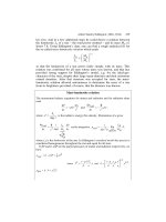
1986) (Figure 13.1). Immediately after the sperm enters the egg, the fertilized
Analysis of Genes and Genomes Richard J. Reece
2004 John Wiley & Sons, Ltd ISBNs: 0-470-84379-9 (HB); 0-470-84380-2 (PB)
380 ENGINEERING ANIMALS 13
Maternal and
paternal pronuclei
Polar
body
Zona
pellucida
Fertilised egg
Day 5
Fertilised egg
Two cells
Four cells
Morula
Blastocyst
Day 5
Day 3
Day 3
Day 2
Day 2
Day 1
Day 1
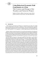
Figure 13.1. Early embryonic development. Microscopy images were obtained from
www.fertilita.org
cell, now called a zygote, contains two nuclei – called pronuclei.Thematernal
and paternal pronuclei then fuse with each other to form a single fertilized
nucleus. The zygote then begins to divide – first into two cells, then four, then
eight and so on, forming a ball of cells called a morula –fromtheLatinfor
mulberry. The morula continues to divide and a cavity forms within it that fills
13.1 PRONUCLEAR INJECTION 381
with fluid from the uterus. At this stage, the zygote is called a blastocyst and the
cavity is called the blastocoele. The cavity divides the cells of the blastocyst into
an inner cell mass (which will become the embryo) and an outer trophoblast
(which will form the placenta). Before implanting into the wall of the uterus, the
blastocyst floats in the uterine cavity for 2 days and sheds the zona pellucida,
allowing its adherence to the uterine wall. The implanted embryo continues to
divide and specialize until birth and beyond. Not all of the newly divided cells
will go on to form parts of the animal; some are programmed to die as part of
the normal developmental process (Sulston and Horvitz, 1977).
Three main methods have been developed to introduce foreign DNA into
animals. The mouse has long been the organism of choice for this type of
manipulation as a laboratory mammal that has relatively well understood and
amenable genetics. The production of altered mouse embryos for the creation
of transgenic mice is certainly well advanced but other animals, particularly
farm animals, have also been modified using similar techniques.
13.1 Pronuclear Injection
As with the methods we have previously discussed for the direct injection
of DNA fragments into Xenopus oocytes (Chapter 12), DNA can be injected
directly into the pronuclei of freshly fertilized mouse eggs (Palmiter and
Brinster, 1986). Immediately following fertilization, the large male and small
female pronuclei are visible under the microscope as discrete entities. DNA
injections are usually made into the larger male pronucleus while the egg is
being held in position using a suction pipette in a micromanipulation device
(Figure 13.2). The injected DNA may integrate into the pronuclear DNA and,
upon fusion with the female pronucleus, will be incorporated into the zygote.
The injected embryos are cultured in vitro until the morula stage and then
implanted into a pseudo-pregnant female mouse that has been previously
mated with a vasectomized male. The stimulus of mating elicits the appropriate
hormonal changes needed to make her uterus receptive. The implanted embryo
is then allowed to develop into a mouse pup. If the foreign DNA has been
successfully transferred to the mouse, then the pup will be heterozygous for the
new DNA. A small piece of the newly born pup’s tail is usually taken for DNA
analysis (Southern blotting, PCR etc.) to check for the presence of the foreign
DNA. Mating two of the heterozygotes can produce homozygous mice, with
one in four of their offspring being homozygous for the transgene.
Pronuclear injection has been used to introduce a variety of foreign DNA
fragments into mice. For example, a linear DNA fragment containing the
promoter of the mouse metallothionein-I gene fused to the structural gene of
382 ENGINEERING ANIMALS 13
Grow in culture
to morula
Implant into pseudo-
pregnant females
Breed
heretozygotes
Test pups for presence
of transgene
Homozygous
transgenic mouse
Suction
pipette
Injection of DNA
into pronucleus
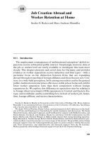
Figure 13.2. The production of transgenic mice by pronuclear injection. DNA is injected
into the larger male pronucleus and grown in culture until several divisions have occurred.
The embryos are then implanted into a pseudo-pregnant female. Assuming that the
transgene integrated before the first cell division, the pups should be heterozygous for the
transgene. Inbreeding of the heterozygotes will generate homozygous individuals
rat growth hormone was microinjected into the pronuclei of fertilized mouse
eggs (Palmiter et al., 1982). Of 21 mice that developed from the injected eggs,
seven carried the fusion gene and six of these grew significantly larger than their
littermates. Several of these transgenic mice were found to have extraordinarily
13.1 PRONUCLEAR INJECTION 383
high levels of growth hormone mRNA in their liver and growth hormone in
their serum. At 74 days of age, the transgenic mice weighed up to 44 g, while
their non-transgenic littermates weighed approximately 29 g. The technique
has also been used to attempt to produce therapeutic proteins within transgenic
animals. For example, human α
1
-antitrypsin (AAT) has been produced in mice
for the treatment of cystic fibrosis lung disease and other conditions in which
connective tissue is broken down irreversibly. AAT is a plasma protein that
inhibits elastase, a key player in the inflammatory response that, unchecked, will
lead to excessive tissue destruction. A DNA fragment containing the genomic
form of the human AAT gene, whose natural promoter had been replaced by
the sheep β-lactoglobulin milk promoter, was injected into the pronucleus of
mice embryos (Archibald et al., 1990). Mice that expressed the transgene in the
mammary gland secreted the human form of the AAT protein into their milk at
high levels (up to 7 mg of protein per mL milk). Subsequently, transgenic sheep
expressing AAT in their milk have been produced in the same way (Wright
et al., 1991). In this case, sheep expressing up to 60 mg of AAT per mL milk
were reported.
One of the major advantages of pronuclear injection is that the foreign
DNA to be inserted does not necessarily need to be contained within a vector.
Linear DNA fragments may be injected into the pronucleus, where they often
integrate as multiple (varying from a few to several hundred) head-to-tail copies
at an apparently random location within the mouse genome. The potential
disadvantages of pronuclear injection include the following.
• The nature of the DNA integration event means that pronuclear injection
can only be used to add genes to the animal. It cannot be used to delete
genes (knock-out), or to alter existing genes within the genome.
• The randomness of the insertion can have dramatic effects on the expression
of the foreign gene depending on the precise site of the insertion within
individual animals. Therefore, the expression of the transgene cannot readily
be controlled.
• The expression of the transgene is not strictly inherited. That is, the offspring
of highly expressing parent animals may show considerably different levels
of expression. In some cases, this may be due to altered genomic methylation
patterns at the site of the transgene (Palmiter, Chen and Brinster, 1982).
• The production of transgenic mice by pronuclear injection can occasionally
result in a mosaic animal, where the transgene is only present in a limited
set of tissues and organs of the animal. This happens when integration of
the transgene is delayed until after the first cell division. There can also be
384 ENGINEERING ANIMALS 13
multiple insertion events at different genomic loci and at different times.
Thus, a single founder can be mosaic for one insertion site but not the other.
13.2 Embryonic Stem Cells
Embryonic stem (ES) cells are undifferentiated cells isolated from the inner cell
mass of a blastocyst (Evans and Kaufman, 1981) (see Figure 13.1). They can be
cultured in vitro by growing them in a dish coated with mouse embryonic skin
cells that have been treated so they will not divide. This coating layer of cells
(called a feeder layer) provides a surface to which the ES cells can attach and, in
addition, releases nutrients into the culture medium. Unlike most other animal
cells, they can be maintained in culture, through successive cell divisions, for
long periods. ES cells in culture remain undifferentiated provided that they are
grown well separated from each other. If they are allowed to clump together,
they begin to differentiate spontaneously. ES cells have the potential to form all
of the cell types, of the mature animal (muscle, nerve, skin etc.) including the
gametes (Nagy et al., 1993). In addition, systems for the specific differentiation
of cultured ES cells have been developed (Keller, 1995). For example, ES cells
cultured in the presence of stromal cells and various cytokines resulted in the
generation of primitive erythrocytes and other haematopoietic precursor cells
(Nakano, Kodama and Honjo, 1994; Kennedy et al., 1997).
The ability of ES cells to be maintained in culture for extended periods,
combined with their ability to differentiate into a variety of different cell types,
makes them an attractive target for genetic manipulation. The basic method for
ES cell based animal production is shown in Figure 13.3. Foreign DNA can be
introduced into the cultured ES cells, using the methods discussed previously
(Chapter 12), and transfected cells selected. The recombinant ES cells are
then introduced into a fresh blastocyst, where they mix with the cells of the
inner cell mass. The blastocyst is then implanted into the uterus of a pseudo-
pregnant female and pups produced. Since the implanted blastocyst contains
two different types of ES cell (normal and recombinant), the resulting offspring
will be chimeric – some cells will contain the transgene, while other will not.
The chimeric pups are then crossed with wild-type animals to generate true
heterozygotes, which can then subsequently be inbred to create a homozygote.
Thus ES cell animal production requires two rounds of breeding to generate
a homozygote.
One of the major advantages of ES cells is that they are relatively efficient
at homologous recombination in comparison to other animal cells. This means
that targeted transgenes can be produced in which specific genes of the genome
are either deleted or altered (Thomas and Capecchi, 1987). Recombination
13.2 EMBRYONIC STEM CELLS 385
Breed homozygous
transgenic mouse
Implant into pseudo-
pregnant females
Culture from inner
cell mass of
mouse blastocyst
Transfect with
foreign DNA
and select
Inject transgenic
ES cells into
inner cell mass
Figure 13.3. Embryonic stem cells. ES cells are harvested from the inner cell mass of
a blastocyst and cultured
in vitro
. Here they can be genetically modified before being
returned to a fresh blastocyst
between homologous sequences in the vector DNA and the genome is used to
target the insertion of the foreign DNA fragment to a specific sequence within
the genome. Although ES cells are able to perform homologous recombination,
a significant level of non-homologous recombination still occurs. Therefore, it
is important to be able to separate the two types of event. A mechanism to
386 ENGINEERING ANIMALS 13
Non-homologous
recombination
Neo
R
tk
Vector
Genome
Neo
R
tk
(a)
Resistant to G418, killed by ganciclovir
Homologous
recombination
Neo
R
tk
Vector
Genome
Neo
R
Resistant to G418 and ganciclovir
Gene
NH
N
N
N
O
NH
2
O
OH
HO
(b)
(c)
Figure 13.4. Selection of gene knockouts in ES cell cultures. (a) Non-homologous
recombination results in the transfer of both the neomycin resistance and thymidine kinase
(
tk
) genes to the host cell. (b) Homologous recombination results in the transfer of only the
neomycin resistance gene to the host cell. (c) The structure of ganciclovir. Cells containing
the
tk
gene may be killed by treatment with ganciclovir, which is phosphorylated by
thymidine kinase, and then undergoes further phosphorylation by cellular kinases. In its
triphosphorylated form, the drug inhibits DNA polymerase by acting as a terminator of
DNA synthesis
delete a gene by homologous recombination is shown in Figure 13.4. A vector is
constructed in which DNA sequences corresponding to the regions immediately
flanking the 5
-and3
-ends of the gene that is to be deleted from the genome are
cloned either side of a selectable marker gene (e.g. the neomycin resistance gene,
whose expression allows the cells to grow in the presence of G418). The vector
also contains the HSV thymidine kinase (tk) gene. A linear DNA fragment
bearing these sequences is transfected into cultured ES cells and selection is
13.2 EMBRYONIC STEM CELLS 387
made in a medium containing G418. Only ES cells that have taken up the
DNA fragment will be able to grow. To distinguish between cells that have
that have integrated the DNA fragment in an homologous fashion and those
that have done so non-homologously, selection is then made on ganciclovir.
Ganciclovir is a synthetic analogue of 2
-deoxyguanosine (Figure 13.4(c)) that
is phosphorylated by thymidine kinase to form a dGTP analogue that inhibits
DNA polymerase activity. If the DNA inserted randomly, then the tk gene
will still be associated with the transgene, and cells will die due to the drug
treatment. If, however, homologous integration has occurred, then the tk gene
will be lost and cells will survive ganciclovir treatment (Mansour, Thomas
and Capecchi, 1988). In addition to supplying a mechanism to delete genes
(knock-out), specific genes may also be replaced with mutated versions of
themselves. The mutant version of the gene is simply cloned into the vector
next to the neomycin resistance gene and then transfected into ES cells. The
regions of homology at the ends of the linear DNA fragment determine the
genomic location (or individual gene) into which the transgene is inserted.
The ability to specifically knock out genes can provide an immensely powerful
approach to assigning gene function in whole animals, especially the mouse
(Osada and Maeda, 1998). Perhaps more importantly, knockouts can provide
excellent model systems for the analysis of human disease. We have previously
discussed the potential difficulties with this type of analysis in other organisms
(Chapter 10), and many of the same problems can also be encountered with
animal knock-outs. Three main classes of knock-out may be generated.
• Lethal. The deletion of the molecular chaperone hsp47 is lethal to mouse
embryos, predominately as a function of defective collagen biosynthesis
(Nagai et al., 2000).
• Observable phenotype. The deletion of the tumour suppressor gene p53
results in the formation of mice that develop normally, but are exquisitely
sensitive to spontaneous tumours early in their lives (Donehower et al.,
1992).
• No observable phenotype. The deletion of Matrilin 1, an extracellular
matrix protein that is expressed in cartilage, yields transgenic mice with no
apparent phenotype in comparison to their wild-type counterparts (Aszodi
et al., 1999).
A lethal phenotype generally reflects the earliest non-redundant role of the
gene, and precludes an analysis of an analysis of gene function later in devel-
opment. The diploid nature of higher organisms means that mutants that fall
into this class may be analysed in their heterozygous (+/−) state. Additionally,
388 ENGINEERING ANIMALS 13
conditional knock-outs may be produced (see below). Knock-outs that fall into
the last category (no observable phenotype) may arise as a result of genes
acting in parallel pathways compensating for each others’ functions. It is also
possible that the techniques are simply too crude to detect any subtle differ-
ences between the wild-type and the knock-out animals. The complexity of
animal genomes also means that a knock-out may have a profound effect in
one strain of mouse, but quite a different effect in another. For example, the
deletion of the gene encoding epidermal growth factor in one mouse strain
(CF-1) results in embryos that die around the time of implantation into the
uterus. If, however, the same knockout is introduced into a different mouse
strain (CD-1), then the animals can survive for up to three weeks after birth
(Threadgill et al., 1995). Ideally, knockout experiments should be performed
in a variety of strain backgrounds, but the length of time required to do that,
and the costs involved, often preclude this analysis.
One problem with this type of approach for producing transgenic ani-
mals, which we have seem previously when looking at engineering in plants
(Chapter 11), is that the selectable maker gene is transferred to the transgenic
animal. The high-level expression of an antibiotic-resistance gene within a
transgenic animal is generally undesirable. The expression of the marker may
induce the abnormal expression of other neighbouring genes, and the potential
for transfer of the marker gene to non-transgenic animals should be avoided.
The marker gene can effectively be removed after the transgene has been
established within the ES cell if its sequences are flanked by loxP sites – the
recognition sequences for the Cre recombinase (Kilby, Snaith and Murray,
1993). Transfection of the transgenic cell line with a plasmid expressing Cre
recombinase catalyses the excision of the DNA between the two loxP sites to
remove the marker gene and leave a single loxP site in its place.
There are many instances where the expression of an inserted transgene is
required only in a specific tissue or set of cells. This can readily be achieved
by constructing the foreign gene such that it is under the control of a tissue-
specific promoter. For example, the promoter of the calcium–calmodulin
dependent kinase II (CaMKIIα) gene drives expression only in the neurons
of the hippocampus (Mayford et al., 1996). Such an approach works well,
provided that a suitable tissue-specific promoter is available (Table 13.1).
Conditional knock-outs can also be produced, again using the loxP-Cre
site-specific recombination system (Gossen and Bujard, 2002). If, for example,
the knock-out of a gene results in an embryonic-lethal phenotype, then it may
be necessary to delete the gene from the genome after the animal has been
born. A method by which this can be achieved is shown in Figure 13.5 (K
¨
uhn
et al., 1995). The normal copy of the gene to be deleted is replaced in the
13.2 EMBRYONIC STEM CELLS 389
Cre
Mx
1
Target gene
loxP loxP
Deletion of target gene
loxP
+ inducer
Figure 13.5. Tissue-specific gene knock-outs. See the text for details
Table 13.1. Some tissue-specific promoters in mice. Adapted from Lewandoski (2001)
Promoter Gene normally
controlled
Tissue or cells
of expression
Reference
Alb Albumin Liver (Postic et al., 1999)
Camk2α Ca
2+
/calmodulin-
dependent protein
kinase II, α
Forebrain (Mayford et al., 1995)
Cryαa Crystallin αA Eye lens (Lakso et al., 1992)
En2 Engrailed Mid/hindbrain (Logan et al., 1993)
Gcg Glucagon Pancreatic α-cells (Herrera, 2000)
Ins2 Insulin II Pancreatic β-cells (Rommel et al., 1994)
KRT5 Keratin 5 Epidermis (Ramirez et al., 1994)
Lck Lymphocyte-specific
tyrosine kinese
T cells (Chaffin et al., 1990)
Msx2 Msh-like homeobox
gene 2
Apical ectodermal
ridge of limb bud
(Liu et al., 1994)
Myog Myogenin Skeletal muscle (Yee and Rigby, 1993)
Nes Nestin Neuronal cells (Zimmerman et al.,
1994)
Pax6 Paired-box gene 6 Retina (Gruss and Walther,
1992)
Wnt1 Wingless related
MMTV integration
site 1
Neural crest (Echelard, Vassileva and
McMahon, 1994)
390 ENGINEERING ANIMALS 13
genome by a version that is flanked by loxP sites (often referred to as a floxed
gene – flanked by loxP). In addition, the transgenic animal is also modified
to carry a copy of the gene encoding the Cre recombinase under the control
of an inducible promoter, e.g. Mx1. Mx1 is part of the mouse viral defence
system and is transcriptionally inert in healthy mice (Hug et al., 1988). The
promoter can, however, be activated by high levels of interferon or by adding
synthetic double-stranded RNA to cells (which induces interferon expression).
Transgenic animals produced in this way retain a functional copy of the gene
to be deleted until they are injected with double-stranded RNA. The effect of
the lost gene may then be investigated.
Rather than constructing a transgenic mouse containing both the tissue-
specific promoter expressing the Cre recombinase and the target gene sur-
rounded by loxP sites, a series of transgenic mice have been constructed that
each contain a different tissue-specific promoter controlling the expressing of
Cre. These can then be used as a ‘bank’ of mice strains to which transgenic
mice containing a particular floxed gene can be crossed. Mating these strains
will result in the formation of progeny in which the gene in inactivated only
in those tissues that express Cre (Gu et al., 1994). This means that a single
transgenic floxed gene can be deleted in a variety of tissues without having to
resort to further in vitro manipulation.
The tetracycline-inducible expression system (see Chapter 8) may be used
to drive Cre expression to regulate knock-out function. In this system, a
transactivator fusion protein composed of the tetracycline repressor (tetR) and
the acidic activation domain of the herpes simplex virus 16 (VP16) protein
regulate the expression of the Cre gene from a promoter containing tet-operator
(tetO) sequences. In the absence of tetracycline, the Cre gene is expressed and
will induce site-specific recombination between two loxP sites. In the presence
of tetracycline, the Cre gene will not be expressed and recombination will not
occur (St-Onge, Furth and Gruss, 1996).
13.3 Nuclear Transfer
Although animal cells become increasingly committed as differentiation and
development proceeds, the DNA contained within each differentiated cell still
retains all the information necessary to form the whole animal. If the nucleus of
a differentiated cell is introduced into an enucleated egg then, under appropriate
conditions, the nucleus can become ‘reprogrammed’ such that development of
the animal reoccurs. The production of cloned animals – all of which have orig-
inated from a single, possibly recombinant, cell line – has several potential uses.
13.3 NUCLEAR TRANSFER 391
• Recombinant protein production. We have discussed previously that the
expression level of recombinant protein production is not strictly inherited
(Chapter 12). Therefore, the ability to create large number of animals each
expressing identical levels of, say, a therapeutic protein can only be achieved
using cloned animals.
• The conservation of endangered species. Rare animals could be cloned to
repopulate dwindling natural levels.
The idea of transferring a nucleus from one cell to another is not new. Over
50 years ago it was discovered that the nuclei of blastocyst frog cells could
be implanted into eggs that lacked a nucleus to created a series of cloned
frogs that were identical to the donor cells (Briggs and King, 1952). It was
found, however, that as the donor cells became more differentiated, it became
increasing difficult to reprogramme them to produce new animals. The few
embryos cloned from differentiated cells that survived to become tadpoles grew
abnormally. This led to the speculation that genetic potential diminished as
a cell differentiated and that it was impossible to clone an organism from
adult differentiated cells. In 1975, however, John Gurdon developed a method
of nuclear transfer using fully differentiated cells and Xenopus eggs (Gurdon,
Laskey and Reeves, 1975). This is a two-step process.
• Production of enucleated eggs. Delicate needles and a powerful microscope
were used to suck the nucleus from a frog oocyte to produce an enucleated
oocyte. With the genetic material removed the enucleated oocyte would not
divide or differentiate even when fertilized.
• Introduction of a new nucleus. Using the same equipment, the nuclei of
keratinized skin cells of adult Xenopus foot-webs were transfered into the
enucleated oocytes. Many of these new cells behaved like normal fertilized
eggs and were capable of producing tadpoles. Since the tadpoles arose from
the cells of the same adult, they all contained the same genetic material
and were clones of each other produced from apparently fully differentiated
cells. This indicates that DNA is not discarded or permanently inactivated
even in highly specialized cells.
A somewhat modified procedure has been used recently to produce cloned
mammals (Figure 13.6). This was first achieved by taking cells from the
blastocyst stage of a sheep embryo and fusing them with enucleated eggs
(Smith and Wilmut, 1989). The reconstituted cells were subjected to a brief
electrical pulse to stimulate embryonic development prior to implantation
into a surrogate ewe. Live sheep have subsequently been produced from the
392 ENGINEERING ANIMALS 13
Clone of
sheep 1
Fuse
cells
Sheep 2
Implant into
surrogate ewe
Sheep 1
udder cell
Sheep 2
oocyte
Enucleate
Culture cells
to blastocyst
Sheep 1
Figure 13.6. Nuclear transfer. The cells of an adult sheep (sheep 1) are fused with the
enucleated eggs of a sheep of a different breed (sheep 2). The fusion between the two is
grown in culture to the blastocyst stage prior to implantation into a surrogate ewe. The
resulting lamb contains the nuclear genome of sheep 1
nuclei of cultured embryonic cells (Campbell et al., 1996), and from cultured
adult breast epithelial cells (Wilmut et al., 1997). This last example produced
probably the most famous sheep in the world – Dolly (Box 13.1). The success
of these experiments appears to be dependent on the synchronization of the
cell cycles of the donor and recipient cells that are to be fused. In the case of
Dolly, quiescence of the donor cell was induced prior to the cell fusion process.
Unsynchronized cells appear to be less successful in forming fruitful fusions.
Box 13.1. The life and death of Dolly.
Dolly was the first mammal clone to be produced from an adult cell. She was
produced following the procedures described below (Wilmut et al., 1997).
• Donor cells. Mammary gland tissue of a 6-year-old Finn Dorset ewe
was used to prepare a primary cell culture. This culture contained a
mixture of mammary epithelial cells (>90 per cent), myoepithelial cells
and fibroblasts. An important step in the success of the cloning process
was to induce these donor cells to exit their growth cycle and enter the
G
0
phase of the cell cycle before nuclear transfer. This was accomplished
13.3 NUCLEAR TRANSFER 393
by reducing the concentration of serum in which they were grown to
starve the cells.
• Recipient cells. Oocytes were obtained from Scottish Blackface ewes
between 28 and 33 hours after injection of gonadotropin releasing
hormone (GnRH) and enucleated using a fine glass pipette.
• Cell fusion. Fusion of the donor cell to the enucleated oocyte and
activation of the oocyte were induced by the electrical pulses – a single
DC pulse to activate the cells and a single AC pulse followed by three DC
pulses to promote cell fusion. 277 individual fused cells were produced.
• Growth and implantation. The fused cells were cultured in ligated
oviducts of sheep. After 6 days of culture, 29 of the 277 reconstructed
embryos had developed into a morula or blastocyst. One, two or three
embryos were transferred to Scottish Blackface ewes and allowed to
develop to term. The 29 morula/blastocysts were transferred to 13 differ-
ent ewes, and from these only one became pregnant. On July 5 1996 after
148 days pregnancy, the normal duration for her breed, Dolly – a Finn
Dorset sheep – was born with a healthy birth weight of 6.6 kg. Dolly, a
sheep derived from a mammary gland cell, was named after the singer
Dolly Parton.
Box Figure 13.1. Dolly, and her lamb Bonnie. Image courtesy of The Roslin Institute
394 ENGINEERING ANIMALS 13
The precise cell type from which Dolly was derived remains unclear. Further
analysis indicated that she was indeed derived from the cells of the mammary
gland of the donor sheep, rather than from a contaminating cell (Ashworth
et al., 1998). She is not, however, an exact clone of the sheep whose cells were
used to create her. The DNA of her mitochondria are derived exclusively
from recipient enucleated oocytes (Evans et al., 1999). Therefore she is a
chimera, containing somatic cell derived nuclear DNA but oocyte derived
mitochondrial DNA. It also is interesting to note that the scientific paper
in which Dolly was introduced to the world (Wilmut et al., 1997) does
not include the words ‘clone’ or ‘cloning’ anywhere within its text. Perhaps
the authors realized the potential impact of their findings and chose less
inflammatory language to describe their results. Dolly subsequently grew
into an adult sheep have bore her own offspring (Box Figure 13.1). Finn
Dorset sheep have an average life expectancy of about 12 years, but in
January 2002 Dolly was reported to be suffering from arthritis, which is
highly unusual for a sheep of her age. On 14 February 2003, aged only six,
Dolly was put to sleep following a diagnosis that she was suffering from a
progressive lung disease.
The method of nuclear transfer to produce viable offspring from differenti-
ated adult cells is not without its problems (Wilmut et al., 2002). It is likely that
not all of the difficulties described below are due to the nuclear transfer process
itself, as some similar abnormalities have been reported after embryo culture.
• The process is extremely inefficient. In the case of Dolly, only one of the
277 cell fusions produced was capable of developing into a lamb. Similar
efficiency levels have also been reported for other whole animal cloning
experiments.
• Many of the embryos produced by nuclear transfer suffer gross abnormali-
ties. In addition to embryonic loss, nuclear transfer is also associated with
very high rates of foetal, perinatal and neonatal loss, and production of
abnormal offspring.
• Although Dolly was born following a normal gestation period and was a
normal weight, many offspring produced by nuclear transfer suffer from
large offspring syndrome (LOS) in which gestation period and birth-weight
are greatly increased (Lazzari et al., 2002). The frequency and severity of
the symptoms of LOS appear to vary widely even under similar experimen-
tal conditions. Early deviations from the normal developmental pattern,
13.3 NUCLEAR TRANSFER 395
particularly with regard to embryonic gene expression, may be involved in
this phenomenon.
• Since Dolly was created from a cell that was potentially 6 years old, what
genetic age was she when she was born? This has been addressed by looking
at the length of the telomeres at the ends of chromosomes. Telomeres
generally shorten as aging progresses, although the precise effects of this
phenomenon are not well understood. Dolly has been found to have short
telomeres when compared with other sheep of the same age (Shiels et al.,
1999). It was recently reported that Dolly developed arthritis, which is
highly unusual in a sheep of her age (Williams, 2002). It remains to be seen
whether this and other potential age-related effects, including Dolly’s death,
are a result of the nuclear transfer process.
• Widespread disruptions in the DNA methylation patterns have been
described in cloned embryos of a number of cloned animals (Fairburn,
Young and Hendrich, 2002). The effects of these changes remain unclear.
• The technique of nuclear transfer is still in its infancy. This means that
the effects of aging and genetic inheritance have not been fully assessed. In
two independent studies, animals cloned from one cell type became obese
in adult life (Tamashiro et al., 2002) whereas those from another cell type
died at an unusually early age (Ogonuki et al., 2002). Further work in this
area is required.
The technique of nuclear transfer by which Dolly was produced has been
replicated or modified to produced clones from adult cells using a variety of
other farm animals, e.g. cows, goats and pigs (Cibelli et al., 1998; Baguisi et al.,
1999; Polejaeva et al., 2000), and in more experimentally amenable laboratory
animals such as mice (Wakayama et al., 1998). In addition, cloned domestic
pets such as cats (Shin et al., 2002) and rabbits (Chesn
´
e et al., 2002) have
also been reported. In early 2003, news reports suggested that the first cloned
human child had been born. Although such claims have not been scrutinized
scientifically, it seems inevitable that a cloned human will be produced at some
stage. The difficulties encountered with cloned animals described above should
serve as a warning to anyone considering the procedure. The temptation to
replace a dead or dying child with an ‘exact copy’ may be more than some
parents can bear, but the potentially disastrous consequences should not be
underestimated.
Aside from the very negative impact of nuclear transfer technology described
above, the process has proved useful for the creation of animals with specific
traits. The ability to recreate a whole animal from cells that have been
396 ENGINEERING ANIMALS 13
extensively manipulated in vitro could have a profound positive impact on
medicine. For example, there is great potential for the replacement of damaged
human organs (e.g. liver, heart) with their equivalents from animals. This
process, termed xenotransplantation, is often unsuccessful because some of the
cell surface carbohydrates are different between humans and animals. With
the exception of catarrhines (Old World monkeys, apes and humans), all
animals possess the enzyme α(1,3)-galactosyl transferase, which catalyses the
formation of the disaccharide galactose-α(1,3)-galactose that is found on the
cell surface. The presence of the disaccharide causes hyperactue rejection of the
organ in humans. This problem can only be partially overcome by temporarily
removing antibodies to galactose-α(1,3)-galactose from the recipient through
affinity adsorption. However, returning antibodies can damage the transplanted
organ and severely limit its survival even in the presence of high levels of
immunosuppressive drugs. Sheep have been produced that lack the GGTA1
gene encoding the α(1,3)-galactosyl transferase enzyme (Denning et al., 2001).
GGTA1 was replaced in tissue culture cells by a copy of the neomycin-resistance
gene, and nuclear transfer was used to generate sheep embryos. Unfortunately,
the foetuses died before birth, so it remains to be seen whether organs from
animals produced in this way may be suitable for human transplantation. More
recently, pigs knocked out for either one (Lai et al., 2002; Dai et al., 2002)
or both (Phelps et al., 2002) alleles of GGTA1 have been produced. Some of
the knock-out pigs are apparently healthy and further work will assess the
suitability of their organs for human transplantation.
13.4 Gene Therapy
Gene therapy is an approach to treat, cure or ultimately prevent disease by
changing the expression of genes within an individual. The idea seems simple – a
healthy copy of a mutated gene is introduced into an affected individual such
that the normal protein can be made, and the disease symptoms thereby
alleviated (Morgan and Anderson, 1993). Although the idea of gene therapy
has been around for some time, actual treatments are still in their infancy.
Most human clinical trials are only in the research stages. Gene therapy is
most applicable to the correction of single gene disorders, especially recessive
diseases where a functional copy of the defective gene will restore the activity
of the mis-functional protein (Table 13.2). The insertion of the transgene to
bring about the desired change can be targeted to either germ (egg and sperm)
or somatic (body) cells.
• Germ-linegenetherapy. The egg or sperm cells are changed with the goal of
passing on the changes to their offspring. Human germ-line gene therapy is
13.4 GENE THERAPY 397
Table 13.2. Examples of some single-gene human genetic disorders
Disorder Symptoms
Autosomal recessive:
Cystic fibrosis Recurrent lung infection, increased mucus
production
α
1
-antitrypsin deficiency Liver failure, emphysema
Phenylketonuria Mental retardation
Tay-Sachs disease Neurological degeneration, blindness, paralysis
Sickle cell anaemia Anaemia
Thalassemia Anaemia
Autosomal dominant:
Neurofibromatosis type 1 Tumours of peripheral nerves
Huntington’s disease Involuntary dance-like movements, dementia
Mytonic dystrophy Heart defects and cataracts
Familial retinoblastoma Tumours of the eye
X-linked:
Haemophilia Deficient blood clotting
Duschenne muscular dystrophy Progressive muscle wasting
Fragile-X syndrome Mental retardation
prohibited in most countries since the consequences of producing a human
with artificially altered genetic traits are far from clear.
• Somaticgenetherapy. The genome of the recipient is altered, but this
change is not passed to the next generation. Somatic gene therapy can be
classed as being performed either in vivo or ex vivo (Figure 13.7). In vivo
therapy involves the addition of a gene directly to a patient. Ex vivo therapy
involves the removal of cells from the patient and their culturing and genetic
manipulation in vitro before the return of the modified cells to the patient.
The type of therapy used depends on the sorts of cell that need to be modified.
If the cells in which the gene defect is apparent can be easily cultured, then the
ex vivo route offers tremendous advantages. For example, all blood cells are
derived from multipotent stem cells inthebonemarrow.ThesedifferfromES
cells that we have previously discussed in that they can only differentiate into a
limited number of different cell types. Multipotent stem cells can, however, be
398 ENGINEERING ANIMALS 13
Ex vivo
In vivo
Figure 13.7.
In vivo
and
ex vivo
human gene therapy. See the text for details
cultured in vitro for extended periods. Therefore, disorders of the blood system
may be treated ex vivo through the isolation and culturing of bone marrow
stem cells. These cells can be modified in vitro and the resulting transgenic
cells can then be returned to the patient. The modified stem cells will then
produce the various modified differentiated cells that may cure the disease. In
vitro manipulation of the cells permits the use of a wide variety of methods
to insert the transgene – e.g. viral infection, injection and other methods (see
Chapter 12). Other cells and tissues are less amenable to ex vivo treatment. For
example, lung epithelial cells – whose function is severely impaired in cystic
fibrosis patients – grow very poorly in culture. Even if they could be cultured,
it would not be possible to repopulate an affected lung with transgenic lung
epithelial cells. Therefore, diseases such as cystic fibrosis must be treated in
vivo so that the cells of the defective lung can be modified. This limits the
type of transgene insertion that can take place. Viruses, e.g. adenovirus that
naturally infect epithelial cells, are usually used to transport the transgene into
the affected cells.
13.5 Examples and Potential of Gene Therapy
The history of human gene therapy trails is not a particularly happy one. With
one notable exception (see below), the effect of introducing a gene into cells
rarely promotes more than a transient relief from the symptoms of the disease
being treated. Worse still, there have been highly publicized cases where gene
13.5 EXAMPLES AND POTENTIAL OF GENE THERAPY 399
therapy trial patients have suffered as a consequence of the treatment itself. For
example, in 1999 an 18-year-old gene therapy trial volunteer from Philadelphia,
Jesse Gelsinger, died following a gene therapy trial (Teichler Zallen, 2000).
Gelsinger had an ornithine transcarbamylase (OTC) disorder, a rare genetic
defect of the liver that renders the body unable to clear ammonia from the
bloodstream. He was treated with an adenoviral vector as a mechanism to insert
a healthy copy of the gene into his liver, but the virus itself elicited a massive
immune reaction that resulted in his death. Cases such as this graphically
illustrate the need for the development of vectors characterized by maximum
transfection efficiency and minimal toxicity.
Some gene therapy successes have, however, been noted. Children born
with severe combined immune deficiency, X-SCID, have a poor prognosis
using traditional medicines. The disease is caused by a mutation on the X
chromosome in the gene encoding the gamma chain (γ c) of the interleukin-2
receptor. Mutations in this gene prevent two types of white blood cell, the
T-cells and natural killer cells, from developing normally (Sugamura et al.,
1996). With little or no defence against infection, sufferers usually die within
the first year of life unless a bone marrow donor can be found. Stem cells
were collected from the bone marrow of an affected infant and treated with
a retrovirus carrying a wild-type copy of the γ c gene (Cavazzana-Calvo et al.,
2000). When the transgenic stem cells were returned to the infant they were
capable of generating all of the cells required for a fully functional immune
system for at least 10 months (Fischer, Hacein-Bey and Cavazzana-Calvo,
2002). Removing the bone marrow cells from the body prior to infection with
the retrovirus eliminates the danger of acute reaction to the virus itself, and also
ensures that the virus only infects the correct cells. Repopulating the immune
system with a relatively small number of transgenic bone marrow cells may
also cause problems. The treatment specifically selects for proliferating cells
and may therefore increase the risk of bone marrow related cancers. It has
been noted that some of patients treated in this way develop leukaemia (one
out of 10 patients successfully treated), attributed to a result of the integration
of the foreign DNA fragments into the genome at random locations. In this
case, the retrovirus inserted the therapeutic gene into the regulatory region
of a gene called Lmo2 on chromosome 11 (G
¨
ansbacher et al., 2003). The
activation of the therapeutic gene appeared to cause the expression of Lmo2
which is an oncogene (Davenport, Neale and Goorha, 2000). Even with these
problems, these experiments represent the only example to date where a patient
is apparently completely cured using gene therapy.
Some of the problems associated with random integration of the transgene
during gene therapy may be addressed by utilizing site-specific recombination
400 ENGINEERING ANIMALS 13
systems. For example, DNA fragments have been constructed such that they
contain a therapeutic gene adjacent to the recognition sequence of a site-specific
recombinase enzyme. If these DNA fragments are injected into the tail veins of
mice together with a DNA fragment encoding the integrase, then site-specific
genomic integration of the transgene occurs (Olivares et al., 2002). This could
be developed from the mouse model into a human therapy.
Other gene therapy trails are currently ongoing for both genetic and non-
heritable diseases.
• Haemophilia B. Sufferers lack the gene for factor IX, a critical agent
in the blood clotting process. Parvoviruses have been used to insert the
missing gene into skeletal muscle cells (High, 2001). The cells then generate
the missing factor, thereby removing the need for daily injections of the
protein itself.
• Cancer. Some cancer treatments may be amenable to gene therapy (Wad-
hwa et al., 2002). Modified viral vectors can be used to prime the immune
system to attack cancer cells, while other approaches employ viruses to
carry suicide genes into the cancer cells.
• HIV. Specifically engineered HIV may eventually be recruited to help
control HIV-1 infection (Statham and Morgan, 1999).
Currently, the promise of gene therapy remains just that. Even single gene
defect diseases can manifest themselves as deficiencies in a wide variety of
different cell types. Being able to correct the defect in one cell type may not be
sufficient to cure the disease fully. However, the development and refinement
of transgene delivery systems, combined with advances in our understanding
of stem cells may generate many more opportunities in the future where gene
therapy may be clinically important.
Glossary
Adenine – a purine base found in DNA and RNA. Adenine base pairs with thymine in
DNA and uracil in RNA
Alanine scanning mutagenesis – the conversion of amino acids within a protein to
alanine to determine the role of specific amino acid side chains
Alkaline lysis – a method for breaking open bacterial cells for the isolation of extra-
chromosomal DNA
Allele – one of several alternative versions of a gene located at the same locus of
a chromosome
α-complementation – in mutants of E. coli which express an inactive version of β-
galactosidase, subunit assembly (and enzyme activity) may be restored by the
presence of a small amino-terminal fragment of the lacZ product (the a-polypeptide)
usually produced from a cloning vector
Antibiotic – a substance able to inhibit or kill microorganisms
Antibody – a protein produced by B lymphocytes that recognizes an antigen and triggers
an immune response
Anticodon – a triplet of nucleotide bases in tRNA that identifies the amino acid
carried and binds to a complementary codon in mRNA during protein synthesis at
a ribosome
Antigen – a protein or substance capable of stimulating an immune response
BAC – bacterial artificial chromosomes
Bacteriophage – a bacterial virus
Base pair – bp – the pairing of A with T and G with C in duplex DNA
Bermuda principle – the rapid, public release of genome DNA sequence data, without
restrictions on use
Blastocyst – an early embryo typically having the form of a hollow fluid-filled cavity
bounded by a single layer of cells
Catabolite repression – the decreased expression of genes when organisms are grown
in glucose
cDNA – a single strand of DNA that is synthesized from, and is therefore complemen-
tary to, an RNA molecule
cDNA library – a collection of double-stranded cDNA molecules contained within
a vector
Cell cycle – the period from one cell division to the next
Cell-cycle checkpoints – systems for interrupting the cell cycle if something has
gone wrong
Analysis of Genes and Genomes Richard J. Reece
2004 John Wiley & Sons, Ltd ISBNs: 0-470-84379-9 (HB); 0-470-84380-2 (PB)
402 GLOSSARY
Centromere – the point or region on a chromosome to which the spindle attaches
during mitosis and meiosis
Chromatid – one of the usually paired and parallel strands of a duplicated chromosome
joined by a single centromere
Chromatin – a complex of DNA and proteins in the nucleus of a cell
Chromatin immunoprecipitation (ChIP) – a method for identifying proteins bound to
particular sequences of DNA
Chromosome – a discrete unit of the genome that is visible as a morphological entity
during cell division. Each chromosome is a single DNA molecule
Chromosome walking – the sequential isolation of clones carrying overlapping DNA
sequences that allows the sequencing of large regions of the chromosome from a
single starting point
Clone – an organism, cell or molecule produced from a single ancestor
Cloning vector – a plasmid or phage that is used to carry inserted foreign DNA
Codon – the triplet of nucleotides that result in the insertion of an amino acid or a
termination signal into a polypeptide
Codon usage – the frequency at which amino acid codons are used for the production
of proteins
Complementary – the sequences on one strand of a nucleic acid molecule can bind to
their complementary partners on another strand. A = T, G = C
Conjugation – the transfer of all or part of a chromosome that occurs during bacte-
rial mating
Conservative replication – a disproved model for DNA synthesis in which the newly
synthesized DNA strands bind to each other
Contig – a continuous sequence of DNA produced from a number of smaller, overlap-
ping fragments
Cosmid – a plasmid onto which phage lambda cos sites have been inserted. Conse-
quently, the plasmid DNA can be packaged in vitro into the lambda phage coat
Cytological map – a type of chromosome map where genes are located on the basis of
the effect that chromosome mutations have on staining patterns
Cytosine – a pyrimidine base found in DNA and RNA. Cytosine bases pairs with
guanine
Denatured – in DNA, the conversion of the double-stranded form to a single-stranded
form. In proteins, the conversion from an active to an inactive form
Differential display – a technique to visualize difference in the expression of genes from
different sources
Dinucleotide – the joining of two nucleotides through the formation of a phosphodi-
ester linkage
Dispersive replication – a disproved model of DNA synthesis in which a random
interspersion of parental and new segments are found in daughter DNA molecules
DNA – deoxyribonucleic acid
DNA ligase – the enzyme that catalyses the formation of a phosphodiester bond
between two DNA chains
DNA polymerase – the enzyme that synthesizes new DNA strands from a DNA template
DNA topoisomerase – an enzyme that changes the linking number of DNA molecules