Báo cáo sinh học: "ChickRH6: a chicken whole-genome radiation hybrid panel" pot
Bạn đang xem bản rút gọn của tài liệu. Xem và tải ngay bản đầy đủ của tài liệu tại đây (844.09 KB, 13 trang )
Genet. Sel. Evol. 34 (2002) 521–533 521
© INRA, EDP Sciences, 2002
DOI: 10.1051/gse:2002021
Original article
ChickRH6: a chicken whole-genome
radiation hybrid panel
Mireille M
ORISSON
∗
, Alexandre L
EMIÈRE
, Sarah B
OSC
,
Maxime G
ALAN
, Florence P
LISSON
-P
ETIT
, Philippe P
INTON
,
Chantal D
ELCROS
, Katia F
ÈVE
, Frédérique P
ITEL
, Valérie F
ILLON
,
Martine Y
ERLE
, Alain V
IGNAL
Laboratoire de génétique cellulaire, Institut national de la recherche agronomique,
31326 Castanet-Tolosan, France
(Received 4 February 2002; accepted 13 May 2002)
Abstract – As a first step towards the development of radiation hybrid maps, we have produced
a radiation hybrid panel in the chicken by fusing female embryonic diploid fibroblasts irradiated
at 6 000 rads with HPRT-deficient hamster Wg3hCl
2
cells. Due to the low retention frequency
of the chicken fragments, a high number of clones was produced from which the best ones
were selected. Thus, 452 fusion clones were tested for retention frequencies with a panel of
46 markers. Based on these results, 103 clones with a mean marker retention of 23.8% were
selected for large scale culture to produce DNA in sufficient quantities for the genotyping of
numerous markers. Retention frequency was tested again with the same 46 markers and the 90
best clones, with a final mean retention frequency of 21.9%, were selected for the final panel.
This panel will be a valuable resource for fine mapping of markers and genes in the chicken,
and will also help in building BAC contigs.
chicken / radiation hybrid / mapping
1. INTRODUCTION
The interest in studying the chicken genome has greatly increased in the past
years, driving international efforts towards detailed physical and genetic maps
(for a review, see [29]). One of the reasons for this interest is the importance
of this species in agriculture. The various current efforts aiming at mapping
QTLs (quantitative trait loci), involved in production traits and the resistance
to pathogens, will benefit greatly from detailed knowledge of the genome.
Other reasons include the importance of the chicken in evolutionary studies as
a model organism for birds, and in developmental biology. Also, the chicken
∗
Correspondence and reprints
E-mail:
522 M. Morisson et al.
has the typical genome organisation of birds, with two chromosome subtypes:
nine pairs of cytogenetically distinguishable macrochromosomes including the
two sex chromosomes Z and W [19] and 30 pairs of small, cytogenetically
indistinguishable microchromosomes. The female is the heterogametic sex
(ZW) and the male is the homogametic sex (ZZ). The estimated haploid genome
size of the chicken is close to 1.2 × 10
9
bp [2].
Apart from having a small size, estimated to being between 7 and 23 Mb [2],
the microchromosomes appear to be more gene dense than the macrochro-
mosomes [3,24,30] and to show a higher rate of recombination [9,25,28].
Twenty-two pairs of them have now been identified by using large insert clones
in FISH (fluorescence in situ hybridisation) experiments ([8,29], Fillon et al.,
in preparation).
A consensus genetic map containing close to 1 900 loci was published [12],
consisting of 50 linkage groups, with some of them containing as little as two
markers. Despite the efforts made to integrate the genetic and cytogenetic maps
of the chicken chromosomes [25,31], 16 of the small linkage groups still have
to be assigned to a chromosome. The fact that the number of linkage groups
is still higher than that of the chromosomes is probably a consequence of the
very high recombination rates associated to the microchromosomes, impeding
linkage association.
Two BAC libraries were constructed in the chicken [5,20] and a large amount
of chicken ESTs were produced [1,34], (see also: ,
).
In this context, whole-genome radiation hybrid (WGRH) panels provide a
complementary approach to the different genome mapping techniques currently
used in chicken. The resolution that can be achieved is higher than that
obtained with recombinant mapping, enabling the ordering of markers other-
wise clustered on the genetic map. Another interesting point is the possibility
to map markers by a simple PCR, avoiding the necessary development of
polymorphism as required for genetic maps. Therefore, the mapping of the now
available high numbers of ESTs can be streamlined. The potential resolution
of a radiation hybrid panel is tailored by the radiation dose, and panels of
different resolutions can be created depending on the needs: aid to BAC contig
construction, high resolution transcript maps of a whole genome, or regional
fine mapping of candidate regions for QTLs.
WGRH panels are now available in many species including human [13,27,
33], mouse [23], rat [22], dog [35], cat [26], cow [37], pig [38] and horse [17].
Zebrafish WGRH panels and RH maps have been published [11,14], demon-
strating that RH technology can also be used for non-mammalian vertebrates.
Recently, a first collection of 48 chicken radiation hybrids was published [18].
However, the number of clones produced, the retention frequencies per clone
and the DNA quantities available were too low and also varying parameters
A chicken radiation hybrid panel 523
such as different radiation doses and recipient cell lines were used. The
heterogeneity of the clones thus obtained, with variations in breakage and
retention frequencies, may cause problems for the mapping process.
We report here the production of a chicken whole-genome radiation hybrid
panel, obtained by fusing irradiated female embryonic diploid fibroblasts to a
HPRT-deficient hamster cell line (Wg3hCl
2
). A radiation dose of 6 000 rads was
chosen as a compromise between resolution power and linkage power to build
a first chicken RH map. Due to the low retention rate of the chicken genome
in the hybrids, a large number of clones (452) was produced, from which the
selection of the best ones was performed by using a set of 46 markers chosen
across the genome from the genetic map. Ninety hybrids were selected to build
the final panel.
2. MATERIALS AND METHODS
2.1. Generation of radiation hybrids
The method was adapted from the one first described by Walter et al. [36]
and widely used [22,38]. The chicken donor cells used for constructing this
panel were normal diploid fibroblasts. They were obtained from six different
9-day-old female chick embryos and propagated in complete RPMI media
[RPMI1640 (Sigma Chemical Co.) supplemented with 15% foetal calf serum
(Life Technologies), streptomycin and penicillin]. During each experiment,
2× 10
7
cells were suspended in 20 mL of RPMI1640 and irradiated at 6 000 rads
by gamma rays from a Cesium-137 source. They were mixed to the same num-
ber of recipient cells of the hypoxanthine guanine phosphoribosyl transferase
(HPRT)-deficient hamster cell line, Wg3hCl
2
[7]. Fusion partners were first
pelleted and then suspended in 1 mL of polyethyleneglycol (Roche Diagnostics
GmbH). After 1 min, 40 mL of RPMI without serum were gradually added
and 1 mL of this fusion mixture was added to forty 75-cm
2
flasks containing
30 mL of the complete RPMI media. Twenty-four hours after fusion, HAT
(hypoxanthine-aminopterin-thymidine) was added to the media. Four days
later, the whole media was changed to discard the non-fused cells. Eight to
ten days after the fusion the first hybrid clones were observed. When fully
grown after 8 to 20 days of culture, they were picked and transferred to 25-
cm
2
flasks. The cells were subsequently transferred to two 75-cm
2
flasks. In
order to limit the number of doubling events and the possible loss of chicken
genome fragments, all the cells in a flask were passaged. Finally, the cells were
harvested from the two 75-cm
2
flasks. Five million were used to extract DNA
and the rest were cryopreserved.
524 M. Morisson et al.
2.2. Estimation of chromosome retention frequency
Hybrids were screened for the presence of chicken DNA by using a set of
46 markers distributed across the genome. All of them were microsatellite
markers except the XhoI-family marker from chromosome W [4]: the primer
sequences are available at
/>The PCR reaction contained 25 ng hybrid DNA, 2 mM MgCl
2
, 0.3 U Taq
DNA polymerase (Life technologies), 1 X buffer (Life technologies), 0.05 X
1% W-1 (Life technologies), 200 µM of each dNTPs, 0.2 µM of each primer
and 1 X loading buffer (350 mM sucrose and 0.2 mM cresol red) in a total
volume of 15 µL.
The PCR protocol used for the marker XhoI was the one described by Clinton
(1994) [4], while for the microsatellite markers, a touch-down PCR protocol [6]
was performed: denaturation at 94
◦
C for 3 min was followed by two cycles at
each annealing temperature of 57
◦
C and 55
◦
C and 33 cycles with an annealing
temperature of 53
◦
C (30 s at the annealing temperature, 20 s at 72
◦
C and 30 s
at 94
◦
C).
PCR products were analysed using 2% agarose gels and were visualised
using ethidium bromide staining. All markers were genotyped in duplicate and
scored for the presence or absence of PCR products.
2.3. Large scale culture (LSC)
One hundred and three hybrids were selected based on their high retention
rate and cultured in a large scale at the Centre d’étude du polymorphisme humain
(Dr Claudia de Toma, Fondation Jean Dausset – CEPH – 27, rue Juliette Dodu,
75010 Paris, France). Cryopreserved seed stocks were used to inoculate one
75-cm
2
flask. The cells were subsequently transferred to eight roller bottles
to produce the final harvest for DNA extraction. The retention frequencies of
these 103 hybrids were then re-evaluated using the same 46 markers as before,
to select the 90 best ones in the final panel.
3. RESULTS
3.1. Generation and characterisation of 452 whole genome radiation
hybrids
Fifteen fusion experiments were carried out to produce 452 chicken whole-
genome radiation hybrids. One clone was recovered per 720 000 chicken
fibroblasts, corresponding to a fusion efficiency of approximately 1.4 × 10
−6
clones per chicken fibroblast.
A chicken radiation hybrid panel 525
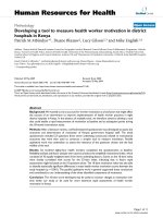
Figure 1. Distribution of the 452 hybrids depending on their retention frequency.
Most of the hybrids (67.7%) showed an average retention frequency below 15%.
Only 32.3% of the hybrids were satisfactory candidates for the final panel.
[0–5[: retention frequencies below 5%.
[5–10[: comprised between 5% and 9.99%.
To determine the extent to which donor fragments were retained in our
hybrids, 46 markers were amplified by PCR from DNA obtained from each
of them. Due to the particularities of the chicken genome structure, care was
taken in the choice of markers to represent all chromosome types. Half of
the markers were from macrochromosomes 1 to 8 and the gonosomes. When
several markers from the same chromosomes were used, they were chosen at
a minimum distance of 8 cM from one another. Sixteen other markers were
from linkage groups assigned to identified microchromosomes. The seven
last markers were from undefined regions of the genome: two were from small
linkage groups, supposedly corresponding to two different microchromosomes,
and five were not linked to any other marker or linkage group in the actual
consensus linkage map of the chicken [12]. We supposed that these last markers
belong to the smallest microchromosomes and we therefore analysed the results
considering two classes of markers: 23 localised on macrochromosomes and
23 localised on microchromosomes.
The average retention rate of the 452 hybrids was 11.3% for the whole gen-
ome, but the overall retention rate for markers located on microchromosomes
was higher (14.8%) as compared to that of macrochromosomes (9.5%).
Thus, only 32.3% of the hybrids showed a whole genome retention frequency
higher than 15% and could be considered as potential candidates for the final
mapping panel (Fig. 1). No higher retention frequency could be observed on
526 M. Morisson et al.
chromosome 4 bearing HPRT [10], since the chromosome 4 markers used in
this study were not close to this gene. The two haploid markers located on
chromosome Z showed retention frequencies lower than the others, due to the
use of female chicken donor cells while the XhoI marker was better retained
among the hybrids reflecting its location close to a centromere (Fig. 2). Indeed,
the cytogenetic assignment for this marker was proposed to be in Wp1.2-q1.1
by Solari et al. [32].
3.2. Pre-selection of 103 candidate hybrids
Considering the low average retention frequency of the chicken-hamster
hybrids, as many as 452 clones were produced, with the aim of a final panel of
90. Our first estimations of the chicken genome loss in the hybrids after large
scale culture, was around 10%. Therefore, we decided to grow the 103 best
candidate hybrids in large scale culture (LSC), in order to be able to choose
90 of them for the definitive panel. As compared to the 452 hybrids, this pre-
selection improved the overall whole genome retention frequency from 11.3%
to 23.8%, the overall macrochromosome retention frequency from 9.5% to
21.5% and the microchromosome one from 14.8% to 28.5% (Fig. 3).
3.3. Selection of the 90 hybrids for the final panel
After LSC, the retention frequencies of the 103 pre-selected hybrids were
estimated using the same 46 markers. The average retention rate was 20.9%
for the whole genome, but the overall retention rate for the markers located
on the macrochromosomes was 18.6% and for the markers located on the
microchromosomes, 25.7% (Fig. 3). The average fragment loss reached 12.5%
for the whole genome: 13.5% for the macrochromosomes and 10% for the
microchromosomes.
We finally selected the 90 hybrids that constitute the definitive panel. The
average retention rates are 21.9% for the whole genome, 20.1% for the mac-
rochromosomes and 25.7% for the microchromosomes (Fig. 3). However,
the retention was not always so uniform, with lower values for the largest
chromosomes: 15.3% for GGA1 and 17.1% for GGA2.
Two control DNAs were included in the panel: the Wg3hCl
2
hamster cell
line DNA and a chicken DNA that is a mix of two female embryonic chicken
DNAs.
4. CONCLUSIONS
A fusion efficiency of approximately 1.4 × 10
−6
clones per chicken fibro-
blast was observed. This fusion efficiency was low in comparison to that of
A chicken radiation hybrid panel 527
Figure 2. Retention frequencies of 46 genome-wide markers typed on the 452 hybrids. The 46 markers were chosen from linkage
groups of the published genetic map [12], 23 are located on macrochromosomes, 16 are on identified microchromosomes, 2 are on
small linkage groups not yet assigned to a chromosome and 5 are genetically unlinked. We thus analysed the results considering two
classes of markers: 23 localised on macrochromosomes and 23 localised on microchromosomes. No higher retention frequency could
be observed on chromosome 4 bearing HPRT. The two haploid markers located on chromosome Z showed retention frequencies lower
than the others, due to the use of female chicken donor cells while the XhoI marker was better retained among the hybrids reflecting
its location close to a centromere. LG W24: linkage group E47/W24; LG W28: linkage group E22/C19/W28; Unlinked: genetically
unlinked markers on the chicken consensus map.
528 M. Morisson et al.
Figure 3. Retention frequencies of the 46 genome-wide markers typed in the 103 candidate hybrids before and after large scale culture
(LSC) and in the final panel of 90 hybrids. The 103 candidate hybrids were cultured on large scale and the retention frequency of the
46 markers was re-evaluated for each of them. The average loss reached 12.5% for the whole genome, but the average loss for the
macrochromosomes was higher (13.5%) than that of the microchromosomes (10.0%). The average retention rates for the final panel
were 21.9% for the whole genome, 20.1% for the macrochromosomes and 25.7% for the microchromosomes. LG W24: linkage group
E47/W24; LG W28: linkage group E22/C19/W28; Unlinked: genetically unlinked markers on the chicken consensus map; LSC: large
scale culture; G: whole genome retention rate; M: macrochromosome retention rate; µ: microchromosome retention rate.
A chicken radiation hybrid panel 529
mammals, e.g. 10
−5
in dogs [35] and 10
−4
in human [36]. This was expected
given the evolutionary distance of the two species, and the results obtained by
others ([18], Bumstead, personal communication).
The average retention rate of the 452 hybrids was 11.3% for the whole
genome, but the overall retention rate for the markers located on the microchro-
mosomes was higher (14.8%) as compared to that of the macrochromosomes
(9.5%). These results were in accordance with those of Kwock et al. [18],
where retention frequencies were 17.8% for microchromosomes and 10.6%
for macrochromosomes. It has been observed that smaller chromosomes are
generally retained at a higher rate, as e.g. in a human WGRH panel [13]. In
the chicken, where the difference in size between the macrochromosomes and
the microchromosomes is more pronounced, this trend is more evident. One
possible explanation is that in a microchromosome, markers are usually at a
closer distance to a centromere, a region where retention frequencies have been
shown to be high [15].
For both practical and economical reasons, reducing the number of hybrid
DNA samples in a panel to the number that can be easily handled in a microplate
is highly desirable. Theoretical data have been reported on the consequences
of hybrid selection on the mapping power of RH panels and it is generally
admitted that a retention frequency over 20% is a minimum prerequisite for a
good efficiency of a panel [16,21,35]. Despite a drastic selection (90 hybrids
selected out of 452 developed), we could not manage to improve the retention
frequency of the two largest macrochromosomes to reach 20%. However, the
overall retention frequency of this panel remains suitable for RH mapping.
DNA extraction produced more than 3 mg of DNA for each hybrid, an amount
sufficient for an estimated 150 000 PCR assuming 20 ng per reaction.
This RH panel is a powerful tool for integrating genetic and physical maps.
Mapping STSs from BAC contig ends will help find the location and orientation
of linkage groups. An obvious exploitation is the mapping of cDNAs and
ESTs onto a framework of microsatellite markers, providing the basis for the
biological characterisation of specific chromosomal regions of interest. Such
RH-mapping data will also be a powerful method in comparative gene mapping
since chromosomal order can be established for expressed genes that are usually
conserved between species but are often recalcitrant to linkage mapping for
a lack of readily detectable allelic variations (for chicken-human comparative
mapping see Schmid et al. [29]).
Approximately 1 000 markers are currently being screened across the RH
panel; a part of them are microsatellite markers anchored on the genetic map and
the others are ESTs. This WGRH panel, named ChickRH6, is available to the
academic community, upon request to the authors (;
).
530 M. Morisson et al.
ACKNOWLEDGEMENTS
We thank Dr Fabienne Pituello for providing 9-day-old chick embryos. This
work was aided by a grant from the Département de Génétique Animale, Institut
national de la recherche agronomique, France.
REFERENCES
[1] Abdrakhmanov I., Lodygin D., Geroth P., Arakawa H., Law A., Plachy J., Korn
B., Buerstedde J M., A large database of chicken bursal ESTs as a resource for
the analysis of vertebrate gene function, Genome Res. 10 (2000) 2062–2069.
[2] Bloom S.E., Delany M.E., Muscarella D.E., Constant and variable features of
the avian chromosomes, in: Etches R.J., Gibbins A.M.V. (Eds.), Manipulation
of the Avian Genome, CRC Press Inc., Orlando, FL, 1993, pp. 39–59.
[3] Clark M.S., Edwards Y.J.K., McQueen H.A., Meek S.E., Smith S., Umrania
Y., Warner S., Williams G., Elgar G., Sequence scanning chicken cosmids: a
methodology for genome screening, Gene 227 (1999) 223–230.
[4] Clinton M., A rapid protocol for sexing chick embryos (Gallus g. domesticus),
Anim. Genet. 25 (1994) 361–362.
[5] Crooijmans R.P.M.A., Vrebalov J., Dijkhof R.J.M., van der Poel J.J., Groenen
M.A.M., Two-dimensional screening of the Wageningen chicken BAC library,
Mamm. Genome 11 (2000) 360–363.
[6] Don R.H., Cox P.T., Wainwright B.J., Baker K., Mattick J.S., “Touchdown” PCR
to circumvent spurious priming during gene amplification, Nucleic Acids Res.
19 (1991) 4008.
[7] Echard G., Gellin J., Gillois M., Localisation des gènes MPI, PKM2, NP sur le
chromosome 3 du porc (Sus scrofa L.) et analyse cytogénétique d’une lignée de
hamster chinois issue de la DON (wg3h), Genet. Sel. Evol. 16 (1984) 261–270.
[8] Fillon V., Morisson M., Zoorob R., Auffray C., Douaire M., Gellin J., Vignal
A., Identification of 16 chicken microchromosomes by molecular markers using
two-colour fluorescence in situ hybridization (FISH), Chromosome Res. 6 (1998)
307–313.
[9] Fillon V., Zoorob R., Yerle M., Auffray C., Vignal A., Mapping of the genetically
independent chicken major histocompatibility complexes B@ and RFP-Y@ to
the same microchromosome by two-color fluorescence in situ hybridization
(FISH), Cytogenet. Cell Genet. 75 (1996) 7–9.
[10] Fukagawa T., Hayward N., Yang J., Azzalin C., Griffin D., Stewart A.F., Brown
W., The chicken HPRT gene: a counter selectable marker for the DT40 cell line,
Nucleic Acids Res. 27 (1999) 1966–1969.
[11] Geisler R., Rauch G J., Baier H., van Bebber F., Broß L., Dekens M.P.S., Finger
K., Fricke C., Gates M.A., Geiger H., Geiger-Rudolph S., Gilmour D., Glaser
S., Gnügge L., Habeck H., Hingst K., Holley S., Keenan J., Kirn A., Knaut H.,
Lashkari D., Maderspacher F., Martyn U., Neuhauss S., Neumann C., Nicolson
T., Pelegri F., Ray R., Rick J.M., Roehl H., Roeser T., Schauerte H.E., Schier
A.F., Schönberger U., Schönthaler H B., Schutle-Merker S., Seydler C., Talbot
W.S., Weiler C., Nüsslein-Volhard C., Haffter P., A radiation hybrid map of the
zebrafish genome, Nat. Genet. 23 (1999) 86–89.
A chicken radiation hybrid panel 531
[12] Groenen M.A.M., Cheng H.H., Bumstead N., Benkel B.F., Briles W.E., Burke
T., Burt D.W., Crittenden L.B., Dodgson J., Hillel J., Lamont S., Ponce de Leon
F.A., Soller M., Takahashi H., Vignal A., A consensus linkage map of the chicken
genome, Genome Res. 10 (2000) 137–147.
[13] Gyapay G., Schmitt K., Fizames C., Jones H., Vega-Czarny N., Spillett D.,
Muselet D., Prud’Homme J.F., Dib C., Auffray C., Morissette J., Weissenbach
J., Goodfellow P.N., A radiation hybrid map of the human genome, Hum. Mol.
Genet. 5 (1996) 339–346.
[14] Hukriede N.A., Joly L., Tsang M., Miles J., Tellis P., Epstein J.A., Barbazuk
W.B., Li F.N., Paw B., Postlethwait J.H., Hudson T.J., Zon L.I., McPherson J.D.,
Chevrette M., Dawid I.B., Johnson S.L., Ekker M., Radiation hybrid mapping of
the zebrafish genome, Proc. Natl. Acad. Sci. USA 96 (1999) 9745–9750.
[15] James M.R., Richard C.W., 3rd, Schott J J., Yousry C., Clark K., Bell J.,
Terwilliger J.D., Hazan J., Dubay C., Vignal A., Agrapart M., Imai T., Nakamura
Y., Polymeropoulos M., Weissenbach J., Cox D.R., Lathrop G.M., A radiation
hybrid map of 506 STS markers spanning human chromosome 11, Nat. Genet. 8
(1994) 70–76.
[16] Jones H.B., Hybrid selection as a method of increasing mapping power for
radiation hybrids, Genome Res. 6 (1996) 761–769.
[17] Kiguwa S.L., Hextall P., Smith A.L., Critcher R., Swinburne J., Millon L.,
Binns M.M., Goodfellow P.N., McCarthy L.C., Farr C.J., Oakenfull E.A., A
horse whole-genome-radiation hybrid panel: Chromosome 1 and 10 preliminary
maps, Mamm. Genome 11 (2000) 803–805.
[18] Kwok C., Korn R.M., Davis M.E., Burt D.W., Critcher R., McCarthy L., Paw
B.H., Zon L.I., Goodfellow P.N., Schmitt K., Characterization of whole genome
radiation hybrid mapping resources for non-mammalian vertebrates, Nucleic
Acids Res. 26 (1998) 3562–3566.
[19] Ladjali-Mohammedi K., Bitgood J.J., Tixier-Boichard M., Ponce de Leon F.A.,
International System for Standardized Avian Karyotypes (ISSAK): standardized
banded karyotypes of the domestic fowl (Gallus domesticus), Cytogenet. Cell
Genet. 86 (1999) 271–276.
[20] Lee M K., Ren C., Ling P., Cox B., Dodgson J.B., Zhang H B., Towards a
BAC-based physical map of the chicken genome, in: Plant & Animal Genome
IX Conference, 2001, San Diego.
[21] Lunetta K.L., Boehnke M., Lange K., Cox D.R., Experimental design and error
detection for polyploid radiation hybrid mapping, Genome Res. 5 (1995) 151–
163.
[22] McCarthy L.C., Bihoreau M T., Kiguwa S.L., Browne J., Watanabe T.K.,
Hishigaki H., Tsuji A., Kiel S., Webber C., Davis M.E., Knights C., Smith
A., Critcher R., Huxtall P., Hudson J.R., Jr., Ono T., Hayashi H., Takagi T.,
Nakamura Y., Tanigami A., Goodfellow P.N., Lathrop G.M., James M.R., A
whole-genome radiation hybrid panel and framework map of the rat genome,
Mamm. Genome 11 (2000) 791–795.
[23] McCarthy L.C., Terrett J., Davis M.E., Knights C.J., Smith A.L., Critcher R.,
Schmitt K., Hudson J., Spurr N.K., Goodfellow P.N., A first-generation whole
genome-radiation hybrid map spanning the mouse genome, Genome Res. 7
(1997) 1153–1161.
532 M. Morisson et al.
[24] McQueen H.A., Siriaco G., Bird A.P., Chicken microchromosomes are hyper-
acetylated, early replicating, and gene rich, Genome Res. 8 (1998) 621–630.
[25] Morisson M., Pitel F., Fillon V., Pouzadoux A., Bergé R., Vit J P., Zoorob R.,
Auffray C., Gellin J., Vignal A., Integration of chicken cytogenetic and genetic
maps: 18 new polymorphic markers isolated from BAC and PAC clones, Anim.
Genet. 29 (1998) 348–355.
[26] Murphy W.J., Menotti-Raymond M., Lyons L.A., Thompson M.A., O’Brien S.J.,
Development of a feline whole genome radiation hybrid panel and comparative
mapping of human chromosome 12 and 22 loci, Genomics 57 (1999) 1–8.
[27] Olivier M., Aggarwal A., Allen J., Almendras A.A., Bajorek E.S., Beasley E.M.,
Brady S.D., Bushard J.M., Bustos V.I., Chu A., Chung T.R., De Witte A., Denys
M.E., Dominguez R., Fang N.Y., Foster B.D., Freudenberg R.W., Hadley D.,
Hamilton L.R., Jeffrey T.J., Kelly L., Lazzeroni L., Levy M.R., Lewis S.C., Liu
X., Lopez F.J., Louie B., Marquis J.P., Martinez R.A., Matsuura M.K., Misherghi
N.S., Norton J.A., Olshen A., Perkins S.M., Perou A.J., Piercy C., Piercy M.,
Qin F., Reif T., Sheppard K., Shokoohi V., Smick G.A., Sun W L., Stewart
E.A., Tejeda J.F., Tran N.M., Trejo T., Vo N.T., Yan S.C.M., Zierten D.L., Zhao
S., Sachidanandam R., Trask B.J., Myers R.M., Cox D.R., A high-resolution
radiation hybrid map of the human genome draft sequence, Science 291 (2001)
1298–1302.
[28] Rodionov A.V., Micro versus macro: a review of structure and functions of avian
micro- and macrochromosomes, Russ. J. Genet. 32 (1996) 517–527.
[29] Schmid M., Nanda I., Guttenbach M., Steinlein C., Hoehn H., Schartl M., Haaf
T., Weigend S., Fries R., Buerstedde J M., Wimmers K., Burt D.W., Smith J.,
A’Hara S., Law A., Griffin D.K., Bumstead N., Kaufman J., Thomson P.A.,Burke
T., Groenen M.A.M., Crooijmans R.P.M.A., Vignal A., Fillon V., Morisson M.,
Pitel F., Tixier-Boichard M., Ladjali-Mohammedi K., Hillel J., Mäki-Tanila A.,
Cheng H.H., Delany M.E., Burnside J., Mizuno S., First report on chicken genes
and chromosomes 2000, Cytogenet. Cell Genet. 90 (2000) 169–218.
[30] Smith J., Bruley C.K., Paton I.R., Dunn I., Jones C.T., Windsor D., Morrice D.R.,
Law A.S., Masabanda J., Sazanov A., Waddington D., Fries R., Burt D.W., Dif-
ferences ingene density on chicken macrochromosomes and microchromosomes,
Anim. Genet. 31 (2000) 96–103.
[31] Smith J., Paton I.R., Bruley C.K., Windsor D., Burke D., Ponce de Leon
F.A., Burt D.W., Integration of the genetic and physical maps of the chicken
macrochromosomes, Anim. Genet. 31 (2000) 20–27.
[32] Solari A.J., Dresser M.E., High-resolution cytological localization of the Xho1
and EcoR1 repeat sequences in the pachytene ZW bivalent of the chicken,
Chromosome Res. 3 (1995) 87–93.
[33] Stewart E.A., McKusick K.B., Aggarwal A., Bajorek E., Brady S., Chu A., Fang
N., Hadley D., Harris M., Hussain S., Lee R., Maratukulam A., O’Connor K.,
Perkins S., Piercy M., Qin F., Reif T., Sanders C., She X., Sun W L., Tabar P.,
Voyticky S., Cowles S., Fan J B., Mader C., Quackenbush J., Myers R.M., Cox
D.R., An STS-based radiation hybrid map of the human genome, Genome Res.
7 (1997) 422–433.
A chicken radiation hybrid panel 533
[34] Tirunagaru V.G., Sofer L., Cui J., Burnside J., An expressed sequence tag
database of T-cell-enriched activated chicken splenocytes: sequence analysis
of 5 251 clones, Genomics 66 (2000) 144–151.
[35] Vignaux F., Hitte C., Priat C., Chuat J C., Andre C., Galibert F., Construction and
optimization of a dog whole-genome radiation hybrid panel, Mamm. Genome
10 (1999) 888–894.
[36] Walter M.A., Spillett D.J., Thomas P., Weissenbach J., Goodfellow P.N., A
method for constructing radiation hybrid maps of whole genomes, Nat. Genet. 7
(1994) 22–28.
[37] Womack J.E., Johnson J.S., Owens E.K., Rexroad C.E., 3rd, Schläpfer J., Yang
Y P., A whole-genome radiation hybrid panel for bovine gene mapping, Mamm.
Genome 8 (1997) 854–856.
[38] Yerle M., Pinton P., Robic A., Alfonso A., Palvadeau Y., Delcros C., Hawken
R., Alexander L., Beattie C., Schook L., Milan D., Gellin J., Construction of a
whole-genome radiation hybrid panel for high-resolution gene mapping in pigs,
Cytogenet. Cell Genet. 82 (1998) 182–188.
To access this journal online:
www.edpsciences.org