Báo cáo y học: "Gene-dosage effects in Down syndrome and trisomic mouse models" pot
Bạn đang xem bản rút gọn của tài liệu. Xem và tải ngay bản đầy đủ của tài liệu tại đây (75.28 KB, 4 trang )
Genome Biology 2004, 5:244
comment
reviews
reports deposited research
interactions
information
refereed research
Minireview
Gene-dosage effects in Down syndrome and trisomic mouse
models
Katheleen Gardiner
Address: Eleanor Roosevelt Institute at the University of Denver, Department of Biochemistry and Molecular Genetics, University of
Colorado Health Sciences Center, 1899 Gaylord Street, Denver, CO 80206, USA. E-mail:
Abstract
The abnormalities found in human Down syndrome (trisomy 21) have been thought to result
from increased expression of genes on chromosome 21 because of their higher gene dosage.
Now, several groups have shown this to be generally the case, but some inter-individual
variability and other exceptions were found.
Published: 30 September 2004
Genome Biology 2004, 5:244
The electronic version of this article is the complete one and can be
found online at />© 2004 BioMed Central Ltd
The gene-dosage hypothesis for Down
syndrome
The Down syndrome (trisomy 21) phenotype is characterized
by abnormalities affecting most organs and organ systems
[1]. Although the extent and severity of the abnormalities is
highly variable among individuals, all have some level of
intellectual disability that is associated with specific brain
regions and the performance of specific cognitive tasks [2,3].
The incidence of Down syndrome remains at approximately
1 in 800 live births, and given the intellectual disabilities, it
is a significant social and medical issue. Although it has been
known for several decades that Down syndrome is caused by
an extra, normal copy of the long arm of human chromo-
some 21 (21q), the molecular and cellular events linking the
presence of an extra chromosome to the phenotypic features
are unknown. The central hypothesis in Down-syndrome
research is that gene dosage results in a 50% increase in
expression of genes on chromosome 21q, and that this
directly or indirectly alters the timing, pattern or extent of
development. Accordingly, an essential question in Down-
syndrome research is: are all trisomic genes overexpressed
in all tissues and at all time points? If not, then which genes
are overexpressed, and when and where? Answers to these
questions are critical for determining which genes are rele-
vant to phenotype development, for linking expression of
specific genes to specific phenotypic features, and to account
for phenotypic variability.
Chromosome 21 encodes over 300 genes and predicted genes
[4]. These include approximately 170 protein-coding genes
with clear orthologs in the mouse genome plus a significant
number of gene models predicted on the basis of spliced
expressed sequence tags (ESTs) with verified expression.
Because analyses of cases of Down syndrome that are due to
partial trisomies of chromosome 21q have not eliminated any
significant segment of 21q from containing genes potentially
impacting cognitive function [5], and because we understand
so little about individual gene functions, essentially all genes
within 21q need to be considered as candidates for relevance to
the Down-syndrome phenotype. Given the large number of
genes involved, determining which genes are overexpressed
requires a large-scale approach, which is complicated by the
small differences in expression level (50%) between normal
and Down syndrome individuals predicted by gene dosage.
Recently, several groups have contributed to progress in this
area, by screening microarrays with RNA from brains of
human fetuses with Down syndrome [6] or a trisomic mouse
model [7], by screening a cDNA array containing mouse
orthologs of human chromosome 21 genes with RNA from
several tissues of a mouse model [8], and by using quantitative
reverse-transcriptase-coupled (RT) PCR analysis of RNA from
different tissues of a mouse model at different ages [9]. The
results of all four studies support the hypothesis that gene-
dosage effects exist in Down syndrome, but they also show that
dosage effects may be specific to particular genes, alleles
and/or tissues, and that background and stochastic or transient
effects may be confounding factors.
Large-scale studies of gene-dosage effects in
Down-syndrome fetuses and trisomic mice
Mao et al. [6] screened two Affymetrix oligonucleotide
arrays (containing probes for approximately 12,000 and
22,000 human genes) with RNA from age-matched Down-
syndrome and euploid control fetuses at 17-20 weeks gesta-
tion (see Table 1). They used RNA from four normal and four
Down-syndrome cerebrums and from four normal and four
Down-syndrome-derived astrocyte cell lines cultured from
cerebral cortex. Samples were analyzed individually in order
to detect variation between individuals. From all analyses, a
global increase in expression level of 25 chromosome 21
genes was observed in the Down-syndrome samples com-
pared to the euploid controls. The increase overall was con-
sistent with predictions if gene expression followed gene
dosage, but levels varied among individuals such that there
were examples of individual genes showing no increases in
comparisons of individual Down-syndrome-euploid pairs
(see also below). Importantly, these variations in expression
levels might be linked to phenotypic variations that would
have been apparent at later developmental time points. Dif-
ferences in expression levels of genes on other chromosomes
- about 85 genes whose expression increased and 100 whose
expression decreased - were also observed.
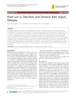
Regions of human chromosome 21 are orthologous to seg-
ments of three mouse chromosomes [10] (Figure 1): the cen-
tromere-proximal 30 megabase (Mb) region of chromosome
21 up to and including the ZNF295 gene is orthologous to
the telomeric region of mouse chromosome 16 [10] and the
next two approximately 1-2 Mb segments of chromosome 21
are orthologous to regions of mouse chromosomes 17 and 10,
respectively. Because of the large region of homology with
mouse chromosome 16, development of segmental trisomy
mouse models for Down syndrome has focused on this
region. Currently, the best mouse models of Down syndrome
are the Ts65Dn mouse (reviewed in [11,12]) and the Ts1Cje
mouse [13]. Ts65Dn mice have three copies of 94 genes
orthologous to human chromosome 21 genes, contained
within chromosome 16 from the Gabpa/App gene cluster to
the distal telomere [4]. Ts1Cje mice are trisomic for 71
orthologs of human chromosome 21 genes, within chromo-
some 16 distal to (and not including) the superoxide dismu-
tase 1 (Sod1) gene to the telomere [13].
Amano et al. [7] screened Affymetrix oligonucleotide arrays
representing about 11,000 mouse genes with RNA from
whole brains of postnatal day zero Ts1Cje mice. Six trisomic
females and six littermate controls were examined, and 38
genes within the trisomic segment showed detectable expres-
sion. Of these, 37 showed a mean increase in expression level
of about 1.5-fold in the trisomic mice, consistent with gene-
dosage effects. But out of all the possible trisomy:euploid
comparisons (6 × 6 = 36), not all individual pairwise compar-
isons showed similar increases. Indeed, of the 37 genes, only
24 showed increases in 18 or more of the 36 possible pairs.
Because these mice are maintained on an inbred background
(C57BL/6J), the only genetic contribution to expression dif-
ferences is the trisomic segment. Expression levels of ten tri-
somic genes were assayed by real-time RT-PCR using RNA
244.2 Genome Biology 2004, Volume 5, Issue 10, Article 244 Gardiner />Genome Biology 2004, 5:244
Table 1
Overview of studies of gene-dosage effects in trisomies
Mao et al. [6] Amano et al. [7] Kahlem et al. [8] Lyle et al. [9]
Species and strain Humans with Down Ts1Cje mice Ts65Dn mice Ts65Dn mice
syndrome
Number of individuals 4 plus 4 controls 6 plus 6 controls 4 plus 4 controls 4 plus 4 controls
Ages 17-20 weeks gestation Postnatal day 0 3-4 months Postnatal day 30 and 11
months
Tissues Cerebrum and cortex-derived Whole brains Cortex, midbrain, cerebellum, Brain, liver, kidney, heart,
astrocyte cell lines heart, testis, liver, kidney, lung, muscle, and lung
and muscle
Technique used Affymetrix microarrays Affymetrix microarrays Custom cDNA array RT-PCR
Number of genes on Over 300 71 94 (77 tested in 9 tissues) 94 (78 tested in 10 tissues)
trisomic segment
Trisomic genes increased 25 (variation among 37 (variation among All genes (of 66 with detectable 607 gene-tissue
in expression individuals) individuals) expression) except in muscle combinations (of 666 with
and 10 gene-tissue combinations detectable expression)
Non-trisomic genes with Approximately185 258 Not determined Not determined
altered expression
from four additional trisomic and normal control mice. All
showed relative expression increases of over 1.34-fold,
including those with increases of about 1.2-fold in the array
experiments, as well as the Prdm15 (Znf295) gene, which
showed no increase in arrays. Also, similar to the results of
Mao et al. [6], some non-trisomic genes showed altered
expression levels: 59 showed levels under 0.7 times normal
and 199 showed levels over 1.2 times normal, out of the
10,000 genes with detectable expression.
Kahlem et al. [8] created custom arrays containing cDNAs
for 77 orthologs of human chromosome 21 genes that are tri-
somic in the Ts65Dn mouse. Arrays were screened with
Ts65Dn RNA from nine tissues, including cerebellum, cortex
and midbrain, in each case pooling RNAs from four individ-
uals, aged 3-4 months. Expression of 66 trisomic genes was
detected in at least one of the nine tissues. In eight tissues,
overall levels of expression were consistent with gene
dosage, with trisomy:euploid ratios ranging from about 1.63
and about 1.73 in cortex and heart, respectively, to about
1.23 in kidney. Only muscle, with ratios of 1.16, failed to
show notable gene-dosage effects. A small number of specific
gene-tissue combinations deviated from dosage effects; 10
combinations showed unchanged or decreased ratios and 15
showed ratios over 2.0. Quantitative RT-PCR analysis of
several genes corroborated the array results in 78% of cases.
To circumvent the sensitivity limitations of microarrays,
which do not always detect genes expressed at low levels, Lyle
et al. [9] used real-time RT-PCR for experiments with Ts65Dn
mice. RNA from brain, liver, kidney, heart, muscle and lung of
postnatal day 30 mice, and brain, liver, kidney and heart of 11-
month-old mice was used, in each case pooling material from
four trisomic or euploid male mice. Assays of 78 trisomic
genes showed an overall mean expression ratio of approxi-
mately 1.5. There were statistically significant variations,
however. For example, 59 gene-tissue combinations (out of
666; 78 genes were tested in 10 tissues, and 114 gene-tissue
combinations had no detectable expression) showed no signif-
icant increase in trisomic mice, and 26 gene-tissue combina-
tions showed over 2.2-fold increases in trisomic mice.
Variability between individuals
Both Mao et al. [6] and Amano et al. [7] observed deviations
from dosage predictions of trisomy:euploid RNA-expression
ratios in some pairwise comparisons between individuals.
Sources of such variation - apart from experimental artifacts
and limitations - include allelic variation in trisomic genes or
in background (euploid) genes that regulate trisomic genes
and possible stochastic or environmental effects. Certainly,
allelic variation has been observed in human gene-expres-
sion patterns [14-16]. Specifically for chromosome 21,
promoter polymorphisms in six genes were reported that
resulted in expression differences of 30-66% among alleles
expressed in HEK293 embryonic kidney cells [17]. If an
individual trisomic for a low-expressing allele is compared
to a euploid individual disomic for a high-expressing allele,
significant expression differences may be undetectable; con-
versely, trisomy for a high-expressing allele could result in
much more than a 50% difference compared with a euploid
for low-expressing alleles. Similar arguments apply to back-
ground variation (in euploid genes) in humans and in
Ts65Dn mice (because the latter are maintained as F1
hybrids of two strains).
The experiments with Ts65Dn mice used RNAs pooled from
multiple individuals [8,9]. Variation between individuals is
expected because of the history of the strain: in the Ts65Dn
mice, the trisomic segment originated from the DBA/2J
strain, but because of extensive early breeding to
C57BL/6JEi, there may be no DBA/2J alleles remaining
except immediately around the breakpoint of the rearrange-
ment. The Ts65Dn strain is now maintained as F1 hybrids of
C57BL/6JEi and C3H/HeSnJ [18]. Thus, the three alleles in
individual mice may all derive from C57BL/6JEi or be
combinations of C57BL/6JEi and C3H/HeSnJ, and inter-
individual variation will be found.
Genetic variation does not exist, however, in the experiments
with Ts1Cje mice, because they are maintained on the inbred
comment
reviews
reports deposited research
interactions
information
refereed research
Genome Biology 2004, Volume 5, Issue 10, Article 244 Gardiner 244.3
Genome Biology 2004, 5:244
Figure 1
Human chromosome 21 and homologous regions in mouse models.
Regions that are syntenic with mouse chromosomes are indicated on the
left; those that are trisomic in the major mouse models are indicated on
the right. See text for further details.
Trisomic regions in
mouse models
Mouse-chromosome
homologies
Ts65Dn
Ts1Cje
16
17
10
About 1.2 Mb
About
30 Mb
About
2.2 Mb
ZNF295
SODI
GABPA/APP
11.2
21.2
22.11
22.12
22.13
22.2
22.3
21
C57BL/6J strain. The reported inter-individual variation in
expression levels [7] must therefore have other causes, such
as stochastic processes or subtle environmental effects, possi-
bly fluctuating in time but frozen in these snapshots of
expression patterns. This is consistent with reports of vari-
able expression levels of 37 genes in the hippocampus of
inbred rats (the Sprague-Dawley strain); 2-3-fold variations
in average expression level were seen among 20 individuals,
with as much as 10-fold variation seen with some genes [19].
Deviation from the expected 1.5-fold increase
and variations in non-trisomic gene expression
Gene-tissue combinations with ratios of trisomy:euploid
expression significantly greater or less than 1.5-fold were
reported in the Ts65Dn experiments [8,9]. Notably, Kahlem
et al. [8] reported that expression of trisomic genes in skele-
tal muscle was increased only 1.16-fold, but Lyle et al. [9]
reported that expression of only 28 of 78 genes was
increased significantly less than 1.5-fold. Is this difference
due to the different techniques used (arrays versus RT-PCR),
or to age effects (day 30 versus 3-4-month-old mice)? Also,
while the former study [8] reported only 5 gene-tissue com-
binations with levels increased over 1.5-fold (excluding
testes), and 15 increased under 1.5-fold (out of 594 combina-
tions), the latter [9] reported 123 combinations with over
1.5-fold increases and 298 with under 1.5-fold increases (out
of 666). Although these differences are reported as statisti-
cally significant, in particular for data from Lyle et al. [9],
the biological significance remains to be determined. For
example, it is not possible to state that the cell will recognize
1.43-fold as different from 1.50-fold for the hormonally
upregulated Neu-associated kinase (Hunk) gene in the post-
natal day 30 brain, or 1.41-fold as not different from normal
for the GABPA transcription factor gene in the kidney [9].
Alterations in expression levels of many non-trisomic genes
were reported by two of the studies [6,7]. Currently, there is
no known functional link between the euploid genes and the
trisomic genes that would predict such dysregulation. Fur-
thermore, 19 of the euploid genes found to have altered
expression by Mao et al. [6] were reported to be expressed
by Amano et al. [7], but none showed altered expression in
the latter study. Such inconsistencies could be due to differ-
ences in the organism, tissue, or developmental time
studied, or to subtle environmental effects.
From the results of these experiments [6-9], it is reasonable
to conclude that expression of trisomic mRNAs in human
Down syndrome and mouse chromosome 16 segmental tri-
somies is governed by gene dosage. Additional large-scale
experiments along these lines, therefore, seem unnecessary
and are unlikely to further illuminate the field. The inter-
individual variation, and the possible specific effects on reg-
ulation of genes in different tissues at different times,
however, argue that dosage still needs to be examined
carefully, but this will be more productive when applied to
specific genes, specific tissues or cell types, among individu-
als, and in relation to phenotypic variability. The preference,
of course, is to examine expression at the protein level.
Acknowledgements
This work was supported by the Fondation Jerome Lejeune.
References
1. Epstein CJ: Down syndrome (trisomy 21). In Metabolic and Mole-
cular Bases of Inherited Disease. Edited by Scriver CA, Beaudet AL, Sly
WS, Valle D. New York: McGraw Hill; 1995, 749-794.
2. Pennington BF, Moon J, Edgin J, Stedron J, Nadel L: The neuropsy-
chology of Down syndrome: evidence for hippocampal dys-
function. Child Dev 2003, 74:75-93.
3. Nadel L: Down’s syndrome: a genetic disorder in biobehav-
ioral perspective. Genes Brain Behav 2003, 2:156-166.
4. Gardiner K, Fortna A, Bechtel L, Davisson MT: Mouse models of
Down syndrome: how useful can they be? Comparison of
the gene content of human chromosome 21 with ortholo-
gous mouse genomic regions. Gene 2003, 318:137-147.
5. Korenberg JR, Chen XN, Schipper R, Sun Z, Gonsky R, Gerwehr S,
Carpenter N, Daumer C, Dignan P, Disteche C, et al.: Down syn-
drome phenotypes: the consequences of chromosomal
imbalance. Proc Natl Acad Sci USA 1994, 91:4997-5001.
6. Mao R, Zielke CL, Zielke HR, Pevsner J: Global up-regulation of
chromosome 21 gene expression in the developing Down
syndrome brain. Genomics 2003, 81:457-467.
7. Amano K, Sago H, Uchikawa C, Suzuki T, Kotliarova SE, Nukina N,
Epstein CJ, Yamakawa K: Dosage-dependent over-expression of
genes in the trisomic region of Ts1Cje mouse model for
Down syndrome. Hum Mol Genet 2004, 13:1333-1340.
8. Kahlem P, Sultan M, Herwig R, Steinfath M, Balzereit D, Eppens B,
Saran NG, Pletcher MT, South ST, Stetten G, et al.: Transcript
level alterations reflect gene dosage effects across multiple
tissues in a mouse model of Down syndrome. Genome Res
2004, 14:1258-1267.
9. Lyle R, Gehrig C, Neergaard-Henrichsen C, Deutsch S, Antonarakis
SE: Gene expression from the aneuploid chromosome in a
trisomy mouse model of Down syndrome. Genome Res 2004,
14:1268-1274.
10. Davisson MT, Bechtel LJ, Akeson EC, Fortna A, Slavov D, Gardiner K:
Evolutionary breakpoints on human chromosome 21. Genomics
2001, 78:99-106.
11. Davisson MT, Costa ACS: Mouse models of Down syndrome. In
Mouse Models in the Study of Genetic Neurological Disorders. Volume 9 of
Advances in Neurochemistry. Edited by Popko B. New York: Plenum
Press; 1999, 297-327.
12. Crnic LS, Pennington BF: Down syndrome: neuropsychology
and animal models. Progr Infancy Res 2000, 1:69-111.
13. Sago H, Carlson EJ, Smith DJ, Kilbridge J, Rubin EM, Mobley WC,
Epstein CJ, Huang TT: Ts1Cje, a partial trisomy 16 mouse
model for Down syndrome, exhibits learning and behavioral
abnormalities. Proc Natl Acad Sci USA 1998, 95:6256-6261.
14. Yan H, Yuan W, Velculescu VE, Vogelstein B, Kinzler KW: Allelic
variation in human gene expression. Science 2002, 297:1143.
15. Cheung VG, Conlin LK, Weber TM, Arcaro M, Jen KY, Morley M,
Spielman RS: Natural variation in human gene expression
assessed in lymphoblastoid cells. Nat Genet 2003, 33:422-425.
16. Morley M, Molony CM, Weber TM, Devlin JL, Ewens KG, Spielman
RS, Cheung VG: Genetic analysis of genome-wide variation in
human gene expression. Nature 2004, 430:743-747.
17. Buckland PR, Coleman SL, Hoogendoorn B, Guy C, Smith SK,
O’Donovan MC: A high proportion of chromosome 21 pro-
moter polymorphisms influence transcriptional activity.
Gene Expr 2004, 11:233-239.
18. Davisson MT, Schmidt C, Reeves RH, Irving NG, Akeson EC, Harris
BS, Bronson RT: Segmental trisomy as a mouse model for
Down syndrome. Prog Clin Biol Res 1993, 384:117-133.
19. Alfonso J, Pollevick GD, Castensson A, Jazin E, Frasch AC: Analysis
of gene expression in the rat hippocampus using real time
PCR reveals high inter-individual variation in mRNA expres-
sion levels. J Neurosci Res 2002, 67:225-234.
244.4 Genome Biology 2004, Volume 5, Issue 10, Article 244 Gardiner />Genome Biology 2004, 5:244