Báo cáo y học: "A computational method to predict genetically encoded rare amino acids in proteins" pdf
Bạn đang xem bản rút gọn của tài liệu. Xem và tải ngay bản đầy đủ của tài liệu tại đây (1.09 MB, 15 trang )
Genome Biology 2005, 6:R79
comment reviews reports deposited research refereed research interactions information
Open Access
2005Chaudhuri and YeatesVolume 6, Issue 9, Article R79
Method
A computational method to predict genetically encoded rare amino
acids in proteins
Barnali N Chaudhuri and Todd O Yeates
Address: UCLA-DOE Institute for Genomics and Proteomics and Department of Chemistry and Biochemistry, University of California, Los
Angeles, USA.
Correspondence: Todd O Yeates. E-mail:
© 2005 Chaudhuri and Yeates; licensee BioMed Central Ltd.
This is an Open Access article distributed under the terms of the Creative Commons Attribution License (
which permits unrestricted use, distribution, and reproduction in any medium, provided the original work is properly cited.
Prediction of rare amino acids<p>A new method for predicting recoding by rare amino acids such as selenocysteine and pyrrolysine was used to survey a set of microbial genomes.</p>
Abstract
In several natural settings, the standard genetic code is expanded to incorporate two additional
amino acids with distinct functionality, selenocysteine and pyrrolysine. These rare amino acids can
be overlooked inadvertently, however, as they arise by recoding at certain stop codons. We report
a method for such recoding prediction from genomic data, using read-through similarity evaluation.
A survey across a set of microbial genomes identifies almost all the known cases as well as a number
of novel candidate proteins.
Background
Codon redefinitions that expand upon the standard genetic
code beyond the 20 canonical amino acids are reported in all
three domains of life [1,2]. Two known genetically encoded
rare amino acids (RAAs) are selenocysteine and pyrrolysine,
the proposed 21
st
and the 22
nd
amino acids, respectively [3-7].
Selenocysteine, a selenium-analog of cysteine, is a potent
nucleophile [5] and has been reported in organisms as diverse
as Escherichia coli and human beings [4,5]. Selenium plays a
dual role in nature as an essential micronutrient in human
health, and as an environmental hazard to humans, livestock
and wildlife [8] when it is present in high amounts. Thus,
selenium is a target for both molecular biology and bioreme-
diation research [8,9]. The distribution of selenium in the
form of selenocysteine residues [5,10] in specific proteins is
not completely understood. Pyrrolysine is a recently discov-
ered amino acid in the methanogenic archaeon Methanosa-
rcina barkeri, where it supposedly plays a critical role in
methyltransferase chemistry as an electrophile [6,7]. Tradi-
tional genomic sequence analyses tend to overlook these
RAAs, leading to mis-annotation in the sequence databases.
Systematic bioinformatic investigations of the genomic data
offer the possibility of understanding which organisms utilize
RAAs, and which proteins in particular incorporate them into
their structures.
Predicting which natural proteins contain the RAA seleno-
cysteine or pyrrolysine on the basis of genomic sequence data
is a difficult problem [2]. The difficulty arises from the dis-
tinction that, unlike other amino acids, RAAs are not coded
for by dedicated codons. Instead, they are incorporated in
special circumstances by the UGA (opal; selenocysteine) and
the UAG (amber; pyrrolysine) codons [3-7], which are ordi-
narily interpreted as stop signals to terminate translation
(Figure 1a). From a genomics point of view, the problem is
how to discriminate between all the true stop signals in
genomic sequence data, and those cases that signal for incor-
poration of a RAA. At the mRNA level, one feature referred to
as the selenocysteine insertion sequence (SECIS) hairpin
motif is understood to signal for selenocysteine insertion. The
situation is greatly complicated, however, by the divergence
of the signal between different proteins and between different
organisms with respect to the sequence and position of the
signaling element, situated in either the 3' or 5' untranslated
Published: 31 August 2005
Genome Biology 2005, 6:R79 (doi:10.1186/gb-2005-6-9-r79)
Received: 8 March 2005
Revised: 20 June 2005
Accepted: 27 July 2005
The electronic version of this article is the complete one and can be
found online at />R79.2 Genome Biology 2005, Volume 6, Issue 9, Article R79 Chaudhuri and Yeates />Genome Biology 2005, 6:R79
Figure 1 (see legend on next page)
U
SECIS
UGA
mRNA
tRNA-sec
Codon
(a)
(b)
(c)
U
Readthrough similari
ty
evaluation
BLAST search
window
Top BLAST
hit
Start
Stop
Extended regio
n
C
Candidate ORF
SelB
35 microbial genomes with SID genes
203,339 predicted ORFs with UGA terminus
Similarity based read-through evaluation
3,594 ORFs selected
Evaluation
of multiple
sequence
alignment
(109 ORFs selected)
Species-
specific
SECIS
check
(bacteria)
92 predictions
7 new candidates
5’
3’
Genome Biology 2005, Volume 6, Issue 9, Article R79 Chaudhuri and Yeates R79.3
comment reviews reports refereed researchdeposited research interactions information
Genome Biology 2005, 6:R79
region of a recoded open reading frame (ORF; archaea/
eukaryotes) or downstream of the recoded UGA (bacteria).
Much less is understood about the newly discovered pyrroly-
sine incorporation machinery. The presence of a PYLIS
(SECIS-equivalent) cis-acting element [2], and competition
between translational termination and read-through, have
been anticipated [11].
A number of earlier studies by Gladyshev and coworkers [12-
16] have addressed the problem of predicting selenopro-
teomes, producing sets of selenoproteins encoded in various
genomes. Systematic selenocysteine predictions in prokaryo-
tes have been based on two criteria: alignment of the 'UGA'
codon in the mRNA sequence with cysteine in homologous
proteins in a pair-wise sequence alignment (henceforth, the
cysteine alignment criterion), and the detection of a consen-
sus SECIS signal in the nucleotide sequences (henceforth, the
SECIS criterion). Both methods performed very well with
near-zero false negatives [13,16]. Nevertheless, certain
aspects of these approaches make them less suitable for gen-
eralized applications. For example, they cannot be applied to
selenoproteins that fail to fit the cysteine alignment criterion
(those selenoproteins that do not have a homolog in the data-
base with a cysteine residue taking the place of the seleno-
cysteines). The SECIS criterion also presents some
limitations. High numbers of false positives arise with the
genome-wide prediction of short, local RNA folding motifs,
such as the SECIS element [17]. The observation that different
organisms have divergent signals for selenocysteine insertion
complicates the problem further [13,16]. Other models that
do not rely on the identification of specific recoding signals,
such as evaluation of the coding potential of the nucleotide
sequence beyond the UGA termini, have been developed for
eukaryotes [14]. To overcome the various difficulties associ-
ated with the detection of rare selenoproteins from genomic
data, a combination of strategies is shown to be advantageous
[2,14]. A database homology search using the entire lengths of
candidate genes with an in-frame UAG codon has been
employed recently for analyzing the nature of pyrrolysine
decoding in methanogens [11].
Here we expand upon ideas developed by Gladyshev and col-
leagues [12-16], and introduce a new, multi-component
scheme for microbial selenocysteine and pyrrolysine predic-
tion. Several criteria are combined in series, including a new
predictive element, 'read-through similarity analysis' (RSA;
Figure 1b). The RSA criterion is applied in the early stage of
the procedure to evaluate the read-through potential of an
ORF based on an analysis of sequence similarity involving the
hypothetical amino acid sequence translated beyond the can-
didate stop codon. This scheme is model-free, in the sense
that it does not rely on any special RNA context, read-through
mechanism, or incorporation of any particular amino acid
residue at the recoding site. Following the RSA analysis, sub-
sequent criteria (for example, cysteine alignment and SECIS)
can be enforced, or overridden in special cases where the
other criteria provide compelling evidence for a bona-fide
read-through situation. Success of this predictive approach is
not, therefore, strictly contingent on the presence of a protein
homolog containing a cysteine substitution in the database or
on a canonical SECIS motif in the case of selenoproteins. In
addition to almost all of the known cases of UGA-encoded
selenocysteines (Table 1), the present method successfully
identifies several proteins with UAG-encoded pyrrolysine
(Table 2), including novel candidates, as well as instances of
genome-wide redefinition of UGA as a particular amino acid,
such as tryptophan in Mycoplasma spp. The generality and
wide applicability of the present approach makes it well
suited to the critical problem of analyzing the rapidly growing
number of new genomes.
Results and discussion
The selenoprotein prediction scheme
Our selenoproteome prediction scheme was developed based
on the expectation that a putative selenoprotein will satisfy
the following, specific conditions. It should show: a signifi-
cant 'read-through similarity' (see below); an alignment of the
selenocysteine residue with semi-invariant cysteine resi-
due(s) in a set of aligned homologs; and a hairpin motif (puta-
tive SECIS) near the candidate ORF, which is consistent with
the hairpin motifs near the other selenoproteins found in the
same organism. The components of the predictive approach
are combined as shown in Figure 1c. The RSA method incor-
porates an analysis of the protein sequences following the
presumptive stop codons in a genome (Figure 1b). Due to the
recoding of UGA as a selenocysteine, the sequence following
the UGA codon would be translated as the carboxy-terminal
part of an extended protein. This makes it possible to identify
candidate selenoproteins in situations where the putative
protein sequence immediately following a UGA codon is sta-
tistically similar to the aligned region of another homologous
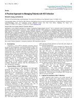
Schematic representation of the selenocysteine insertion machinery and the selenoprotein detection schemeFigure 1 (see previous page)
Schematic representation of the selenocysteine insertion machinery and the selenoprotein detection scheme. (a) A cartoon diagram of selenocysteine
incorporation during protein translation inside the cell. The selenocysteine-specific elongation factor (SelB; pink) is shown interacting with the
selenocysteine insertion sequence (SECIS) hairpin element in the mRNA and tRNA-sec (SelC). The anticodon of SelC tRNA interacts with and recognizes
the 'UGA' codon. The ribosome and other components of the translational machinery are omitted for clarity. (b) Schematic representation of the 'read-
through similarity analysis' approach. The top BLAST hit is shown in blue. The window lengths used for the BLAST search and read-through similarity
evaluation are marked in the drawing. (c) A flow chart describing how the different components of the predictive scheme are combined for selenoprotein
prediction. ORF, open reading frame.
R79.4 Genome Biology 2005, Volume 6, Issue 9, Article R79 Chaudhuri and Yeates />Genome Biology 2005, 6:R79
Table 1
A list of predicted selenoproteins encoded by UGA read-through
Accession ID Organism Computationally identified selenoproteins* annotated by their homologs
AE000657 Aquifex aeolicus 1. gi|12515210|gb|AAG56295.1|AE005358_3 formate dehydrogenase-N, nitrate-inducible,
alpha subunit [Escherichia coli]
2. gi|51589698|emb|CAH21328.1| selenide, water dikinase [Yersinia pseudotuberculosis IP
32953]
AE017125 Helicobacter hepaticus 1.gi|27362035|gb|AAO10941.1|AE016805_198 formate dehydrogenase, alpha subunit [Vibrio
vulnificus CMCP6]
2. gi|46914191|emb|CAG20971.1| putative selenophosphate synthase [Photobacterium
profundum]
AE017143 Haemophilus ducreyi 35000HP 1. gi|26108424|gb|AAN80626.1|AE016761_201 selenide, water dikinase [Escherichia coli
CFT073]
AE004439 Pasteurella multocida 1. gi|2983532|gb|AAC07107.1| formate dehydrogenase, alpha subunit [Aquifex aeolicus VF5]
2. gi|5103639|dbj|BAA79160.1| 194 amino acid long hypothetical protein
[Aeropyrum pernix K1]
AE005674 Shigella flexneri 2a 1. gi|12515215|gb|AAG56300.1|AE005358_8 orf; unknown function [Escherichia
coli O157:H7 EDL933]
2. gi|1788928|gb|AAC75627.1| quinolinate synthetase, B protein; quinolinate syn-
thetase, B protein, catalytic and NAD/flavoprotein subunit [Escherichia coli >K12]
3. gi|2983532|gb|AAC07107.1| formate dehydrogenase, alpha subunit [Aquifex aeolicus VF5]
4. gi|2983532|gb|AAC07107.1| formate dehydrogenase, alpha subunit [Aquifex aeolicus VF5]
5. gi|3868721|gb|AAD13462.1| selenopolypeptide subunit of formate dehydrogenase H;
formate dehydrogenase H, selenopolypeptide subunit [Escherichia coli K12]
AE014073 Shigella flexneri 2a 1. gi|2983532|gb|AAC07107.1| formate dehydrogenase, alpha subunit [Aquifex aeolicus VF5]
2. gi|1788928|gb|AAC75627.1| quinolinate synthetase, B protein; quinolinate syn-
thetase, B protein, catalytic and NAD/flavoprotein subunit [Escherichia coli K12]
3. gi|2983532|gb|AAC07107.1| formate dehydrogenase, alpha subunit [Aquifex aeolicus VF5]
4. gi|3868721|gb|AAD13462.1| selenopolypeptide subunit of formate dehydrogenase H;
formate dehydrogenase H, selenopolypeptide subunit [Escherichia coli K12]
AE006469 Sinorhizobium meliloti 1. gi|2983532|gb|AAC07107.1| formate dehydrogenase, alpha subunit [Aquifex aeolicus VF5]
AE008691 Thermoanaerobacter tengcongensis 1. gi|41816370|gb|AAS11237.1| glycine reductase complex selenoprotein GrdA [Treponema
denticola ATCC 35405]
2. gi|51857693|dbj|BAD41851.1| glycine reductase complex selenoprotein B [Symbiobacterium
thermophilum IAM 14863]
3. gi|46914191|emb|CAG20971.1| putative selenophosphate synthase [Photobacterium
profundum]
AE014075 Escherichia coli CFT073 1. gi|2983532|gb|AAC07107.1| formate dehydrogenase, alpha subunit [Aquifex aeolicus VF5]
2. gi|56130341|gb|AAV79847.1| formate dehydrogenase H [Salmonella enterica subsp. enterica
serovar Paratyphi A str. ATCC 9150]
3. gi|2983532|gb|AAC07107.1| formate dehydrogenase, alpha subunit [Aquifex aeolicus VF5]
BA000007 Escherichia coli O157H7 1. gi|56130341|gb|AAV79847.1| formate dehydrogenase H [Salmonella enterica subsp. enterica
serovar Paratyphi A str. ATCC 9150]
2. gi|2983532|gb|AAC07107.1| formate dehydrogenase, alpha subunit [Aquifex aeolicus VF5]
3. gi|2983532|gb|AAC07107.1| formate dehydrogenase, alpha subunit [Aquifex aeolicus VF5]
U00096 Escherichia coli K12 1. gi|5105267|dbj|BAA80580.1| 114 amino acid long hypothetical protein
[Aeropyrum pernix K1]
2. gi|2983532|gb|AAC07107.1| formate dehydrogenase, alpha subunit [Aquifex aeolicus VF5]
3. gi|56130341|gb|AAV79847.1| formate dehydrogenase H [Salmonella enterica subsp. enterica
serovar Paratyphi A str. ATCC 9150]
4. gi|2983532|gb|AAC07107.1| formate dehydrogenase, alpha subunit [Aquifex aeolicus VF5]
AE014299 Shewanella oneidensis 1. gi|2983532|gb|AAC07107.1| formate dehydrogenase, alpha subunit [Aquifex aeolicus VF5]
AE015451 Pseudomonas putida KT2440 1. gi|2983532|gb|AAC07107.1| formate dehydrogenase, alpha subunit [Aquifex aeolicus VF5]
AE004091 Pseudomonas aeruginosa 1. gi|2983532|gb|AAC07107.1| formate dehydrogenase, alpha subunit [Aquifex aeolicus VF5]
AE016958 Mycobacterium avium paratuberculosis 1. gi|13880045|gb|AAK44759.1| hypothetical protein MT0536 [Mycobacterium
tuberculosis CDC1551]
Genome Biology 2005, Volume 6, Issue 9, Article R79 Chaudhuri and Yeates R79.5
comment reviews reports refereed researchdeposited research interactions information
Genome Biology 2005, 6:R79
2. gi|2983532|gb|AAC07107.1| formate dehydrogenase, alpha subunit [Aquifex aeolicus VF5]
AE017042 Yersinia pestis biovar Mediaevalis 1. gi|2983532|gb|AAC07107.1| formate dehydrogenase, alpha subunit [Aquifex aeolicus VF5]
AE009952 Yersinia pestis KIM 1. gi|2983532|gb|AAC07107.1| formate dehydrogenase, alpha subunit [Aquifex aeolicus VF5]
AL590842 Yersinia pestis CO92 1. gi|2983532|gb|AAC07107.1| formate dehydrogenase, alpha subunit [Aquifex aeolicus VF5]
AE017180 Geobacter sulfurreducens 1. gi|19918170|gb|AAM07420.1| 4-carboxymuconolactone decarboxylase [Methanosarcina
acetivorans str. C2A]
2. gi|21956737|gb|AAM83670.1|AE013608_5 glutaredoxin 3 [Yersinia pestis KIM]
3. gi|37201109|dbj|BAC96933.1| thiol-disulfide isomerase and thioredoxins [Vibrio vulnificus
YJ016]
4. gi|2983532|gb|AAC07107.1| formate dehydrogenase, alpha subunit [Aquifex aeolicus VF5]
5. gi|34105000|gb|AAQ61356.1| conserved hypothetical protein [Chromobacterium violaceum
ATCC 12472]; gi|53758707|gb|AAU92998.1| HesB/YadR/YfhF family protein [Methylococcus
capsulatus str. Bath];
6. gi|46914191|emb|CAG20971.1| Putative selenophosphate synthase [Photobacterium
profundum]
7. gi|32448022|emb|CAD77542.1| peroxiredoxin [Pirellula sp.]
8. gi|29605647|dbj|BAC69712.1 hypothetical protein [Streptomyces avermitilis MA-4680]
(SelW)
9. gi|34482757|emb|CAE09757.1| sulfur transferase precursor [Wolinella succinogenes]
AE017226 Treponema denticola ATCC 35405 1. gi|51857694|dbj|BAD41852.1| glycine reductase complex selenoprotein A [Symbiobacterium
thermophilum IAM 14863]
2. gi|51857693|dbj|BAD41851.1| glycine reductase complex selenoprotein B [Symbiobacterium
thermophilum IAM 14863]
3. gi|56380162|dbj|BAD76070.1| glutathione peroxidase [Geobacillus kaustophilus HTA426]
4. gi|51857693|dbj|BAD41851.1| glycine reductase complex selenoprotein B [Symbiobacterium
thermophilum IAM 14863]
5. gi|26108424|gb|AAN80626.1|AE016761_201 selenide, water dikinase [Escherichia coli
CFT073]
6. gi|52209545|emb|CAH35498.1| thioredoxin 1 [Burkholderia pseudomallei K96243]
AL111168 Campylobacter jejuni 1. gi|27362035|gb|AAO10941.1|AE016805_198 formate dehydrogenase, alpha subunit [Vibrio
vulnificus CMCP6]
2. gi|54018125|dbj|BAD59495.1| hypothetical protein [Nocardia farcinica IFM 10152]; (SelW)
AL513382 Salmonella typhi 1. gi|3868721|gb|AAD13462.1| selenopolypeptide subunit of formate dehydrogenase H;
formate dehydrogenase H, selenopolypeptide subunit [Escherichia coli K12]
2. gi|2983532|gb|AAC07107.1| formate dehydrogenase, alpha subunit [Aquifex aeolicus VF5]
AE006468 Salmonella typhimurium LT2 1. gi|2983532|gb|AAC07107.1| formate dehydrogenase, alpha subunit [Aquifex aeolicus VF5]
2. gi|2983532|gb|AAC07107.1| formate dehydrogenase, alpha subunit [Aquifex aeolicus VF5]
3. gi|3868721|gb|AAD13462.1| selenopolypeptide subunit of formate dehydrogenase H;
formate dehydrogenase H, selenopolypeptide subunit [Escherichia coli K12]
BA000016 Clostridium perfringens 1. gi|28202985|gb|AAO35429.1| conserved protein [Clostridium tetani E88];
gi|20906561|gb|AAM31712.1| HesB protein [Methanosarcina mazei Goe1]
2. gi|46914191|emb|CAG20971.1| putative selenophosphate synthase [Photobacterium
profundum]
BX470251 Photorhabdus luminescens 1. gi|2983532|gb|AAC07107.1| formate dehydrogenase alpha subunit [Aquifex aeolicus VF5]
BX571656 Wolinella succinogenes 1. gi|27362035|gb|AAO10941.1|AE016805_198 formate dehydrogenase, alpha subunit [Vibrio
vulnificus CMCP6]
L42023 Haemophilus influenzae 1. gi|2983532|gb|AAC07107.1| formate dehydrogenase, alpha subunit [Aquifex aeolicus VF5]
2. gi|26108424|gb|AAN80626.1|AE016761_201 selenide, water dikinase [Escherichia coli
CFT073]
CR354531 Photobacterium profundum 1. gi|58428447|gb|AAW77484.1| conserved hypothetical protein [Xanthomonas
oryzae pv. oryzae KACC10331]
CR354532 Photobacterium profundum 1. gi|41816370|gb|AAS11237.1| glycine reductase complex selenoprotein GrdA [Treponema
denticola ATCC 35405]
2. gi|51589698|emb|CAH21328.1| selenide, water dikinase [Yersinia pseudotuberculosis IP
32953]
Table 1 (Continued)
A list of predicted selenoproteins encoded by UGA read-through
R79.6 Genome Biology 2005, Volume 6, Issue 9, Article R79 Chaudhuri and Yeates />Genome Biology 2005, 6:R79
3. gi|41816370|gb|AAS11237.1| glycine reductase complex selenoprotein GrdA [Treponema
denticola ATCC 35405]
4. gi|41818450|gb|AAS12639.1| glycine reductase complex selenoprotein GrdB2 [Treponema
denticola ATCC 35405]
AE009439 Methanopyrus kandleri (archaea) 1. gi|2622673|gb|AAB86026.1| formate dehydrogenase, alpha subunit homolog
[Methanothermobacter thermautotrophicus]; gi|2622681|gb|AAB86033.1| tungsten
formylmethanofuran dehydrogenase, subunit B [Methanothermobacter thermautotrophicus]
2. gi|57160335|dbj|BAD86265.1| probable formate dehydrogenase, alpha subunit
[Thermococcus kodakaraensis KOD1]
3. gi|33566318|emb|CAE37231.1| putative iron-sulfur binding protein [Bordetella parapertussis]
4. gi|44921146|emb|CAF30381.1| heterodisulfide reductase, subunit A [Methanococcus
maripaludis]
5. gi|44921142|emb|CAF30377.1| coenzyme F420-non-reducing hydrogenase, subunit delta
[Methanococcus maripaludis]; gi|2622243|gb|AAB85627.1| methyl viologen-reducing
hydrogenase, delta subunit homolog FlpD [Methanothermobacter thermautotrophicus];
gi|20904385|gb|AAM29752.1| heterodisulfate reductase, subunit A [Methanosarcina mazei
Goe1]
6. gi|45047811|emb|CAF30938.1| coenzyme F420-reducing hydrogenase subunit alpha
[Methanococcus maripaludis]
7. gi|39576202|emb|CAE80367.1| selenide, water dikinase [Bdellovibrio bacteriovorus HD100]
L77117 Methanococcus jannaschii (archaea) 1. gi|44921146|emb|CAF30381.1| heterodisulfide reductase subunit A [Methanococcus
maripaludis]
2. gi|45047811|emb|CAF30938.1| coenzyme F420-reducing hydrogenase subunit alpha
[Methanococcus maripaludis]
3. gi|50875900|emb|CAG35740.2| methyl-viologen-reducing hydrogenase, delta subunit
[Desulfotalea psychrophila LSv54]
4. gi|2622240|gb|AAB85625.1| methyl viologen-reducing hydrogenase, delta subunit
[Methanothermobacter thermautotrophicus]; gi|44921142|emb|CAF30377.1| coenzyme F420-
non-reducing hydrogenase subunit delta [Methanococcus maripaludis]
5. gi|2622673|gb|AAB86026.1| formate dehydrogenase, alpha subunit homolog
[Methanothermobacter thermautotrophicus]; gi|45048129|emb|CAF31247.1| tungsten containing
formylmethanofuran dehydrogenase, subunit B [Methanococcus maripaludis] (overlaps with #4)
6. gi|26108424|gb|AAN80626.1|AE016761_201 selenide, water dikinase [Escherichia coli
CFT073]
7. gi|53758707|gb|AAU92998.1| HesB/YadR/YfhF family protein [Methylococcus capsulatus str.
Bath]
8. gi|45047727|emb|CAF30854.1| formate dehydrogenase, alpha subunit [Methanococcus
maripaludis]
BX950229 Methanococcus maripaludis (archaea) 1. gi|2622673|gb|AAB86026.1| formate dehydrogenase, alpha subunit homolog
[Methanothermobacter thermautotrophicus]; gi|19886584|gb|AAM01476.1| Formylmethanofuran
dehydrogenase subunit B [Methanopyrus kandleri AV19]
2. gi|2622673|gb|AAB86026.1| formate dehydrogenase, alpha subunit homolog
[Methanothermobacter thermautotrophicus]
3. gi|2622240|gb|AAB85625.1| methyl viologen-reducing hydrogenase, delta subunit
[Methanothermobacter thermautotrophicus]; gi|39981962|gb|AAR33424.1| heterodisulfide
reductase subunit [Geobacter sulfurreducens PCA]
4. gi|2622673|gb|AAB86026.1| formate dehydrogenase, alpha subunit homolog
[Methanothermobacter thermautotrophicus]
5. gi|2622673|gb|AAB86026.1| formate dehydrogenase, alpha subunit homolog
[Methanothermobacter thermautotrophicus]; gi|19918286|gb|AAM07526.1| formylmethanofuran
dehydrogenase, subunit B [Methanosarcina acetivorans str. C2A]
6. gi|19886593|gb|AAM01482.1| Heterodisulfide reductase, subunit A, polyferredoxin
[Methanopyrus kandleri AV19]
Organism names, National Center for Biotechnology Information accession numbers for the genomes and the top PSI-BLAST hit(s) from our
database are shown. Seven novel candidate selenoproteins are shown in bold type. *Each entry corresponds to a computationally identified read-
through protein in the organism indicated to the left. FASTA files for these recoded protein sequences are provided in the Additional file 2. For each
recoded protein, the GI number and the functional annotation for a homologous protein are given.
Table 1 (Continued)
A list of predicted selenoproteins encoded by UGA read-through
Genome Biology 2005, Volume 6, Issue 9, Article R79 Chaudhuri and Yeates R79.7
comment reviews reports refereed researchdeposited research interactions information
Genome Biology 2005, 6:R79
Table 2
Methyltransferases predicted to encode pyrrolysine by UAG read-through in a set of methanogenic archaea
Organism Computationally identified pyrrolysine-proteins* annotated by their homologs
Methanosarcina acetivorans (AE010299) 1. gi|56678713|gb|AAV95379.1| trimethylamine methyltransferase family protein [Silicibacter pomeroyi DSS-
3]
2. gi|14247242|dbj|BAB57633.1| menaquinone biosynthesis methyltransferase [Staphylococcus aureus subsp.
Aureus Mu50]
3. gi|36785418|emb|CAE14364.1| protein methyltranferase [Photorhabdus luminescens subsp. laumondii
TTO1]
4. gi|56679325|gb|AAV95991.1| trimethylamine methyltransferase family protein [Silicibacter pomeroyi DSS-
3]
5. i|20904823|gb|AAM30145.1| SAM-dependent methyltransferases [Methanosarcina mazei Goe1]
6. gi|56312282|emb|CAI06927.1| predicted methyltransferase [Azoarcus sp. EbN1]
7. gi|45047608|emb|CAF30735.1| generic methyltransferase [Methanococcus maripaludis]
8. gi|20905508|gb|AAM30766.1| methylcobalamin: Coenzyme M methyltransferase [Methanosarcina mazei
Goe1]
9. Predicted ORF monomethylamine methyltransferase [Methanosarcina mazei Goe1]
†
10. Predicted ORF monomethylamine methyltransferase [Methanosarcina mazei Goe1]
†
11. Predicted ORF dimethylamine methyltransferase [Methanosarcina mazei Goe1]
†
12. Predicted ORF dimethylamine methyltransferase [Methanosarcina mazei Goe1]
†
13. Predicted ORF dimethylamine methyltransferase [Methanosarcina mazei Goe1]
†
Methanosarcina mazei (AE008384) 1. gi|19914316|gb|AAM03972.1| trimethylamine methyltransferase [Methanosarcina acetivorans str. C2A]
2. gi|19914320|gb|AAM03976.1| dimethylamine methyltransferase [Methanosarcina acetivorans str. C2A]
3. gi|19914753|gb|AAM04365.1| trimethylamine methyltransferase [Methanosarcina acetivorans str. C2A]
4. gi|19913899|gb|AAM03597.1| monomethylamine methyltransferase [Methanosarcina acetivorans str. C2A]
5. gi|19914755|gb|AAM04366.1| dimethylamine methyltransferase [Methanosarcina acetivorans str. C2A]
6. gi|19914320|gb|AAM03976.1| dimethylamine methyltransferase [Methanosarcina acetivorans str. C2A]
7. gi|19913899|gb|AAM03597.1| monomethylamine methyltransferase [Methanosarcina acetivorans str. C2A]
Methanosarcina barkeri (draft genome) 1. gi|19914320|gb|AAM03976.1| dimethylamine methyltransferase [Methanosarcina acetivorans str. C2A]
2. gi|19913899|gb|AAM03597.1| monomethylamine methyltransferase [Methanosarcina acetivorans str. C2A]
3. gi|19914316|gb|AAM03972.1| trimethylamine methyltransferase [Methanosarcina acetivorans str. C2A]
4. gi|19914320|gb|AAM03976.1| dimethylamine methyltransferase [Methanosarcina acetivorans str. C2A]
5. gi|19914334|gb|AAM03988.1| protein-L-isoaspartate (D-aspartate) O-methyltransferase [Methanosarcina
acetivorans str. C2A]
6. gi|19913899|gb|AAM03597.1| monomethylamine methyltransferase [Methanosarcina acetivorans str. C2A]
7. gi|19913899|gb|AAM03597.1| monomethylamine methyltransferase [Methanosarcina acetivorans str. C2A]
Methanococcoides burtonii (draft
genome)
1. gi|19914320|gb|AAM03976.1| dimethylamine methyltransferase [Methanosarcina acetivorans str. C2A]
2. gi|19914753|gb|AAM04365.1| trimethylamine methyltransferase [Methanosarcina acetivorans str. C2A]
3. gi|5458504|emb|CAB49992.1| methlytransferase, putative [Pyrococcus abyssi]
4. gi|5458504|emb|CAB49992.1| methlytransferase, putative [Pyrococcus abyssi] (overlaps with #3)
5. gi|19914320|gb|AAM03976.1| dimethylamine methyltransferase [Methanosarcina acetivorans str. C2A]
6. gi|19914753|gb|AAM04365.1| trimethylamine methyltransferase [Methanosarcina acetivorans str. C2A]
7. gi|19913899|gb|AAM03597.1| monomethylamine methyltransferase [Methanosarcina acetivorans str. C2A
*Each entry corresponds to a computationally identified read-through protein in the organism indicated to the left. FASTA files for these recoded
protein sequences are provided in the Additional data files. For each recoded protein, the GI number and the functional annotation for a
homologous protein are given.
†
These open reading frames (ORFs) in M. acitovorans were predicted during a repeat search using a BLAST database
containing putative methylamine methyltransferase ORFs in M. mazei as identified by our method. Although the M. acitovorans genome was annotated
for several pyrrolysine-containing methylamine methyltranferases, this was not the case with the M. mazei genome. Thus, several methyltransferases
that are specific to these methanosarcina species could not be detected in our original calculation due to the lack of read-through homologs. Such
repeat searches were not performed for the two unfinished genomes.
R79.8 Genome Biology 2005, Volume 6, Issue 9, Article R79 Chaudhuri and Yeates />Genome Biology 2005, 6:R79
protein in a protein sequence database. The statistical detec-
tion of sequence homology in relatively short regions
following the presumptive stop codon is achieved using a
modified interpretation of standard dynamic alignment
methods [18,19] (see Materials and methods section).
A search for selenoproteins was restricted to those organisms
that contain at least one of the genes that are required for syn-
thesizing selenoproteins [3,4]. A set of 35 microbial genomes
that have one or more of the three essential components of
the selenocysteine insertion device (SID; SelA, the seryl tRNA
selenium transferase; SelB, the elongation factor; and SelC,
the sec-tRNA gene) were used (see Additional data file 1 for a
list). The labile selenium donor selenophosphate synthetase
(SelD) was not included as part of the SID because it can be a
selenoprotein itself.
The RSA method was applied to all the predicted theoretical
ORFs (length ≥ 90 residues) that contain an in-frame UGA
stop codon. Out of a total 203,339 ORFs analyzed, 3,594 sat-
isfied the test for likely similarity in the read-through region.
These were subjected to further analysis.
Multiple sequence alignments (MSAs) were used as a subse-
quent step in analyzing the candidate selenoproteins, follow-
ing the cysteine alignment criterion [13]. Cysteine residues
often play special functional roles in proteins, such as in
nucleophilic attack, or in metal coordination. A seleno-
cysteine residue can substitute for a cysteine residue in these
functional roles [10]. Functionally important residues usually
form the most conserved features in a MSA. Therefore, we
expect selenocysteine to align with conserved or semi-con-
served residues (cysteines and selenocysteines) in
homologous proteins. The MSA analysis step detected 109
candidate ORFs for further scrutiny.
As a final test, candidate selenoprotein genes were subjected
to SECIS-element detection. Unlike archaea or eukaryotes,
bacterial SECIS sequences are less conserved, thus complicat-
ing a search for a canonical SECIS profile [13], although a
consensus bacterial SECIS model has been recently reported
[16]. We used a fast, heuristic-based search [20] for a short
hairpin motif common to a set of short, un-aligned mRNA
segments downstream of the 'UGA' codon of the candidate
selenoprotein ORFs in each bacterial organism (see Materials
and methods section). The underlying assumption is that the
SECIS elements in all the candidate mRNA strings within a
given organism will have somewhat conserved primary
(sequence) and secondary (base-paired) structures, so they
can be recognized by the SID machinery in that organism.
Thus, non-SECIS sequences should be distinguishable from
well-aligned SECIS elements within an organism. This step
was very useful in rejecting false positives when two or more
bona fide selenoproteins were detected in an organism. In
archaeal microbes, SECIS motif detection was not performed
by the above method, as the SECISearch [12,13] program
described earlier was sufficient.
The predicted selenoproteins
The multi-step selenoprotein prediction scheme was highly
successful in detecting a large number of known selenopro-
teins in a range of organisms (Table 1; Figure 2a). A compar-
ison of the number of selenoproteins detected by our method
versus the existing selenoprotein entries in the database of
recoded proteins for those organisms (RECODE [21]) is
shown in Figure 2a. About 96% (estimated sensitivity) of the
RECODE entries (53 out of 55) were successfully predicted.
Approximately 90% (estimated specificity) of the selenopro-
teins predicted here belong to previously known families.
Amongst the proteins identified, it was noteworthy that a
remarkably high number (approximately 48%) of
selenoproteins fall within the formate dehydrogenase (FDH)
protein family (Figure 2b). FDH is a member of the molyb-
dopterin-dependant FDH/DMSO reductase superfamily of
homologous enzymes in the SCOP classification [22]. Several
ORFs showed the presence of -CxxC- or -CxxCxxC- motifs
typical of a special subset of redox proteins in which one of the
cysteines is replaced with a selenocysteine. Consistent with
earlier reports [13,23], a set of selenoproteins was identified
in a group of methanogenic archaea (Table 1), including
Methanococcus jannaschii, Methanopyrus kandleri and
Methanococcus maripaludis. Apart from an almost complete
coverage of all the known selenoproteins, our method identi-
fies seven additional likely selenoproteins (Table 1) for fur-
ther experimental validation.
Although our method was highly successful in detecting
almost all of the selenoproteins in the known database, it
could not detect two known selenoproteins. The first one was
a SelD gene in Campylobacter jejuni that could not be identi-
fied due to a sequence error in the genomic data [16]. The
second one was the radical S-adenosylmethionine (SAM)
domain protein in Geobacter sulfurreducens. Here, the
selenocysteine residue is situated too close to the carboxyl
terminus, thus causing a very low RSA Z-value (1.8). This is a
true false negative and illustrates a shortcoming of relying on
read-through similarity.
One advantage of the generalized RSA approach over the
existing SECIS search-based methods is its ability to detect
selenoproteins with non-standard SECIS motifs. This
requires overlooking the SECIS criterion, which is made pos-
sible in the present approach by the power and selectivity of
the other two criteria (RSA and cysteine alignment). We were
able to detect all four known selenoproteins in the piezophile
Photobacterium profundum [24], two of which could not be
detected by the SECIS criterion [16] due to the presence of a
divergent SECIS element. In addition, a fifth candidate
selenoprotein is identified here (Figure 2c), which had a
divergent SECIS element and whose predicted selenocysteine
residues line up with cysteine in all four homologous proteins
Genome Biology 2005, Volume 6, Issue 9, Article R79 Chaudhuri and Yeates R79.9
comment reviews reports refereed researchdeposited research interactions information
Genome Biology 2005, 6:R79
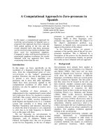
An overview of the predicted selenoproteomeFigure 2
An overview of the predicted selenoproteome. (a) A Venn diagram representation of the overlap between the known selenoproteins in the RECODE
database (bold line) and the results of our prediction method (plain line) over the same set of organisms as included in RECODE. (b) A pie chart
illustrating the types of selenoproteins in our predicted dataset. The dataset was divided into the following groups: formate dehydrogenase (FDH) family
enzymes; archaeal methanogenesis selenoproteins (excluding the FDH family); selenophosphate synthetase (SelD); other known selenoproteins (for
example, thioredoxin, hesB); glycine reductase genes (GRD); and new candidate selenoproteins. (c) A section of the multiple sequence alignments (MSA)
of the newly predicted candidate selenoprotein from P. profundum with its four homologs found in our database. Note the alignment of putative
selenocysteine (U denotes selenocysteine) with cysteine residues in the MSA. (d) The MSA of a selenoprotein formylmethanofuran dehydrogenase from
M. maripaludis in which the recoded selenocysteine aligns with a set of conserved aspartate residues rather than the cysteine residues. The MSA
illustrations were prepared using ALSCRIPT [39].
2
53
6
(a)
(c)
FDH
(b)
48%
13%
9%
10%
12%
Others
SelD
GRD
New
Methano
genesis
8%
New
(d)
R79.10 Genome Biology 2005, Volume 6, Issue 9, Article R79 Chaudhuri and Yeates />Genome Biology 2005, 6:R79
identified. Putative SECIS motifs for these four selenopro-
teins and the additional candidate in P. profundum are pre-
sented in Figure 3a.
A second advantage of the RSA-based approach is the poten-
tial ability to detect selenoproteins that are not represented in
the database by a homologous protein with a cysteine in the
position corresponding to the presumptive stop codon. A
close look at the multiple sequence alignments of certain
selenoprotein homologs in the Conserved Domain database
[25] indicated that nucleophilic serine, aspartate and gluta-
mate residues sometimes replace the catalytic cysteine func-
tionality. Unlike the previously described cysteine alignment
criterion [13], the RSA-based approach does not analyze
cysteine/selenocysteine alignment in an early stage. The
presence of these conserved, non-cysteine residues aligned
with putative selenocysteine can, therefore, be analyzed while
inspecting the MSA, followed by an analysis of the SECIS fea-
ture. The protein formylmethanofuran dehydrogenase in M.
maripaludis provides an example of a verified selenoprotein
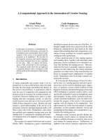
Representatives of the putative selenocysteine insertion sequence (SECIS) hairpin elements in various genomes as identified by the present studyFigure 3
Representatives of the putative selenocysteine insertion sequence (SECIS) hairpin elements in various genomes as identified by the present study. (a) The
SECIS elements from the genes coding for the following proteins from P. profundum: 1, glycine reductase GrdA; 2, glycine reductase GrdB2; 3, glycine
reductase GrdA; 4, selenophosphate synthetase (SelD); 5, a hypothetical protein. (b) The SECIS elements from the genes coding for the following proteins
from E. coli: 1, formate dehydrogenase; 2, formate dehydrogenase-N; 3, formate dehydrogenase-O.
(a)
(b)
Genome Biology 2005, Volume 6, Issue 9, Article R79 Chaudhuri and Yeates R79.11
comment reviews reports refereed researchdeposited research interactions information
Genome Biology 2005, 6:R79
that is detected by our method without invoking the cysteine/
selenocysteine alignment criterion (Figure 2d). The subject
selenocysteine aligns with a set of aspartate residues in the
MSA. However, glycine reductase A (GrdA), a selenoprotein
whose homologs do not have cysteine in place of seleno-
cysteine [13], could not be identified using our method on a
test run. This failure resulted from a crucial lack of significant
read-through similarity between GrdA and the other proteins
homologous to GrdA.
A small number of ORFs (see Additional data file 2) were
found with the translated UGA codon (U) aligned with strictly
invariant nucleophilic residues (aspartate, glutamate or ser-
ine) in the MSA. None of these ORFS belong to previously
known selenoprotein families or had a convincing SECIS
motif adjacent to the UGA codon. Because of the lack of any
additional evidence, it is not possible to further separate the
true read-through events from the false-positives that might
arise from statistical uncertainty or sequencing error. Never-
theless, some of these ORFs could be genuine read-through
cases.
Putative pyrrolysine recoding in archaea
The RSA method was also used to search for proteins poten-
tially containing the pyrrolysine residue, the so-called 22
nd
amino acid (Table 2). The pyrrolysine amino acid residue was
recently discovered to be encoded by the UAG (amber) codon
in the monomethylamine methyltransferase enzyme in Meth-
anosarcina barkeri, where it serves as an electrophile to
methylate the cobalt-corrinoid cofactor [6,7,26]. First, a
search for homologs of the PylS gene (which codes for the
pyrrolysine-specific aminoacyl tRNA synthetase [6,7]) in the
available genomic data identified several methanogenic
archaea as organisms likely to encode pyrrolysine containing
proteins. These organisms include: Methanosarcina barkeri
fusaro, Methanosarcina acetivorans, Methanosarcina
mazei and Methanococcoides burtonii. Putative pyrrolysine-
containing methylamine methyltransferses from methano-
genesis pathways have been reported in this same set of
organisms [11,26]. Within these four organisms, a total of 34
ORFs containing putative pyrrolysine residues were found to
exhibit significant read-through similarity to homologous
methyltransferases (Table 2). Out of 2,086 and 3,611 theoret-
ical ORFs (see Materials and methods section) analyzed in the
complete genomes of M. mazei and M. acetivorans, 87 and
97, respectively, showed significant read-through similarity.
We have listed all those ORFs with an in-frame UAG codon
that exhibit high RSA similarity, as well as well-aligned MSA
for M. acetivorans and M. mazei (Additional data file 2).
Apart from previously described transposases [11], the list
contains several other candidate proteins, including a novel
homolog of the cobalamin biosynthesis protein CobN (Figure
4c).
Overall distribution of the recoded proteins
A marked tendency was noted for the selenoproteins to occur
in certain pathways and functional categories (Figure 2b).
The majority of detected selenoproteins in bacteria were
FDHs that convert formate to carbon dioxide in anaerobic
environments [27]. Other known selenoproteins include
SelD, GrdA and GrdB (from the anaerobic glycine reduction
pathway), HesB (associated with the nitrogen fixation genes),
and several oxidoreductases (for example, thioredoxin and
peroxiredoxin). In archaea, selenocysteine usage appears to
be confined to a small group of enzymes in the anaerobic
methanogenesis pathway [23] (such as FDH and formylmeth-
anofuran dehydrogenase from the FDH family, and heterodi-
sulfide reductase) that have conceivably co-evolved under
similar evolutionary constraints in a number of methano-
gens. Pyrrolysine-encoding is found in methyltransferases
[26] from a pathway that converts methylamines to methane
in Methanosarcina sp. and in the Antarctic archaeon M.
burtonii. A high incidence of unusual stop codon reassign-
ments, both selenocysteines and pyrrolysines, in methano-
genesis enzymes in ancient archaea is intriguing.
Relative merit of the RSA-based approach
The selenoprotein identification scheme presented herein (an
'RSA-first, SECIS-later' approach) differs from the previously
reported methods in several ways. Earlier studies (based on
the SECIS search approach) provided an estimated rate of
false SECIS hits to be 3 to 15 per 10 Mb [12,17] in eukaryotes,
greatly surpassing the number of true selenoproteins. An
improved result has been obtained by using a statistical pro-
file computed from a training dataset of aligned known SECIS
elements in metazoa [17]. A recent bacterial SECIS-search
method analyzed 48,472 SECIS hits in a set of 29 organisms
(representing 1.5% of all the UGA codons analyzed), out of
which 28,974 (approximately 60%) were selected for further
analysis of protein sequence conservation in the UGA flank-
ing regions [16]. Still, difficulties remain for approaches that
rely on detecting small RNA signal sequences as an early step
in analysis, especially in situations such as new genomes,
where the nature of the signal may not be understood in
advance. Examining presumptive protein sequences as a
prior step mitigates these difficulties. In the present study,
although RSA was applied to fairly small segments of the pro-
tein sequences following the UGA codon, it was quite efficient
in identifying candidates representing read-through events
(Figure 1c). This ability of RSA to limit the predicted set to a
relatively small, manageable number of likely candidates
(3,594 out of 203,339, approximately 1.7%) facilitated further
detailed calculations in genome-wide analyses. Of the small
set of 109 ORFs selected by the subsequent MSA analysis, 92
(approximately 84%) were selected afterward as putative
selenoproteins. Thus, an analysis of protein sequences is able
to filter out most of the false-positives, without using any
mRNA context information. Our combined 'RSA-first,
SECIS-later' method is, therefore, applicable to cases (for
example, P. profundum) where a divergent signal makes a
R79.12 Genome Biology 2005, Volume 6, Issue 9, Article R79 Chaudhuri and Yeates />Genome Biology 2005, 6:R79
SECIS-based search unsuitable [16]. In the present approach,
it becomes possible to scrutinize putative non-canonical
SECIS signals. In addition, our method provides a useful way
to search for selenoproteins lacking homologs containing cor-
responding cysteine residues [13] (Figure 2d).
The RSA approach was likewise successful in predicting puta-
tive pyrrolysine-proteins in archaea. Out of the 9,515
theoretical ORFs analyzed for putative pyrrolysine residues in
four methanogens, 321 ORFs (3.4%) displayed significant
read-through similarity. Unlike the case for selenoproteins, a
reliable benchmarking of pyrrolysine-protein predictions
against a known dataset was not possible. The predicted
result encompasses the previously reported methylamine
methyltransferases [26], however, and includes a number of
likely candidates for further experiments. Intriguingly, the
putative pyrrolysine residues do not align so exclusively with
a particular, conserved amino acid in homologous proteins
(Figure 4a-c) [11]. The RSA method appears, therefore, to be
generally useful as an initial predictor for pyrrolysine pro-
teins. In addition, the RSA approach offers wider utility for
identifying cases of genome-wide stop codon redefinition (for
example, in Mycoplasma spp.; see Materials and methods
section) or special instances of stop codon read-through (for
example, UAG read-through in a pilus biosynthesis gene in E.
coli [28] (data not shown)).
Conclusion
To summarize, we have developed a novel computational
scheme for predicting selenocysteine and pyrrolysine resi-
dues in proteins and have applied the method to microbes
with complete genomes. In addition to confirming well-
known examples, our method predicts new prospective can-
didates for further experimental validation. A worldwide web
site has been developed for the interested user community
[29]. The method should be a useful tool for predicting rare
amino acids, as well as other read-through events, and for
correcting gene annotations in the growing genomic
databases.
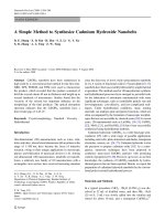
Sections of the multiple sequence alignments of the putative pyrrolysine-containing proteinsFigure 4
Sections of the multiple sequence alignments of the putative pyrrolysine-containing proteins. (a) A protein known to use UAG read-through, methylamine
methyltransferase from M. acetivorans. (b) A putative methyltransferase from M. burtonii. (c) A predicted read-through ORF homologous to a cobalamin
biosynthesis protein CobN (gi|20906100|gb|AAM31298.1|, Methanosarcina mazei Goe1) from M. acetivorans. Note the alignment of presumed pyrrolysine
residues (denoted as X) with various amino acids.
11:gi|13881726|
10:gi|48429756|
9:gi|1499975|
8:gi|4981851|
7:gi|20516906|
6:gi|20904392|
5:gi|57158963|
4:gi|5458504|
3:gi|3257551|
2:gi|18893258|
1:ORF
AVGEAVPFVSRHFGAVLMAFTLCFVTDPAAIFRETRRLLADG
ASAYEIPFPDKYFDFSLNMVTICFLDYPEKAIREAKRVSN
AKGEDLPFKDEEFDFFLINTVLEFAENPKKMIEEAKRVLKRG
GTAENLPLKDESFDFALMVTTICFVDDPERALKEAYRILKKG
GVAENLPFEDSSFDLVLMVTTICFVDDPLRALKECYRVLKND
GVAETLPFKRQSFDLVLIVASLSLFKDPVQALREAAGVLKPG
GTAENLPFEEDSMDYLLMVTTICFVDDPEKALKEAYRVLKPG
GVAEDLPFPDNSLECILMVTTICFVDDPEKAIKEAYRVLKPN
GIAEDLPFPDSSLSCILMVTTICFVDDVEKSIKEAYRVLKPG
GVAENLPFEDNSLDCILMVTTICFVDDPEKAIKEAYRVLKPG
GVGESLPFKGSSMKLALIVTSLCFMD-AKKVLXEAYRMLAPE
11:gi|20906197|
10:gi|56676827|
9:gi|56677214|
8:gi|56678713|
7:gi|14523932|
6:gi|15075296|
5:gi|15074998|
4:gi|56680780|
3:gi|56678737|
2:gi|56679325|
1:ORF
VGSPELGLISASVAKLAQFYGLPAFVAGT
FGTPEPALGSLVMGQLARRLNMPLRCAGNFSTSKAPDGQAMQ
FGTPEHFTASLVAGQLARRIGLPWRCAG-GSAANINDAQAAN
FGTPEASLVTYGAGQLARRLGLPFRSAGSFCGSKLPDAQAAY
FGTPENAKANIIAGQLARRYKLPYRTSN-ANASNAVDLQAAY
GGSGEQALLTAGCAQMHQFYRLPGGAAAGIADAKLPDMQAGW
FGTPEPSLVSYGAAQLARRLGLPFRTGGSLCGSKVPDAQAAH
GGGGEQAILMAGAAQMGRRWDLPTSSIAGITDAKRLDAQYGA
GGSGEQALLTAGCAQMHRFYDLPGGAAAGIADSKLPDMQAGW
FGTPEYMRATQMTGQMARFYGLPVRSSG-VCAANVPDGQAMW
VGSPELGLISASVAKLAQFYGLPAFVAGTXSDAKIPDNQAGH
11:gi|60493616|
10:gi|2621409|
9:gi|2621757|
8:gi|44920813|
7:gi|2622471|
6:gi|2621372|
5:gi|2621801|
4:gi|2622026|
3:gi|20906098|
2:gi|20906100|
1:ORF
-TQSPAALEEMTAIMLESARKGLWKASAEQVAELAKLHTETV
-GRNSYALISITGTMLTAAYEGHWNPDEATLRLLASTWASLT
TGNRAYSMISITGTLLTAAHKGFWKADEATLRLVANTWASTV
-ENNPYAYQSMTARMLETARKGCWDASGETLKNLANEFIQSV
KSLQPYSYASTVAWLLEASRRGLWSTDSATLQRLADEYIAAV
-KSNPYARASIIARLIETVRKGYWSPSDQVKASLANEYINMV
SGNRAYAMISITGTMLNVIYMGYWKTDETITRNIANRWAKMI
QGRNSYAMISMTGTMLTAIHRGYWNPDDATKRLIATTWAQAI
-QNNPAAYQSITARMLEAVRHDYWAPSEDVIESLATEYEQSV
-QNNPYAYQSMTARMLETARKGSWDASDDVLKSLAEEYAESV
-ANNPWARQSMEARMLEAIRKGYWDADXETIDALTREYVESV
(a)
(b)
(c)
Genome Biology 2005, Volume 6, Issue 9, Article R79 Chaudhuri and Yeates R79.13
comment reviews reports refereed researchdeposited research interactions information
Genome Biology 2005, 6:R79
Materials and methods
All the complete genomes were obtained from the National
Center for Biotechnology Information (NCBI) [30]. Unfin-
ished M. barkeri and M. burtonii genomes were obtained
from The Institute for Genomic Research [31]. A list of
accession numbers is provided in the Additional data files. A
Perl script was written to perform all the computations (avail-
able upon request). All computations were performed in a
local cluster of Linux computers. tRNA genes were computa-
tionally identified using the tRNASCAN-SE program [32].
Genes encoding SelA and SelB were detected directly from
annotated genomes from NCBI.
All theoretical ORFs (≥ 90 residues) that begin with a start
codon (ATG, TTG or GTG) and end with a stop codon (TAA,
TAG or TGA) and contain one in-frame TGA (for seleno-
cysteine) or TAG (for pyrrolysine) codon were extracted from
the genomic data for analysis. In order to detect two short
SelW proteins in G. sulfurreducens and C. jejuni, a reduced
(80 residue) length constraint was used.
Read-through similarity analysis (RSA)
For each of the predicted ORFs, the BLAST program [33,34]
was used to search for homologous proteins in a customized
sequence database. The BLAST search space was restricted to
a window of a maximum 100 residue length, pivoting at the
stop codon. The BLOSUM62 matrix was used throughout and
the selenocysteine residue was treated as 'any amino acid' (X).
The BLAST database contained a maximum of 650,870 pro-
tein sequences from all the annotated complete microbial
genomes from NCBI (dated 4 December 2005). A self-exclud-
ing BLAST database was used for the homology search in each
organism. Top hits (E-value ≤ 10
-1
) that encompassed either
side of the stop codon were identified. For each of the
selected, truncated ORF sequences ({x
1
, x
2
, , x
i
, , x
u
, , x
n
}
where n = min{u + 60, u + t}; u = position of the stop codon;
t = position of the subsequent stop codon) and the corre-
sponding top hit sequence from the BLAST search ({y
1
, y
2
,
y
j
, , y
m
}), a (n + 1) by (m + 1) dynamic alignment matrix was
calculated with an affine gap penalty function [18,19]. N-ter-
minal overhangs for both the sequences were not penalized;
the 0
th
row and the 0
th
column were initialized with zero
values.
For each cell (i,j) in the matrix:
a(i,j) = s(i,j) + max { a(I - 1,j - 1),
b(i-1,j-1),
c(i-1,j-1) }
b(i,j) = max { -(h + g) + a(i,j - 1),
-g + b(i,j - 1),
-(h + g) + c(i,j - 1) }
c(i,j) = max { -(h + g) + a(I - 1,j),
-(h + g) + b(I - 1,j),
-g + c(I - 1,j) }
score (i,j) = max {a(i,j), b(i,j), c(i,j)}; s(i,j) → BLOSUM62
matrix; h = 12, g = 2
Best_score
ORF
= max{score(n,j), j = 1, ,m} (1)
Because we were exclusively interested in the significance of
the alignment at the carboxy-terminal extension region
beyond the stop position, the highest score from the n
th
col-
umn (that is the alignment of the terminal residue x
n
of the
truncated ORF with the {y
1
, , y
m
} residues) was taken as the
maximal score (Best_score
ORF
) instead of the usual Smith-
Waterman score. A Z-value was computed by shuffling the
terminal extension region 100 times, re-computing the scores
in the terminal block of the matrix ({x
stop
, ,x
n
} and {y
1
, ,y
m
})
and averaging the maximal score (<Best_score
rand
>). A test
calculation with 10,000 times shuffling for one genome pro-
duced similar results. The values from randomized sequences
were used to calculate a Z-value:
Z
ORF
=
(Best_score
ORF
- <Best_score
rand
>)/standard_deviation (2)
A generally weak dependence on length and amino acid com-
position makes the Z-values (which follow an extreme value
distribution) useful for evaluating the significance of align-
ment scores [35]. We have used a fairly conservative Z-value
cutoff (Z
c
= 8.0) [35] to decide the statistical significance of a
C-terminal alignment. Selection criteria had to be relaxed for
two legitimate selenoproteins, a sulfur transferase in G.
sulfurreducens (Z-value 7.9) and a coenzyme F420-reducing
hydrogenase subunit in M. maripaludis (Z-value 4.6).
Multiple sequence alignment
For each of the selected candidate ORFs (Z
c
≥ 8), a sensitive,
iterative PSI-BLAST search was performed using position-
specific scoring matrices. The top 10 hits (E ≤ 10
-3
) were used
to construct a MSA with ClustalW [36]. Amino acids lining up
with the putative selenocysteine residue were examined.
Selenocysteines that aligned with two or more cysteine resi-
dues were selected for further analysis.
SECIS element analysis
In accordance with a recent analysis of bacterial SECIS ele-
ments [16], a 111 nucleotide long mRNA stretch surrounding
the UGA codon position (-10 to +100) was extracted from
each of the selected ORFs passing the previous tests in each
bacterial organism. The extracted set of RNA sequences for
each organism was used to detect a common, single hairpin
R79.14 Genome Biology 2005, Volume 6, Issue 9, Article R79 Chaudhuri and Yeates />Genome Biology 2005, 6:R79
motif using the rapid, heuristic-based RNAPROFILE pro-
gram [20]. A test calculation predicted the known SECIS ele-
ment of the gene encoding FDH from E. coli correctly [37]
(Figure 3b). The putative SECIS hairpin motifs were manu-
ally inspected for consistency.
Control analysis
To evaluate the performance of the RSA step, we analyzed the
Mycoplasma genitalium organism that utilizes UGA to code
for tryptophan throughout its genome. M. genitalium is a
small genome with 470 genes [38], the majority of which have
a homolog in our database, thus minimizing database effects
in our calculation. We applied the RSA method to all the the-
oretical ORFs with one in-frame TAA (313) or TAG (137) or
TGA (780) codon. A self-excluding BLAST database of micro-
bial proteins was used. In M. genitalium, over 78% of the TGA
cases (91% when a self-included database was used) were
identified by the RSA method as recoding events with a Z-
value of 8 or higher. These cases aligned overwhelmingly with
tryptophan residues in homologs. In contrast, only about 2%
to 3% of the ORFs contatining a TAA or TAG stop codon
passed the same RSA test.
We also applied the selenoprotein detection scheme to the
Aeropyrum pernix (BA000002) genome, which does not
contain any selenocysteine insertion genes. Out of 26 of 1,288
ORFs with in-frame 'UGA' that were selected by RSA (approx-
imately 2%), none were selected in the subsequent MSA test.
A web-server for RSA analysis
A web-based service is available for RSA analysis of submitted
DNA sequences [29]. The server was designed to analyze an
ORF with one in-frame stop codon (UAA, UAG or UGA). A
larger, non-redundant BLAST database (to be updated regu-
larly) is used by the web server. The Z-value score and the
MSA for the ORF are returned to the user.
Sensitivity and specificity
Sensitivity = true positive/(true positive + false negative)
Specificity = true positive/(true positive + false positive)
Estimates of true positives, false negatives and false positives
were based on predictions performed on the set of organisms
whose selenoproteins have been described in the RECODE
[21] database (Figure 2a). The number of true positives is
taken to be the number of predictions that are already known
selenoproteins in the RECODE database. False negatives are
those known selenoproteins not predicted by our method.
False positives are difficult to estimate. As an extreme esti-
mate, we have taken as an upper bound all those predictions
that are not in the known database. The actual false positive
rate is probably considerably lower than this estimate.
Additional data files
The following additional data are available with the online
version of this paper. Additional data file 1 is a list of all the
genomes analyzed together with the NCBI accession number.
Additional data file 2 contains all the predicted recoded pro-
teins from the complete genomes analyzed in this study in
FASTA format.
Additional File 1A list of all the genomes analyzed together with the NCBI accession numberA list of all the genomes analyzed together with the NCBI accession numberClick here for fileAdditional File 2All the predicted recoded proteins from the complete genomes ana-lyzed in this study in FASTA formatAll the predicted recoded proteins from the complete genomes ana-lyzed in this study in FASTA formatClick here for file
Acknowledgements
This work was supported by the DOE office of Biological and Environmen-
tal Research. The authors thank T Holton for assistance with the web-
based server preparation.
References
1. Gesteland RF, Atkins JF: Recoding: dynamic reprogramming of
translation. Annu Rev Biochem 1996, 65:741-768.
2. Namy O, Rousset JP, Napthine S, Brierley I: Reprogrammed
genetic decoding in cellular gene expression. Mol Cell 2004,
13:157-168.
3. Bock A: Biosynthesis of selenoproteins: an overview. Biofactors
2000, 11:77-78.
4. Stadtman TC: Selenocysteine. Annu Rev Biochem 1996, 65:83-100.
5. Hatfield DL, Gladyshev VN: How selenium has altered our
understanding of the genetic code. Mol Cell Biol 2002,
22:3565-3576.
6. Srinivasan G, James CM, Krzycki JA: Pyrrolysine encoded by UAG
in Archaea: charging of a UAG-decoding specialized tRNA.
Science 2002, 296:1459-1462.
7. Hao B, Gong W, Ferguson TK, James CM, Krzycki JA, Chan MK: A
new UAG-encoded residue in the structure of a methanogen
methyltransferase. Science 2002, 296:1462-1466.
8. Rayman MP: The importance of selenium to human health.
Lancet 2000, 356:233-241.
9. Frankenberger WT Jr, Arshad M: Bioremediation of selenium-
contaminated sediments and water. Biofactors 2001,
14:241-254.
10. Jacob C, Giles GI, Giles NM, Sies H: Sulfur and selenium: the role
of oxidation state in protein structure and function. Angew
Chem Int Ed Engl 2003, 42:4742-4758.
11. Zhang Y, Baranov PV, Atkins JF, Gladyshev VN: Pyrrolysine and
selenocysteine use dissimilar decoding strategies. J Biol Chem
2005, 280:20740-20751.
12. Kryukov GV, Castellano S, Novoselov SV, Lobanov AV, Zehtab O,
Guigo R, Gladyshev VN: Characterization of mammalian
selenoproteomes. Science 2003, 300:1439-1443.
13. Kryukov GV, Gladyshev VN: The prokaryotic selenoproteome.
EMBO Rep 2004, 5:538-543.
14. Castellano S, Novoselov SV, Kryukov GV, Lescure A, Blanco E, Krol
A, Gladyshev VN, Guigo R: Reconsidering the evolution of
eukaryotic selenoproteins: a novel nonmammalian family
with scattered phylogenetic distribution. EMBO Rep 2004,
5:71-77.
15. Zhang Y, Fomenko DE, Gladyshev VN: The microbial selenopro-
teome of the Sargasso Sea. Genome Biol 2005, 6:R37.
16. Zhang Y, Gladyshev VN: An algorithm for identification of bac-
terial selenocysteine insertion sequence elements and
selenoprotein genes. Bioinformatics 2005, 21:2580-2589.
17. Lambert A, Lescure A, Gautheret D: A survey of metazoan selen-
ocysteine insertion sequences. Biochimie 2002, 84:953-959.
18. Gotoh O: An improved algorithm for matching biological
sequences. J Mol Biol 1982, 162:705-708.
19. Setubal C, Meidanis J: Introduction to Computational Molecular Biology
Boston: PWS Publishing Company; 1997.
20. Pavesi G, Mauri G, Stefani M, Pesole G: RNAProfile: an algorithm
for finding conserved secondary structure motifs in una-
ligned RNA sequences. Nucleic Acids Res 2004, 32:3258-3269.
21. Baranov PV, Gurvich OL, Hammer AW, Gesteland RF, Atkins JF:
RECODE 2003. Nucleic Acids Res 2003, 31:87-89.
22. Murzin AG, Brenner SE, Hubbard T, Chothia C: SCOP: a structural
classification of proteins database for the investigation of
sequences and structures. J Mol Biol 1995, 247:536-540.
Genome Biology 2005, Volume 6, Issue 9, Article R79 Chaudhuri and Yeates R79.15
comment reviews reports refereed researchdeposited research interactions information
Genome Biology 2005, 6:R79
23. Cobucci-Ponzano B, Rossi M, Moracci M: Recoding in archaea.
Mol Microbiol 2005, 55:339-348.
24. Vezzi A, Campanaro S, D'Angelo M, Simonato F, Vitulo N, Lauro FM,
Cestaro A, Malacrida G, Simionati B, Cannata N, et al.: Life at depth:
Photobacterium profundum genome sequence and expres-
sion analysis. Science 2005, 307:1459-1461.
25. Marchler-Bauer A, Anderson JB, DeWeese-Scott C, Fedorova ND,
Geer LY, He S, Hurwitz DI, Jackson JD, Jacobs AR, Lanczycki CJ, et al.:
CDD: a curated Entrez database of conserved domain
alignments. Nucleic Acids Res 2003, 31:383-387.
26. Krzycki JA: Function of genetically encoded pyrrolysine in cor-
rinoid-dependent methylamine methyltransferases. Curr
Opin Chem Biol 2004, 8:484-491.
27. Jormakka M, Byrne B, Iwata S: Formate dehydrogenase: a versa-
tile enzyme in changing environments. Curr Opin Struct Biol
2003, 13:418-423.
28. Jalajakumari MB, Thomas CJ, Halter R, Manning PA: Genes for bio-
synthesis and assembly of CS3 pili of CFA/II enterotoxigenic
Escherichia coli: novel regulation of pilus production by
bypassing an amber codon. Mol Microbiol 1989, 3:1685-1695.
29. Read-through Similarity Analysis [-
mbi.ucla.edu/~neel/RSA.php]
30. National Center for Biotechnology Information [ftp://
ftp.ncbi.nih.gov/genomes/Bacteria]
31. The Institute for Genomic Research []
32. Lowe TM, Eddy SR: tRNAscan-SE: a program for improved
detection of transfer RNA genes in genomic sequence.
Nucleic Acids Res 1997, 25:955-964.
33. Altschul SF, Gish W, Miller W, Myers EW, Lipman DJ: Basic local
alignment search tool. J Mol Biol 1990, 215:403-410.
34. Altschul SF, Madden TL, Schaffer AA, Zhang J, Zhang Z, Miller W, Lip-
man DJ: Gapped BLAST and PSI-BLAST: a new generation of
protein database search programs. Nucleic Acids Res 1997,
25:3389-3402.
35. Comet JP, Aude JC, Glemet E, Risler JL, Henaut A, Slonimski PP,
Codani JJ: Significance of Z-value statistics of Smith-Water-
man scores for protein alignments. Comput Chem 1999,
23:317-331.
36. Thompson JD, Higgins DG, Gibson TJ: CLUSTAL W: improving
the sensitivity of progressive multiple sequence alignment
through sequence weighting, position-specific gap penalties
and weight matrix choice. Nucleic Acids Res 1994, 22:4673-4680.
37. Liu Z, Reches M, Groisman I, Engelberg-Kulka H: The nature of the
minimal 'selenocysteine insertion sequence' (SECIS) in
Escherichia coli. Nucleic Acids Res 1998, 26:896-902.
38. Fraser CM, Gocayne JD, White O, Adams MD, Clayton RA, Fleis-
chmann RD, Bult CJ, Kerlavage AR, Sutton G, Kelley JM, et al.: The
minimal gene complement of Mycoplasma genitalium. Sci-
ence 1995, 270:397-403.
39. Barton GJ: ALSCRIPT: a tool to format multiple sequence
alignments. Protein Eng 1993, 6:37-40.