Báo cáo y học: "Identification of novel Y chromosome encoded transcripts by testis transcriptome analysis of mice with deletions of the Y chromosome long arm" potx
Bạn đang xem bản rút gọn của tài liệu. Xem và tải ngay bản đầy đủ của tài liệu tại đây (4.38 MB, 15 trang )
Genome Biology 2005, 6:R102
comment reviews reports deposited research refereed research interactions information
Open Access
2005Touréet al.Volume 6, Issue 12, Article R102
Research
Identification of novel Y chromosome encoded transcripts by testis
transcriptome analysis of mice with deletions of the Y chromosome
long arm
Aminata Touré
¤
*
, Emily J Clemente
¤
†
, Peter JI Ellis
†
,
Shantha K Mahadevaiah
*
, Obah A Ojarikre
*
, Penny AF Ball
‡
,
Louise Reynard
*
, Kate L Loveland
‡
, Paul S Burgoyne
*
and Nabeel A Affara
†
Addresses:
*
Division of Developmental Genetics, MRC National Institute for Medical Research, Mill Hill, London, NW7 1AA, UK.
†
University
of Cambridge, Department of Pathology, Tennis Court Road, Cambridge, CB2 1QP, UK.
‡
Monash Institute of Medical Research, Monash
University, and The Australian Research Council Centre of Excellence in Biotechnology and Development, Melbourne, Victoria 3168 Australia.
¤ These authors contributed equally to this work.
Correspondence: Paul S Burgoyne. E-mail:
© 2005 Touré et al.; licensee BioMed Central Ltd.
This is an open access article distributed under the terms of the Creative Commons Attribution License ( which
permits unrestricted use, distribution, and reproduction in any medium, provided the original work is properly cited.
Y chromosome - encoded mouse testis transcripts<p>Microarray analysis of the changes in the testis transcriptome resulting from deletions of the male-specific region on the mouse chro-mosome long arm (MSYq) identified novel Y chromosome-encoded transcripts.</p>
Abstract
Background: The male-specific region of the mouse Y chromosome long arm (MSYq) is
comprised largely of repeated DNA, including multiple copies of the spermatid-expressed Ssty gene
family. Large deletions of MSYq are associated with sperm head defects for which Ssty deficiency
has been presumed to be responsible.
Results: In a search for further candidate genes associated with these defects we analyzed changes
in the testis transcriptome resulting from MSYq deletions, using testis cDNA microarrays. This
approach, aided by accumulating mouse MSYq sequence information, identified transcripts derived
from two further spermatid-expressed multicopy MSYq gene families; like Ssty, each of these new
MSYq gene families has multicopy relatives on the X chromosome. The Sly family encodes a protein
with homology to the chromatin-associated proteins XLR and XMR that are encoded by the X
chromosomal relatives. The second MSYq gene family was identified because the transcripts
hybridized to a microarrayed X chromosome-encoded testis cDNA. The X loci ('Astx') encoding
this cDNA had 92-94% sequence identity to over 100 putative Y loci ('Asty') across exons and
introns; only low level Asty transcription was detected. More strongly transcribed recombinant loci
were identified that included Asty exons 2-4 preceded by Ssty1 exons 1, 2 and part of exon 3.
Transcription from the Ssty1 promotor generated spermatid-specific transcripts that, in addition to
the variable inclusion of Ssty1 and Asty exons, included additional exons because of the
serendipitous presence of splice sites further downstream.
Conclusion: We identified further MSYq-encoded transcripts expressed in spermatids and
deriving from multicopy Y genes, deficiency of which may underlie the defects in sperm
Published: 2 December 2005
Genome Biology 2005, 6:R102 (doi:10.1186/gb-2005-6-12-r102)
Received: 17 June 2005
Revised: 19 September 2005
Accepted: 27 October 2005
The electronic version of this article is the complete one and can be
found online at />R102.2 Genome Biology 2005, Volume 6, Issue 12, Article R102 Touré et al. />Genome Biology 2005, 6:R102
development associated with MSYq deletions.
Background
The mammalian Y chromosome seems predisposed to accu-
mulating multiple copies of genes that play a role in sperma-
togenesis [1-7]. Determining the precise functions of such
multicopy genes is inherently difficult. In humans and mouse,
indications as to function have so far derived from the analy-
sis of naturally occurring deletion mutants. In the mouse,
deletions in MSYq (the Y chromosome long arm, excluding
the pseudo-autosomal region) affect sperm development
(spermiogenesis) and function, with the severity of the sperm
defects being correlated with the extent of the deletion [3,8-
14]. Mouse MSYq appears to be composed predominantly of
highly repeated DNA sequences [15-22], and when the
present project was initiated the only known MSYq encoded
testis transcripts derived from the complex multicopy Ssty
gene family [3,23-25]. This gene family, with two distinct sub-
families, namely Ssty1 and Ssty2, is expressed in the testis
during spermiogenesis and, in the absence of other candi-
dates, it had been postulated that Ssty deficiency is responsi-
ble for the defective sperm development in MSYq deficient
mice.
In this study we utilized custom-made testis cDNA microar-
rays to identify further Y encoded testis transcripts that are
absent or reduced in level as a consequence of MSYq
deficiencies.
Results
Identifying MSYq encoded testis transcripts by
microarray analysis
Three MSYq-deficient mouse models were used in this study
(Figure 1): XY
RIII
qdel, with deletion of about two-thirds of
MSYq; XY
Tdym1
qdelSry, with deletion of about nine tenths of
MSYq; and XSxr
a
Y*
X
, in which the only Y specific sequences
are provided by the Y short arm derived sex-reversal factor
Sxr
a
, and thus lack all of MSYq. These models are hereafter
abbreviated as 2/3MSYq
-
, 9/10MSYq
-
and MSYq
-
.
To analyze the testis transcriptomes of these MSYq deficient
mice we used microarrayed testis cDNA clones enriched for
cDNAs deriving from spermatogenic cells (see Materials and
methods, below). Total testis RNA from each of the MSYq-
deficient mice (labeled with the fluorochrome Cy3) and from
matched controls (labeled with Cy5) was hybridized to the
array; four technical replicates were obtained for each model.
The initial raw data were filtered to select only clones for
which fluorescence intensity data were available from at least
two of the technical replicates for each model. After this
filtering, 14,681 clones remained from the complete set. As
expected, scatter plots of these filtered data show that expres-
sion of the vast majority of clones is unchanged between the
MSYq deficient and control mice (Figure 2a).
For transcripts encoded by single or multiple copy Y genes
mapping to MSYq, substantially reduced levels should occur
in one or more of the deletion models. In order to focus on
potential MSYq encoded transcripts we restricted our analy-
sis to clones exhibiting a twofold or greater reduction in fluo-
rescence intensity relative to control and a t test probability of
under 1% for the comparison between the replicates for the
Cy3 and Cy5 fluorescence intensities. Twenty-three clones
were identified as substantially reduced by these criteria in
one or more of the MSYq deletion models. BLAST (basic local
alignment search tool) comparisons were used to identify
matching cDNAs (if previously identified) and/or the chro-
mosomal locations of the encoding sequences. Sixteen of the
23 proved to be Ssty cDNA clones, thus demonstrating the
efficacy of the strategy. Of the remaining seven clones, five
proved to be Y encoded Sly cDNAs (see below), one was X
encoded and one was autosomally encoded (Figure 2a, b).
Sly transcription is reduced in proportion to the extent
of MSYq deficiency
All five of the additional Y encoded clones were found to have
homology to regions of the cDNA clone BC049626 previously
identified in a large-scale cDNA sequencing project [26]
(Additional data file 1). This cDNA clone initially had no chro-
mosomal assignment, but it was subsequently ascribed to a
gene, Sly, that maps to MSYq (MGI:2687328; Mouse Chro-
mosome Y Mapping Project [Jessica E Alfoldi, Helen Skalet-
sky, Steve Rozen and David C Page at the Whitehead Institute
for Biomedical Research, Cambridge, MA, USA, and the
Washington University Genome Sequencing Center, St.
Louis, MO, USA]). Sly is a member of a multicopy family, and
in December 2004 a total of 65 Sly family members were pre-
dicted based on the Y sequence data. There is a related mult-
icopy X gene family that includes Xmr and Xlr [27,28].
To confirm the downregulation of the Sly transcript in the
MSYq deletion mice, we probed a northern blot of total testis
RNA from the three MSYq deficient models and their controls
with microarray clone MTnH-K10 (Figure 3a) and subse-
quently with the full BC049626 cDNA (not shown). The
hybridization with both probes revealed high transcript levels
in the control testes, a clear reduction in 2/3MSYq
-
testes, and
further marked reductions in 9/10MSYq
-
and MSYq
-
testes.
The reductions as estimated by phosphorimager analysis with
clone MTnH-K10 (with the microarray estimates in brackets)
were as follows: 2/3MSYq
-
, 47% (37%); 9/10MSYq
-
, 81%
(84%); and MSYq
-
, 83% (91%). Because Sly has substantial
homology to Xmr, which is also strongly transcribed in the
testis, we considered that the remaining hybridization in the
Genome Biology 2005, Volume 6, Issue 12, Article R102 Touré et al. R102.3
comment reviews reports refereed researchdeposited research interactions information
Genome Biology 2005, 6:R102
two models with severe MSYq deficiency could be due to
cross-hybridization with Xmr transcripts. We therefore
designed primers for reverse transcriptase polymerase chain
reaction (RT-PCR) that distinguished between the two tran-
scripts and used these to amplify template from testis cDNAs
derived from the three MSYq deficient models and controls
(Figure 3b). A 266 base-pair (bp) product was amplified from
all samples, and sequencing confirmed that this derived from
Xmr. A 227 bp product was also amplified from the control,
2/3MSYq
-
and 9/10MSYq
-
testes (although clearly reduced),
but there was no amplified product from MSYq
-
testes.
Sequencing of this 227 bp product from 2/3MSYq
-
and 9/
10MSYq
-
testes confirmed that it derived from Sly. Thus, only
MSYq
-
completely lacks Sly transcripts.
Identification of another family of Y encoded testis
transcripts reduced in MSYq deficient mice
In December 2004, a BLAST analysis of the array clone
8832_f_22 against the mouse genome registered 41 hits on
the mouse X chromosome and 710 hits on the mouse Y chro-
mosome. The arrayed cDNA clone was apparently X encoded,
there being eight putative loci (for example, gi:4640881,
3118-6344) with four exons that would encode matching
cDNAs; the remaining nine hits were from incomplete loci or
short sequence fragments. To investigate the coding potential
of the Y sequences identified in the initial analysis, a further
BLAST analysis was carried out with a complete X locus, and
this identified 123 putative Y chromosomal loci (for example,
gi:33667254, 73667-76894) that retain the same intron/exon
structure as the X loci, and with 92-94% sequence identity
across exons and introns.
The arrayed X encoded cDNA clone shared homology with
three previously identified testis cDNA clones - two appar-
ently X encoded (CF198098, AK076884) and one Y encoded
(AK016790) - and a BLAST of mouse expressed sequence tags
(ESTs) identified further related testis transcripts together
with others derived from small intestine, aorta and eight cell
embryos. The testis cDNAs and ESTs are summarized in Fig-
ure 4a. The arrayed X encoded cDNA, together with the two
exactly matching transcripts BF019211 and CF198098, derive
from purified spermatocyte cDNA libraries [29]. Of the Y
encoded testis transcripts, EST AV265093 matches only a
short stretch of exon 4 and the two remaining transcripts,
namely AK016790 and BY716467, show only segments of
homology to the arrayed clone. Nevertheless, amplification
from testis cDNA with an Asty exon 1/exon 4 primer pair gen-
erated products of 437 bp and 622 bp, and sequencing con-
firmed that these derived from Y chromosomal loci, the
smaller product lacking exon 3. We refer to the similar X and
Y loci encoding these X and Y transcripts as Astx and Asty
(Amplified spermatogenic transcripts X encoded/Y encoded),
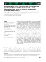
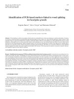
Sex chromosome complements of the mice with MSYq deficiencies and relevant control miceFigure 1
Sex chromosome complements of the mice with MSYq deficiencies and relevant control mice. (a) XY
RIII
control, illustrating the previously documented
male specific gene content of the mouse Y chromosome. The short arm (shown expanded) carries seven single copy genes, one duplicated gene (Zfy), and
multiple copies of Rbmy. MSYq carries multiple copies of the Ssty gene family. (b) XY
Tdym1
control. This male has a normal Y gene complement except that
a 11 kb deletion has removed the testis determinant Sry; the Sry deletion is complemented by an Sry transgene located on an autosome. (c) The variant
Y
RIII
qdel has a deletion removing about two-thirds of MSYq. (d) The variant Y
Tdym1
qdel has a large deletion removing about nine-tenths of MSYq, together
with the small 11 kb deletion removing Sry (complemented by an Sry transgene). (e) XSxr
a
Y*
X
mice are male because of the presence of the Y
RIII
short arm
derived, sex reversal factor Sxr
a
attached distal to the X pseudo-autosomal region (PAR). Sxr
a
comprises most of the Y short arm except for a substantial
reduction in copies of Rbmy. The Y*
X
chromosome is in effect an X chromosome with a deletion from just proximal to Amel (close to the X PAR
boundary) to within the DXHXF34 sequence cluster adjacent to the X centromere. It provides a second PAR, which is essential in order to avoid meiotic
arrest. CEN, centromere; kb, kilobase; TEL, telomere.
Del Sry
Sxr
a
7 copies
Rbmy
Sry
Zfy2
Uty
Eif2s3y
Smcy
Ube1y
Zfy1
Usp9y
Dby
XSxr
Y
*xa
XY
Del 2/3 MSYq
Del Sry
Del 9/10 MSYq
qdel
XY
RIII
XY
XY
RIII
Large X deletion
Ssty
‡50 copies
Rbmy
CEN
Sry
Zfy2
Uty
Eif2s3y
Smcy
Ube1y
Zfy1
TEL
Dby
Usp9y
PAR PAR
XY
>100
copies
(a) (b)
Tdy m1
(2/3 MSYq
-
) (9/10 MSYq )
-
(c) (d)
Tdy m1
(MSYq )
-
(e)
qdel
R102.4 Genome Biology 2005, Volume 6, Issue 12, Article R102 Touré et al. />Genome Biology 2005, 6:R102
respectively (for Astx and Asty transcript sequences, see
Additional data file 2).
Intriguingly, the Y encoded transcripts AK016790 and
BY716467, in addition to the sequence matching Astx/Asty
exons 2 and 3 (and part of the intervening intron), proved to
have exonic sequence matching Ssty1 exon 1 and part of exon
3, together with further sequence matching another testis
cDNA AK015935 (Figure 4a). BLAST searches identified
'recombinant' Y genomic loci (comprising partial Ssty1 and
Asty loci followed by sequence that includes exons matching
AK015935) that could encode these transcripts (Figure 4b,
Additional data file 3). We refer to these loci as Asty(rec).
The close homology between Astx and Asty suggested that the
arrayed Astx cDNA clone would cross-hybridize with Asty
and Asty(rec) RNAs. We designed primers from Astx/Asty
exon 4 for RT-PCR that we thought should specifically
amplify either Astx or Asty, but further analysis (see below)
identified Asty(rec) transcripts that also include exon 4. RT-
PCR analysis using these primers showed that Asty and/or
Asty(rec), rather than Astx transcripts, are reduced in MSYq
deficient mice (Figure 4c). Probing the northern blot of total
testis RNA from the three MSYq deficient models and their
controls with an Asty exon 4 probe revealed a transcript of
about 1 kilobase (kb), together with other larger transcripts
with sizes ranging up to more than 9.5 kb. All bands exhibited
progressive reduction in intensity with increasing MSYq defi-
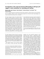
Microarray analysis of the testis transcriptomes of the three MSYq deficient modelsFigure 2
Microarray analysis of the testis transcriptomes of the three MSYq deficient models. (a) Scatter plots showing transcription levels for the testis transcripts
of MSYq deficient models relative to their controls. Expression in the MSYq deficient mice (y-axis, Cy3 label, arbitrary units) for each clone is plotted
versus expression in age- and strain-matched normal testis control (x-axis, Cy5 label, arbitrary units). Data from four technical replicates are combined.
Data are normalized on the median signal for each channel and then filtered (as described in Materials and methods) to show only clones with data for two
technical replicates from each model. The data points showing significant reduction are plotted as enlarged triangles (Y clones green or red, and one X
clone black - for clone identities see b). (b) The 23 cDNA clones identifying transcripts that were significantly reduced in one or more of the MSYq
deficient models. The clones deriving from the two Y families and the one X family are color coded.
MSYq-del fluorescence
Control fluorescence
9/10 MSYq
-
10
100
1,000
10,000
MSYq
-
10
100
1,000
10,000
100 1,000 10,000 100,00010
Array clone cDNA Gene Chromosome.
8846_o_17 YMT2/B Ssty1 Y
MTnC-O10 PC11 Ssty2 Y
Sxrb-03_A03 PC11 Ssty2 Y
MTn10-E22 PC11 Ssty2 Y
MTn10-H11 PC11 Ssty2 Y
13874_f_09 PC11 Ssty2 Y
8846_g_14 PC11 Ssty2 Y
Sxrb-04_G10 PC11 Ssty2 Y
Sxrb-02_I24 PC11 Ssty2 Y
Sxrb-01_M19 PC11 Ssty2 Y
MTnH-B21 PC11 Ssty2 Y
8849_j_10 PC11 Ssty2 Y
MTnB-F20 PC11 Ssty2 Y
8850_c_06 PC11 Ssty2 Y
X-Yhomol_c_08 PC11 Ssty2 Y
X-Yhomol_c_02 PC11 Ssty2 Y
MTnH-K10 BC049626 Sly Y
MTn14-G18 BC049626 Sly Y
MTnB-M09 BC049626 Sly Y
MTnF-J16 BC049626 Sly Y
MTnE-N09 BC049626 Sly Y
8832_f_22 CF198098 X
Sxrb-05_O09 2
(b)
10
100
1,000
10,000
2/3 MSYq
-
(a)
Genome Biology 2005, Volume 6, Issue 12, Article R102 Touré et al. R102.5
comment reviews reports refereed researchdeposited research interactions information
Genome Biology 2005, 6:R102
ciency (Figure 4d), and thus we are confident that it is not due
to cross-hybridization to Astx. The size for the two transcripts
identified by RT-PCR is of course unknown. However, given
that the microarrayed Astx cDNA is 1.5 kb, it is reasonable to
assume that the two Asty transcripts should be approximately
1.3 kb (lacking exon 3) and 1.5 kb; faint bands approximating
these sizes are present. In further attempts to determine the
origins of these multiple sized transcripts we probed the blot
with a probe matching part of exon 6 of the Asty(rec) tran-
script AK016790 (Figure 4b) and found that the bands of
about 7.5 kb and above hybridized to this probe (not shown).
Thus, there are transcripts derived from the 'recombinant'
loci that also include Asty exon 4. Because the recombinant
loci lack Asty exon 1 we then probed the blot with an Asty
probe from exon 1, but no convincing hybridization was
obtained. We conclude that the transcripts detected by the
exon 4 probe that dose with the extent of the MSYq deletions
derive predominantly from the Asty(rec) loci.
Multiple copies of Sly and Asty are present on MSYq
We next used the microarrayed Sly clone MTnH-K10 and the
Asty exon 4 probe to hybridize to Southern blots of DNA sam-
ples from the MSYq deficient and control mice. The Sly probe
revealed multiple hybridizing bands in control males that
were reduced in intensity in 2/3MSYq
-
males and exhibited
no detectable hybridization in 9/10MSYq
-
or MSYq
-
males, or
in females (Figure 5a, c). However, after long exposure of a
further blot that included DNA from XX and XO females,
multiple bands were detected in 9/10MSYq
-
and MSYq
-
males
that were also present in XX and XO females (Figure 5e). In
the females the band intensities dosed with the number of X
chromosomes and thus presumably were due to cross-hybrid-
ization with Xlr/Xmr. However, at least three bands that are
absent from females were retained in 9/10MSYq
-
males, but
were absent from MSYq
-
males. Furthermore, a genomic PCR
for the first Sly intron amplified from 9/10MSYq
-
but not
from MSYq
-
DNA. This is consistent with the finding of Sly
transcripts by RT-PCR in 9/10MSYq
-
males but not in MSYq
-
males. The Asty exon 4 probe also detected multiple hybrid-
izing bands in the control males that reduced in intensity in
the 2/3MSYq
-
males (Figure 5b). There was a faintly hybrid-
izing band in females, presumably due to cross-hybridization
with Astx. The blot with all three MSYq deficient DNAs exhib-
ited no hybridizing bands in 9/10MSYq
-
and MSYq
-
that were
of the sizes of the four predominant Y-specific bands seen in
the controls, but there was some unexplained intense hybrid-
ization at the level of the presumed Astx band and above.
Sly, Asty and Asty(rec) are expressed in the testis during
spermiogenesis
Probing a multiple mouse tissue polyA northern blot (Figure
6a) with Sly clone MtnH-K10 indicated that Sly transcription
is restricted to the testis, but the Asty exon 4 probe also
detected transcripts in heart, consistent with there being a Y
encoded EST from aorta (CA584558). In northern blots of
RNAs from 12.5 days postpartum (dpp) to 30.5 dpp testes
(Figure 6b), Sly and Asty/Asty(rec) transcripts were first
detectable at 20.5 dpp, suggesting they are restricted to sper-
matid stages. To confirm spermatid expression we used the
same probes for RNA in situ on testis sections, and for Sly
(Figure 6c, d) high level spermatid specific expression was
confirmed. For Asty/Asty(rec) hybridization was at a level
not markedly above background, but it did appear to locate to
round spermatids, particularly with respect to hybridization
within the nucleus; however, there was a similar localization
to round spermatids in the sense control (Figure 6e). We then
found that the previously described transcript BU936708
derives from Y 'loci' that transcribe through Asty loci in the
antisense orientation and include antisense sequence from
Asty exon 4 (Additional data file 3); this transcript will
hybridize to the sense control probe. Thus, we suspect that
the round spermatid nuclear localization with the antisense
probe does reflect the presence of Asty/Asty(rec) transcripts.
However, it is apparent from the northern analysis and in situ
analysis that Asty/Asty(rec) transcripts are much less abun-
dant than those of Sly, which is consistent with there being
five Sly clones but no Asty/Asty(rec) clones on the testis
cDNA microarray.
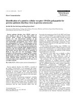
Transcription of Sly is reduced or absent in the MSYq deficient malesFigure 3
Transcription of Sly is reduced or absent in the MSYq deficient males. (a)
Northern blot of total testis RNA probed with the microarrayed Sly
cDNA clone MTnH-K10 and with an actin probe as a loading control.
Hybridization to the Sly probe is clearly reduced in 2/3MSYq
-
males and is
further markedly reduced in the two models with more severe MSYq
deficiency. (b) Reverse transcriptase polymerase chain reaction analysis of
testis cDNA with primers that distinguish between Sly and Xmr transcripts
and with Hprt primers as an amplification control. Some Sly transcripts are
retained in 9/10MSYq
-
males but they are absent in MSYq
-
males.
(b)
(a)
-1.35 Kb
XY
Tdym1
, Sry
XY
RIII
Actin
Sly
2
/
3
MSYq
-
MSYq
-
9
/
10
MSYq
-
Hprt
Xmr
Sly
XY
RIII
2
/
3
MSYq
-
MSYq
-
9
/
10
MSYq
-
-
R102.6 Genome Biology 2005, Volume 6, Issue 12, Article R102 Touré et al. />Genome Biology 2005, 6:R102
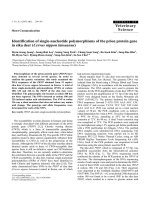
Identification of the novel MSYq encoded transcripts Asty and Asty(rec)Figure 4
Identification of the novel MSYq encoded transcripts Asty and Asty(rec). (a) Testis transcripts identified from a BLAST (basic local alignment search tool)
with the arrayed X encoded Astx cDNA clone 8832_f_22. BF019211, CF198098 and AK076884 are X encoded transcripts, whereas the rest are Y
encoded transcripts. The Y encoded transcripts AK016790 and BY716467 include exons that do not derive from Asty (including two exons matching
Ssty1). We refer to these transcripts as Asty(rec) because they derive from novel 'recombinant' loci on MSYq. For all the transcripts the percentage
sequence identity is given for those regions that match the microarrayed clone. (b) The structure of the Asty(rec) locus encoding AK016790. The exons
included in AK016790 are indicated by filled color coded rectangles and the inter-exonic distances are given in base pairs. The position of other Ssty1 and
Asty exons not included in the AK016790 transcript are also indicated. (Note that the Ssty1 exon 3 is truncated in the Asty(rec) locus.) The two exons
colored red are those that match the previously described transcript AK015935. (c) Reverse transcriptase polymerase chain reaction of testis cDNA with
primers designed to specifically amplify Asty/Asty(rec) or Astx transcripts. It is clear that it is the Asty/Asty(rec) transcripts that are preferentially reduced in
the MSYq deficient males. (d) Northern blot of total testis RNA probed with an Asty exon 4 probe and with an actin probe as a loading control.
Transcripts ranging in size from about 1 kilobase (kb) to more than 9.5 kb are detected with the Asty exon 4 probe; the approximately 7.5 kb and larger
transcripts definitely derive from the Asty/Asty(rec) loci (see text) and (in contrast to AK016790) must include Asty exon 4.
100%
BF019211
f22
12
3
4
1 641 827 1008
CF198098
100%
AK076884
97%
exon 2 exon 3
AK016790
Intronic sequence
Ssty1 exons 1/3 2 exons matching AK015935
95%
93%
BY716467
95%
93%
AV265093
92%
(a)
2 3 41
1 2 3
1-140 136-328 327-1285
220bp 830bp
1-294 295-510 511-694 695-1194
484bp 691bp 524bp
1 2 3 4 5 6
1-137 138-268 269 - 486 487-670 671-794 795-1592
1869bp 485bp 695bp 5677bp 300bp
(b)
Asty(rec)
(AK016790)
Ssty1
Asty
(c)
Astx
Asty
-
XY
RIII
2
/
3
MSYq
-
9
/
10
MSYq
-
MSYq
-
XY
Tdym1
, Sry
-
-
-
-
Asty
(d)
-1.35
-9.5
-7.5
-4.4
-2.4
Kb
XY
Tdym1
, Sry
XY
RIII
2
/
3
MSYq
-
MSYq
-
9
/
10
MSYq
-
Genome Biology 2005, Volume 6, Issue 12, Article R102 Touré et al. R102.7
comment reviews reports refereed researchdeposited research interactions information
Genome Biology 2005, 6:R102
Because we believe the transcripts detected by the Asty exon
4 probe derive predominantly from the Asty(rec) loci that are
almost certainly driven by the spermatid specific Ssty1 pro-
motor, we made attempts to determine whether true Asty
transcripts are also spermatid specific. For this we used an
Asty exon 1-4 primer pair (previously used to provide evi-
dence for Asty transcripts) to amplify testis cDNA samples
from 1.5 dpp to adult. Transcripts were weakly detected by
these primers at 14.5 and 18.5 dpp, but the predominant
expression was at 22.5 dpp onward when there were tran-
scripts of two sizes (Figure 6f).
We know these primers can also amplify Astx transcripts, and
so we sequenced cloned RT-PCR products to confirm their
identity. This confirmed the presence of the two previously
identified Asty transcripts (one of which lacks exon 3). The
longer transcript was detected from 14.5 dpp onward, and the
shorter transcript from 22.5 dpp onward. Because spermatids
first appear at about 20 dpp, we conclude that the shorter
Asty isoform appears to be spermatid specific, but that the
longer one is not spermatid specific.
The protein encoding potential of Sly and Asty family
members and their X relatives
Of the 65 loci of the Sly gene family, 34 have retained coding
potential for a protein that is related to three previously
described chromatin associated nuclear proteins: XLR and
XMR, which are encoded by members of a complex multicopy
X gene family [28,30-32], and the autosomally encoded
SYCP3 protein, via a conserved COR1 region [33,34] (Figure.
Multiple copies of Sly and Asty/Asty(rec) map to MSYqFigure 5
Multiple copies of Sly and Asty/Asty(rec) map to MSYq. (a, b) Southern of EcoRI digested DNAs showing hybridization of Sly probe MTnH-K10 and Asty
exon 4 probe, respectively, to multiple male specific bands, with reduced hybridization to all bands in 2/3MSYq
-
males. (c, d) Southern of EcoRI-digested
DNAs from 2/3MSYq
-
, 9/10MSYq
-
, and MSYq
-
males together with control XY
RIII
, showing reduced hybridization in 2/3MSYq
-
males and an apparent
absence of hybridization to Y specific bands in the males with more extensive MSYq deficiency. (e) Two week exposure of a Southern blot of EcoRI
digested DNAs from 9/10MSYq
-
and MSYq
-
males together with XO and XX females, and an underloaded XY
RIII
control. X derived fragments now cross-
hybridize with the Sly probe (dosing with the number of X chromosomes), but in addition there are at least three male specific bands (outlined) retained
in the 9/10MSYq
-
males that are absent in the MSYq
-
males.
A.
15 -
7 -
4 -
Kb
2.9 -
Asty
15 -
7 -
4 -
Kb
2.9 -
1.8 -
-
XY
2
/
3
MSYq
-
Sly
Asty
-
-
-
Sly
Sly
XO
XX
MSYq
-
XY
-
(b)
(a)
(c)
(d)
(e)
XX
2
/
3
MSYq
-
XY
MSYq
-
9
/
10
MSYq
-
9
/
10
MSYq
-
R102.8 Genome Biology 2005, Volume 6, Issue 12, Article R102 Touré et al. />Genome Biology 2005, 6:R102
Figure 6 (see legend on next page)
14
15
15
16
12-3
4
7
8
9
11
P
PPPPPPPPD
12
13
mm
2
Z
Z
LELEPLPLBB
IN
STAGES OF THE CYCLE
IN
10
4
6
5
A4m
A1
A1
A1
A1A1
A2s
A1mA1s
A3
A2m
15
16
I II-III XVIVIV VIIIVII XIIX XII
= Strong
signal
= Weak
signal
= No signal= Moderate
signal
Actin
9.5 -
7.5 -
4.4 -
2.4 -
1.35 -
0.24 -
Asty
- Heart
-Brain
- Spleen
- Lung
- Liver
- Smooth musc
le
- Kidney
- Testis
1.35 -
Sly
Kb
(a)
(d)
- 9.5 dpp
- 12.5 dpp
- 22.5 dpp
- 34.5 dpp
- adult
- 18.5 dpp
- 14.5 dpp
Asty
Actinb
Asty exon 4 sense
Asty exon 4 antisense
Asty exon 4 antisense
(f)
(e)
Sly antisense
Sly sense
Sly antisense
(c)
4.7 -
1.9 -
- 12.5 dpp
- 16.5 dpp
- 20.5 dpp
- 22.5 dpp
- 26.5 dpp
- 30.5 dpp
-XXSxr
b
Asty
Actin
Sly
1.9 -
Kb
(b)
Genome Biology 2005, Volume 6, Issue 12, Article R102 Touré et al. R102.9
comment reviews reports refereed researchdeposited research interactions information
Genome Biology 2005, 6:R102
7a, b). The putative SLY protein is most similar to XMR
(amino acid identities: XMR 48%, XLR 46% and SCP3 30%),
but within the COR1 region it is most similar to XLR (amino
acid identities: XLR 79%, XMR 50% and SCP3 37%).
All eight Astx loci have a modest open reading frame (ORF) in
exon 4, six of which would encode a protein of 106 amino
acids and two would encode a protein with a carboxyl-termi-
nal extension to 122 amino acids (Figure 8a). Surprisingly,
given the greater than 92% sequence identity of the Asty and
Astx loci, this exon 4 ORF is not conserved in any of the puta-
tive Asty loci so far identified. There are six Asty loci with an
ORF in exon 1 that could encode proteins of 116 or 120 amino
acids (Figure 8b). However, the Asty sequence we obtained by
RT-PCR from testis cDNA (Additional data file 2) did not
match the sequence of this subset of loci with an exon 1 ORF;
thus, it appears likely that the Y-linked members of this fam-
ily no longer produce a functional protein. Assessing whether
any of the novel Asty(rec) transcripts have significant protein
encoding potential will require detailed characterization of
this complex family of transcripts.
Discussion
The objective of the present study was to try to establish
whether members of the Ssty gene family are the only Y genes
present on MSYq that are expressed in the testis during
sperm development. Our strategy was to use testis cDNA
microarrays to identify transcripts that are reduced or absent
in the testes of mice with MSYq deficiencies, and then to
determine their chromosomal assignments. This strategy led
to the identification of further testis transcripts unrelated to
Ssty, which proved also to be encoded by multicopy Y loci on
MSYq and expressed in spermatids.
The first of these, Sly (Sycp3-like Y-linked; MGI:2687328) is
most closely related to the multicopy Xlr/Xmr gene family,
which is located on the X chromosome. It had previously been
reported, based on Southern blot evidence, that multiple Xlr
related sequences are present on the X and Y chromosomes,
but it was concluded that most if not all of these Xlr related
sequences were likely to represent nontranscribed pseudo-
genes [35]. However, another X linked family member,
namely Xmr, was subsequently described that is specifically
expressed in the testis [28], and our present findings estab-
lish that Sly transcripts, encoded by multiple loci on MSYq,
are also abundantly expressed in the testis in spermatids.
The XLR protein is a thymocyte nuclear protein that has been
shown to colocalize with SATB1, a protein that binds to AT-
rich sequences at the base of chromatin loops [31,36]. XMR is
a testis specific nuclear protein that concentrates in the sex
body of pachytene spermatocytes as the chromatin begins to
condense [28]. SYCP3 is an autosomally encoded protein that
is part of the synaptonemal complex of chromosomes in mei-
osis. XLR, XMR and SYCP3, together with the putative FAM9
proteins encoded by the human X chromosome, have been
grouped into a superfamily (InterPro accession number
IPR006888, PFAM accession number PF04803) because
they all share a conserved Cor1 domain. The Cor1 domain of
the putative SLY protein is very similar (79% identity) to that
of XLR, and we already have preliminary evidence that an
SLY protein is produced; we predict that this SLY protein will
also associate with chromatin loops.
Because SYCP3 is a fundamental component of the synapton-
emal complex during meiosis, it is not surprising that it has
been identified in a wide range of vertebrates. A comparison
of mammalian Cor1 domain proteins (Figure 9) indicates that
Sycp3 is the ancestral gene and that some time before the
divergence of human and mouse lineages the gene came onto
the X chromosome and became multicopy; these copies then
evolved rapidly and independently. At some point after the
divergence of mouse and rat, one of the X copies was brought
on to the Y chromosome, creating a new X-Y homologous
subfamily. This subfamily subsequently became massively
amplified in copy number on both the X and Y chromosomes.
Like Sly, Xmr and its close relatives are highly expressed in
spermatids [37], and our recent finding that a major
transcriptional consequence of MSYq deficiency is the upreg-
ulation of a number of spermatid-expressed X genes, includ-
ing Xmr, leads us to suspect that amplification of Xmr and Sly
is a result of 'genomic conflict' between sex linked meiotic
drivers and suppressors [37,38].
Sly and Asty/Asty(rec) are expressed in spermatidsFigure 6 (see previous page)
Sly and Asty/Asty(rec) are expressed in spermatids. (a) A multi-tissue northern blot of polyA
+
RNA probed with Sly clone MTnH-K10 and Asty exon 4
probe. Sly transcripts are restricted to the testis, but Asty/Asty(rec) transcripts are also seen in the heart. (b) A northern blot of total testis RNA from
testes of mice aged 12.5-30.5 days postpartum (dpp) hybridized with the same Sly and Asty probes. No transcripts are detectable before 20.5 dpp,
suggesting that the transcripts are restricted to spermatid stages. (c) RNA in situ analysis confirming that Sly transcription in the testis is predominantly if
not exclusively in spermatids. (d) Diagram summarizing the expression of Sly in spermatids throughout the spermatogenic cycle. The spermatogenic stages
starting from the basal layer and reading left to right are: spermatogonia: A1 through to B; meiotic prophase spermatocytes: preleptotene (PL), leptotene
(LE), zygotene (Z), pachytene (P), diplotene (D); meiotic divisions (mm); postmeiotic spermatid stages: 1-13. (e) RNA in situ analysis with the Asty exon 4
probe that should detect Asty and Asty(rec) transcripts. The most convincing signal is in round spermatid nuclei, but this is seen with the antisense and
sense (control) probes. However, the previously reported cDNA clone BU936708 (Additional data file 3) contains Asty exon 4 sequence in the antisense
oritentation and which would hybridize with the sense probe. (f) Exon 1-4 Asty reverse transcriptase polymerase chain reaction from 9.5 dpp to adult
testes. Sequencing of the cloned amplification products confirmed the presence of full-length Asty transcripts at 14.5 dpp, and a shorter transcript lacking
exon 3 from 22.5 dpp.
R102.10 Genome Biology 2005, Volume 6, Issue 12, Article R102 Touré et al. />Genome Biology 2005, 6:R102
The second MSYq encoded spermatid transcript, Asty, is also
encoded by a multicopy Y gene. Asty has a very high degree of
homology (92-94% identity across exons and introns) to a
multicopy X gene (Astx), suggesting that it may be a recent
arrival on the mouse Y chromosome. Intriguingly, BLAST
searches with the microarrayed Astx clone sequence failed to
detect similar sequences in the human genome, but in the rat
there were four X-linked sequences matching the last third of
the mouse Astx/Asty loci. Future analysis of these putative rat
loci, in particular to determine whether related sequences are
present on the rat Y chromosome, may help to delineate the
evolutionary history of this gene family. In addition to these
Asty loci, we have identified novel 'recombinant' loci, appar-
ently driven by the Ssty1 promotor, that incorporate a subset
of Ssty1 and Asty exons, and through alternative splicing and
serendipitous splicing of more downstream sequences they
have the potential to produce a range of novel transcripts in
addition to the previously described transcripts AK016790
and AK015935.
Because all eight copies of Astx have retained a similar ORF
in exon 4, it is reasonable to predict that they encode a pro-
tein. Interestingly, none of the more than 100 putative Asty
loci have retained the Astx exon 4 ORF, despite the greater
than 92% sequence identity of these loci. Six copies of Asty do
have protein encoding potential in exon 1; however, the only
Asty transcripts we have thus far identified do not derive from
this subset of loci with exon 1 protein encoding potential.
Overall, this evidence suggests that Astx is probably trans-
lated and that Asty is not. This does not, however, rule out a
functional role for Asty, or indeed the more strongly tran-
scribed Asty(rec), in sperm differentiation, especially given
the increasing literature on functional RNAs. A particularly
pertinent example in the present context is provided by the
The protein encoding potential of SlyFigure 7
The protein encoding potential of Sly. (a) Sly encodes a putative protein with a COR1 region. This COR 1 region is shared with two closely related
proteins, namely XMR and XLR, and with the less closely related synaptonemal complex protein SYCP3. (b) A comparison of the predicted protein
sequence for SLY with the predicted proteins for other Sly related Y loci that are each represented by more than one copy (information collated from
sequence information available in December 2004).
XMR (Mus) MSIKKLWVIPKDGYLLLLDYDSDEE EEQAHSEVKRPAFGKHENMPPHVEADEDIRDEQDSMLDKSGEN VSF 71
SLY (Mus) MRR-MALKKLKVIPKEGYLLLLDFDDEDDDIKVSEEALSEVKSPAFDKNENISPQAEADEDMGDEVDSMLDKSEVNNPAIGK 81
XLR (Mus)
MENWDLSSD EMQDGNAPELDVIEEHNPVTRDDENAN 36
SYCP3 (Mus) MLRGCGDSDSSPEPLSKHLKMVPGGRKHSG-KSGKPPLVDQPKKAFDFEKDDKDLSGSEEDVADEKAPVIDKHGKK R 76
SEEWQRFARSVETP-MENWNLL SGEQQVRN-ASELDLMEVQNPVTHDDGNANPEEVVGDT RKKINNKLCEQ KFDMDIQKFN 152
DENISPQVKGDEDMGHEVGSMLDKSGDDIYKTLHIKRKWMETYVKESFKGSNQKLERFCKTNERERKNINNKFCEQYITTFQKSDMDVQKFN 173
PEEVVGDTRS PVQNILGKFEGDINKRLHIKRKRMETYIKDSFKDSNVKLEQLWKTNKQERKKINNKFCEQYITTFQKFDMDVQKFN 122
SAGIIEDVGG EVQNMLEKFGADINKALLAKRKRIEMYTKASFKASNQKIEQIWKTQQEEIQKLNNEYSQQFMNVLQQWELDIQKFE 162
EEQEKSVNNYQKEQQALKLSECSQSPTMEAIEDMHEKSMEGLMNMETNNYDMLFDVDGEETL 214
EEKEKSVNSCQKEQQALKLSKCSQNQTLEAVKEMHEKSMEVLMNLGTKN 222
EEQEKSVNNYQKEQQALKLSKCSQSQTLEAIKDMHENYMEGLMNLETNNYNMLFDVDGELRKEMSVFKKDLMKHTLKYSSSFPSSD 208
EQGEKLSNLFRQQQKIFQQSRIVQSQRMFAMKQIHEQFIKSLEDVEKNNDNLFTGTQSELKKEMAMLQKKVMMETQQQEMANVRKSLQSMLF 254
(a)
COR 1 domain
COR 1 domain
(b)
XP487005(SLY)
MRRMALKKLKVIPKEGYLLLLDFDDEDDDIKVSEEALSEVKSPAFDKNENISPQAEADEDMGDEVDSMLDKSE 73
XP356452 MRRMSLKKLKVIPKEGYLLLLDFDDEDDDIKVSEEALSEVKSPAFDKNENISPQAEGDEDMGDEVDSMLDKSE 73
XP356439 MRRMALKKLKVIPKEGYLLLLDFDDEDDDIKVSEEALSEVKSPAFDKNENISPQAEADEDMGDEVDSMLDKSE 73
XP486989 MSYYCVLMRRMALKKLKVIPKEGYLLLLDFDDEDDDIKVSEEALSEVKSPAFDKNENISPQAEADEDMGDEVDSMLDKSE 80
XP486869 MSYYCVLMRRMALKKLKVIPKEGYLLLLDFDDEDDDIKVSEEALSEVKSPAFDKNENISPQAEADEDMGDEVDSMLDKSE 80
****:***************************************************.****************
XP487005 VNNPAIGKDENISPQVKGDEDMGHEVGSMLDKSGDDIYKTLHIKRKWMETYVKESFKGSNQKLERFCKTNERERKNINNK 153
XP356452 VNNPAIGKDENISPQVKGDEDMGHEVGSMLDKSGDDIYKTLHIKRKWMETYVKESFKGSNQKLERFCKTNERERKNINNK 153
XP356439 VNYPAIGKDENISPQVKGNEDMGHEVGSMLDKSGDDIYKTLHIKRKWMETYVKESFKGSNQKLERFCKTNERERKNINNK 153
XP486989 VNNPAIGKDENISPQVKGDEDMGHEVGSMLDKSGDDIYKTLHIKRKWMETYVKESFKCSNQKLERFCKTNERERKNINNK 160
XP486869 VNNPAIGKDENISPQVKGVEDMGHEVGSMLDKSGDDIYKTLHIKRKWMETYVKESFKCSNQKLERFCKTNERERKNINNK 160
** *************** ************************************** **********************
XP487005 FCEQYITTFQKSDMDVQKFNEEKEKSVNSCQKEQQALKLSKCSQNQTLEAVKEMHEKSMEVLMNLGTKN 222 [10 Loci]
XP356452 FCEQYITTFQKSDMDVQKFNEEKEKSVNSCQKEQQALKLSKCSQNQTLEAVKEMHEKSMEVLMNLGTKN 222 [ 3 Loci]
XP356439 FCEQYISTFQKSDMDVQKFNEEKEKSVNSCQKEQQALKLSKCSQNQTLEAVKEMHEKSMEVLMNLGTKN 222 [ 3 Loci]
XP486989 FCEQYITTFQKSDMDVQKFNEEKEKSVNSCQKEQQALKLSKCSQNQTLEAVKEMHEKSMEVLMNLGTKY 229 [ 3 Loci]
XP486869 FCEQYITTFQKSDMDVQKFNEEKEKSVNSCQKEQQALKLSKCSQNQTLEAVKEMHEKSMEVLMNLGTKY 229 [ 2 Loci]
******:*************************************************************
Genome Biology 2005, Volume 6, Issue 12, Article R102 Touré et al. R102.11
comment reviews reports refereed researchdeposited research interactions information
Genome Biology 2005, 6:R102
Stellate/suppressor of Stellate system in Drosophila, in which
transcripts encoded by multicopy loci on the Y regulate
expression of a protein expressed in spermatids and encoded
by related multicopy loci on the X chromosome [39], via an
antisense/small interfering RNA mechanism. It has been pos-
tulated that these RNA mediated regulatory interactions
between multiple X and Y loci in Drosophila arose as a conse-
quence of a past postmeiotic genomic conflict [38,40] and
there are clear parallels with the regulatory interactions we
have uncovered between MSYq-encoded loci and spermatid-
expressed X genes [37]. In this regard it is important that we
have established that Astx transcripts are present in sperma-
tids (Additional data file 4) as well as being expressed in sper-
matocytes (BF019211, CF198098).
The identification of additional MSYq encoded, spermatid
expressed transcripts provides alternatives to Ssty deficiency
as to the cause of the abnormalities in sperm shape associated
with MSYq deficiencies. Three features suggest that Sly defi-
ciency is the more likely cause. First, and importantly, reten-
tion of one or more transcribed Sly copies in 9/10MSYq
-
males (in contrast to Asty and Ssty [8]) provides a potential
explanation for the less severe sperm abnormalities in these
males, as compared with MSYq
-
males that completely lack
Sly. Second, Sly is predicted to encode a chromatin associated
protein, and the related proteins encoded by Xlr and Xmr are
expressed at sites of chromatin restructuring. Thus, it is rea-
sonable to suppose that Sly deficiency might affect sperm
head shape by disturbing chromatin organization in the
nucleus. Third, Sly is the most strongly transcribed in sper-
matids. On the other hand, we consider the sex ratio distor-
tion seen in 2/3MSYq
-
males to be a consequence of
disturbing the balance between sex-linked meiotic drivers
and suppressors involved in X-Y gene conflict [37], and
because this balance may have been achieved by MSYq
encoded proteins or RNAs, or both, all MSYq gene families
remain plausible candidates.
Conclusion
The highly repetitive nature of the mouse Y chromosome long
arm presents formidable challenges for the determination of
its functional gene content. Our strategy of using expression
array data to highlight transcriptionally active loci among the
sea of partial and degenerate gene copies has proved success-
ful in identifying further MSYq encoded transcripts, defi-
A comparison of the protein encoding potentials of Astx and AstyFigure 8
A comparison of the protein encoding potentials of Astx and Asty. (a) The predicted protein for the conserved open reading frame in exon 4 of the arrayed
Astx clone (f22) and for the four protein variants predicted from the eight putative X chromosomal loci. (b) The two predicted ASTY protein variants
encoded by two overlapping open reading frames in exon 1 present in six Asty loci.
ASTX(f22)
MFRLLHILLKMPRMTWFLVIFVLFLCCCFLLTFEEDTLLACCYVSHLLALETNIPLLKCFTFPTLFGKHN 70
ASTX(1) MFRLLHILLKMPRMAWFLVIFVLFLCCCFLLTFEEDTLLACCYVSHLLALETNIPLLKCFTFPTLFGKHN 70
ASTX(2) MFRLLHILLKMPRMTWFLVIFVLFLCCCFLLTFEEDTLLACCYVSHLLALETNIPLLKCFTFPTLFGKHN 70
ASTX(3) MFRLLHILLKMPRMTWFLVIFVLFLCCCFLLTFEEDTLLACCYVSHLLALETNISLLKSFTFPTLFGKHN 70
ASTX(4) MFRLLHILLKMPRMTWFLVIFVLFLCCCFLLTFEEDTLLACCYVSHLLALETNIPLLKCFTFPTLFGKHN 70
************** *************************************** *** ***********
ECTSFFPIVSHTYHYVIQLYNCNHFDQHSQEYKFYV 106
ECTSFFPIVSKTYHYVIQLYNCNHFDQYSHEYKFYV 106
ECTSFFPIVSKTYHYVIQLYNCNHFDQYSHEYKFYV 106
ECTSFFPIVSKTYHYVIQLYNCNHFDQYSHEYKFYV 106
ECTSFFPIVSKTYHYVIQLYNCNHFDQYSHEYKFMCKLLPVLCYFPPDDSF1 122
********** **************** * ****
ASTY(1) MGSFISSKHEITIKNTHHLNVCGRHDNNLRSLLRGCNVVV 40
ASTY(2) MWCLIRKQSEHSPKTWLQFHTSSSSQAYVLLLGSFISSKHEITIKNTHHLNVSGRHDNNLRSLLRGCNVVF 71
******************** ******************
SGLVSSLNICPSTHSSAWRPGCLCLLEKREFQYCLISFQIFFAEFRKSTYFMWCVDSRQQLFQYSNRLDGYLWSMKPLVR 12
0
SGLVSSLNICLSTHSSAWRPGCLCLLEKRESQYCLICLQIFFCRI 11
6
********** ******************* ***** ****
(a)
(b)
R102.12 Genome Biology 2005, Volume 6, Issue 12, Article R102 Touré et al. />Genome Biology 2005, 6:R102
ciency of which may contribute to the abnormal sperm head
development and function seen in males with MSYq deficien-
cies. This type of approach is likely to form an important com-
ponent of future functional analysis of mammalian Y
chromosomes and other repetitive chromosomal regions.
Materials and methods
Mice
All mice were produced on a random bred albino MF1 strain
(NIMR colony) background. The mice used to provide RNA
for microarray analysis were the three MSYq deficient geno-
types (Figure 1) that we have previously analyzed with respect
to Ssty expression and sperm abnormalities, together with
appropriate age- and strain-matched controls.
Dendrogram showing the relationship between the SLY protein and other identified or predicted mammalian Cor1 domain proteinsFigure 9
Dendrogram showing the relationship between the SLY protein and other identified or predicted mammalian Cor1 domain proteins. The autosomally
encoded SYCP3 is the presumed ancestral gene because it has been identified in a wide range of vertebrate species. The remaining proteins other than SLY
are X encoded. For the rat, the putative X-encoded proteins have not yet been named and the labels given are simply a reflection of their position within
the dendrogram. The massive expansion in copy number is only seen in the branch containing XMR, XMR-rel., XLR and SLY. This amplification therefore
occurred subsequent to the divergence of mouse and rat lineages, concurrent with the appearance of Sly on the Y chromosome.
SYCP3 (Human)
SYCP3 (Chimp)
SYCP3 (Rat)
SYCP3 (Hamster)
SYCP3 (Mouse)
XMR (Mouse)
XLR3b (Mouse)
XLR4 (Mouse)
‘XLR4’ (Rat)
XLR5 (Mouse)
‘XLR5’ (Rat)
FAM9A (Human)
FAM9B (Human)
XMR-rel. (Mouse)
XLR3a (Mouse)
XLR (Mouse)
SLY (Mouse)
‘XLR’ (Rat)
‘XLR’ (Rat)
‘XLR’ (Rat)
SYCP3 (Human)
SYCP3 (Chimp)
SYCP3 (Rat)
SYCP3 (Hamster)
SYCP3 (Mouse)
XMR (Mouse)
XLR3b (Mouse)
XLR4 (Mouse)
‘XLR4’ (Rat)
XLR5 (Mouse)
‘XLR5’ (Rat)
FAM9A (Human)
FAM9B (Human)
XMR-rel. (Mouse)
XLR3a (Mouse)
XLR (Mouse)
SLY (Mouse)
‘XLR’ (Rat)
‘XLR’ (Rat)
‘XLR’ (Rat)
NP 995321.
NP 777611.
XP 219321.
NP 113681.
NP 033555.
XP 621007.
NP 035855.
NP 963288.
XP 343832.
XP 229219.
XP 217535.
NP 710161.
XP 509310.
NP 037173.
CAA54560.1
NP 035647.
NP 035856.
NP 035857.
NP 067340.
XP 219738.
Genome Biology 2005, Volume 6, Issue 12, Article R102 Touré et al. R102.13
comment reviews reports refereed researchdeposited research interactions information
Genome Biology 2005, 6:R102
XY
RIII
qdel males (2/3 MSYq
-
)
These mice have an RIII strain Y chromosome with a deletion
removing approximately two thirds of MSYq. The sperm have
mild distortions of head shape; the mice are nevertheless fer-
tile with a distortion of the sex ratio in favor of females [3].
XY
RIII
males are the appropriate controls.
XY
Tdym1
qdelSry males (9/10 MSYq
-
)
These mice have a 129 strain Y chromosome with a deletion
removing approximately nine-tenths of MSYq, and also a
small deletion (Tdy
m1
) removing the testis determinant Sry
from the short arm, this deficiency being complemented by
an autosomally located Sry transgene. These males are sterile
with virtually all of the sperm having grossly distorted heads
[8]. XY
Tdym1
Sry males are the appropriate controls.
XSxr
a
Y*X males (MSYq
-
)
In these mice the only Y specific material is provided by the Y
short arm derived Sxr
a
factor, which is attached to the X chro-
mosome distal to the pseudo-autosomal region (PAR); the Y*
X
chromosome provides a second PAR, thus allowing fulfill-
ment of the requirement for PAR synapsis [41]. These males
lack the entire Y specific (non-PAR) gene content of Yq; they
also have a 7.5-fold reduction in copies of the Rbmy gene fam-
ily, located on the short arm adjacent to the centromere. The
males are sterile and have even more severe sperm head
defects than do XY
-
qdelSry males [8,41]. Our recent work
indicates that Rbmy deficiency is unlikely to be a significant
cause of the abnormal sperm development [42]. Because Sxr
a
originated from a Y
RIII
chromosome, the appropriate controls
are again XY
RIII
.
Sample collection and microarray analysis
Testes were obtained from two of each of the MSYq deficient
males and the two control genotypes, at 2 months of age.
Total RNA was isolated using TRI reagent (Sigma-Aldrich,
Poole, Dorset, UK) and cleaned using RNEasy columns (Qia-
gen, Crawley, West Sussex, UK), in accordance with to the
manufacturers' protocols. Microarray hybridizations and
analysis were carried out as described by Ellis and coworkers
[43], except that microarray data normalization was based on
the global median signal for Cy3 and Cy5 channels rather
than on a panel of control genes. This form of normalization
is valid because the majority of genes do not vary between the
mutant and control samples for any of the models (Figure 2a).
Briefly, 10 µg total RNA was fluorescently labeled using an
indirect protocol, and the test and control samples were
allowed to co-hybridize to the array. Four technical replicate
slides were obtained for each test/control comparison. Cy3
was used to label the test (mutant) sample and Cy5 the control
(normal) sample in all cases. Fluorescence intensities provide
measures of the relative abundance for each hybridizing testis
transcript for each genotype, whereas Cy3/Cy5 ratios for
individual clones provide a measure of the levels in each of
the MSYq deficient models relative to their matched controls.
The arrayed clones consisted of two subtracted adult mouse
testis libraries [43], six testis cell type separated libraries
(IMAGE clone plate numbers 8825-8830, 8831-8836, 8846-
8850, 9339-9342, 13869-13871, 13872-13874 [29]), and
appropriate controls [43]. From sequence analysis performed
to date, the combined gene set included cDNAs derived from
more than 4,000 genes, often represented by multiple clones,
enriched for cDNAs deriving from spermatogenic cells. Slides
were scanned using an ArraywoRx CCD-based scanner and
the resulting images quantitated using GenePix. Raw data
were processed in Excel to remove data from spots flagged as
bad or not found, and from features with a background sub-
tracted intensity of under 100 in both channels. Global
median normalization, t tests, and fold change filtering were
performed using GeneSpring. Full details of the array experi-
ment were submitted to the ArrayExpress database (acces-
sion number E-MEXP-251).
Southern blot analysis
The probes used for Southern analysis were Sly cDNA clone
BC049626 [26] and an Asty 314 bp exon 4 probe amplified
with primers CAGCAAGGAGAGTGGGGAGTA and CAGT-
GGGATGTTGGTTTCTAATG. Genomic DNA was extracted
from tail biopsies and 15 µg (or 4 µg for the control XY on the
long exposure Southern blot) was digested with EcoRI, elec-
trophoresed through a 0.8% agarose gel and transferred to a
Hybond-N membrane (Amersham Biosciences, Little Chal-
font, Buckinghamshire, UK). After UV crosslinking (Strata-
Linker™. Stratagene, La Jolla, CA, USA), the membrane was
hybridized overnight with
32
P-labelled probes either at low
stringency (55°C; hybridization buffer: 6 × salt sodium citrate
(SSC), 5 × Denhart's, 0.1% sodium dodecyl sulfate (SDS), 100
µg/ml salmon sperm DNA; two 30 minute washes (one with
0.5 × SSC and 0.1% SDS, and one with 0.1 × SSC and 0.1%
SDS)) or at high stringency (60°C; hybridization buffer: 6 ×
SSC, 5 × Denhart's, 0.5% SDS, 100 µg/ml salmon sperm
DNA; two 30 minute washes (one with 0.5 × SSC and 0.1%
SDS, and one with 0.1 × SSC and 0.1%SDS)). The membrane
was exposed to X-ray film or Phosphorimager screen
overnight.
Northern analysis
The probes used for northern analysis were as follows: Sly
cDNA clones BC049626 [26] and MtnH_K10 from the micro-
array (Additional data file 1), and the Asty 314 bp exon 4
probe used for northern analysis. An actin probe that recog-
nizes α- and β-actin transcripts [44] served as a control for
RNA integrity. Total RNA (20 µg) was electrophoresed in a
1.4% formaldehyde/agarose gel and transferred to Hybond-N
membrane (Amersham) using 20 × SSC buffer. The RNA was
cross-linked to the membrane with UV (StrataLinker™), the
membrane was fixed for 1 hour at 80°C, and hybridized over-
night at 60°C with
32
P-labelled probes in hybridization buffer
(6 × SSC, 5 × Denharts, 0.1% SDS, 50 mmol/l sodium phos-
phate, 100 µg/ml salmon sperm DNA). After two 60°C
washes (30 min with 0.5 × SSC and 0.1% SDS, and 30 min
R102.14 Genome Biology 2005, Volume 6, Issue 12, Article R102 Touré et al. />Genome Biology 2005, 6:R102
with 0.1 × SSC and 0.1% SDS) the membrane was exposed to
X-ray film for 5 hours and subsequently to a Phosphorimager
screen to allow quantitation of hybridization using Image-
Quant software.
RT-PCR analysis
RNA samples were treated for DNA contamination using
DNAse I amplification grade kit (Invitrogen, Paisley, UK). For
the Sly/Xmr RT-PCR, 2 µg total RNA was reverse transcribed
in a 40 µl reaction using standard procedures. A 2.5 µl aliquot
was then added to a 25 µl PCR reaction. RT-PCR for Astx/
Asty was performed using the Qiagen OneStep RT-PCR kit,
following the manufacturers' instructions. In both cases
amplification was for 30 cycles at an annealing temperature
of 60°C.
The primers used were as follows: Sly and Xmr, forward
primer GTGCGGTTTGGAAGTGT and reverse primer
CTCAAGCAGAAGCAGATG; Asty and Astx exon 4, forward
Asty primer GRGGAGTAGAACTCATCATC and forward Astx
primer GGGGAGTAGAACTCATCTTTA, with common
reverse primer CAGGAGATGACTAACATAGCA; Asty exon 1
to exon 4, forward GGCCTTGCTCTTATGTCATC and reverse
CGATGATGAGTGACCTAAAGAT; and Astx exon 1/2 (span-
ning intron 1) to 3/4 (spanning intron 3), forward GCTCCA-
GAAGACAGAGATAC and reverse
AGACTTCAAACCTCATGCAGT.
Sequencing
RT-PCR product was purified using the QiaQuick kit (Qiagen)
and cloned using the pGEM-T easy vector system I kit (Invit-
rogen). Sequencing reactions were performed from 5' and 3'
ends using standard protocols (BigDye; Amersham). Com-
pleted reactions were analyzed by the Cambridge Department
of Genetics sequencing service using a 3130xl capillary sys-
tem (Applied Biosystems, Warrrington, Chesire, UK). Cycling
conditions for the sequencing reactions were 96°C, 55°C and
60°C for 10 s, 5 s and 4 minutes, respectively.
RNA in situ analysis
In situ hybridization using digoxigenin-labeled cRNA probes
from Sly clone MtnH_K10 and Asty and Astx exon 4 were
used to localize each mRNA on Bouin-fixed paraffin-embed-
ded mouse testis sections using procedures previously
described [45], with hybridization and washing temperatures
up to 55°C. Both antisense and sense (negative control)
cRNAs were used on each sample, in every experiment, and
for each set of conditions tested.
RNA fluorescence in situ hybridization
RNA fluorescence in situ hybridization (FISH) was per-
formed basically as described previously for Cot1 RNA FISH
[46] using an X chromosomal BAC clone RP23-83P7 that
contains the Astx locus encoding the cDNA AK076884
(BACPAC Resources, Oakland, CA, USA). Hybridization reac-
tions consisted of 100 ng biotin-labeled BAC probe, 3 µg
mouse Cot1 DNA and 10 µg salmon sperm DNA, and were
carried out overnight at 37°C. Staging of spermatogenic cells
was based on DAPI fluorescence morphology, together with
immunolabelling for the synaptonemal complex protein
SYCP3 (rabbit anti-SYCP3; Abcam, Cambridge, UK) and the
phosphorylated form of histone H2AX (mouse anti-gamma
H2AX; Upstate, Dundee, UK), as previously described
[46,47]. The chromosomal source of the RNA FISH signals
was first checked by DNA FISH with a digoxigenin labeled
RP23-83P7 BAC probe prepared using the Digoxigenin Nick
Translation Kit (Roche Diagnostics, Lewes, East Sussex, UK).
Hybridizations were carried out as for RNA FISH with strin-
gency washes as described previously [46], and the DNA
FISH signals were developed using anti-DIG-FITC (Chemi-
con, Chandlers Ford, Hampshire, UK), diluted 1:10, for 1 hour
at 37°C. Further confirmation of the X chromosomal source
of the Astx transcripts was provided by X chromosome paint-
ing, as previously described [46].
Additional data files
The following additional data are available with the online
version of this paper: a file providing sequence information
for the Sly-related clones from the microarray (Additional
data file 1); a file providing sequence information for the
microarrayed Astx clone and for the Asty RT-PCR products
(Additional data file 2); a diagram providing sequence infor-
mation on the 'recombinant' loci encoding the transcripts
AK016790 and AK015935 (Additional data file 3); and images
showing Astx transcriptional analysis (Additional data file 4).
Additional data file 1A file providing sequence information for the Sly-related clones from the microarrayA file providing sequence information for the Sly-related clones from the microarrayClick here for fileAdditional data file 2A file providing sequence information for the microarrayed Astx clone and for the Asty RT-PCR productsA file providing sequence information for the microarrayed Astx clone and for the Asty RT-PCR productsClick here for fileAdditional data file 3A diagram providing sequence information on the 'recombinant' loci encoding the transcripts AK016790 and AK015935A diagram providing sequence information on the 'recombinant' loci encoding the transcripts AK016790 and AK015935Click here for fileAdditional data file 4Images showing Astx transcriptional analysisImages showing Astx transcriptional analysisClick here for file
Acknowledgements
We thank Aine Rattigan for PCR genotyping, James Turner for help with
the RNA FISH analysis, and Anthony Brown, David Carter and Rob Furlong
at the Department of Pathology Centre for Microarray Resources for
printing and QC of microarray slides. AT was supported by a European
Community 'Marie Curie' individual fellowship. The study was funded in
part by BBSRC and the Wellcome Trust (E.J.C., P.J.I.E.), the NHMRC of
Australia (Fellowship #143792 to K.L.L.) and the ARC (P.A.F.B., K.L.L.).
References
1. Skaletsky H, Kuroda-Kawaguchi T, Minx PJ, Cordum HS, Hillier L,
Brown LG, Repping S, Pyntikova T, Ali J, Bieri T, et al.: The male-
specific region of the human Y chromosome is a mosaic of
discrete sequence classes. Nature 2003, 423:825-837.
2. Rozen S, Skaletsky H, Marszalek JD, Minx PJ, Cordum HS, Waterston
RH, Wilson RK, Page DC: Abundant gene conversion between
arms of palindromes in human and ape Y chromosomes.
Nature 2003, 423:873-876.
3. Conway SJ, Mahadevaiah SK, Darling SM, Capel B, Rattigan ÁM, Bur-
goyne PS: Y353/B: a candidate multiple-copy spermiogenesis
gene on the mouse Y chromosome. Mamm Genome 1994,
5:203-210.
4. Ma K, Inglis JD, Sharkey A, Bickmore WA, Hill RE, Prosser EJ, Speed
RM, Thomson EJ, Jobling M, Taylor K, et al.: A Y chromosome gene
family with RNA-binding protein homology:candidates for
the Azoospermia factor AZF controlling human
spermatogenesis. Cell 1993, 75:1287-1295.
5. Mahadevaiah SK, Odorisio T, Elliott DJ, Rattigan A, Szot M, Laval SH,
Washburn LL, McCarrey JR, Cattanach BM, Lovell-Badge R, et al.:
Mouse homologues of the human AZF candidate gene RBM
are expressed in spermatogonia and spermatids, and map to
Genome Biology 2005, Volume 6, Issue 12, Article R102 Touré et al. R102.15
comment reviews reports refereed researchdeposited research interactions information
Genome Biology 2005, 6:R102
a Y deletion interval associated with a high incidence of
sperm abnormalities. Hum Mol Genet 1998, 7:715-727.
6. Lahn BT, Page DC: Functional coherence of the human Y
chromosome. Science 1997, 278:675-680.
7. Reijo R, Lee T-Y, Salo P, Alagappan R, Brown LG, Rosenberg M,
Rozen S, Jaffe T, Straus D, Hovatta O, et al.: Diverse sperma-
togenic defects in humans caused by Y chromosome
deletions encompassing a novel RNA-binding protein gene.
Nature Genet 1995, 10:383-393.
8. Touré A, Szot M, Mahadevaiah SK, Rattigan A, Ojarikre OA, Bur-
goyne PS: A new deletion of the mouse Y chromosome long
arm associated with loss of Ssty expression, abnormal sperm
development and sterility. Genetics 2004, 166:901-912.
9. Moriwaki K, Suh D-S, Styrna J: Genetic factors effecting sperm
morphology in the mouse. Mouse News Lett 1988, 82:138.
10. Styrna J, Bilinska B, Krzanowskaa H: The effect of a partial Y chro-
mosome deletion in B10.BR-Ydel mice on testis morphol-
ogy, sperm quality and efficiency of fertilization. Reprod Fertil
Dev 2002, 14:101-108.
11. Styrna J, Imai HT, Moriwaki K: An increased level of sperm
abnormalities in mice with a partial deletion of the Y
chromosome. Genet Res 1991, 57:195-199.
12. Styrna J, Klag J, Moriwaki K: Influence of partial deletion of the Y
chromosome on mouse sperm phenotype. J Reprod Fertil 1991,
92:187-195.
13. Suh D-S, Styrna J, Moriwaki K: Effect of Y chromosome and H-2
complex derived from Japanese wild mouse on sperm
morphology. Genet Res Camb 1989, 53:17-19.
14. Xian M, Azuma S, Naito K, Kunieda T, Moriwaki K, Toyoda Y: Effect
of a partial deletion of Y chromosome on in vitro fertilizing
ability of mouse spermatozoa. Biol Reprod 1992, 47:549-553.
15. Bergstrom DE, Yan H, Sonti MM, Narayanswami S, Bayleran JK, Simp-
son EM: An expanded collection of mouse Y chromosome
RDA clones. Mamm Genome 1997, 8:510-512.
16. Eicher EM, Hutchison KW, Philips SJ, Tucker PK, Lee B: A repeated
segment on the mouse Y chromosome is composed of ret-
roviral related Y enriched and Y specific sequences. Genetics
1989, 122:181-192.
17. Fennelly J, Harper K, Laval S, Wright E, Plumb M: Co-amplification
to tail-to-tail copies of MuRVY and IAPE retroviral genomes
on the Mus musculus Y chromosome. Mamm Genome 1996,
7:31-36.
18. Navin A, Prekeris R, Lisitsyn NA, Sonti MM, Grieco DA, Narayan-
swami S, Lander ES, Simpson EM: Mouse Y-specific repeats iso-
lated by whole chromosome representational difference
analysis. Genomics 1996, 36:349-353.
19. Nishioka Y, Dolan BM, Fiorellino A, Prado VF: Nucelotide
sequence analysis of a mouse Y chromosomal DNA frag-
ment containing Bkm and LINE elements. Genetica 1992,
85:7-15.
20. Nishioka Y, Dolan BM, Prado VF, Zahed L, Tyson H: Comparison
of mouse Y-chromosomal repetitive sequences isolated
from Mus musculus, M. spicilegus, and M. spretus. Cytogenet Cell
Genet 1993, 64:54-58.
21. Nishioka Y, Dolan BM, Zahed L: Molecular characterization of a
mouse Y chromosomal repetitive sequence amplified in dis-
tantly related species in the genus Mus. Genome 1993,
36:588-593.
22. Nishioka Y, Lamothe E: Isolation and characterization of a
mouse Y chromosomal repetitive sequence. Genetics 1986,
113:417-432.
23. Bishop CE, Hatat D: Molecular cloning and sequence analysis of
a mouse Y chromosome RNA transcript expressed in the
testis. Nucleic Acids Res 1987, 15:2959-2969.
24. Prado VF, Lee C-H, Zahed L, Vekemans M, Nishioka Y: Molecular
characterization of a mouse Y chromosomal repetitive
sequence that detects transcripts in the testis. Cytogenet Cell
Genet 1992, 61:87-90.
25. Touré A, Grigoriev V, Mahadevaiah SK, Rattigan Á, Ojarikre OA, Bur-
goyne PS: A protein encoded by a member of the multi-copy
Ssty gene family located on the long arm of the mouse Y
chromosome is expressed during sperm development.
Genomics 2004, 83:140-147.
26. Strausberg RL, Feingold EA, Grouse LH, Derge JG, Klausner RD, Col-
lins FS, Wagner L, Shenmen CM, Schuler GD, Altschul SF, et al.: Gen-
eration and initial analysis of more than 15,000 full-length
human and mouse cDNA sequences. Proc Natl Acad Sci USA
2002, 99:16899-16903.
27. Cohen DI, Hedrick SM, Nielsen EA, D'Eustachio P, Ruddle F, Stein-
berg AD, Paul WE, Davis MM: Isolation of a cDNA clone corre-
sponding to an X-linked gene family (XLR) closely linked to
the murine immunodeficiency disorder xid. Nature 1985,
314:369-372.
28. Calenda A, Allenet B, Escalier D, Bach J-F, Garchon H-J: The meio-
sis-specific Xmr gene product is homologous to the
lymphocyte Xlr protein and is a component of the XY body.
EMBO J 1994, 13:100-109.
29. McCarrey JR, O'Brien DA, Skinner MK: Construction and prelim-
inary characterization of a series of mouse and rat testis
cDNA libraries. J Androl 1999, 20:635-639.
30. Garchon HJ, Davis MM: The XLR gene product defines a novel
set of proteins stabilized in the nucleus by zinc ions. J Cell Biol
1989, 108:779-787.
31. Escalier D, Allenet B, Badrichani A, Garchon HJ: High level expres-
sion of the Xlr nuclear protein in immature thymocytes and
colocalization with the matrix-associated region-binding
SATB1 protein. J Immunol 1999, 162:292-298.
32. Escalier D, Garchon H-J: XMR is associated with the asynapsed
segments of sex chromosomes in the XY body of mouse pri-
mary spermatocytes. Chromosoma 2000, 109:259-265.
33. Dobson M, Pearlman RE, Karaiskakis A, Spyropoulos B, Moens PB:
Synaptonemal complex proteins: occurrence, epitope map-
ping and chromosome disjunction. J Cell Sci 1994,
107:2749-2760.
34. Lammers JHM, Offenberg HH, Van Aalderen M, Vink ACG, Dietrich
AJJ, Heyting C: The gene encoding a major component of syn-
aptonemal complexes of rat is related to X-linked lym-
phocyte-regulated genes. Mol Cell Biol 1994, 14:1137-1146.
35. Garchon HJ, Loh E, Ho WY, Amar L, Avner P, Davies MM: The XLR
sequence family: dispersion on the X and Y chromosomes of
a large set of closely related sequences most of which are
pseudogenes. Nucleic Acids Res 1989, 17:9871-9888.
36. de Belle I, Cai S, Kohwi-Shigematsu T: The genomic sequences
bound to special AT-rich sequence-binding protein 1
(SATB1) in vivo in Jurkat T cells are tightly associated with
the nuclear matrix at the bases of the chromatin loops. J Cell
Biol 1998, 141:335-348.
37. Ellis PJ, Clemente EJ, Ball P, Toure A, Ferguson L, Turner JM, Loveland
KL, Affara NA, Burgoyne PS: Deletions on mouse Yq lead to
upregulation of multiple X- and Y-linked transcripts in
spermatids. Hum Mol Genet 2005, 14:2705-2715.
38. Hurst LD: Is Stellate a relict meiotic driver?. Genetics 1992,
130:229-230.
39. Aravin AA, Naumova NM, Tulin AV, Vagin VV, Rozovsky YM,
Gvozdev VA: Double-stranded RNA-mediated silencing of
genomic tandem repeats and transposable elements in the
D. melanogaster germline. Curr Biol 2001, 11:1017-1027.
40. Hurst LD: Further evidence consistent with Stellate 's involve-
ment in meiotic drive. Genetics 1996, 142:641-643.
41. Burgoyne PS, Mahadevaiah SK, Sutcliffe MJ, Palmer SJ: Fertility in
mice requires X-Y pairing and a Y-chromosomal 'spermio-
genesis' gene mapping to the long arm. Cell 1992, 71:391-398.
42. Szot M, Grigoriev V, Mahadevaiah SK, Ojarikre OA, Tour A, Von
Glasenapp E, Rattigan A, Turner JM, Elliott DJ, Burgoyne PS: Does
Rbmy have a role in sperm development in mice?. Cytogenet
Genome Res 2003, 103:330-336.
43. Ellis PJ, Furlong RA, Wilson A, Morris S, Carter D, Oliver G, Print C,
Burgoyne PS, Loveland KL, Affara NA: Modulation of the mouse
testis transcriptome during postnatal development and in
selected models of male infertility. Mol Hum Reprod 2004,
10:271-281.
44. Minty AJ, Caravatti M, Robert B, Cohen A, Daubas P, Weydert A,
Gros F, Buckingham ME: Mouse actin mRNAs. Construction
and characterization of a recombinant plasmid molecule
containing a complementary DNA transcript of mouse
alpha-actin mRNA. J Biol Chem 1981, 256:1008-1014.
45. Meinhardt A, O'Bryan MK, McFarlane JR, Loveland KL, Mallidis C,
Foulds LM, Phillips DJ, de Kretser DM: Localization of follistatin
in the rat testis. J Reprod Fertil 1998, 112:233-241.
46. Turner JM, Mahadevaiah SK, Fernandez-Capetillo O, Nussenzweig A,
Xu X, Deng CX, Burgoyne PS: Silencing of unsynapsed meiotic
chromosomes in the mouse. Nat Genet 2005, 37:41-47.
47. Mahadevaiah SK, Turner JMA, Baudat F, Rogakou EP, de Boer P,
Blanco-Rodriguez J, Jasin M, Keeney S, Bonner WM, Burgoyne PS:
Recombinational DNA double-strand breaks in mice pre-
cede synapsis. Nature Genet 2001, 27:271-276.