Báo cáo y học: "The AP-2 family of transcription factors." potx
Bạn đang xem bản rút gọn của tài liệu. Xem và tải ngay bản đầy đủ của tài liệu tại đây (112.76 KB, 8 trang )
Genome Biology 2005, 6:246
comment
reviews
reports deposited research
interactions
information
refereed research
Protein family review
The AP-2 family of transcription factors
Dawid Eckert, Sandra Buhl, Susanne Weber, Richard Jäger and
Hubert Schorle
Address: Department of Developmental Pathology, Institute of Pathology, Sigmund-Freud Strasse 25, 53125 Bonn, Germany.
Correspondence: Hubert Schorle. E-mail:
Summary
The AP-2 family of transcription factors consists of five different proteins in humans and mice: AP-2␣,
AP-2, AP-2␥, AP-2␦ and AP-2⑀. Frogs and fish have known orthologs of some but not all of
these proteins, and homologs of the family are also found in protochordates, insects and
nematodes. The proteins have a characteristic helix-span-helix motif at the carboxyl terminus,
which, together with a central basic region, mediates dimerization and DNA binding. The amino
terminus contains the transactivation domain. AP-2 proteins are first expressed in primitive
ectoderm of invertebrates and vertebrates; in vertebrates, they are also expressed in the
emerging neural-crest cells, and AP-2
␣
-/-
animals have impairments in neural-crest-derived facial
structures. AP-2 is indispensable for kidney development and AP-2␥ is necessary for the
formation of trophectoderm cells shortly after implantation; AP-2␣ and AP-2␥ levels are elevated
in human mammary carcinoma and seminoma. The general functions of the family appear to be
the cell-type-specific stimulation of proliferation and the suppression of terminal differentiation
during embryonic development.
Published: 28 December 2005
Genome Biology 2005, 6:246 (doi:10.1186/gb-2005-6-13-246)
The electronic version of this article is the complete one and can be
found online at />© 2005 BioMed Central Ltd
Gene organization and evolutionary history
The AP-2 family of transcription factors (Ensembl Family
ENSF00000001105) consists in humans and mice of five
members, AP-2␣, AP-2, AP-2␥, AP-2␦ and AP-2⑀; frogs and
fish have some of these proteins, and homologs are also
known in invertebrates. The chromosomal locations and
accession numbers of the family are given in Tables 1 and 2,
respectively. All mammalian AP-2 proteins except AP-2␦ are
encoded by seven exons and share a characteristic domain
structure (reviewed in [1]; for AP-2␦ see [2] and for AP-2⑀
see [3,4]). Orthologs show a similarity between 60 and 99%
at the amino-acid level, whereas paralogs show a similarity
between 56 and 78%.
Analysis of the phylogenetic tree (Figure 1) reveals that the
vertebrate AP-2 proteins are grouped together and are
divided into five groups. The single Xenopus AP-2 is most
closely related to mammalian AP-2␣ proteins. As the genes
AP-2

and AP-2
␦
are found on the same chromosome in
chickens, rodents and humans (Table 1), it is likely that they
are the result of an internal duplication. According to the
phylogenetic tree, AP-2
␦
genes appear to have separated
from the rest of the family early in the vertebrate clade and
to have evolved separately (Figure 1). A BLAST search of the
puffer fish Fugu rubripes fourth genome assembly database
[5] suggests that there are orthologs of AP-2
␣
, AP-2

, AP-2
␥
and AP-2
⑀
but not AP-2
␦
genes in bony fish, although only
orthologs of AP-2
␣
and AP-2

have been found in zebrafish.
In the genome of the protochordate Ciona intestinalis a
single AP-2 gene has been predicted; the phylogenetic tree
shows that the protein evolved before the split of the AP-2␣,
AP-2, AP-2␥ and AP-2⑀ proteins, with the highest sequence
similarity with the AP-2␣ group, suggesting that AP-2␣
might be most similar to the ancestor of AP-2 proteins. This
hypothesis is further supported by the conserved epithelial
expression patterns of murine AP-2
␣
[6], Xenopus AP-2 [7]
and the amphioxus and lamprey AP-2 [8] genes. As
expected, the two Caenorhabditis elegans and the single
Drosophila melanogaster AP-2 proteins show the weakest
phylogenetic relationship with vertebrate and protochor-
date AP-2 transcription factors; they form an outgroup to
the other AP-2 family members (Figure 1). Given that no
AP-2 gene has been identified in yeast, the family probably
originated late in evolution and expanded considerably in
the vertebrates.
Characteristic structural features
All AP-2 proteins share a highly conserved helix-span-helix
dimerization motif at the carboxyl terminus, followed by a
central basic region and a less conserved domain rich in
proline and glutamine at the amino terminus (Figure 2). The
proteins are able to form hetero- as well as homodimers. The
helix-span-helix motif together with the basic region medi-
ates DNA binding [9,10], and the proline- and glutamine-
rich region is responsible for transactivation. AP-2 has been
shown to bind to the palindromic consensus sequence
5Ј-GCCN
3
GGC-3Ј, found in various cellular and viral
enhancers (reviewed in [1]); a binding-site selection assay
in vitro also revealed the additional binding motifs
5Ј-GCCN
3
GGC-3Ј, 5Ј-GCCN
4
GGC-3Ј and 5Ј-GCCN
3/4
GGG-3Ј
[11]. Other binding sites differing from these sequence
motifs, for example, the SV40 enhancer element
5Ј-CCCCAGGC-3Ј [12], indicate that AP-2 proteins may bind
to a range of G/C-rich elements with variable affinities.
246.2 Genome Biology 2005, Volume 6, Issue 13, Article 246 Eckert et al. />Genome Biology 2005, 6:246
Table 1
Chromosomal locations of AP-2 genes from selected species
AP-2
␣
AP-2

AP-2
␥
AP-2
␦
AP-2
⑀
Other AP-2 genes*
H. sapiens 6p24 6p12 20q13.2 6p12.1 1p34.3
P. troglodytes 6p22.3 6p12 21 - -
M. musculus 13 A5-B1 1 A2-A4 2 H3-H4 1 A3 4 D2.2
R. norvegicus 17p12 9q13 3q42 9q13 5q36
G. gallus 23-3-
X. tropicalis scaffold_278 - - - -
D. rerio 24 20 - - -
C. elegans II
D. melanogaster 3L
*The AP-2 genes of C. elegans and D. melanogaster are not orthologous to any of the five mammalian genes. Data taken from the database entries for the
accession numbers given in Table 2. No information on mapping is available for the C. intestinalis AP-2 gene.
Table 2
Accession numbers for AP-2 proteins from selected species
AP-2␣ AP-2 AP-2␥ AP-2␦ AP-2⑀ Other AP-2 proteins*
H. sapiens NP_003211 NP_003212 NP_003213 NP_758438 NP_848643
P. troglodytes - XP_518532 XP_526337 - -
M. musculus NP_035677 NP_033360 NP_033361 NP_694794 NP_945198
R. norvegicus XP_225238 XP_217356 NP_958823 XP_236975 XP_233526
G. gallus NP_990425 NP_990226 - XP_426224 -
X. tropicalis AAD53289 - - - -
X. laevis AAA49972 - - - -
D. rerio NP_789829 NP_001019836 - - -
C. elegans NP_4951819
D. melanogaster NP_730664
C. intestinalis BAE06307 and BAE06308
*The AP-2 genes of C. elegans, D. melanogaster and C. intestinalis are not orthologous to any of the five mammalian genes.
comment
reviews
reports deposited research
interactions
information
refereed research
Genome Biology 2005, Volume 6, Issue 13, Article 246 Eckert et al. 246.3
Genome Biology 2005, 6:246
Figure 1
Phylogenetic tree of the AP-2 family. Amino-acid sequence alignments were performed using ClustalW implemented in Sequence Data Explorer of the
MEGA3 software [67]. The phylogenetic tree was created using the neighbor-joining method (gaps setting: pairwise deletion; distance method: number of
differences). Numbers at selected nodes indicate the percentage frequencies of branch association on the basis of 1,000 bootstrap repetitions. The scale
bar indicates the number of residue changes. Asterisks indicate predicted proteins; brackets denote subfamilies in vertebrates. Species: Caenorhabditis
elegans (nematode); Ciona intestinalis (sea squirt); Drosophila melanogaster (fruit fly); Danio rerio (zebrafish); Gallus gallus (chicken); Homo sapiens (human);
Mus musculus (mouse); Pan troglodytes (chimpanzee); Rattus norvegicus (rat); Xenopus laevis and Xenopus tropicalis (frog).
H. sapiens
AP-2α
α
P. troglodytes
AP-2α*
M. musculus
AP-2α
R. norvegicus
AP-2α
G. gallus
AP-2α
X. laevis
AP-2
X. tropicalis
AP-2
D. rerio
AP-2α
D. rerio
AP-2β
G. gallus
AP-2β
P. troglodytes
AP-2β*
R. norvegicus
AP-2β*
H. sapiens
AP-2β
M. musculus
AP-2β
H. sapiens
AP-2γ
P. troglodytes
AP-2γ*
M. musculus
AP-2γ
R. norvegicus
AP-2γ
H. sapiens
AP-2ε
M. musculus
AP-2ε
R. norvegicus
AP-2ε*
C. intestinalis
AP-2
G. gallus
AP-2δ*
H. sapiens
AP-2δ
M. musculus
AP-2δ
R. norvegicus
AP-2δ*
D. melanogaster
AP-2
C. elegans
AP-2 F28C6.2
C. elegans
AP-2 F28C6.1
β
γ
ε
δ
50
99
100
99
96
100
87
90
97
96
86
100
100
100
100
100
99
87
55
100
100
100
99
100
100
99
99
Target genes with AP-2-binding sites in their promoter
sequences are involved in biological processes such as cell
growth and differentiation and include, for example, those
encoding insulin-like growth factor binding protein 5 (IGF-
BP5) with the binding site 5Ј-GCCAGGGGC-3Ј [13], prothy-
mosin-␣ (5Ј-GCCGGTGGGC-3Ј) [14] and the estrogen
receptor (5Ј-GCCTGCGGGG-3Ј) [15].
Most AP-2 proteins have a PY motif (XPPXY) and other
highly conserved critical residues in the transactivation
domain; by contrast, the PY motif is missing in AP-2␦ but
the amino- and carboxy-terminal ends of the core sequence
of the transactivation domain are still conserved. In addi-
tion, the binding affinity of AP-2␦ to conserved AP-2-
binding sites is much lower than that of other AP-2 proteins
[2]. This suggests that AP-2␦ might transactivate genes in
vivo by a different mechanism from that used by other AP-2
proteins, probably through interactions with a novel group
of coactivators and through a different affinity for AP-2-
binding sites. Alternatively, AP-2␦ might act as a negative
regulator, inhibiting or modulating the transactivation capa-
bility or DNA-binding affinity of the other AP-2 family
members. The crystal structure of the AP-2 proteins has not
yet been solved.
Localization and function
AP-2 transcription factors are localized predominantly in the
nucleus, where they bind to target sequences and regulate
transcription of target genes. AP-2 proteins have also been
shown to interfere with other signal transduction pathways;
for example, it has been proposed that they modulate the
pathway downstream of the developmental signaling molecule
Wnt by associating with the Adenomatous polyposis coli
(APC) tumor suppressor protein in the nucleus [16].
The activity of AP-2 proteins can be controlled at multiple
levels: their transactivation potential, their DNA binding,
their subcellular localization [17-19] and their degradation
[20,21] can all be modified. Mechanisms of regulation
include post-translational modifications, such as protein
kinase A-mediated phosphorylation [22,23], sumoylation
[24] and redox regulation [25,26], as well as physical inter-
action with various proteins (see Table 3 for a comprehen-
sive list). Interacting proteins either modulate the activity of
AP-2 proteins or are influenced in their function by binding
to AP-2 proteins.
The tissue distribution and developmental functions of AP-2
transcription factors have been studied extensively in several
species. Drosophila AP-2 (dAP-2) is expressed in the maxil-
lary segment and neural structures during embryogenesis,
and in the central nervous system (CNS) and the leg, anten-
nal and labial imaginal disks during larval development
[27,28]. Mutation of the dAP-2 gene leads to defects in pro-
boscis development and leg-joint formation [29,30].
The multiple overlapping and diverging expression patterns
of AP-2 family proteins suggest that, following the expansion
of the family during vertebrate evolution, redundant and
non-redundant functions of the individual AP-2 family
members evolved. Although the single AP-2 protein in the
cephalochordate amphioxus is expressed mainly in non-
neuronal ectoderm, in the lamprey, a primitive vertebrate,
AP-2 has co-opted a second expression domain, the neural
crest [8]. The single AP-2 homolog described so far in
Xenopus is expressed in the epidermis and neural crest and
has been shown to be critical for the development of these
structures [7,31-33]. In zebrafish, the two AP-2 family
members, tfap2a and tfap2b [34], are coexpressed in the
neural tube, the ectoderm and the pronephric ducts of the
developing kidney, but only tfap2a is expressed in neural
crest cells [35,36]. Positional cloning revealed that the
zebrafish point mutants named mont blanc [35] and lockjaw
[36] encode tfap2a; the mutant animals display impaired
development of neural-crest derivatives, such as the facial
skeleton, the peripheral nervous system and pigment cells
[37,38]. It is also interesting to note that AP-2 proteins are
expressed in the primitive ectoderm of both invertebrates
and vertebrates, suggesting an evolutionarily conserved role
for the family in the formation of this tissue.
In mice, three of the five AP-2 family members (AP-2
␣
, AP-
2

and AP-2
␥
) are coexpressed in neural-crest cells, the
peripheral nervous system, facial and limb mesenchyme,
various epithelia of the developing embryo and the extra-
embryonic trophectoderm [2,39-41]. AP-2
␦
expression is
restricted mainly to the developing heart, CNS and retina
[39], whereas AP-2
⑀
expression is detected in cells of the
246.4 Genome Biology 2005, Volume 6, Issue 13, Article 246 Eckert et al. />Genome Biology 2005, 6:246
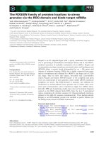
Figure 2
A schematic representation of the protein structure of an AP-2␣ dimer,
showing the proline- and glutamine (P/Q)-rich transactivation domain (89
amino acids, red), the PY motif within this domain (5 amino acids, green),
the basic domain (20 amino acids, yellow) and the helix-span-helix motif
(131 amino acids, blue). The helix-span-helix motif is responsible for
dimerization of the proteins and mediates DNA binding together with the
basic domain. Modified from SwissProt, ID: P34056 [68].
Transactivation
H
2
N
H
2
N
PY
COOH
COOH
Dimerization
DNA binding
Basic
domain
Helix-span-helix
motif
P/Q-rich
domain
olfactory bulb [3,4]. Despite the overlapping expression
patterns of AP-2
␣
, AP-2

and AP-2
␥
, disruption of these AP-2
genes reveals non-redundant roles during development.
Mutation of AP-2
␣
predominantly affects the cranial neural
crest and the limb mesenchyme, leading to disturbances of
facial and limb development in a manner reminiscent of the
defects described in dAP2 mutant flies [42,43]. AP-2

and
AP-2
␥
, on the other hand, are essential for kidney develop-
ment [44,45] or placentation of the embryo [46,47],
respectively. In humans, mutations generating a dominant
negative allele of AP-2

have been shown to be the cause of
Char syndrome (Online Mendelian Inheritance in Man
(OMIM) ID 169100 [48]); the hallmarks of this syndrome
are patent ductus arteriosus (abnormal persistence of a
normal fetal heart structure after birth) with facial dysmor-
phism and abnormal fifth digits [49,50].
Comparing all mutant phenotypes, it can be seen that loss of
AP-2 transcription factor activity generally impairs prolifer-
ation and induces premature differentiation and/or apopto-
sis in various cell types during development. This conclusion
is further substantiated by results from a screen for AP-2␣
target genes [51] and supported by gain-of-function studies
in Xenopus and mice [31,52,53]. As uncontrolled prolifera-
tion leads to malignancies, AP-2 transcription factors are not
only implicated in normal development, but also seem to be
involved in cellular neoplasia, and enhanced AP-2 levels
have been reported in various types of cancer [19,54-60]. In
a murine breast-cancer model, tumor progression is
enhanced after transgenic overexpression of AP-2
␥
[55].
Thus, AP-2 proteins can be viewed as gatekeepers control-
ling the balance between proliferation and differentiation
during embryogenesis.
comment
reviews
reports deposited research
interactions
information
refereed research
Genome Biology 2005, Volume 6, Issue 13, Article 246 Eckert et al. 246.5
Genome Biology 2005, 6:246
Table 3
Proteins that physically interact with AP-2 transcription factors
Domain of AP-2
Protein Description proteins that interacts* Function of interaction Reference
APC Adenomatous polyposis coli Basic region Inhibition of -catenin/TCF/LEF-dependent transcription [16]
tumor suppressor
CITED2 Coactivator DD Transcriptional activation [69]
CITED4 Coactivator n.d. Transcriptional activation [70]
CDP CCAAT displacement protein DBD, DD Repression of the hamster histone H3.2 promoter [71]
DEK Oncoprotein, chromatin remodeling n.d. Transcriptional activation [72]
E1A Transforming protein of adenovirus DBD, DD Repression of AP-2 target genes [73]
c-Myc Onco-protein Carboxyl terminus Impairment of Myc/Max DNA-binding and transactivation [14]
PARP PolyADP-ribose polymerase Carboxyl terminus Transcriptional activation [74]
PAX-6 Transcription factor n.d. Stimulation of gelatinase B activation [75]
PC4 Coactivator Transcriptional activation [24]
P300/CBP Coactivator Amino terminus Transcriptional activation [69]
p53 Tumor suppressor n.d. Augmentation of p53-dependent transcription [76]
RAP74 Subunit of transcription factor TFIIF Central region Unknown [74]
containing DBD
Rb Retinoblastoma tumor suppressor Amino terminus
†
Repression of the hamster histone H3.2 promoter; [77,78]
transcriptional activation of the E-cadherin gene
SP1 Transcription factor Basic region Transcriptional activation of the ovine CYP11A1 gene [79]
SV40T Transforming protein of SV40 virus n.d. Blocks DNA binding of AP-2 protein [12]
UBC9 E2-conjugating enzyme DBD, DD Sumoylation [80]
WWOX Tumor suppressor Amino terminus Cytoplasmic localization PPPY motif [17]
PY motif
YB-1 Transcription factor n.d. Stimulation of gelatinase A transcription [81]
YY1 Transcription factor DBD, DD Stimulation of the hamster histone H3.2 promoter [82]
*Abbreviations: DBD, DNA-binding domain; DD, dimerization domain; n.d., not determined.
†
It is currently not entirely clear whether Rb binds AP-2
only via the amino terminus [78], or whether the DNA-binding domain is also necessary [77].
Frontiers
The lethal phenotypes of the AP-2 mutants generated so far
have precluded an analysis of the roles of AP-2 transcription
factors in adult tissues. We and others are currently exploit-
ing the power of conditional mouse mutants to overcome
these restrictions [61-63]. Such approaches will not only
shed light on normal AP-2 functions but will probably also
lead to unique insights into human disorders.
Complementary approaches currently include the identifica-
tion of AP-2 target genes; this might give a better under-
standing of developmental disturbances and pave the way to
novel treatment options [51,64]. At the molecular level, one
major challenge will be the identification of specific AP-2
homo- or hetero-dimeric complexes bound to a particular
promoter and the identification of the specific properties of
each complex with respect to gene regulation. Also, the sig-
naling pathways responsible for induction of AP-2 genes are
currently under investigation. A cross-species comparison of
the various AP-2 promoters may give insights into the evolu-
tion of tissue specificity and help to determine important
enhancer elements. Moreover, given that CpG islands are
present in AP-2 promoters, epigenetic regulation such as
DNA methylation also needs to be considered.
AP-2 transcription factors are currently being studied exten-
sively in human cancer, and they may be of diagnostic value,
as has been demonstrated for mammary or testicular carci-
noma [19,54,56,65,66]. It is tempting to speculate that AP-2
transcription factors might not only be molecular markers
for certain types of cancer, but could also be causally
involved in their etiologies and would therefore represent a
potential target for therapeutic intervention.
Acknowledgements
We thank Roland Dosch and Michael Pankratz for critical reading of the
manuscript. This work was supported by funding from the Deutsche
Forschungsgemeinschaft (# 503/6 and 503/7) that was awarded to H.S.
References
1. Hilger-Eversheim K, Moser M, Schorle H, Buettner R: Regulatory
roles of AP-2 transcription factors in vertebrate develop-
ment, apoptosis and cell-cycle control. Gene 2000, 260:1-12.
2. Zhao F, Satoda M, Licht JD, Hayashizaki Y, Gelb BD: Cloning and
characterization of a novel mouse AP-2 transcription factor,
AP-2delta, with unique DNA binding and transactivation
properties. J Biol Chem 2001, 276:40755-40760.
3. Wang HV, Vaupel K, Buettner R, Bosserhoff AK, Moser M: Identifi-
cation and embryonic expression of a new AP-2 transcrip-
tion factor, AP-2 epsilon. Dev Dyn 2004, 231:128-135.
4. Feng W, Williams T: Cloning and characterization of the mouse
AP-2 epsilon gene: a novel family member expressed in the
developing olfactory bulb. Mol Cell Neurosci 2003, 24:460-475.
5. IMCB - Fugu Genome Project [ />6. Mitchell PJ, Timmons PM, Hebert JM, Rigby PW, Tjian R: Transcrip-
tion factor AP-2 is expressed in neural crest cell lineages
during mouse embryogenesis. Genes Dev 1991, 5:105-119.
7. Snape AM, Winning RS, Sargent TD: Transcription factor AP-2 is
tissue-specific in Xenopus and is closely related or identical
to keratin transcription factor 1 (KTF-1). Development 1991,
113:283-293.
8. Meulemans D, Bronner-Fraser M: Amphioxus and lamprey AP-2
genes: implications for neural crest evolution and migration
patterns. Development 2002, 129:4953-4962.
9. Williams T, Tjian R: Analysis of the DNA-binding and activa-
tion properties of the human transcription factor AP-2.
Genes Dev 1991, 5:670-682.
10. Williams T, Tjian R: Characterization of a dimerization motif
in AP-2 and its function in heterologous DNA-binding pro-
teins. Science 1991, 251:1067-1071.
11. Mohibullah N, Donner A, Ippolito JA, Williams T: SELEX and
missing phosphate contact analyses reveal flexibility within
the AP-2[alpha] protein: DNA binding complex. Nucleic Acids
Res 1999, 27:2760-2769.
12. Mitchell PJ, Wang C, Tjian R: Positive and negative regulation of
transcription in vitro: enhancer-binding protein AP-2 is
inhibited by SV40 T antigen. Cell 1987, 50:847-861.
13. Duan C, Clemmons DR: Transcription factor AP-2 regulates
human insulin-like growth factor binding protein-5 gene
expression. J Biol Chem 1995, 270:24844-24851.
14. Gaubatz S, Imhof A, Dosch R, Werner O, Mitchell P, Buettner R,
Eilers M: Transcriptional activation by Myc is under negative
control by the transcription factor AP-2. EMBO J 1995,
14:1508-1519.
15. Newman SP, Bates NP, Vernimmen D, Parker MG, Hurst HC:
Cofactor competition between the ligand-bound oestrogen
receptor and an intron 1 enhancer leads to oestrogen
repression of ERBB2 expression in breast cancer. Oncogene
2000, 19:490-497.
16. Li Q, Dashwood RH: Activator protein 2alpha associates with
adenomatous polyposis coli/beta-catenin and inhibits beta-
catenin/T-cell factor transcriptional activity in colorectal
cancer cells. J Biol Chem 2004, 279:45669-45675.
17. Aqeilan RI, Palamarchuk A, Weigel RJ, Herrero JJ, Pekarsky Y, Croce
CM: Physical and functional interactions between the Wwox
tumor suppressor protein and the AP-2gamma transcrip-
tion factor. Cancer Res 2004, 64:8256-8261.
18. Mazina OM, Phillips MA, Williams T, Vines CA, Cherr GN, Rice RH:
Redistribution of transcription factor AP-2alpha in differen-
tiating cultured human epidermal cells. J Invest Dermatol 2001,
117:864-870.
19. Pellikainen J, Naukkarinen A, Ropponen K, Rummukainen J, Kataja V,
Kellokoski J, Eskelinen M, Kosma VM: Expression of HER2 and its
association with AP-2 in breast cancer. Eur J Cancer 2004,
40:1485-1495.
20. Li M, Wang Y, Hung MC, Kannan P: Inefficient proteasomal-
degradation pathway stabilizes AP-2alpha and activates
HER-2/neu gene in breast cancer. Int J Cancer 2005.
doi:10.1002/ijc.21426.
21. Nyormoi O, Wang Z, Doan D, Ruiz M, McConkey D, Bar-Eli M:
Transcription factor AP-2alpha is preferentially cleaved by
caspase 6 and degraded by proteasome during tumor
necrosis factor alpha-induced apoptosis in breast cancer
cells. Mol Cell Biol 2001, 21:4856-4867.
22. Garcia MA, Campillos M, Marina A, Valdivieso F, Vazquez J: Tran-
scription factor AP-2 activity is modulated by protein kinase
A-mediated phosphorylation. FEBS Lett 1999, 444:27-31.
23. Park K, Kim KH: The site of cAMP action in the insulin induc-
tion of gene expression of acetyl-CoA carboxylase is AP-2. J
Biol Chem 1993, 268:17811-17819.
24. Zhong L, Wang Y, Kannan P, Tainsky MA: Functional characteri-
zation of the interacting domains of the positive coactivator
PC4 with the transcription factor AP-2alpha. Gene 2003,
320:155-164.
25. Grether-Beck S, Felsner I, Brenden H, Krutmann J: Mitochondrial
cytochrome c release mediates ceramide-induced activator
protein 2 activation and gene expression in keratinocytes.
J Biol Chem 2003, 278:47498-47507.
26. Huang Y, Domann FE: Redox modulation of AP-2 DNA binding
activity in vitro. Biochem Biophys Res Commun 1998, 249:307-312.
27. Bauer R, McGuffin ME, Mattox W, Tainsky MA: Cloning and char-
acterization of the Drosophila homologue of the AP-2 tran-
scription factor. Oncogene 1998, 17:1911-1922.
28. Monge I, Mitchell PJ: DAP-2, the Drosophila homolog of tran-
scription factor AP-2. Mech Dev 1998, 76:191-195.
29. Kerber B, Monge I, Mueller M, Mitchell PJ, Cohen SM: The AP-2
transcription factor is required for joint formation and cell
survival in Drosophila leg development. Development 2001,
128:1231-1238.
246.6 Genome Biology 2005, Volume 6, Issue 13, Article 246 Eckert et al. />Genome Biology 2005, 6:246
30. Monge I, Krishnamurthy R, Sims D, Hirth F, Spengler M, Kammer-
meier L, Reichert H, Mitchell PJ: Drosophila transcription factor
AP-2 in proboscis, leg and brain central complex develop-
ment. Development 2001, 128:1239-1252.
31. Luo T, Matsuo-Takasaki M, Thomas ML, Weeks DL, Sargent TD:
Transcription factor AP-2 is an essential and direct regula-
tor of epidermal development in Xenopus. Dev Biol 2002,
245:136-144.
32. Winning RS, Shea LJ, Marcus SJ, Sargent TD: Developmental regu-
lation of transcription factor AP-2 during Xenopus laevis
embryogenesis. Nucleic Acids Res 1991, 19:3709-3714.
33. Luo T, Lee YH, Saint-Jeannet JP, Sargent TD: Induction of neural
crest in Xenopus by transcription factor AP2alpha. Proc Natl
Acad Sci USA 2003, 100:532-537.
34. Knight RD, Javidan Y, Zhang T, Nelson S, Schilling TF: AP2-depen-
dent signals from the ectoderm regulate craniofacial devel-
opment in the zebrafish embryo. Development 2005,
132:3127-3138.
35. Holzschuh J, Barrallo-Gimeno A, Ettl AK, Durr K, Knapik EW,
Driever W: Noradrenergic neurons in the zebrafish hindbrain
are induced by retinoic acid and require tfap2a for expres-
sion of the neurotransmitter phenotype. Development 2003,
130:5741-5754.
36. Knight RD, Nair S, Nelson SS, Afshar A, Javidan Y, Geisler R, Rauch
GJ, Schilling TF: lockjaw encodes a zebrafish tfap2a required
for early neural crest development. Development 2003,
130:5755-5768.
37. Barrallo-Gimeno A, Holzschuh J, Driever W, Knapik EW: Neural
crest survival and differentiation in zebrafish depends on
mont blanc/tfap2a gene function. Development 2004, 131:1463-
1477.
38. Knight RD, Javidan Y, Nelson S, Zhang T, Schilling T: Skeletal and
pigment cell defects in the lockjaw mutant reveal multiple
roles for zebrafish tfap2a in neural crest development. Dev
Dyn 2004, 229:87-98.
39. Zhao F, Lufkin T, Gelb BD: Expression of Tfap2d, the gene
encoding the transcription factor Ap-2 delta, during mouse
embryogenesis. Gene Expr Patterns 2003, 3:213-217.
40. Moser M, Ruschoff J, Buettner R: Comparative analysis of AP-2
alpha and AP-2 beta gene expression during murine
embryogenesis. Dev Dyn 1997, 208:115-124.
41. Chazaud C, Oulad-Abdelghani M, Bouillet P, Decimo D, Chambon P,
Dolle P: AP-2.2, a novel gene related to AP-2, is expressed in
the forebrain, limbs and face during mouse embryogenesis.
Mech Dev 1996, 54:83-94.
42. Schorle H, Meier P, Buchert M, Jaenisch R, Mitchell PJ: Transcrip-
tion factor AP-2 essential for cranial closure and craniofacial
development. Nature 1996, 381:235-238.
43. Zhang J, Hagopian-Donaldson S, Serbedzija G, Elsemore J, Plehn-
Dujowich D, McMahon AP, Flavell RA, Williams T: Neural tube,
skeletal and body wall defects in mice lacking transcription
factor AP-2. Nature 1996, 381:238-241.
44. Moser M, Dahmen S, Kluge R, Grone H, Dahmen J, Kunz D, Schorle
H, Buettner R: Terminal renal failure in mice lacking tran-
scription factor AP-2 beta. Lab Invest 2003, 83:571-578.
45. Moser M, Pscherer A, Roth C, Becker J, Mucher G, Zerres K,
Dixkens C, Weis J, Guay-Woodford L, Buettner R et al.: Enhanced
apoptotic cell death of renal epithelial cells in mice lacking
transcription factor AP-2beta. Genes Dev 1997, 11:1938-1948.
46. Auman HJ, Nottoli T, Lakiza O, Winger Q, Donaldson S, Williams T:
Transcription factor AP-2gamma is essential in the extra-
embryonic lineages for early postimplantation develop-
ment. Development 2002, 129:2733-2747.
47. Werling U, Schorle H: Transcription factor gene AP-2
␥␥
essential for early murine development. Mol Cell Biol 2002,
22:3149-3156.
48. OMIM - Online Mendelian Inheritance in Man
[ />49. Zhao F, Weismann CG, Satoda M, Pierpont ME, Sweeney E, Thomp-
son EM, Gelb BD: Novel TFAP2B mutations that cause Char
syndrome provide a genotype-phenotype correlation. Am J
Hum Genet 2001, 69:695-703.
50. Satoda M, Zhao F, Diaz GA, Burn J, Goodship J, Davidson HR, Pier-
pont ME, Gelb BD: Mutations in TFAP2B cause Char syn-
drome, a familial form of patent ductus arteriosus. Nat Genet
2000, 25:42-46.
51. Pfisterer P, Ehlermann J, Hegen M, Schorle H: A subtractive gene
expression screen suggests a role of transcription factor
AP-2 alpha in control of proliferation and differentiation. J
Biol Chem 2002, 277:6637-6644.
52. Zhang J, Brewer S, Huang J, Williams T: Overexpression of tran-
scription factor AP-2alpha suppresses mammary gland
growth and morphogenesis. Dev Biol 2003, 256:127-145.
53. Jager R, Werling U, Rimpf S, Jacob A, Schorle H: Transcription
factor AP-2gamma stimulates proliferation and apoptosis
and impairs differentiation in a transgenic model. Mol Cancer
Res 2003, 1:921-929.
54. Pauls K, Jager R, Weber S, Wardelmann E, Koch A, Buttner R,
Schorle H: Transcription factor AP-2gamma, a novel marker
of gonocytes and seminomatous germ cell tumors. Int J
Cancer 2005, 115:470-477.
55. Jager R, Friedrichs N, Heim I, Buttner R, Schorle H: Dual role of
AP-2gamma in ErbB-2-induced mammary tumorigenesis.
Breast Cancer Res Treat 2005, 90:273-280.
56. Hoei-Hansen CE, Nielsen JE, Almstrup K, Sonne SB, Graem N,
Skakkebaek NE, Leffers H, Meyts ER: Transcription factor AP-
2gamma is a developmentally regulated marker of testicu-
lar carcinoma in situ and germ cell tumors. Clin Cancer Res
2004, 10:8521-8530.
57. Hurst HC: Update on HER-2 as a target for cancer therapy:
the ERBB2 promoter and its exploitation for cancer treat-
ment. Breast Cancer Res 2001, 3:395-398.
58. Beger M, Butz K, Denk C, Williams T, Hurst HC, Hoppe-Seyler F:
Expression pattern of AP-2 transcription factors in cervical
cancer cells and analysis of their influence on human papillo-
mavirus oncogene transcription. J Mol Med 2001, 79:314-320.
59. Turner BC, Zhang J, Gumbs AA, Maher MG, Kaplan L, Carter D,
Glazer PM, Hurst HC, Haffty BG, Williams T: Expression of AP-2
transcription factors in human breast cancer correlates
with the regulation of multiple growth factor signalling
pathways. Cancer Res 1998, 58:5466-5472.
60. Bosher JM, Totty NF, Hsuan JJ, Williams T, Hurst HC: A family of
AP-2 proteins regulates c-erbB-2 expression in mammary
carcinoma. Oncogene 1996, 13:1701-1707.
61. Nelson DK, Williams T: Frontonasal process-specific disrup-
tion of AP-2alpha results in postnatal midfacial hypoplasia,
vascular anomalies, and nasal cavity defects. Dev Biol 2004,
267:72-92.
62. Brewer S, Feng W, Huang J, Sullivan S, Williams T: Wnt1-Cre-
mediated deletion of AP-2alpha causes multiple neural
crest-related defects. Dev Biol 2004, 267:135-152.
63. Werling U, Schorle H: Conditional inactivation of transcription
factor AP-2gamma by using the Cre/loxP recombination
system. Genesis 2002, 32:127-129.
64. Luo T, Zhang Y, Khadka D, Rangarajan J, Cho KW, Sargent TD:
Regulatory targets for transcription factor AP2 in Xenopus
embryos. Dev Growth Differ 2005, 47:403-413.
65. Friedrichs N, Jager R, Paggen E, Rudlowski C, Merkelbach-Bruse S,
Schorle H, Buettner R: Distinct spatial expression patterns of
AP-2alpha and AP-2gamma in non-neoplastic human breast
and breast cancer. Mod Pathol 2005, 18:431-438.
66. Hoei-Hansen CE, Nielsen JE, Almstrup K, Hansen MA, Skakkebaek
NE, Rajpert-DeMeyts E, Leffers H: Identification of genes differ-
entially expressed in testes containing carcinoma in situ. Mol
Hum Reprod 2004, 10:423-431.
67. Kumar S, Tamura K, Nei M: MEGA3: Integrated software for
Molecular Evolutionary Genetics Analysis and sequence
alignment. Brief Bioinform 2004, 5:150-163.
68. Swiss-Prot [ />69. Braganca J, Eloranta JJ, Bamforth SD, Ibbitt JC, Hurst HC, Bhat-
tacharya S: Physical and functional interactions among AP-2
transcription factors, p300/CREB-binding protein, and
CITED2. J Biol Chem 2003, 278:16021-16029.
70. Braganca J, Swingler T, Marques FI, Jones T, Eloranta JJ, Hurst HC,
Shioda T, Bhattacharya S: Human CREB-binding protein/p300-
interacting transactivator with ED-rich tail (CITED) 4, a new
member of the CITED family, functions as a co-activator for
transcription factor AP-2. J Biol Chem 2002, 277:8559-8565.
71. Wu F, Lee AS: CDP and AP-2 mediated repression mecha-
nism of the replication-dependent hamster histone H3.2
promoter. J Cell Biochem 2002, 84:699-707.
72. Campillos M, Garcia MA, Valdivieso F, Vazquez J: Transcriptional
activation by AP-2alpha is modulated by the oncogene DEK.
Nucleic Acids Res 2003, 31:1571-1575.
comment
reviews
reports deposited research
interactions
information
refereed research
Genome Biology 2005, Volume 6, Issue 13, Article 246 Eckert et al. 246.7
Genome Biology 2005, 6:246
73. Somasundaram K, Jayaraman G, Williams T, Moran E, Frisch S,
Thimmapaya B: Repression of a matrix metalloprotease gene
by E1A correlates with its ability to bind to cell type-specific
transcription factor AP-2. Proc Natl Acad Sci USA 1996, 93:3088-
3093.
74. Kannan P, Yu Y, Wankhade S, Tainsky MA: PolyADP-ribose poly-
merase is a coactivator for AP-2-mediated transcriptional
activation. Nucleic Acids Res 1999, 27:866-874.
75. Sivak JM, West-Mays JA, Yee A, Williams T, Fini ME: Transcription
factors Pax6 and AP-2alpha interact to coordinate corneal
epithelial repair by controlling expression of matrix
metalloproteinase gelatinase B. Mol Cell Biol 2004, 24:245-257.
76. McPherson LA, Loktev AV, Weigel RJ: Tumor suppressor activity
of AP2alpha mediated through a direct interaction with
p53. J Biol Chem 2002, 277:45028-45033.
77. Wu F, Lee AS: Identification of AP-2 as an interactive target
of Rb and a regulator of the G1/S control element of the
hamster histone H3.2 promoter. Nucleic Acids Res 1998,
26:4837-4845.
78. Batsche E, Muchardt C, Behrens J, Hurst HC, Cremisi C: RB and
c-Myc activate expression of the E-cadherin gene in epithelial
cells through interaction with transcription factor AP-2. Mol
Cell Biol 1998, 18:3647-3658.
79. Pena P, Reutens AT, Albanese C, D’Amico M, Watanabe G, Donner A,
Shu IW, Williams T, Pestell RG: Activator protein-2 mediates
transcriptional activation of the CYP11A1 gene by interac-
tion with Sp1 rather than binding to DNA. Mol Endocrinol
1999, 13:1402-1416.
80. Eloranta JJ, Hurst HC: Transcription factor AP-2 interacts with
the SUMO-conjugating enzyme UBC9 and is sumolated in
vivo. J Biol Chem 2002, 277:30798-30804.
81. Mertens PR, Alfonso-Jaume MA, Steinmann K, Lovett DH: A syner-
gistic interaction of transcription factors AP2 and YB-1 reg-
ulates gelatinase A enhancer-dependent transcription. J Biol
Chem 1998, 273:32957-32965.
82. Wu F, Lee AS: YY1 as a regulator of replication-dependent
hamster histone H3.2 promoter and an interactive partner
of AP-2. J Biol Chem 2001, 276:28-34.
246.8 Genome Biology 2005, Volume 6, Issue 13, Article 246 Eckert et al. />Genome Biology 2005, 6:246