Báo cáo y học: "microRNA expression in the prefrontal cortex of individuals with schizophrenia and schizoaffective disorder" ppsx
Bạn đang xem bản rút gọn của tài liệu. Xem và tải ngay bản đầy đủ của tài liệu tại đây (411.57 KB, 11 trang )
Genome Biology 2007, 8:R27
comment reviews reports deposited research refereed research interactions information
Open Access
2007Perkinset al.Volume 8, Issue 2, Article R27
Research
microRNA expression in the prefrontal cortex of individuals with
schizophrenia and schizoaffective disorder
Diana O Perkins
*
, Clark D Jeffries
†‡
, L Fredrik Jarskog
*
, J
Michael Thomson
§
, Keith Woods
§
, Martin A Newman
§
, Joel S Parker
¶
,
Jianping Jin
¥
and Scott M Hammond
§
Addresses:
*
Department of Psychiatry, University of North Carolina at Chapel Hill, CB 7160, Chapel Hill, NC 27599, USA.
†
School of Pharmacy,
University of North Carolina at Chapel Hill, CB 7360, Chapel Hill, NC 27599, USA.
‡
Renaissance Computing Institute, University of North
Carolina at Chapel Hill, Chapel Hill, NC 27599, USA.
§
Department of Cell and Developmental Biology, University of North Carolina at Chapel
Hill, CB 7090, Chapel Hill, NC 27599, USA.
¶
Constella Group, LLC, Meridian Parkway, Durham, NC 27713, USA.
¥
Department of Molecular
Biology, University of North Carolina at Chapel Hill, CB 7104, Chapel Hill, NC 27599, USA.
Correspondence: Clark D Jeffries. Email:
© 2007 Perkins et al.; licensee BioMed Central Ltd.
This is an open access article distributed under the terms of the Creative Commons Attribution License ( which
permits unrestricted use, distribution, and reproduction in any medium, provided the original work is properly cited.
MicroRNAs in schizophrenia<p>Transcriptional profiling reveals a possible association between schizophrenia and altered miRNA expression</p>
Abstract
Background: microRNAs (miRNAs) are small, noncoding RNA molecules that are now thought
to regulate the expression of many mRNAs. They have been implicated in the etiology of a variety
of complex diseases, including Tourette's syndrome, Fragile × syndrome, and several types of
cancer.
Results: We hypothesized that schizophrenia might be associated with altered miRNA profiles.
To investigate this possibility we compared the expression of 264 human miRNAs from
postmortem prefrontal cortex tissue of individuals with schizophrenia (n = 13) or schizoaffective
disorder (n = 2) to tissue of 21 psychiatrically unaffected individuals using a custom miRNA
microarray. Allowing a 5% false discovery rate, we found that 16 miRNAs were differentially
expressed in prefrontal cortex of patient subjects, with 15 expressed at lower levels (fold change
0.63 to 0.89) and 1 at a higher level (fold change 1.77) than in the psychiatrically unaffected
comparison subjects. The expression levels of 12 selected miRNAs were also determined by
quantitative RT-PCR in our lab. For the eight miRNAs distinguished by being expressed at lower
microarray levels in schizophrenia samples versus comparison samples, seven were also expressed
at lower levels with quantitative RT-PCR.
Conclusion: This study is the first to find altered miRNA profiles in postmortem prefrontal cortex
from schizophrenia patients.
Background
Schizophrenia is a common neuropsychiatric disorder affect-
ing one percent of the general population. The personal,
familial, and societal costs of the disease are enormous, with
chronic symptoms that result in marked functional disability.
Published: 27 February 2007
Genome Biology 2007, 8:R27 (doi:10.1186/gb-2007-8-2-r27)
Received: 24 November 2006
Revised: 25 January 2007
Accepted: 27 February 2007
The electronic version of this article is the complete one and can be
found online at />R27.2 Genome Biology 2007, Volume 8, Issue 2, Article R27 Perkins et al. />Genome Biology 2007, 8:R27
In fact, approximately three percent of all person-years lived
with disability are due to schizophrenia [1].
It is clear that schizophrenia has a strong genetic component,
although its genetic basis remains unknown [2]. Consistent
with a disease mechanism that involves post-transcriptional
dysregulation of gene expression, postmortem studies find
altered levels of mRNA and proteins rather than a specific
abnormal protein [3]. Postmortem studies also find differ-
ences between schizophrenia and unaffected comparison
subjects in the relationship of such mRNAs and cognate pro-
teins [4,5].
microRNAs (miRNAs) are a class of noncoding RNAs
(ncRNAs) that in animals regulate gene expression by inhib-
iting mRNA translation. Each miRNA is initially processed
from a large (approximately 200 nucleotide (nt) to several
thousand nt) RNA transcript, the 'primary miRNA' (pri-
miRNA) to a smaller (approximately 58-137 nt) hairpin pre-
cursor miRNA (pre-miRNA) by a protein complex, the
'microprocessor', and then by DICER1 (alias Dicer) to the
mature miRNA [6]. The mature miRNA joins with the RNA-
induced silencing complex (RISC), and then binds the RISC
to a partially complementary target region in an mRNA to
accelerate mRNA degradation or inhibit translation. Some
474 RNA hairpins (pre-miRNAs) are known to be transcribed
in humans, yielding 471 distinct, mature miRNAs, and there
are in addition over 800 predicted human miRNAs. The asso-
ciated control systems might regulate expression of thou-
sands of human genes [7-9]. In particular, seminal
experiments have shown that miRNAs regulate a variety of
key biological functions, including cell proliferation and dif-
ferentiation [10-15], insulin secretion [16], and apoptosis
[17]. Emerging evidence suggests that miRNAs also regulate
brain development [18,19], dendritic spine morphology [20],
and neurite outgrowth [21], that is, certain processes that are
hypothesized to be associated with schizophrenia
neuropathology.
In addition to critical regulatory roles in development and
cellular functions, miRNAs have now been implicated in sev-
eral human diseases [22]. For example, the etiology of some
cases of Tourette's syndrome, a disorder characterized by
vocal and motor tics, has been shown to be related to either
the absence of or a mutation in the miR-189 target site in the
3' untranslated region (UTR) of gene SLITRK1 [23]. Fragile X
syndrome, one of the most common genetic disorders affect-
ing brain function, is characterized by deficits that range from
learning disabilities in individuals with normal intelligence to
severe intellectual deficits and behavioral disturbances. The
genetic basis is most commonly a CGG repeat expansion in
the 5' UTR of FMRP causing transcriptional silencing [24].
FMRP might regulate the translation of mRNAs through
association with RISCs and miRNAs, and, in particular, might
regulate translation of mRNAs locally in the dendrites [24-
26].
Given the critical role that miRNAs might play in regulating
brain development early in life and mediating synaptic plas-
ticity later in life, we have hypothesized that the etiopathology
of schizophrenia might be associated with altered expression
or function of miRNAs [27]; the association might be causa-
tive or part of compensatory reactions to some other causa-
tive agents. As a first step we compared the expression of
human miRNAs from postmortem prefrontal cortex (PFC) of
individuals with schizophrenia to that of unaffected
individuals.
Results
General description of prefrontal cortical miRNA
expression
From the 265 distinct, human miRNAs included on our array,
244 were detected (1.5-fold over background) in the PFC tis-
sue of ≥60% of the study subjects. These included robust
detection of miRNAs previously known to be expressed in the
brain (for example, let-7a to let-7i) as well as brain-specific
miRNAs (for example, miR-124a and miR-125b) (Additional
data file 1) [11,28].
miRNA expression in schizophrenia versus unaffected
comparison subjects
Assuming a false discovery rate (FDR) of 5%, 16 miRNAs were
differentially expressed in PFC of schizophrenia subjects (n =
13) or schizoaffective disorder (n = 2) versus PFC of 21 psychi-
atrically unaffected individuals (Table 1). Of the 16 distin-
guished miRNAs, 15 were expressed at lower (fold change
0.63 to 0.89) and one at higher (fold change 1.77) levels than
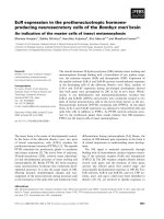
in the psychiatrically unaffected comparison subjects. A heat
map based on cluster analysis illustrates the differentiated
expression levels of these probes (Figure 1). Controlling on
brain pH, postmortem interval (PMI), and hemisphere, and
excluding the two subjects with schizoaffective disorder from
the analyses did not substantially affect these results (Addi-
tional data file 2).
Quantitative RT-PCR verification of microarray results
The expression levels of 12 selected miRNAs were also deter-
mined by quantitative RT-PCR (qRT-PCR) in our lab (Addi-
tional data file 3). For the eight miRNAs distinguished by
being expressed at lower microarray levels in schizophrenia
samples versus comparison samples, seven were also
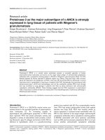
expressed at lower levels with qRT-PCR (Figure 2). For four
of the seven, the difference in expression was significant with
p < 0.05, consistent with microarray findings for the same
miRNAs. The eighth miRNA, miR-7, was found by qRT-PCR
to have higher levels in schizophrenia than comparison sub-
jects, but the difference in expression was not significantly
different between groups (p = 0.23); we have not determined
a cause for this one discrepancy of PCR versus microarray
results. We also compared expression of four miRNAs that
were not differentially expressed in the microarray results,
Genome Biology 2007, Volume 8, Issue 2, Article R27 Perkins et al. R27.3
comment reviews reports refereed researchdeposited research interactions information
Genome Biology 2007, 8:R27
and found none to be differentially expressed by qRT-PCR (p
> 0.05).
Effect of haloperidol exposure on miRNA expression
Since all of the schizophrenia subjects were treated or had
previously been treated with antipsychotics and none of the
psychiatrically unaffected subjects were reported to have
such a treatment history, we endeavored to evaluate the effect
of antipsychotic treatment on miRNA expression. We com-
pared expression of 179 rat miRNAs in haloperidol-treated
and -untreated rats. With a FDR of 5% we found that three
miRNAs were expressed at higher levels in the haloperidol-
treated rats: miR-199a, miR-128a, and miR-128b. None of
these miRNAs was differentially expressed in the PFC of
schizophrenia patients (Additional data file 4).
miRNA and Affymetrix U133A probe relationships
We considered whether the observed pattern of lower expres-
sion of some miRNAs in schizophrenia subjects was related to
lower pri-miRNA transcription. We adopted the previously
published method of Thomson and colleagues [29], where the
pri-miRNA expression was determined from existing
archived mRNA microarray results from the PFC of the same
study subjects. A total of 52 of the miRNAs included in this
study could be mapped to a primary transcript that was
present among the mRNA transcripts accessible with the
Affymetrix U133A array. All but three of the miRNAs with
corresponding U133A probes were from the introns of pro-
tein-coding genes (host genes). The mean expression of only
two of the Affymetrix U133A probes was significantly differ-
ent between groups (ELM2 hosting miR-330 with p = 0.03;
MYH6 hosting miR-208 with p = 0.03). However, these dif-
ferences were not significant after correction for multiple
comparisons (p > 0.05).
We then focused on the five miRNAs expressed at signifi-
cantly lower levels in schizophrenia that also had a U133A
probe that included the pri-miRNA transcript (miR-26b,
miR-9-3p (alias miR-9*), miR-24, miR-7, and miR-30e). The
ratio of mature miRNA to primary miRNA transcripts was
lower for schizophrenia versus controls for all 5 miRNAs, and
the difference in ratios reached statistical significance for 3 of
the 5 (miR-26b, p = 0.009; miR-9-3p, p = 0.002; and miR-
24, p = 0.037). For the one miRNA that was expressed at a
significantly higher level in schizophrenia subjects, miR-
106b, the ratio was also significantly higher (p = 0.003 and p
= 0.006 for the two associated Affymetrix pri-miRNA
probes). In the remaining 46 miRNAs with a corresponding
Affymetrix U133A probe for their pri-miRNA transcripts, the
ratio of miRNA to host mRNA was significantly lower for two
pri-miRNAs (primary transcripts for miR-218, p = 0.021, and
miR-9, p = 0.006) and significantly higher for five (miR-482,
p = 0.015; miR-190, p = 0.018; miR-105, p = 0.02; miR-148b,
p = 0.027; miR-218, p = 0.02). Thus, we found that the
miRNA:U133A probe ratios of the schizophrenia group were
significantly different from those of the comparison group for
4 of the 6 differentiated miRNAs but only 7 of the 46 nondif-
ferentiated miRNAs (p = 0.013, Fisher's exact test) (Addi-
tional data file 5).
Common motifs near the pre-miRNA:pri-miRNA
junction
We hypothesized that the system regulating processing of the
pri-miRNA to pre-miRNA might involve a motif within the
Table 1
Differentially expressed miRNAs from the prefrontal cortex of subjects with schizophrenia compared to psychiatrically healthy subjects
miRNA Fold change Chromosome location(s)
hsa-miR-26b 0.63 2q35
hsa-miR-30b 0.68 8q24.22
hsa-miR-29b 0.69 1q32.2, 7q32.3
hsa-miR-195 0.73 17p13.1
hsa-miR-92 0.76 13q31.3, Xq26.2
hsa-miR-30a-5p 0.79 6q13
hsa-miR-30d 0.80 8q24.22
hsa-miR-20b 0.81 Xq26.2
hsa-miR-29c 0.82 1q32.2
hsa-miR-29a 0.82 7q32.2
hsa-miR-212 0.82 17p13.3
hsa-miR-106b 1.77 7q22.1
hsa-miR-7 0.70 9q21.32, 15q26.1, 19p13.3
hsa-miR-24 0.79 9q22.32, 19p13.12
hsa-miR-30e 0.89 1p34.2
hsa-miR-9-3p 0.77 1q22, 5q14.3, 15q26.1
R27.4 Genome Biology 2007, Volume 8, Issue 2, Article R27 Perkins et al. />Genome Biology 2007, 8:R27
pri-miRNA and upstream of the single-stranded RNA
(ssRNA)-double-stranded RNA (dsRNA) junction that would
lend selectivity to this process. Specifically, we hypothesized
that an upstream motif of some kind is shared by the 15 miR-
NAs that were found to be downregulated in our tests of schiz-
ophrenia PFC samples. To seek bioinformatic indications, we
focused on source pre-miRNAs that were isolated (no other
pre-miRNAs within 1,000 bases), yielding 11 distinguished,
isolated pre-miRNAs: miR-7-1, miR-7-2, miR-7-3, miR-9-1,
miR-9-2, miR-9-3, miR-26b, miR-30a, miR-30b, miR-30d,
miR-30e. Of these, miR-9-1 and miR-30a can yield two
mature miRNAs; the others yield one. Furthermore, miR-7-2,
miR-9-2, miR-9-3, miR-30b, and miR-30d are intergenic,
and the others are intronic in coding genes.
Using a combination of approaches, we found the motif
UGAGNCUU upstream of pre-miRNA sequences for miR-
26b, miR-30a, miR-30b, and miR-7-1. We also found GUCN-
CUUC upstream of pre-miRNAs miR-9-1, miR-9-2, miR-9-3,
miR-7-3, and miR-30e. Thus, both 8 nt motifs are found
upstream of 9 of the 11 isolated, distinguished pre-miRNAs.
Lastly, instances of UGUUNNAAGAUG were found upstream
of pre-miRNAs for miR-30d and miR-7-2 at the same dis-
tance, 108 bases, and not within 500 bases upstream of any
other human, isolated pre-miRNAs. For displays of the motifs
and the bases between motifs and junctions, see Additional
data file 6; clustering of the number of bases in each such
interval is displayed in Figure 3. Bioinformatic searches by us
have found neither shared motifs that are positioned at simi-
larly clustered distances from the junctions nor strong gen-
eral homology among the 11 upstream regions.
Importantly, the same 8 nt motifs UGAGNCUU and GUCNC-
UUC are absent from the 500-base 5' regions of most undis-
tinguished pre-miRNAs. That is, the same motifs are also
upstream of only 13 isolated, undistinguished pre-miRNAs
among a total of 192 isolated, human pre-miRNAs, and some
of the 13 are sequentially similar as mature miRNAs to the 11
distinguished ones. However, carefully designed and exe-
cuted in vivo experiments would be needed to determine
whether the above or any other motifs are actually functional;
the above motifs are intriguing, but their bioinformatic
An miRNA expression map shows differentiated genes as determined by SAM analysisFigure 1
An miRNA expression map shows differentiated genes as determined by SAM analysis. Yellow indicates low expression and blue indicates high expression,
relative to the median.
miR-10 6b
miR-21 2
miR-24
miR-30 e
miR-20 b
miR-26 b
miR-29 c
miR-29 a
miR-30 a-5p
miR-30 d
miR-30 b
miR-29 b
miR-19 5
miR-9- 3p
miR-7
miR-92
CTRL 1025
CTRL 1020
CTRL 1034
SZ 1016
SZ 1009
CTRL 1024
CTRL 1014
CTRL 1008
CTRL 1029
CTRL 1022
CTRL 1003
CTRL 1021
CTRL 1033
CTRL 1057
CTRL 1028
CTRL 1032
CTRL 1066
CTRL 1035
CTRL 1013
SZ 1040
SZ 1001
SZ 1042
CTRL 1030
SZ 1038
SZ 1037
SZ 1060
SA 1039
SZ 1052
CTRL 1026
SZ 1065
SZ 1044
CTRL 1047
CTRL 1006
SZ 1036
SZ 1043
SA 1061
Genome Biology 2007, Volume 8, Issue 2, Article R27 Perkins et al. R27.5
comment reviews reports refereed researchdeposited research interactions information
Genome Biology 2007, 8:R27
miRNA microarray fold changes can be compared with delta-delta C(t) functions of qRT-PCR data (see Materials and methods)Figure 2
miRNA microarray fold changes can be compared with delta-delta C(t) functions of qRT-PCR data (see Materials and methods). The comparisons are over
four samples from schizophrenia patients and four samples from psychiatrically unaffected comparison subjects. Seven of the eight comparisons are
consistent.
Regarding the 11 isolated miRNAs distinguished in schizophrenia, this figure shows the distances (numbers of bases) from shared 5' motifs we discovered (two 8 nt and two 12 nt motif sequences in the pri-miRNA) to the ssRNA-dsRNA junctions at starts of pre-miRNAsFigure 3
Regarding the 11 isolated miRNAs distinguished in schizophrenia, this figure shows the distances (numbers of bases) from shared 5' motifs we discovered
(two 8 nt and two 12 nt motif sequences in the pri-miRNA) to the ssRNA-dsRNA junctions at starts of pre-miRNAs. Pre-miR-30a and -9-1 have double
motif instances; second instances are in the rectangle. Ignoring the second instances as redundant leaves some motif distances in sharp clusters.
0.1
0.3
0.5
0.7
0.9
1.1
1.3
1.5
1.7
1.9
miR-26b** miR-30b*
miR-29b
miR- 195 miR-92** miR-7 miR-24** miR- 30e**
Expression ratio
Microarray qRT-PCR
* For qRT-PCR mean Δ C(t) schizophrenia versus comparison subject p < 0.10
** For qRT-PCR mean Δ C(t) schizophrenia versus comparison subject p < 0.05
0
100
200
300
400
500
30b TGAGNCTT
9-1 GTCNCTTC
26b TGAGNCTT
30d TGTTTCAAGATG
7-2 TGTTCAAAGATG
30a TGAGNCTT
30a TGAGNCTT
9-1 GTCNCTTC
9-3 GTCNCTTC
7-1 TGAGNCTT
7-3 GTCNCTTC
30e GTCNCTTC
9-2 GTCNCTTC
R27.6 Genome Biology 2007, Volume 8, Issue 2, Article R27 Perkins et al. />Genome Biology 2007, 8:R27
properties are certainly not a proof of common regulation of
coordinated pre-miRNA excision.
Discussion
miRNAs, with their key roles in regulating both synaptic plas-
ticity and brain development, are candidate genetic contribu-
tors to the etiopathology of schizophrenia. miRNA expression
for 16 miRNAs was significantly different in the PFC of schiz-
ophrenia versus comparison subjects, with all but one of the
differentiated miRNAs decreased in the schizophrenia sub-
jects. To our knowledge this study is the first to associate
altered expression of miRNAs with schizophrenia. Possibly
the association is etiologic, but it could also be part of a com-
plex response to other factors.
A hypothesized role for altered miRNA biogenesis
Our follow-up analyses were designed to generate hypotheses
about possible mechanisms that could explain the downregu-
lation of miRNAs reported in this study. For miRNAs hosted
in introns of coding genes, we found that the ratios of micro-
array expression levels of miRNA versus mRNA (of host gene)
were significantly different for miRNA distinguished by
schizophrenia. That is, 4 of the 6 hosted, distinguished miR-
NAs exhibited the difference, but only 7 of the 46 undistin-
guished miRNAs did so. This suggests a role for altered
biogenesis of miRNAs rather than altered transcription of pri-
miRNAs. In addition, our bioinformatic investigations found
2 common motifs located at approximately 100 or
approximately 400 bases from the pri-miRNA:pre-miRNA
junction in 9 of the 11 isolated, distinguished miRNAs; but the
same motifs are absent in almost all of the undistinguished
miRNAs. We speculate (see Figure 4) that these motifs might
represent binding sites for factors like heteronuclear ribonu-
clear proteins (hnRNPs) [30], known to chaperone other
RNA events.
The bioinformatic similarities involving motifs, though not
yet investigated in vivo, are consistent with the hypothesis
that the coordinated downregulation of 15 miRNAs reported
in this study might be related to alternative processing during
the pre-miRNA biogenesis process, rather than altered pri-
miRNA transcription. There is evidence that, in some cases,
miRNA biogenesis regulates mature miRNA levels. Thomson
et al. [29] found that in mice, levels of mature miRNAs hsa-
let-7g and hsa-let-7f-2/miR-98 increased over 4,000-fold in
day 14.5 embryos from levels in embryonic stem cells. How-
ever, over the same developmental period the primary tran-
script pri-miRNA expression levels did not change, and pre-
miRNA levels were essentially undetectable. Also, the same
Thomson analysis indicates that the widespread downregula-
tion of miRNAs observed in cancer [31,32] might be due to a
failure in miRNA processing that is post-transcriptional
(transcription of pri-miRNA). Discovery of parallel mecha-
nisms of regulation of other sets of miRNAs, such as the 15
downregulated miRNAs in schizophrenia, would, therefore,
be of considerable interest.
Further study is required to test the hypothesis that altered
regulation of miRNA biogenesis might be involved in the
Transcription yields a continuous supply of some types of pri-miRNA transcripts, capped and polyadenylatedFigure 4
Transcription yields a continuous supply of some types of pri-miRNA transcripts, capped and polyadenylated. hnRNPs are hypothesized to shape the pri-
miRNA into linear and hairpin sections. A signaling system somehow recruits and activates unknown factors that select particular pre-miRNA hairpins on
a particular pri-miRNA for excision and processing in the miRNA pathway. We hypothesize that this system might include a binding motif. RNASEN and
DGCR8 are products of genes 29102 and 54487.
DGCR8
RNASEN
Large complex
hnRNPs
Degradation
mechanisms
pre-miRNA
pri-miRNA
Pol II or Pol III
transcription
AAUAAA
Export
Nucleoplasm
Cytoplasm
Nuclear membrane
Motif binding
Genome Biology 2007, Volume 8, Issue 2, Article R27 Perkins et al. R27.7
comment reviews reports refereed researchdeposited research interactions information
Genome Biology 2007, 8:R27
etiopathology of schizophrenia, and whether the above motifs
are involved in regulating miRNA processing from pri-
miRNA to pre-miRNA.
As a final note, DiGeorge critical region 8 (DGCR8), involved
in miRNA biogenesis as a component of the microprocessor,
is located in a genomic region of chromosome 22q11 where
microdeletions have been associated with a 30-fold increased
risk of schizophrenia [33,34]. Microdeletions in 22q11 occur
in approximately 1 in 3,000 live births but are present in 0.5%
to 3% of individuals with schizophrenia [35,36]. Possibly,
DGCR8 polymorphisms that alter expression or function
through haploinsufficiency or other genetic variants might
also contribute to the etiopathology of schizophrenia by
impacting miRNA biogenesis and regulation of gene
expression.
Common potential mRNA targets
Dysregulation of miRNA levels would be anticipated to affect
the translation of multiple protein coding genes. Bioinfor-
matic strategies are now developed to identify potential
miRNA target sites in the 3' UTR of a protein coding gene, for
example the program miRanda [7]. The potential targets of
miRNAs often include hundreds of genes because the reverse
complement of some 'seeds' (bases 2 through 8 of the mature
miRNA) appears in multiple locations in many pre-mRNA 3'
UTRs. However, only a few of these potential target sites have
been verified as potent in vivo [37]. With the understanding
that identification of mRNA targets is speculative, we
explored whether there might be common mRNA targets for
the 15 distinguished, downregulated miRNAs and whether
these targeted genes are over-represented in any Kyoto
Encyclopedia of Genes and Genomes (KEGG) pathway
through the KEGG website [38].
The differentially expressed miRNAs are currently annotated
in the Memorial Sloan-Kettering Cancer Center Computa-
tional Biology Center web site. These 15 miRNAs are identi-
fied using miRanda to potentially target the 3' UTRs of over
4,600 genes, with 1,539 targeted by 2 or more of them [39].
Using the programs offered by the Database for Annotation,
Visualization, and Integrated Discovery (DAVID) to identify
over-represented pathways, we found that the genes that
were commonly targeted by the miRNAs were significantly
clustered in 12 KEGG pathways (Table 2) [40]. It is of interest
that the most significantly differentiated pathways are
involved in synaptic plasticity at the level of dendritic spines.
For example, the MAPK and phosphatidylinositol signaling
pathways are involved in the regulation of dendritic spine
morphogenesis, size, and shape [41,42] and act through reg-
ulation of the actin cytoskeleton [43]. In addition, the focal
adhesion pathways mediated through extracellular matrix
receptor interactions have also been shown to control den-
dritic spine plasticity [44]. Translation of mRNA into proteins
that are important to synaptic plasticity can occur locally in
dendrites [45]. Thus, the miRNAs differentiated in this study
might be involved in the regulation of synaptic plasticity, and
in that manner associated with characteristics of synaptic
plasticity in schizophrenia.
Conclusion
Although the functions of most human miRNAs have yet to be
discovered, miRNAs have emerged as key regulators of gene
expression. The findings of this study implicate a role for
miRNAs in schizophrenia, and lead us to the hypothesis that
there is altered processing of miRNAs during the miRNA bio-
genesis process in schizophrenia. This hypothesis is analo-
gous to that for altered miRNA transcription in cancer by
Thomson et al. [29].
Table 2
KEGG pathways of gene categories that are over-represented by targets of two or more miRNAs distinguished by schizophrenia
KEGG Pathway Term N% P value
HSA04810:REGULATION OF ACTIN CYTOSKELETON 43 2.81 1.7E-07
HSA04510:FOCAL ADHESION 45 2.94 3.5E-07
HSA04010:MAPK SIGNALING PATHWAY 41 2.68 3.9E-05
HSA04512:ECM-RECEPTOR INTERACTION 17 1.11 0.0029
HSA04070:PHOSPHATIDYLINOSITOL SIGNALING 18 1.17 0.0076
HSA04020:CALCIUM SIGNALING PATHWAY 28 1.83 0.0093
HSA00271:METHIONINE METABOLISM 6 0.39 0.0099
HSA04540:GAP JUNCTION 16 1.04 0.0109
HSA04530:TIGHT JUNCTION 18 1.17 0.0173
HSA04910:INSULIN SIGNALING PATHWAY 21 1.37 0.0193
HSA04630:JAK-STAT SIGNALING PATHWAY 22 1.44 0.0326
HSA04710:CIRCADIAN RHYTHM 5 0.33 0.0370
N, number of potential target genes in pathway; %, percent of pathway genes that are targeted by differentiated miRNAs.
R27.8 Genome Biology 2007, Volume 8, Issue 2, Article R27 Perkins et al. />Genome Biology 2007, 8:R27
Materials and methods
Postmortem tissue
This study was approved by the Institutional Review Board of
the University of North Carolina School of Medicine. Post-
mortem human brain tissue was obtained from the Harvard
Brain Tissue Resource Center [46]. Tissue consisted of frozen
blocks (300-500 mg/block) from the PFC (Brodmann area
nine from 15 individuals with schizophrenia and 21 unaf-
fected comparison subjects (Table 3)). The tissue was group-
matched for age, gender, PMI, and hemisphere. Postmortem
neuropathological examinations were performed by an expe-
rienced neuropathologist, and all subjects included in the
collection were free of neurodegenerative pathology.
Postmortem neurotoxicological studies showed no evidence
of illicit substance use at the time of death.
Animals
Experimental protocols were approved by the UNC Institu-
tional Animal Care and Use Committee. Singly housed, male
Sprague-Dawley rats (150-200 g; Charles River, Raleigh, NC,
Table 3
Demographics
Subject Age (years) PDx Sex PMI pH Hemisphere
1003 51-60 Ctrl F 24 5.8 Right
1006 51-60 Ctrl M 24.2 6.53 Left
1008 61-70 Ctrl F 22.5 6.26 Left
1013 31-40 Ctrl M 18.75 6.68 Right
1014 31-40 Ctrl M 20 5.97 Left
1020 71-80 Ctrl M 20.53 6.05 Right
1021 31-40 Ctrl M 25.67 6.33 Right
1022 80+ Ctrl M 7.42 6.39 Right
1024 71-80 Ctrl M 20.92 6.74 Left
1025 71-80 Ctrl F 23.91 6.67 Right
1026 31-40 Ctrl M 28.83 6.53 Left
1028 61-70 Ctrl F 24.25 6.4 Right
1029 61-70 Ctrl F 7.42 6.03 Right
1030 41-50 Ctrl M 18.33 6.78 Left
1032 41-50 Ctrl M 24.13 6.01 Left
1033 80+ Ctrl M 28.58 6.42 Right
1034 31-40 Ctrl M 16.6 6.24 Left
1035 31-40 Ctrl M 24.5 6.26 Left
1047 61-70 Ctrl M 15.3 6.88 Right
1057 31-40 Ctrl M 28 6.5 Right
1066 21-30 Ctrl M 18.25 7.06 Left
1001 61-70 SZ M 22.1 6.43 Right
1009 71-80 SZ F 24 6.08 Right
1016 61-70 SZ M 22.35 6.55 Right
1036 41-50 SZ M 19 6.05 Left
1037 31-40 SZ M 28 6.25 Left
1038 41-50 SZ M 18.1 6.26 Left
1039 71-80 SA F 13.4 6.81 Left
1040 41-50 SZ M 18.5 6.31 Left
1042 21-30 SZ M 16 6.75 Right
1043 41-50 SZ M 27.1 6.64 Right
1044 41-50 SZ M 19.25 6.57 Right
1052 80+ SZ F 23.25 5.91 Right
1060 71-80 SZ F 21.75 6.65 Right
1061 41-50 SA F 33.78 6.63 Left
1065 41-50 SZ M 19.08 6.6 Left
Ctrl, control; F, female; M, male; PDx, primary diagnosis; PMI, postmortem interval hours; SA, schizoaffective; SZ, schizophrenia.
Genome Biology 2007, Volume 8, Issue 2, Article R27 Perkins et al. R27.9
comment reviews reports refereed researchdeposited research interactions information
Genome Biology 2007, 8:R27
USA) received daily intraperitoneal injections of haloperidol
1 mg/kg/d (n = 6) or saline 0.9% (n = 6) for 4 weeks. One hour
after the final dose, rats were briefly anesthetized with ether
and sacrificed; their brains were removed and hemisected.
Right anterior medial frontal cortex was dissected out and
frozen on dry ice. All tissue was kept frozen at -80°C until use.
miRNA microarray procedures
miRNA microarray expression analysis was performed as
previously described [47]. Tissue disruption by Dounce
homogenization was followed by total RNA isolation with
TRIZOL™ reagent (Invitrogen, Carlsbad, California, USA).
RNA (5 μg) was labeled with T4-RNA ligase and precipitated
with 0.3 M sodium acetate, 2 volumes ethanol, and re-sus-
pended in water.
Oligonucleotide probes were synthesized in duplicate for 264
human miRNAs antisense to the mature sequence reported in
the Sanger miRNA registry [48]. Probes were spotted in
duplicate on Corning (Corning, New York, USA) GAPS-2
coated slides using a robotic spotter and cross-linked by UV.
Hybridization and washing were performed as described. All
arrays were from the same batch, and the microarrays were
run on the same day by the same two persons. Our prior
research indicates that our in-house miRNA microarrays
have excellent reliability and validity [49].
Microarray data analysis began with data extraction from the
GPR files. Data points were eliminated if foreground was not
1.5 times local background and a probe was removed if >40%
of the data points were missing. A total of 239 miRNA
remained after this pre-processing. Data were background
subtracted, log-transformed, and missing values were
imputed using k-NN [50]. For comparisons across samples,
data were normalized using rank invariant normalization
[51]. The per-sample mean of the two rank invariant normal-
ized probes was used for analyses. Univariate calculations of
differential expression were estimated using Statistical Anal-
ysis of Microarrays (SAM; two-class, unpaired test; 500 per-
mutations; FDR of 5%) [52]. All analysis procedures were
done using R [53]. Cluster analysis was done with
GeneCluster
©
[54] and displayed using TreeView
©
[55] (Fig-
ure 1).
mRNA microarray analysis procedures
Previous to our research, mRNA microarray profiling of PFC
tissue from these same subjects (but different samples) was
performed at the Harvard Brain Tissue Resource Center with
Affymetrix U133A
©
arrays using standard methods and qual-
ity control procedures. The cel files and information on sam-
ple acquisition, preparation, and microarray analysis are
publicly available and were downloaded from the Center's
National Brain Databank.
The U133A microarrays were normalized using GC Robust
Multi-Array (GCRMA), and analysis of probe expression lev-
els was done with SAM. We used the March 2006 version of
the UCSC Human (Homo sapiens) Genome Browser [56] to
determine the U133A probes that corresponded to miRNA
locations in host genes.
qRT-PCR procedures
Total RNA (5 μg) was DNase I (Promega, Madison, Wiscon-
sin, USA) treated according to the manufacturer's instruc-
tions, phenol:chloroform extracted, ethanol precipitated, and
dissolved in DEPC-treated dH
2
O (DEPC; diethylpyrocar-
bonate). RNA (5 μg) was polyadenylated using Poly(A)
polymerase (Ambion, Austin, Texas, USA) according to the
manufacturer's instructions, phenol:chloroform extracted,
ethanol-precipitated, and dissolved in DEPC-treated dH
2
O. A
modified cDNA was made as follows: 5 μg of polyadenylated
RNA was reverse-transcribed using Superscript II reverse
transcriptase (Invitrogen, Carlsbad, California, USA) with 2.5
μg of random hexamers and 500 ng of oligo(dT) adapter
primer (5'-GCGAGCACAGAATTAATACGACTCACTATAG-
GTTTTTTTTTTTTVN-3') according to the manufacturer's
instructions. The reaction was terminated by incubation at
70°C for 10 minutes and diluted into 2 ml of dH
2
O (5 ng/μl).
Quantitative PCR was used to measure the mature miRNA
transcript as follows: 5 μl of cDNA was mixed with 5 pmol of
both the forward and reverse primers in a final volume of 12.5
μl and mixed with 12.5 μl of 2× SYBR Green PCR master mix
(Applied Biosystems, Foster City, California, USA). Primer
sequences are in Additional data file 7. All reactions were run
in triplicate on a DNA Engine Opticon 2 (Bio-Rad Laborato-
ries, Hercules, California, USA). The amplification protocol
for mature miRNA PCR was performed according to the high-
stringency protocol of Shi and Chiang [57] except the reverse
primer Mir-qPCR-3-3' (5'-GCAGCA CAGAATTAATACGACT-
CAC-3') was used in conjunction with an exact sequence-spe-
cific primer to each miRNA. Mature miRNA expression used
the reference gene U6 snRNA (U6-F, 5'-CGCTTC GGCAGCA-
CATATAC-3'; U6-R, 5'-TTCACGAATTTGCGTGTCAT-3').
The expression was determined for eight subjects, four with
schizophrenia and four healthy subjects (Additional data file
3). Expression was calculated using the delta-delta C(t)
method: 2
ΔCT healthy-ΔCT schizophrenia
with ΔC
T
= (C
T
miRNA - C
T
reference RNA U6) [58].
Additional data files
The following additional data are available with the online
version of this paper. Additional data file 1 is a table listing the
tested miRNAs and their expression levels and fold changes.
Additional data file 2 is a table showing data conditioning on
PMI, pH, and hemisphere. Additional data file 3 is a table of
qRT-PCR results. Additional data file 4 includes characteriza-
tion and microarray results on rats treated with haloperidol.
Additional data file 5 is a table of host mRNA data. Additional
data file 6 lists putative motifs within regions upstream of
some distinguished pre-miRNAs. Additional data file 7 is a
table listing primer sequences.
R27.10 Genome Biology 2007, Volume 8, Issue 2, Article R27 Perkins et al. />Genome Biology 2007, 8:R27
Additional data file 1Tested miRNAs and their expression levels and fold changesTested miRNAs and their expression levels and fold changesClick here for fileAdditional data file 2Data conditioning on PMI, pH, and hemisphereData conditioning on PMI, pH, and hemisphereClick here for fileAdditional data file 3qRT-PCR resultsqRT-PCR resultsClick here for fileAdditional data file 4Characterization and microarray results on rats treated with haloperidolCharacterization and microarray results on rats treated with haloperidolClick here for fileAdditional data file 5Host mRNA dataHost mRNA dataClick here for fileAdditional data file 6Putative motifs within regions upstream of some distinguished pre-miRNAsPutative motifs within regions upstream of some distinguished pre-miRNAsClick here for fileAdditional data file 7Primer sequencesPrimer sequencesClick here for file
Acknowledgements
Many thanks are due to the referees for insightful and valuable comments
that led to significant improvements. We are also very thankful to the Har-
vard Brain Tissue Resource Center, which is supported in part by PHS grant
number R24 MH068855, for tissue. This project was supported in part by
NIGMS Public Health Service grant GM070674 (SMH), NIH grant MH-
01752 (LFJ), Elsa U Pardee Foundation and NIH Public Health Service 5-
P20-RR020751-01-02 (CDJ), the Foundation of Hope (DOP), and the
American Cancer Society (JMT). The contents of this paper are solely the
responsibility of the authors and do not necessarily represent the official
view of any granting agency.
References
1. Rossler W, Salize HJ, van Os J, Riecher-Rossler A: Size of burden
of schizophrenia and psychotic disorders. Eur
Neuropsychopharmacol 2005, 15:399-409.
2. Sullivan PF: The genetics of schizophrenia. PLoS Med 2005,
2:e212.
3. Harrison PJ, Weinberger DR: Schizophrenia genes, gene expres-
sion, and neuropathology: on the matter of their
convergence. Mol Psychiatry 2005, 10:40-68. image 45
4. Prabakaran S, Swatton JE, Ryan MM, Huffaker SJ, Huang JT, Griffin JL,
Wayland M, Freeman T, Dudbridge F, Lilley KS, et al.: Mitochondrial
dysfunction in schizophrenia: evidence for compromised
brain metabolism and oxidative stress. Mol Psychiatry 2004,
9:684-697.
5. Dracheva S, Elhakem SL, McGurk SR, Davis KL, Haroutunian V:
GAD67 and GAD65 mRNA and protein expression in cere-
brocortical regions of elderly patients with schizophrenia. J
Neurosci Res 2004, 76:581-592.
6. Denli AM, Tops BB, Plasterk RH, Ketting RF, Hannon GJ: Processing
of primary microRNAs by the microprocessor complex.
Nature 2004, 432:231-235.
7. Bentwich I: Prediction and validation of microRNAs and their
targets. FEBS Lett 2005, 579:5904-5910.
8. Berezikov E, Guryev V, van de Belt J, Wienholds E, Plasterk RH, Cup-
pen E: Phylogenetic shadowing and computational identifica-
tion of human microRNA genes. Cell 2005, 120:21-24.
9. Lewis BP, Burge CB, Bartel DP: Conserved seed pairing, often
flanked by adenosines, indicates that thousands of human
genes are microRNA targets. Cell 2005, 120:15-20.
10. Shcherbata HR, Hatfield S, Ward EJ, Reynolds S, Fischer KA, Ruohola-
Baker H: The microRNA pathway plays a regulatory role in
stem cell division. Cell Cycle 2006, 5:172-175.
11. Sempere LF, Freemantle S, Pitha-Rowe I, Moss E, Dmitrovsky E,
Ambros V: Expression profiling of mammalian microRNAs
uncovers a subset of brain-expressed microRNAs with possi-
ble roles in murine and human neuronal differentiation.
Genome Biol 2004, 5:R13.
12. Chen CZ, Li L, Lodish HF, Bartel DP: MicroRNAs modulate
hematopoietic lineage differentiation. Science 2004, 303:83-86.
13. Muljo SA, Ansel KM, Kanellopoulou C, Livingston DM, Rao A, Rajew-
sky K: Aberrant T cell differentiation in the absence of Dicer.
J Exp Med 2005, 202:261-269.
14. Esau C, Kang X, Peralta E, Hanson E, Marcusson EG, Ravichandran LV,
Sun Y, Koo S, Perera RJ, Jain R, et al.: MicroRNA-143 regulates
adipocyte differentiation. J Biol Chem 2004, 279:52361-52365.
15. Chen JF, Mandel EM, Thomson JM, Wu Q, Callis TE, Hammond SM,
Conlon FL, Wang DZ: The role of microRNA-1 and microRNA-
133 in skeletal muscle proliferation and differentiation. Nat
Genet 2006, 38:228-233.
16. Poy MN, Eliasson L, Krutzfeldt J, Kuwajima S, Ma X, Macdonald PE,
Pfeffer S, Tuschl T, Rajewsky N, Rorsman P, et al.: A pancreatic
islet-specific microRNA regulates insulin secretion. Nature
2004, 432:226-230.
17. Xu P, Guo M, Hay BA: MicroRNAs and the regulation of cell
death. Trends Genet 2004, 20:617-624.
18. Giraldez AJ, Cinalli RM, Glasner ME, Enright AJ, Thomson JM, Basker-
ville S, Hammond SM, Bartel DP, Schier AF: MicroRNAs regulate
brain morphogenesis in zebrafish. Science 2005, 308:833-838.
19. Krichevsky AM, King KS, Donahue CP, Khrapko K, Kosik KS: A
microRNA array reveals extensive regulation of microRNAs
during brain development. Rna 2003, 9:1274-1281.
20. Schratt GM, Tuebing F, Nigh EA, Kane CG, Sabatini ME, Kiebler M,
Greenberg ME: A brain-specific microRNA regulates dendritic
spine development. Nature 2006, 439:283-289.
21. Vo N, Klein ME, Varlamova O, Keller DM, Yamamoto T, Goodman
RH, Impey S: A cAMP-response element binding protein-
induced microRNA regulates neuronal morphogenesis. Proc
Natl Acad Sci USA
2005, 102:16426-16431.
22. Hammond SM: MicroRNA therapeutics: a new niche for anti-
sense nucleic acids. Trends Mol Med 2006, 12:99-101.
23. Abelson JF, Kwan KY, O'Roak BJ, Baek DY, Stillman AA, Morgan TM,
Mathews CA, Pauls DL, Rasin MR, Gunel M, et al.: Sequence vari-
ants in SLITRK1 are associated with Tourette's syndrome.
Science 2005, 310:317-320.
24. Bagni C, Greenough WT: From mRNP trafficking to spine dys-
morphogenesis: the roots of fragile X syndrome. Nat Rev
Neurosci 2005, 6:376-387.
25. Jin P, Alisch RS, Warren ST: RNA and microRNAs in fragile X
mental retardation. Nat Cell Biol 2004, 6:1048-1053.
26. Caudy AA, Myers M, Hannon GJ, Hammond SM: Fragile X-related
protein and VIG associate with the RNA interference
machinery. Genes Dev 2002, 16:2491-2496.
27. Perkins DO, Jeffries C, Sullivan P: Expanding the 'central dogma':
the regulatory role of nonprotein coding genes and implica-
tions for the genetic liability to schizophrenia. Mol Psychiatry
2005, 10:69-78.
28. Nelson PT, Baldwin DA, Kloosterman WP, Kauppinen S, Plasterk RH,
Mourelatos Z: RAKE and LNA-ISH reveal microRNA expres-
sion and localization in archival human brain. Rna 2006,
12:187-191.
29. Thomson JM, Newman M, Parker JS, Morin-Kensicki EM, Wright T,
Hammond SM: Extensive post-transcriptional regulation of
microRNAs and its implications for cancer. Genes Dev 2006,
20:2202-2207.
30. Buratti E, Baralle FE: Influence of RNA secondary structure on
the pre-mRNA splicing process. Mol Cell Biol 2004,
24:10505-10514.
31. Takamizawa J, Konishi H, Yanagisawa K, Tomida S, Osada H, Endoh
H, Harano T, Yatabe Y, Nagino M, Nimura Y, et al.: Reduced
expression of the let-7 microRNAs in human lung cancers in
association with shortened postoperative survival. Cancer Res
2004, 64:3753-3756.
32. Lu J, Getz G, Miska EA, Alvarez-Saavedra E, Lamb J, Peck D, Sweet-
Cordero A, Ebert BL, Mak RH, Ferrando AA, et al.: MicroRNA
expression profiles classify human cancers. Nature 2005,
435:834-838.
33. Debbane M, Glaser B, David MK, Feinstein C, Eliez S: Psychotic
symptoms in children and adolescents with 22q11.2 deletion
syndrome: Neuropsychological and behavioral implications.
Schizophr Res 2006, 84:187-193.
34. Bassett AS, Chow EW, AbdelMalik P, Gheorghiu M, Husted J, Weks-
berg R: The schizophrenia phenotype in 22q11 deletion
syndrome. Am J Psychiatry 2003, 160:1580-1586.
35. Karayiorgou M, Gogos JA: The molecular genetics of the 22q11-
associated schizophrenia. Brain Res Mol Brain Res 2004,
132:95-104.
36. Horowitz A, Shifman S, Rivlin N, Pisante A, Darvasi A: A survey of
the 22q11 microdeletion in a large cohort of schizophrenia
patients. Schizophr Res 2005, 73:263-267.
37. Sethupathy P, Corda B, Hatzigeorgiou AG: TarBase: A compre-
hensive database of experimentally supported animal micro-
RNA targets. Rna 2006, 12:192-197.
38. KEGG Genes Database [ />39. Enright AJ, John B, Gaul U, Tuschl T, Sander C, Marks DS: Micro-
RNA targets in Drosophila. Genome Biol 2003, 5:R1.
40. Dennis G Jr, Sherman BT, Hosack DA, Yang J, Gao W, Lane HC, Lem-
picki RA: DAVID: database for annotation, visualization, and
integrated discovery. Genome Biol 2003, 4:P3.
41. Thomas GM, Huganir RL: MAPK cascade signalling and synaptic
plasticity. Nat Rev Neurosci 2004, 5:173-183.
42. Nahorski SR, Young KW, John Challiss RA, Nash MS: Visualizing
phosphoinositide signalling in single neurons gets a green
light. Trends Neurosci 2003, 26:444-452.
43. Tada T, Sheng M:
Molecular mechanisms of dendritic spine
morphogenesis. Curr Opin Neurobiol 2006, 16:95-101.
44. Shi Y, Ethell IM: Integrins control dendritic spine plasticity in
hippocampal neurons through NMDA receptor and Ca2+/
calmodulin-dependent protein kinase II-mediated actin
Genome Biology 2007, Volume 8, Issue 2, Article R27 Perkins et al. R27.11
comment reviews reports refereed researchdeposited research interactions information
Genome Biology 2007, 8:R27
reorganization. J Neurosci 2006, 26:1813-1822.
45. Sutton MA, Schuman EM: Local translational control in den-
drites and its role in long-term synaptic plasticity. J Neurobiol
2005, 64:116-131.
46. Harvard Brain Tissue Resource Center [in
bank.mclean.org/]
47. Igloi GL: Nonradioactive labeling of RNA. Anal Biochem 1996,
233:124-129.
48. miRBase [ />49. Thomson JM, Parker J, Perou CM, Hammond SM: A custom micro-
array platform for analysis of microRNA gene expression.
Nat Methods 2004, 1:47-53.
50. Troyanskaya O, Cantor M, Sherlock G, Brown P, Hastie T, Tibshirani
R, Botstein D, Altman RB: Missing value estimation methods for
DNA microarrays. Bioinformatics 2001, 17:520-525.
51. Li C, Hung Wong W: Model-based analysis of oligonucleotide
arrays: model validation, design issues and standard error
application. Genome Biol 2001, 2:RESEARCH0032.
52. Tusher VG, Tibshirani R, Chu G: Significance analysis of micro-
arrays applied to the ionizing radiation response. Proc Natl
Acad Sci USA 2001, 98:5116-5121.
53. R Project [ />54. GeneCluster [ />genecluster2/gc2.html]
55. Saldanha AJ: Java Treeview - extensible visualization of micro-
array data. Bioinformatics 2004, 20:3246-3248.
56. Kent WJ, Sugnet CW, Furey TS, Roskin KM, Pringle TH, Zahler AM,
Haussler D: The human genome browser at UCSC. Genome
Res 2002, 12:996-1006.
57. Shi R, Chiang VL: Facile means for quantifying microRNA
expression by real-time PCR. Biotechniques
2005, 39:519-525.
58. Livak KJ, Schmittgen TD: Analysis of relative gene expression
data using real-time quantitative PCR and the 2(-Delta Delta
C(T)) Method. Methods 2001, 25:402-408.