Báo cáo sinh học: " Recombinant adeno-associated virus type 2-mediated gene delivery into the Rpe65-/- knockout mouse eye results in limited rescue" ppsx
Bạn đang xem bản rút gọn của tài liệu. Xem và tải ngay bản đầy đủ của tài liệu tại đây (1.88 MB, 15 trang )
BioMed Central
Page 1 of 15
(page number not for citation purposes)
Genetic Vaccines and Therapy
Open Access
Research
Recombinant adeno-associated virus type 2-mediated gene delivery
into the Rpe65
-/-
knockout mouse eye results in limited rescue
Chooi-May Lai
†1
, Meaghan JT Yu
†2
, Meliha Brankov
2
, Nigel L Barnett
3
,
Xiaohuai Zhou
4
, T Michael Redmond
5
, Kristina Narfstrom
6
and P
Elizabeth Rakoczy*
1
Address:
1
Centre for Ophthalmology and Visual Science, The University of Western Australia, Perth, Western Australia, 6009, Australia,
2
Department of Molecular Ophthalmology, Lions Eye Institute and The University of Western Australia, Perth, Western Australia, 6009, Australia,
3
Vision Touch and Hearing Research Centre, School of Biomedical Sciences, University of Queensland, Brisbane, Queensland, 4072, Australia,
4
Virus Core Facility, Gene Therapy Center, University of North Carolina, North Carolina, 27599, USA,
5
Laboratory of Retinal Cell and Molecular
Biology, National Eye Institute, National Institutes of Health, Bethesda, Maryland, 20892, USA and
6
Vision Science Group, Department of
Veterinary Medicine and Surgery, College of Veterinary Medicine, University of Missouri-Columbia, Columbia, Missouri, 65211, USA
Email: Chooi-May Lai - ; Meaghan JT Yu - ;
Meliha Brankov - ; Nigel L Barnett - ; Xiaohuai Zhou - ; T
Michael Redmond - ; Kristina Narfstrom - ; P
Elizabeth Rakoczy* -
* Corresponding author †Equal contributors
Abstract
Background: Leber's congenital amaurosis (LCA) is a severe form of retinal dystrophy. Mutations in the RPE65 gene,
which is abundantly expressed in retinal pigment epithelial (RPE) cells, account for approximately 10–15% of LCA cases.
In this study we used the high turnover, and rapid breeding and maturation time of the Rpe65
-/-
knockout mice to assess
the efficacy of using rAAV-mediated gene therapy to replace the disrupted RPE65 gene. The potential for rAAV-mediated
gene treatment of LCA was then analyzed by determining the pattern of RPE65 expression, the physiological and
histological effects that it produced, and any improvement in visual function.
Methods: rAAV.RPE65 was injected into the subretinal space of Rpe65
-/-
knockout mice and control mice. Histological
and immunohistological analyses were performed to evaluate any rescue of photoreceptors and to determine longevity
and pattern of transgene expression. Electron microscopy was used to examine ultrastructural changes, and
electroretinography was used to measure changes in visual function following rAAV.RPE65 injection.
Results: rAAV-mediated RPE65 expression was detected for up to 18 months post injection. The delivery of
rAAV.RPE65 to Rpe65
-/-
mouse retinas resulted in a transient improvement in the maximum b-wave amplitude under
both scotopic and photopic conditions (76% and 59% increase above uninjected controls, respectively) but no changes
were observed in a-wave amplitude. However, this increase in b-wave amplitude was not accompanied by any slow down
in photoreceptor degeneration or apoptotic cell death. Delivery of rAAV.RPE65 also resulted in a decrease in retinyl
ester lipid droplets and an increase in short wavelength cone opsin-positive cells, suggesting that the recovery of RPE65
expression has long-term benefits for retinal health.
Conclusion: This work demonstrated the potential benefits of using the Rpe65
-/-
mice to study the effects and
mechanism of rAAV.RPE65-mediated gene delivery into the retina. Although the functional recovery in this model was
not as robust as in the dog model, these experiments provided important clues about the long-term physiological benefits
of restoration of RPE65 expression in the retina.
Published: 27 April 2004
Genetic Vaccines and Therapy 2004, 2:3
Received: 23 December 2003
Accepted: 27 April 2004
This article is available from: />© 2004 Lai et al; licensee BioMed Central Ltd. This is an Open Access article: verbatim copying and redistribution of this article are permitted in all media
for any purpose, provided this notice is preserved along with the article's original URL.
Genetic Vaccines and Therapy 2004, 2 />Page 2 of 15
(page number not for citation purposes)
Background
Leber's congenital amaurosis (LCA) comprises a heteroge-
neous group of retinal dystrophies. It is characterized by
severe visual loss from birth, nystagmus, poor pupillary
reflexes, retinal pigmentary or atrophic changes, and
markedly diminished electroretinography (ERG)
responses [1-3]. Mutations in Rpe65, a gene that is pre-
dominantly expressed in retinal pigment epithelial (RPE)
cells, cause about 10–15% of all LCA cases [4-6]. RPE65 is
abundantly expressed in RPE cells, where it is involved in
regenerating the visual pigment chromophore,11-cis reti-
nal, from all-trans retinol, the latter being a product of
photoreceptor phototransduction [7-9]. This recycling
process, known as the visual cycle, is central to vision as
11-cis retinal is used by the photoreceptors to convert light
photons into neuronal signals [8,9].
In vivo analyses, using the spontaneous-mutation RPE65
dog and Rpe65
-/-
mouse models of LCA, have shown that
loss of RPE65 leads to severely depressed electroretinogra-
phy (ERG) responses [7,10-14] and behavioral impair-
ments indicative of diminished vision [15,16]. In
addition, morphological studies have shown that the lack
of RPE65 is associated with a gradual degeneration of the
photoreceptor cells and a characteristic accumulation of
lipid inclusion bodies in the RPE cells, the latter from an
over accumulation of intermediary visual cycle pigments
such as retinyl esters [7,17].
The animal models of LCA not only provide an insight
into the nature of the associated disease, but have also
been used to test potential therapies for its treatment
[16,18-23]. A number of recent studies, using both the
RPE65 dog and Rpe65
-/-
mouse models, have demon-
strated that there is some promise for a future treatment of
LCA being developed. Assessment of both RPE transplan-
tation and oral/intraperitoneal administration of 9-cis ret-
inal in the Rpe65
-/-
mouse have both shown that improved
ERG responses can be produced [18-20]. In addition, it is
well established that the subretinal delivery and expres-
sion of normal, non-mutated RPE65 in the RPE cells of
RPE65 dogs results in functional recovery of vision, as
seen by improvements in both ERG and behavioral
responses, the latter indicative of the presence of limited
vision [16,21-23]. The functional recovery produced in
the RPE65 dog model was generated by using recom-
binant adenoassociated virus (rAAV) to deliver and
express normal, non-mutated RPE65 cDNA [16,22,23].
The use of rAAV-mediated gene therapy has attracted
much interest as it demonstrated a number of characteris-
tics that may be beneficial in a clinical setting. These
include a low immune response; long-term transgene
expression providing minimal surgical intervention; and
localized, specific transgene expression which minimizes
the potential of unwanted, systemic side effects.
We wished to further examine the suitability of rAAV-
mediated gene therapy for treating LCA. In this study, we
used the high turnover, and rapid breeding and matura-
tion time of mice to assess the efficacy of using rAAV-
mediated gene therapy to replace the missing RPE65 gene
in the Rpe65
-/-
knockout strain. The potential for rAAV-
mediated gene treatment of LCA was then analyzed by
determining the pattern of RPE65 expression and the
physiological and histological effects that it produced.
Methods
Virus preparation
The EcoRI/KpnI fragment of mouse RPE65 cDNA (Gen-
Bank Accession Number: NM_029987) was inserted into
the pCI mammalian expression vector (Promega Corp.,
WI, USA) to produce a pCI.RPE65 subclone. A 3800 bp
cassette, consisting of the RPE65 cDNA flanked by a 5'
human cytomegalovirus (CMV) promoter and a 3' SV40
late polyadenylation signal sequence, was removed from
pCI.RPE65 by BglII/BspHI restriction enzyme digest. This
cassette was then inserted between the inverted terminal
repeats of the serotype 2 rAAV plasmid pSSV9 [24]. The
insertion was achieved by blunt end ligation of the 3800
bp CMV.RPE65 cassette with the large fragment of pSSV9
following XbaI digestion [24]. The identity of the
pSSV9.CMV.RPE65 vector was confirmed by restriction
enzyme analysis. The expression of RPE65 protein from
pSSV9.CMV.RPE65 was confirmed by western blot analy-
sis of pSSV9.CMV.RPE65-transfected, human embryonic
kidney (HEK) 293 cells using a rabbit anti-RPE65 polyclo-
nal antibody [25].
pSSV9.CMV.RPE65, AAV helper (Ad8) and adenovirus
helper plasmid DNA were co-transfected into HEK293
cells. The excision and replication of the resultant
rAAV.RPE65 DNA was verified by Hirt analysis [26]. Upon
successful verification, cesium chloride gradient purified
pSSV9.CMV.RPE65 DNA was either co-transfected with
Ad8 and adenovirus helper plasmid DNA into HEK293
cells and the resulting virus (rAAV.RPE65) purified by
cesium chloride gradient density as previously described
[24], or was sent to the Vector Core Facility (University of
North Carolina, NC, USA) for large-scale virus produc-
tion, where the virus (rAAV.RPE65.1) was purified using
iodixanol gradient followed by heparin-affinity chroma-
tography according to published methods [27]. The titers
of rAAV.RPE65 and rAAV.RPE65.1 were both 6 × 10
13
par-
ticles/ml.
Animals
All procedures were approved by the University of West-
ern Australia Animal Experimentation Ethics Committee
and were in compliance with the Association for Research
in Vision and Ophthalmology Statement for the Use of
Animals in Ophthalmic and Vision Research. Mice were
Genetic Vaccines and Therapy 2004, 2 />Page 3 of 15
(page number not for citation purposes)
housed in cages in rooms maintained at constant temper-
ature (22°C) and humidity (50%) and with a 12:12 hr
light-dark cycle. Food (Glen Forest Rodent Chow, Aus-
tralia) and water were given ad libitum.
Subretinal injection of Rpe65
-/-
mice
Rpe65
-/-
mice were anesthetized by intraperitoneal injec-
tion of ketamine (30 mg/kg) and xylazine (8 mg/kg), and
their pupils dilated with topical application of a mixture
containing 2.5% phenylephrine hydrochloride and 1%
tropicamide (Alcon, Australia). The conjunctiva was cut
and the sclera exposed. A shelving puncture of the sclera
was made with a 30-gauge needle. A 32-gauge needle
attached to a 5 µl Hamilton syringe was passed tangen-
tially through the site of the sclera puncture under an
operating microscope. A 1 µl solution containing 6 × 10
10
particles of rAAV.RPE65 or rAAV.RPE65.1 was then deliv-
ered into the subretinal space of the mouse eye, the diam-
eter of which is about 3.5 mm. Successful delivery of virus
into the subretinal space was confirmed by the presence of
a 1 to 1.3 mm diameter circular bleb when examined by
indirect ophthalmoscopy (approximately 30% of the reti-
nal area). The needle was kept in the subretinal space for
1 min, and then withdrawn gently. Finally, a layer of anti-
biotic ointment was applied to the injected eye. Addi-
tional Rpe65
-/-
mice were injected with 1 µl of the control
construct rAAV.GFP. All mice used in this study were
injected upon reaching maturity, at 3 weeks of age.
Electroretinography
rAAV.RPE65-injected and uninjected Rpe65
-/-
mice were
analyzed by electroretinography (ERG) at 1–2 mo (n = 15
rAAV.RPE65-injected, n = 10 uninjected), 7 mo (n = 12
rAAV.RPE65-injected, n = 4 uninjected) and 11 mo (n =
12 rAAV.RPE65-injected, n = 6 uninjected) post-injection.
Following dark-adaptation of the mice, full-field scotopic
flash ERGs were recorded. The mice were anesthetized as
described earlier and maintained at 37°C with a homeo-
thermic electric blanket. Their pupils were dilated with
0.5% tropicamide (Alcon) and the cornea was protected
with carmellose sodium (Celluvisc, Allergan, Australia).
The ERG was recorded between a platinum electrode
touching the cornea and a reference electrode in the
pinna. A ground electrode was attached to the mouse's
back. The flash stimulus was presented by a xenon strobe
light placed 0.3 m in front of the mouse. Four consecutive
responses were amplified and averaged using a MacLab/2e
bioamplifier/data recorder running "Scope" software
(ADInstruments, NSW, Australia). The interstimulus
interval was increased from 30 sec (dimmest flash) to 5
min (brightest flash). Stimulus-response characteristics
were generated by attenuating the maximum flash inten-
sity (1.52 log cd s/m
2
) with neutral density filters over a
range of 3 log units. After the final scotopic recording, the
animals were light adapted for 10 min using a background
light of 1.4 log cd/m
2
and photopic ERGs obtained. The a-
wave amplitude was measured from the baseline to the
trough of the a-wave response and the b-wave amplitude
was measured from the trough of the a-wave to the peak
of the b-wave. Data were expressed as the mean wave
amplitude ± standard error of the mean (SEM; µVolts).
Two-way repeated measures analysis of variance
(ANOVA) was performed on log transformed data to
compare the responses from the rAAV.RPE65-injected and
uninjected Rpe65
-/-
retinas. A post-hoc Bonferroni test was
used to isolate significant differences (P < 0.05) between
rAAV.RPE65-injected and uninjected Rpe65
-/-
mice
responses at each stimulus intensity. All mice were sub-
jected to the same conditions for ERG measurements.
Histological analysis
rAAV.RPE65-injected, age-matched uninjected Rpe65
-/-
mice and age-matched control C57BL/6J mice were eutha-
nased at various time points post-injection, and their eyes
enucleated and fixed in 10% neutral buffered formalin for
2.5 hr. The eyes were then washed in PBS before being
placed in 70% ethanol and embedded in paraffin, with
care being taken to orientate the eyes so that the injection
site was at a known and consistent location. Serial sec-
tions (5 µm) were cut on a Reichert-Jung 2040 microtome
(Leica Microsytems, Australia), mounted on silanated
glass slides, deparaffinized and rehydrated. All analyses
that were performed using these eyes were carried out on
sections from the region corresponding to the injection
site.
For histological analysis and quantification, the sections
were stained with hematoxylin and eosin, and the
number of photoreceptor cells counted. Average cell num-
bers for each retina were established by counting the
number of cells in a 100 µm section of the outer nuclear
layer (using an eyepiece graticule and viewing the stained
section with a 100X oil-immersion lens). Digital images
of the outer nuclear layer of each section were recorded.
Between three and five 100 µm regions within the subret-
inal bleb from each section were selected for counting.
Care was taken to avoid the outer quarter to third of the
retina where the retinal layers became thinner. Counts
were made every 30–50 sections (150–250 µm) such that
15–20 counts were made per eye. The mean of these
counts was then calculated to give an average number of
photoreceptors per 100 µm for each eye. The counting was
performed by 3 independent observers who were not
given the identity of the samples.
Immunohistochemical analysis
Serial sections from rAAV.RPE65-injected and uninjected
Rpe65
-/-
eyes were rehydrated through graded alcohols,
and then bleached by incubation in 0.25% potassium per-
manganate for 20 min followed by 1% oxalic acid for 5
Genetic Vaccines and Therapy 2004, 2 />Page 4 of 15
(page number not for citation purposes)
min [28]. The sections were rinsed several times in Tris
buffer (50 mM, pH 7.2) containing 1% NaCl, then
blocked in 10% normal goat serum for 1 hr. The sections
were incubated at 4°C overnight with a rabbit anti-RPE65
antibody [25], rinsed three times in Tris buffer and then
incubated for 2 hr at room temperature with alkaline
phosphatase-conjugated, goat anti-rabbit IgG (1:100,
Gibco Invitrogen, CA, USA). Immunodetection was car-
ried out using SIGMA FAST Red TR/Naphthol AS-MX
(Sigma Chemical Co., MO, USA) chromogen for 10–15
min, resulting in the formation of a red/pink precipitate.
The sections were counterstained lightly with Meyer's
hematoxylin and mounted in an aqueous mounting
medium for analysis.
For flatmount immunohistochemistry, eyes were enucle-
ated and the injection site was marked with indelible ink
and then fixed whole for 30 min in 4% paraformalde-
hyde. The anterior segment of each eye was removed and
the neuroretina separated from the sclera-choroid-RPE
layers. The separated layers were placed in separate wells
of a 96-well plate and blocked with 10% normal rabbit
serum at room temperature for 1 hr. The layers were then
incubated overnight at 4°C with primary antibodies, rab-
bit anti-RPE65 antibody and rabbit anti-short wavelength
cone (SWC) opsin antibody, washed with Tris-buffered
saline (TBS) and incubated for 2 hr at 4°C with goat anti-
rabbit IgG conjugated with FITC (fluorescein isothiocy-
anate; Sigma Chemical Co.). After 3 washes in TBS, radial
cuts were made to the neuroretina and the sclera-choroid-
RPE layers which were mounted separately on slides with
GVA mounting solution (Zymed, CA, USA) and cover-
slipped prior to examination. Areas within the injection
subretinal bleb in rAAV.RPE65-injected Rpe65
-/-
mice or
the equivalent location in C57BL/6J and uninjected
Rpe65
-/-
mice were examined by fluorescence microscopy.
The number of SWC opsin-positive photoreceptors was
counted in five 100 µm
2
areas within the subretinal bleb
and the results analyzed and graphed.
Apoptosis detection assay and analysis
rAAV.RPE65-injected (n = 2), age-matched uninjected (n
= 2) Rpe65
-/-
, and age-matched C57BL/6J control mice
were euthanased at 7 mo post-injection (8 mo of age) and
their eyes enucleated, processed and sectioned as
described previously. An Apoptosis detection assay was
performed on these sections using the Dead End™ Colori-
metric TUNEL Systems (Promega Corp.). The assay was
performed as described in the manufacturer's instruc-
tions. When complete, the sections were counterstained
with 0.5% methyl green for 10 min, briefly washed in
water then 1-butanol, dehydrated with xylene, and
mounted with DePeX mounting medium (BDH Labora-
tory Supplies, England, UK). Images of the outer nuclear
layer were captured with an Olympus DP-7 digital camera
(Olympus, NY, USA) mounted on a light microscope
(Olympus BX60) using a 100X oil immersion lens. The
relative level of apoptosis was then determined by
expressing the number of TUNEL-positive nuclei as a per-
centage of the total nuclei over a 60 µm region of the ret-
ina. Three to five 60 µm regions were counted from each
section, depending on the size of section, with care being
taken to avoid the outer, thinning quarter of the retinas.
Electron microscopy of the RPE layer of injected Rpe65
-/-
mice
rAAV.RPE65-injected and uninjected eyes from Rpe65
-/-
mice at 20 mo post-injection (21 mo of age) were first per-
fused with fixative (2.5% glutaraldehyde in cacodylate
buffer, pH 7.4), then enucleated and fixed for a further 24
hr in fixative at 4°C. Following careful removal of the cor-
nea and lens, the tissues covering the injection site and
outside the injection site were trimmed into 1 mm
3
blocks
and re-immersed into fresh fixative for a further 24 hr at
4°C. After post-fixing in 1% osmium tetroxide, the tissues
were processed for transmission electron microscopy
(TEM) by conventional methods and embedded in Arald-
ite. Semi-thin sections (1 µm) were stained with 0.5%
toluidine blue in 5% borax and examined with a light
microscope. After selecting the areas of interest, the blocks
were trimmed under a dissecting microscope. Ultra-thin
sections (70 nm) were then prepared on an ultramicro-
tome (LKB Nova, Sweden), stained with Reynolds lead cit-
rate and examined in a Philips 410LS Transmission
Electron Microscope at an accelerating voltage of 80 kV.
Results
rAAV-mediated gene delivery to the Rpe65
-/-
mouse retina
The presence of RPE65 expression following subretinal
injection of rAAV.RPE65 and rAAV.RPE65.1 was moni-
tored by immunohistochemistry using a rabbit anti-
RPE65 antibody. The efficiency and specificity of the
RPE65 antibody was confirmed using retinal sections
from C57BL/6J and uninjected Rpe65
-/-
mice. RPE65
immunoreactivity was readily detectable in the cytoplasm
of RPE cells in C57BL/6J mice (data not shown), but was
completely absent from those of uninjected Rpe65
-/-
mice
(Fig. 1B). No RPE65 immunoreactivity was seen in the
photoreceptor layers of either C57BL/6J or uninjected
Rpe65
-/-
mouse retinas.
In a preliminary study, Rpe65
-/-
mice were injected with
either rAAV.RPE65 or rAAV.RPE65.1. Analysis of RPE/
choroid and retina flatmounts of rAAV.RPE65-injected
Rpe65
-/-
mice showed that the area of RPE65 expression
covered approximately 30% of the surface of the RPE/
choroid flatmounts (corresponding to the size of the bleb
created) with no RPE65 expression present in the flat-
mounted neuroretina. The RPE65 expression appeared
contiguous within the injection area, although some
Genetic Vaccines and Therapy 2004, 2 />Page 5 of 15
(page number not for citation purposes)
RPE65-positive immunohistochemical labelling in the retinas of Rpe65
-/-
mice after injection with rAAV.RPE65Figure 1
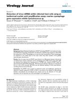
RPE65-positive immunohistochemical labelling in the retinas of Rpe65
-/-
mice after injection with rAAV.RPE65
Labeling in the retinal pigment epithelium is seen at 7 mo post-injection (A). The signal continues for some distance (more
than 600 µm) away from the injection site. This labeling is not seen in the uninjected, age-matched control Rpe65
-/-
mouse (B).
At 11 mo post-injection positive labeling is seen both close to (C), and more distant from (400 µm, D), the injection site (C)
although the signal is more discrete. This pattern of labeling near to (E) and distant from (>300 µm, F) the injection site per-
sists at 18 mo post-injection (E, F). Scale bar: A = 100 µm; B-F = 50 µm. Small arrows point to positively labeled cells, large
arrows point to injection site.
Genetic Vaccines and Therapy 2004, 2 />Page 6 of 15
(page number not for citation purposes)
small, scattered areas with no signal were seen (data not
shown). In contrast, in rAAV-RPE65.1-injected Rpe65
-/-
mice, RPE65 expression was not only present in the RPE/
choroid flatmounts (again in an area of approximately
30% of the retina), but was also present in the neuroretina
flatmounts where more RPE65-immunostained cells were
detected. The RPE65 expression in both the RPE/choroids
and neuroretina flatmounts of the rAAV-RPE65.1 injected
eyes was weaker, and appeared more dispersed, probably
due to the fewer number of cells transduced when com-
pared to rAAV.RPE65-injected RPE/choroids flatmounts
(data not shown). On the basis that rAAV.RPE65 was
more efficient in transducing RPE cells,, and in order to
target transduction of RPE cells only, subsequent studies
were conducted using rAAV.RPE65.
A histological analysis of RPE65 immunoreactivity in the
rAAV.RPE65-injected mice over time demonstrated that
strong RPE65 positive RPE cells were visible from the
injection site to up to 300–600 µm away, but still within
the bleb created, at 1–2 mo (data not shown), 7 mo (Fig.
1A), 11 mo (Fig. 1C and 1D) and 18 mo (Fig. 1E and 1F)
post-injection. However, the extent of RPE65 expression
appeared to decrease at the latest time point. No RPE65
immunoreactivity was seen in either uninjected, age-
matched control Rpe65
-/-
mice, or Rpe65
-/-
mice injected
with the control rAAV.GFP construct (data not shown).
There was no evidence of infiltrating immune cells in any
of the eyes examined.
Electroretinography
ERG analysis of Rpe65
-/-
mice showed an improvement in
the response of rAAV.RPE65-injected animals compared
with uninjected, age-matched controls. A comparison of
scotopic and photopic ERG responses from injected and
uninjected mice is presented in Fig. 2. At 1–2 mo post-
rAAV.RPE65 injection, an increase in the ERG b-wave
amplitude was apparent (Fig. 2A and 2B, upper trace). A
two-way repeated measures ANOVA of the stimulus-
response characteristics (Fig. 3) demonstrated a signifi-
cant (P < 0.001) difference in the scotopic b-wave ampli-
tude between the control and rAAV.RPE65-injected mice.
Post-hoc Bonferroni tests revealed a significant (P < 0.005)
increase of the scotopic b-wave at all flash intensities
above -0.9 log neutral density units (Fig. 3B). There was
also a significant interaction between stimulus intensity
and rAAV.RPE65-injection in the photopic b-wave ampli-
tude (P < 0.05). The post-hoc Bonferroni tests also revealed
a significant (P < 0.05) increase of the photopic b-wave at
the brightest flash intensities (Fig. 3B). No statistically sig-
nificant improvement in a-wave amplitude was seen at
this time point (Fig. 3A, P > 0.05). At 7 mo and 11 mo
post-injection, no differences were found in the ERG a-
wave (Fig. 2B and 2C) or b-wave amplitudes (Fig. 2B,2C,
3C and 3D) recorded from rAAV.RPE65-injected mouse
eyes when compared with responses recorded from unin-
jected, age-matched controls under either scotopic or pho-
topic conditions. Additional rAAV.GFP-injected Rpe65
-/-
control mice showed ERG signals equivalent to those of
uninjected controls (data not shown).
Morphological effects of rAAV.RPE65 injection in Rpe65
-/-
mouse retinas
Histological analysis of the retinas of uninjected Rpe65
-/-
mice showed a slow, progressive degeneration of photore-
ceptors. In brief, at the early age of 1–2 mo, the retinas of
uninjected Rpe65
-/-
mice appeared normal, except for the
less organized appearance of the photoreceptor outer seg-
ments (Fig. 4A). The outer nuclear layer of uninjected
Rpe65
-/-
mice aged 6–12 mo were visibly thinner and the
outer segments appeared highly disorganized when com-
pared to age-matched C57BL/6J controls. At 12 mo and
older (Fig. 4B), the difference in the outer nuclear layer
thickness was very significant when compared to age-
matched C57BL/6J mice (Fig. 4C) and by 21 mo of age,
the outer nuclear layer was completely absent (data not
shown). The morphologic difference was quantified by
counting the number of photoreceptor nuclei in the eyes
of uninjected Rpe65
-/-
mice at 2, 5, 7, 11, 17 and 24 mo
post-injection and comparing them to those of age-
matched C57BL/6J mice. A statistically significant
decrease (P < 0.05, Student's t-test) in photoreceptor
number was obtained for uninjected Rpe65
-/-
mice older
than 3 mo (Fig. 4D), reflecting the progressive loss of pho-
toreceptor cells in these mice. Subsequent comparison of
rAAV.RPE65-injected with age-matched, uninjected con-
trol Rpe65
-/-
mice indicated that no statistically significant
difference in the number of photoreceptors around the
injection site was seen at any of the time points (Fig. 4D;
P > 0.05, Student's t-test), suggesting that there was no
photoreceptor rescue or slow down in photoreceptor loss
following rAAV.RPE65 injection. The lack of photorecep-
tor rescue was reflected by the lack of difference in the
number of apoptotic cells in rAAV.RPE65-injected eyes of
Rpe65
-/-
mice (Fig. 5A) when compared to the
contralateral uninjected eyes (Fig. 5B). At 7 mo post-injec-
tion (8 mo of age), the number of apoptotic cells in both
the uninjected and rAAV.RPE65-injected Rpe65
-/-
appeared higher than those in age-matched C57BL/6J
controls (Fig. 5C). Analysis of the 60 µm regions of unin-
jected and rAAV.RPE65-injected Rpe65
-/-
eyes at 7 mo post
injection (8 mo of age) showed that 5.8 ± 1.9% and 2.7 ±
1.7%, respectively, of the remaining photoreceptors were
apoptotic (P > 0.01, Student's t-test, Fig. 5D).
Electron microscopy of rAAV.RPE65-injected and unin-
jected Rpe65
-/-
mouse eyes at 20 mo post injection (21 mo
of age) revealed the presence of retinyl ester lipid droplets
that are characteristic of Rpe65
-/-
mice [7]. However, a
direct comparison of the RPE in the rAAV.RPE65-injected
Genetic Vaccines and Therapy 2004, 2 />Page 7 of 15
(page number not for citation purposes)
Representative ERG responses recorded from rAAV.RPE65 injected and uninjected Rpe65
-/-
miceover timeFigure 2
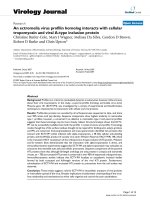
Representative ERG responses recorded from rAAV.RPE65 injected and uninjected Rpe65
-/-
miceover time
Rpe65
-/-
mice at 1–2 mo (A), 7 mo (B) and 11 mo (C) post-injection. Each panel shows representative responses from
rAAV.RPE65-injected (upper traces) and age-matched, uninjected control (lower traces) mice recorded under scotopic (left
panels) or photopic (right panels) conditions.
Photopic
050100150
-25
0
25
50
75
100
050100150
-25
0
25
50
75
100
Time (ms)
050100150
-25
0
25
50
75
100
Scotopic
050100150
Amplitude (
µ
V)
-25
0
25
50
75
100
050100150
Amplitude (
µ
V)
-25
0
25
50
75
100
Time (ms)
050100150
Amplitude (
µ
V)
-25
0
25
50
75
100
A
B
C
Genetic Vaccines and Therapy 2004, 2 />Page 8 of 15
(page number not for citation purposes)
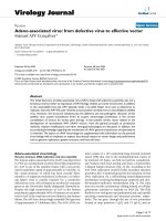
Intensity response characteristics of scotopic and photopic ERGFigure 3
Intensity response characteristics of scotopic and photopic ERG Intensity response characteristics of scotopic (left
panel) and photopic (right panel) ERGs recorded from rAAV.RPE65 injected (o) and age-matched, uninjected control (•)
Rpe65
-/-
mice. Intensity response characteristics of the ERG a-waves (A) and b-waves (B) at 1–2 mo post-injection (n = 15
rAAV.RPE65 injected, n = 10 uninjected). Intensity response characteristics of the ERG b-waves at 7 mo (C, n = 12
rAAV.RPE65-injected, n = 4 uninjected) and 11 mo (D, n = 12 rAAV.RPE65 injected, n = 6 uninjected) post-injection. Data are
mean values ± SEM. * = P < 0.05.
-3 -2 -1 0
0
20
40
60
80
-3 -2 -1 0
b-wave amplitude (
µ
V)
0
20
40
60
80
Photopic
-3 -2 -1 0
0
20
40
60
80
-3 -2 -1 0
0
20
40
60
80
Stimulus intensity (log ND)
-3 -2 -1 0
0
20
40
60
80
Scotopic
-3 -2 -1 0
a-wave amplitude (
µ
V)
0
20
40
60
80
Rpe65
-/-
rAAV.RPE65 injected
Stimulus intensity (log ND)
-3 -2 -1 0
b-wave amplitude (
µ
V)
0
20
40
60
80
-3 -2 -1 0
b-wave amplitude (
µ
V)
0
20
40
60
80
A
B
C
D
*
*
*
*
*
*
*
Genetic Vaccines and Therapy 2004, 2 />Page 9 of 15
(page number not for citation purposes)
mice (Fig. 6A) with the uninjected, age-matched control
(Fig. 6B) showed a striking difference between the
amounts of lipid inclusions present in these eyes. In con-
trast, electron microscopy of sections taken from outside
the subretinal bleb of rAAV.RPE65-injected eyes showed
no difference between the numbers of lipid inclusions
when compared to sections from uninjected eyes (data
not shown). In addition to the reduction in numbers of
lipid droplets, the layer of basal infoldings was also thin-
ner in rAAV.RPE65-injected eyes (Fig. 6A and 6B).
Immunostaining using the anti-SWC opsin antibody
demonstrated the presence of SWC opsin-positive cells
scattered throughout the flatmounted neuroretinas of 8
month old C57BL/6J mice (Fig. 7A). The number of SWC
opsin-positive cells was significantly lower in uninjected
Rpe65
-/-
mouse retinas, with only a small number of SWC
opsin-positive cells being seen in the neuroretinas of
either 3 week (Fig. 7B) or 3 month old Rpe65
-/-
mice (data
not shown). By 8 months of age no SWC opsin-positive
cells were visible in the neuroretinas of uninjected Rpe65
-
/-
mice (Fig. 7C). Examination of flatmounted neuroreti-
nas of 8-month-old rAAV.RPE65-injected Rpe65
-/-
mice
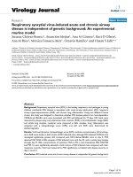
Comparisons of photoreceptor numbersFigure 4
Comparisons of photoreceptor numbers Photomicrographs of theouter retina of a 1 mo uninjected Rpe65
-/-
mouse (A),
a 14 mo injected Rpe65
-/-
mouse (B) and a 14 mo C57BL/6J mouse (C). (D) Graphical presentation of the mean number of cells
per 100 µm length of the outer nuclear layer (ONL) of C57BL/6J (▲), uninjected Rpe65
-/-
(◆) and rAAV.RPE65 injected Rpe65
-
/-
(') at the various ages shown. All points are calculated from the cell numbers averaged over 3 animals unless indicated (*n =
1). Arrow indicates time of injection. Scale bar: A-C = 20 µm.
Genetic Vaccines and Therapy 2004, 2 />Page 10 of 15
(page number not for citation purposes)
revealed the presence of numerous SWC opsin-positive
cells in an area coinciding with the subretinal bleb and, at
a higher density, around the injection site (Fig. 7D).
Counting and analysis of the number of SWC opsin-posi-
tive cells in the C57BL/6J control (n = 5), uninjected
Rpe65
-/-
mice (n = 5) and rAAV.RPE65-injected Rpe65
-/-
eyes (n = 5) showed that the reappearance of the SWC
opsin-positive cells in rAAV.RPE65-injected Rpe65
-/-
mice
was significant, reaching up to 50% of that seen in age-
matched C57BL/6J mice (Fig. 7E).
Discussion
We report here the results from our study examining the
effects of rAAV-mediated RPE65 expression in the retinas
of Rpe65
-/-
mice. Subretinal injection with rAAV.RPE65
purified by cesium chloride density gradient resulted in
Comparison of apoptotic cells numbersFigure 5
Comparison of apoptotic cells numbers Photomicrographs of the outer nuclear layer of 8 mo uninjected Rpe65
-/-
(A),
rAAV.RPE65 injected Rpe65
-/-
(B) and C57BL/6J mice (C) stained for apoptotic nuclei (arrows). (D) Graphical presentation of
the percentage of photoreceptor nuclei that are apoptotic in uninjected Rpe65
-/-
, rAAV.RPE65-injected Rpe65
-/-
and uninjected
C57BL/6J mice. Apoptotic and total photoreceptor nuclei were counted along 60 µm lengths of the outer nuclear layer of mice
at 7 mo post-injection (8 mo of age). Average total photoreceptor counts: uninjected Rpe65
-/-
= 106.8 ± 22.9, rAAV.RPE65
injected Rpe65
-/-
= 134 ± 30.3, uninjected C57 = 213.5 ± 3.3. All data are mean ± S.D. Scale bar: A-C = 20 µm.
Genetic Vaccines and Therapy 2004, 2 />Page 11 of 15
(page number not for citation purposes)
transduction of approximately 30% of the retina and pro-
duced long-term, detectable RPE65 protein expression
(up to 18 mo post-injection when the experiment was ter-
minated) in the RPE cells within the subretinal bleb of
Rpe65
-/-
knockout mice. The longevity of this RPE65
expression agrees with previous studies where rAAV.GFP
reporter gene constructs gave long-term detectable GFP
signals in a variety of animals [24,29-32]. In agreement
with previous rAAV.GFP research, the levels and extent of
RPE65 expression from the rAAV.RPE65 injection
appeared to decrease at the later time points [31]. At this
stage the reason for this decrease in transgene expression
is unclear. The reduction of RPE65 expression could be
due to the protein not being recognized as self, but this is
unlikely as there was no evidence of any infiltrating
immune cells in the injected eyes. It could also be due to
silencing mechanisms, such as promoter silencing [33,34]
or transgene silencing [35,36], or might be due to the lack
of integration [37]. Further work would be required to
confirm or disprove these suggestions, the conclusions of
which are important if rAAV is to be used for mediating
long-term transgene expression.
Following subretinal injection the most common sites of
transgene expression have been the RPE and photorecep-
tors cells. The extent and relative ratio of the transduction
level in these two cells types tends to vary depending on
factors such as the method of virus purification [31,38]
and the virus serotype being used [39]. Consistent with
results using rAAV.GFP [31,38], the expression of RPE65
following injection with cesium chloride density gradient
purified rAAV.RPE65 was only in the RPE cells, with no
expression being visible in the photoreceptors. Our labo-
ratory has examined a number of possible contributing
factors [31,38], but the precise reason for these differences
has yet to be elucidated.
Electron micrograph of injected and uninjected Rpe65
-/-
mouse retinaFigure 6
Electron micrograph of injected and uninjected
Rpe65
-/-
mouse retina Electron micrograph of the RPE of
an rAAV.RPE65-injected Rpe65
-/-
mouse at 20 mo post injec-
tion (A) and an age-matched, uninjected Rpe65
-/-
control (21
mo of age; B). The injected animal shows an accumulation of
retinyl ester lipid droplets in the RPE layer (small arrows)
that is not as prevalent as that in the uninjected control. The
layer of basal infoldings was also thinner in the injected
mouse (large arrows). Scale bar = 5 µm.
Comparison of short wavelength cone opsin-positive cellsFigure 7
Comparison of short wavelength cone opsin-positive
cells Photomicrographs (×40) of flatmounted neuroretinas
stained for SWC opsin in an 8 mo C57BL/6J (A), a 3 wk unin-
jected Rpe65
-/-
(B), an 8 mo Rpe65
-/-
(C) and an 8 mo
rAAV.RPE65-injected Rpe65
-/-
(D) mouse. (E) Graphical
presentation of the mean number of SWC opsin-positive
cells per 100 µm
2
calculated for 8 mo C57BL/6J,
rAAV.RPE65-injected and uninjected Rpe65
-/-
mice.
Genetic Vaccines and Therapy 2004, 2 />Page 12 of 15
(page number not for citation purposes)
Although rAAV.RPE65 was delivered to an area covering
30% of the Rpe65
-/-
mouse retina, pan-retinal ERG
responses (responses over the entire retina) were meas-
ured. Under this circumstance, the finding of an improved
ERG response in rAAV.RPE65-injected Rpe65
-/-
mice is of
great importance for the future treatment of LCA as it sug-
gests a partial restoration of visual function. The delivery
of rAAV.RPE65 to the Rpe65
-/-
mouse retinas resulted in
improvements in the maximum b-wave amplitude under
both scotopic (76% increase above uninjected controls)
and photopic (59% increase above uninjected controls)
conditions. However, the increase in b-wave magnitude
was only seen at the initial early 1–2 mo post-injection
time point of the study. The ability of rAAV.RPE65 deliv-
ery to Rpe65
-/-
mouse retinas to restore visual function,
though limited and transient, agrees with the now well
established data that rAAV.RPE65 gene therapy in the
RPE65 dog model produces an improved visual response
[16,21-23]. Although supporting the RPE65 dog ERG
data, in this current study of rAAV.RPE65-injected Rpe65
-/
-
mice it was difficult to determine the exact nature of the
ERG response, and in particular whether it was rod and/or
cone driven. In other studies performed on either
untreated [7,13,40], 11-cis retinal-treated [41] or double
mutant (Rpe65
-/-
Rho
-/-
, Rpe65
-/-
Cnga3
-/-
) Rpe65
-/-
mice [14],
ERG responses have been attributed to both rods and
cones. In the current work it is not possible to draw defin-
itive conclusions as to whether the improved ERG
responses seen is of rod, cone or a combined origin.
One of the interesting observations of this study was that
the long-term changes in some retinal cells lasted well
beyond measurable functional outcome. Electron micros-
copy indicated that the levels of lipid inclusions, an indi-
cator of retinyl ester accumulation and halted visual cycle
[7,22], was diminished in rAAV.RPE65-injected mice at
20 mo post-injection, suggesting that rAAV.RPE65 is still
able to elicit a biological effect at these later time points.
The remarkable recovery and long-term immunostaining
of SWC-opsin in the functionally important cones sug-
gests that cone function might be recoverable following
rAAV.RPE65 gene therapy. However considering that the
total number of photoreceptors continues to decrease, the
restoration of SWC-opsin immunostaining may not nec-
essarily represent protection against cone degeneration
but only demonstrates the recovery of SWC-opsin in
cones in the presence of an active visual cycle. Although
these results were encouraging, ERG measurements were
not able to differentiate between rod and cone function.
The transient improvement in ERG response, the decrease
in lipid droplet accumulation and the positive SWC-opsin
immunostaining upon rAAV.RPE65 administration were
not accompanied by a statistically significant decrease in
the rate of photoreceptor degeneration or apoptotic cell
death. It appears, therefore, that the delivery of
rAAV.RPE65, while being able to induce restarting of the
visual cycle and phototransduction in the remaining pho-
toreceptors in Rpe65
-/-
mice, was unable to slow or halt the
photoreceptor degeneration that afflicts these mice. The
finding of improved function without photoreceptor res-
cue is not unique, as a similar observation has been
reported after subretinal injection of an rAAV encoding
Prph2 in a retinal degeneration slow mouse model
[42,43]. Vision is maintained through the close and pre-
cise interaction between all the retinal cells. For example
both in humans and in a transgenic mouse model for
retinitis pigmentosa, the degeneration of rod photorecep-
tors eventually leads to the loss of cone photoreceptors as
well [44]. At present one of the limitations of gene therapy
is that only cells present within the retinal bleb can be
targeted and within this treated region not all RPE cells
undergo successful transduction and subsequent RPE65
production. The inability to restore the visual cascade in
all RPE cells and hence the failure to restore function to
the corresponding photoreceptors may be behind the lack
of general photoreceptor rescue. In addition, the level and
extent of RPE65 expression resulting from the
rAAV.RPE65 injection might have been insufficient to
support the survival of a significant number of functional
photoreceptor cells. The restricted transduction area of
subretinal rAAV injection, as seen from the confinement
of transgene expression and reduction of lipid inclusion
to RPE cells within the subretinal bleb, highlights the need
to maximize both the efficiency of the rAAV construct
delivery and the transgene expression in vivo. The effi-
ciency of transgene delivery would be particularly impor-
tant in diseases characterized by pan-retinal degeneration
as they would presumably require the rAAV-mediated
treatment to be present across the entire retina, ideally in
every RPE cell in the retina.
A recent study in the RPE65 dog model, where the volume
of virus that can be injected is not tightly restricted as in
the small mouse eye, demonstrated that delivering larger
volumes, and therefore higher titers of virus, gives a higher
degree of functional rescue [21]. It may be, therefore, that
the intensity of RPE65 expression is also important along
with the size of the area being transduced, especially given
the abundance of endogenous RPE65 protein in RPE cells
of normal animals [45]. Fortunately rAAV biology, prepa-
ration, delivery and expression are being continuously
improved [37,46-48] and no doubt future studies will see
these limitations being overcome.
We performed our subretinal rAAV.RPE65 injections on
Rpe65
-/-
mice immediately after weaning, that is, at 3
weeks of age. At this age there is no histological evidence
of photoreceptor degeneration having started in the
Rpe65
-/-
mice [7]. However, analysis of RPE65 expression
Genetic Vaccines and Therapy 2004, 2 />Page 13 of 15
(page number not for citation purposes)
during embryogenesis and development has shown that
rat RPE65 mRNA expression is detectable at E17 and pro-
tein at post natal days 4–5 [45,49]. If RPE65 expression
commences so soon as to be visible in embryogenesis it is
possible also that, in the absence of RPE65, the deleteri-
ous effects of RPE65 loss would also begin at this early
stage. In other words, the cascade of events leading to
eventual photoreceptor degeneration may be beginning at
a much earlier age than our chosen age of injection. Thus,
while rAAV.RPE65 expression can have a visual cycle effect
within the time scale it is injected, introducing RPE65
expression alone at a later stage is insufficient to halt the
photoreceptor degeneration cascade. If this hypothesis is
true, the implications for rAAV-mediated gene therapy as
a clinical option may be significant, as it may be essential
to deliver rAAV.RPE65 at the time when RPE65 expression
should be commencing in order to completely compen-
sate for its loss. A recent article by [20] demonstrated that
the early addition of 9-cis retinal caused a long-term
reduction in lipids, and thus indicating that early inter-
vention in LCA disease progression is potentially impor-
tant. However, the feasibility of this approach from a
clinical perspective is unclear. Genotypically, Rpe65
mutations are recessive and thus heterozygous parents
may be unaware of their carrier status until they bear an
Rpe65
-/-
homozygous child. Moreover, there is evidence
that photoreceptor degeneration has already commenced
before birth in fetuses afflicted with RPE65 mutations
[50], suggesting that in utero delivery would be needed to
fully prevent the effects of the RPE65 absence. In light of
this information it is likely that neonatal treatment, even
if performed soon after birth, may be only partially
successful as a therapeutic option. Perhaps delivery of
additional agents, either anti-apoptosis gene therapy
[51,52], or oral retinoid supplements [18,20] may be
needed to attain full disease prevention.
In conclusion, the data produced from this study demon-
strated that subretinal injection of rAAV.RPE65 could pro-
duce a limited functional rescue of vision in Rpe65
-/-
mice.
This functional rescue was seen in the form of an
improved, albeit transient, ERG signal and a decreased
level of lipid inclusions in treated eyes at later time points.
In particular, the latter suggests that once optimized, rAAV
may offer a long-term treatment option for LCA patients.
Much work is still required, including improving our
knowledge of the effects of RPE65 loss and the mecha-
nisms that lead to the photoreceptor degeneration, as well
as optimizing the timing, efficiency and specificity of the
current rAAV gene technology. Although gene therapy on
the Rpe65
-/-
mouse model may not have generated as
much success as the dog model for LCA [16,21,22,53], the
use of the Rpe65
-/-
mouse model has its merit in that it
could be used in future studies to address how cones and
rods are specifically affected by absence of RPE65 and to
provide more information on the function of RPE65.
Competing interests
None of the authors of this paper have competing
interests.
Authors' contributions
CML and MJTY prepared the clones and cesium chloride
density gradient purified rAAV.RPE65, and designed, per-
formed, analyzed and interpreted the data presented in
Figs. 1, 4, 5, 6 and 7. MB performed the surgical proce-
dures, NLB performed the ERG measurements and analy-
sis of the data presented in Figs. 2 and 3. XZ prepared the
heparin column purified rAAV.RPE65, TMR provided the
Rpe65
-/-
mouse model, KN participated in the ERG data
interpretation and PER provided the conceptual design of
the project, initiated the collaborations and assisted in
data analysis. All authors have had intellectual contribu-
tion to the preparation of this manuscript and have read
and approved it.
Acknowledgements
This project was financially supported by the Foundation for Fighting Blind-
ness (USA), Retina Australia and the National Health and Medical Research
Council (Australia). We thank Dr R. Samulski for the pSSV9 plasmid and Dr
D. Zhang, Mr. B. Rae, Mr. S. Moore and Dr T. Robertson for their technical
assistance and advice.
References
1. Perrault I, Rozet JM, Gerber S, Ghazi I, Leowski C, Ducroq D, Souied
E, Dufier JL, Munnich A, Kaplan J: Leber congenital amaurosis.
Mol Genet Metab 1999, 68:200-208.
2. Perrault I, Rozet JM, Ghazi I, Leowski C, Bonnemaison M, Gerber S,
Ducroq D, Cabot A, Souied E, Dufier JL, Munnich A, Kaplan J: Differ-
ent functional outcome of RetGC1 and RPE65 gene muta-
tions in Leber congenital amaurosis. Am J Hum Genet 1999,
64:1225-1228.
3. Dharmaraj SR, Silva ER, Pina AL, Li YY, Yang JM, Carter CR, Loyer
MK, El-Hilali HK, Traboulsi EK, Sundin OK, Zhu DK, Koenekoop RK,
Maumenee IH: Mutational analysis and clinical correlation in
Leber congenital amaurosis. Ophthalmic Genet 2000, 21:135-150.
4. Gu SM, Thompson DA, Srikumari CR, Lorenz B, Finckh U, Nicoletti
A, Murthy KR, Rathmann M, Kumaramanickavel G, Denton MJ, Gal A:
Mutations in RPE65 cause autosomal recessive childhood-
onset severe retinal dystrophy. Nat Genet 1997, 17:194-197.
5. Marlhens F, Bareil C, Griffoin JM, Zrenner E, Amalric P, Eliaou C, Liu
SY, Harris E, Redmond TM, Arnaud B, Claustres M, Hamel CP: Muta-
tions in RPE65 cause Leber's congenital amaurosis. Nat Genet
1997, 17:139-141.
6. Lorenz B, Gyurus P, Preising M, Bremser D, Gu S, Andrassi M, Gerth
C, Gal A: Early-onset severe rod-cone dystrophy in young chil-
dren with RPE65 mutations. Invest Ophthalmol Vis Sci 2000,
41:2735-2742.
7. Redmond TM, Yu S, Lee E, Bok D, Hamasaki D, Chen N, Goletz P,
Ma JX, Crouch RK, Pfeifer K: Rpe65 is necessary for production
of 11-cis-vitamin A in the retinal visual cycle. Nat Genet 1998,
20:344-351.
8. Saari JC: Retinoids in photosensitive systems. Retinoids in Photo-
sensitive systems. Edited by: Sporn MB, Roberts AB and Goodmans DS.
New York, Raven Press Ltd; 1994:351-384.
9. Rando RR: The biochemistry of the visual cycle. Chem Rev 2001,
101:1881-1896.
10. Wrigstad A, Narfstrom K, Nilsson SE: Slowly progressive changes
of the retina and retinal pigment epithelium in Briard dogs
Genetic Vaccines and Therapy 2004, 2 />Page 14 of 15
(page number not for citation purposes)
with hereditary retinal dystrophy. A morphological study.
Doc Ophthalmol 1994, 87:337-354.
11. Aguirre GD, Baldwin V, Pearce-Kelling S, Narfstrom K, Ray K, Acland
GM: Congenital stationary night blindness in the dog: com-
mon mutation in the RPE65 gene indicates founder effect.
Mol Vis 1998, 4:23.
12. Veske A, Nilsson SE, Narfstrom K, Gal A: Retinal dystrophy of
Swedish briard/briard-beagle dogs is due to a 4-bp deletion
in RPE65. Genomics 1999, 57:57-61.
13. Ekesten B, Gouras P, Salchow DJ: Ultraviolet and middle wave-
length sensitive cone responses in the electroretinogram
(ERG) of normal and Rpe65 -/- mice. Vision Res 2001,
41:2425-2433.
14. Seeliger MW, Grimm C, Stahlberg F, Friedburg C, Jaissle G, Zrenner
E, Guo H, Reme CE, Humphries P, Hofmann F, Biel M, Fariss RN, Red-
mond TM, Wenzel A: New views on RPE65 deficiency: the rod
system is the source of vision in a mouse model of Leber con-
genital amaurosis. Nat Genet 2001, 29:70-74.
15. Narfstrom K, Wrigstad A, Ekesten B, Nilsson SE: Hereditary Reti-
nal Dystrophy in the Briard Dog: Clinical and Hereditary
Characteristics. Veterinary & Comparative Ophthalmology 1994,
4:85-92.
16. Acland GM, Aguirre GD, Ray J, Zhang Q, Aleman TS, Cideciyan AV,
Pearce-Kelling SE, Anand V, Zeng Y, Maguire AM, Jacobson SG,
Hauswirth WW, Bennett J: Gene therapy restores vision in a
canine model of childhood blindness. Nat Genet 2001, 28:92-95.
17. Wrigstad A, Nilsson SE, Narfstrom K: Ultrastructural changes of
the retina and the retinal pigment epithelium in Briard dogs
with hereditary congenital night blindness and partial day
blindness. Exp Eye Res 1992, 55:805-818.
18. Van Hooser JP, Aleman TS, He YG, Cideciyan AV, Kuksa V, Pittler SJ,
Stone EM, Jacobson SG, Palczewski K: Rapid restoration of visual
pigment and function with oral retinoid in a mouse model of
childhood blindness. Proc Natl Acad Sci U S A 2000, 97:8623-8628.
19. Gouras Peter, Kong Jian, Tsang Stephen H.: Retinal Degeneration
and RPE Transplantation in Rpe65-/- Mice. Invest. Ophthalmol.
Vis. Sci. 2002, 43:3307-3311.
20. Van Hooser JP, Liang Y, Maeda T, Kuksa V, Jang GF, He YG, Rieke F,
Fong HK, Detwiler PB, Palczewski K: Recovery of visual functions
in a mouse model of Leber congenital amaurosis. J Biol Chem
2002, 277:19173-19182.
21. Ford M, Bragadottir R, Rakoczy PE, Narfstrom K: Gene transfer in
the RPE65 null mutation dog: relationship between con-
struct volume, visual behavior and electroretinographic
(ERG) results. Doc Ophthalmol 2003, 107:79-86.
22. Narfstrom K, Katz ML, Bragadottir R, Seeliger M, Boulanger A, Red-
mond TM, Caro L, Lai CM, Rakoczy PE: Functional and structural
recovery of the retina after gene therapy in the RPE65 null
mutation dog. Invest Ophthalmol Vis Sci 2003, 44:1663-1672.
23. Narfstrom K, Katz ML, Ford M, Redmond TM, Rakoczy E, Bragadottir
R: In vivo gene therapy in young and adult RPE65-/- dogs pro-
duces long-term visual improvement. J Hered 2003, 94:31-37.
24. Rolling F, Shen WY, Tabarias H, Constable I, Kanagasingam Y, Barry
CJ, Rakoczy PE: Evaluation of adeno-associated virus-mediated
gene transfer into the rat retina by clinical fluorescence
photography. Hum Gene Ther 1999, 10:641-648.
25. Redmond TM, Hamel CP: Genetic analysis of RPE65: from
human disease to mouse model. Methods Enzymol 2000,
316:705-724.
26. Rolling F, Samulski RJ: AAV as a viral vector for human gene
therapy. Generation of recombinant virus. Mol Biotechnol 1995,
3:9-15.
27. Zolotukhin S, Byrne BJ, Mason E, Zolotukhin I, Potter M, Chesnut K,
Summerford C, Samulski RJ, Muzyczka N: Recombinant adeno-
associated virus purification using novel methods improves
infectious titer and yield. Gene Ther 1999, 6:973-985.
28. Rakoczy PE, Sarks SH, Daw N, Constable IJ: Distribution of cathe-
psin D in human eyes with or without age-related
maculopathy. Exp Eye Res 1999, 69:367-374.
29. Bennett J, Maguire AM, Cideciyan AV, Schnell M, Glover E, Anand V,
Aleman TS, Chirmule N, Gupta AR, Huang Y, Gao GP, Nyberg WC,
Tazelaar J, Hughes J, Wilson JM, Jacobson SG: Stable transgene
expression in rod photoreceptors after recombinant adeno-
associated virus-mediated gene transfer to monkey retina.
Proc Natl Acad Sci U S A 1999, 96:9920-9925.
30. Bainbridge JW, Mistry A, Schlichtenbrede FC, Smith A, Broderick C,
De Alwis M, Georgiadis A, Taylor PM, Squires M, Sethi C, Charteris
D, Thrasher AJ, Sargan D, Ali RR: Stable rAAV-mediated trans-
duction of rod and cone photoreceptors in the canine retina.
Gene Ther 2003, 10:1336-1344.
31. Shen WY, Lai CM, Lai YK, Zhang D, Zaknich T, Sutanto EN, Consta-
ble IJ, Rakoczy PE: Practical considerations of recombinant
adeno-associated virus-mediated gene transfer for treat-
ment of retinal degenerations. J Gene Med 2003, 5:576-587.
32. Sarra GM, Stephens C, Schlichtenbrede FC, Bainbridge JW, Thrasher
AJ, Luthert PJ, Ali RR: Kinetics of transgene expression in
mouse retina following sub-retinal injection of recombinant
adeno-associated virus. Vision Res 2002, 42:541-549.
33. Scharfmann R, Axelrod JH, Verma IM: Long-term in vivo expres-
sion of retrovirus-mediated gene transfer in mouse fibrob-
last implants. Proc Natl Acad Sci U S A 1991, 88:4626-4630.
34. Loser P, Jennings GS, Strauss M, Sandig V: Reactivation of the pre-
viously silenced cytomegalovirus major immediate-early
promoter in the mouse liver: involvement of NFkappaB. J
Virol 1998, 72:180-190.
35. Chen WY, Townes TM: Molecular mechanism for silencing
virally transduced genes involves histone deacetylation and
chromatin condensation. Proc Natl Acad Sci U S A 2000,
97:377-382.
36. Gaetano C, Catalano A, Palumbo R, Illi B, Orlando G, Ventoruzzo G,
Serino F, Capogrossi MC: Transcriptionally active drugs
improve adenovirus vector performance in vitro and in vivo.
Gene Ther 2000, 7:1624-1630.
37. Malik AK, Monahan PE, Allen DL, Chen BG, Samulski RJ, Kurachi K:
Kinetics of recombinant adeno-associated virus-mediated
gene transfer. J Virol 2000, 74:3555-3565.
38. Shen WY, Lai YK, Lai CM, Rakoczy PE: Impurity of recombinant
adeno-associated virus type 2 affects the transduction char-
acteristics following subretinal injection in the rat. Vision Res
2004, 44:339-348.
39. Rabinowitz JE, Rolling F, Li C, Conrath H, Xiao W, Xiao X, Samulski
RJ: Cross-packaging of a single adeno-associated virus (AAV)
type 2 vector genome into multiple AAV serotypes enables
transduction with broad specificity. J Virol 2002, 76:791-801.
40. Rohrer B, Goletz P, Znoiko S, Ablonczy Z, Ma JX, Redmond TM,
Crouch RK: Correlation of regenerable opsin with rod ERG
signal in Rpe65-/- mice during development and aging. Invest
Ophthalmol Vis Sci 2003, 44:310-315.
41. Ablonczy Z, Crouch RK, Goletz PW, Redmond TM, Knapp DR, Ma
JX, Rohrer B: 11-cis-retinal reduces constitutive opsin phos-
phorylation and improves quantum catch in retinoid-defi-
cient mouse rod photoreceptors. J Biol Chem 2002,
277:40491-40498.
42. Ali RR, Sarra GM, Stephens C, Alwis MD, Bainbridge JW, Munro PM,
Fauser S, Reichel MB, Kinnon C, Hunt DM, Bhattacharya SS, Thrasher
AJ: Restoration of photoreceptor ultrastructure and function
in retinal degeneration slow mice by gene therapy. Nat Genet
2000, 25:306-310.
43. Sarra GM, Stephens C, de Alwis M, Bainbridge JW, Smith AJ, Thrasher
AJ, Ali RR: Gene replacement therapy in the retinal degener-
ation slow (rds) mouse: the effect on retinal degeneration
following partial transduction of the retina. Hum Mol Genet
2001, 10:2353-2361.
44. Naash MI, Hollyfield JG, al-Ubaidi MR, Baehr W: Simulation of
human autosomal dominant retinitis pigmentosa in trans-
genic mice expressing a mutated murine opsin gene. Proc Natl
Acad Sci U S A 1993, 90:5499-5503.
45. Hamel CP, Tsilou E, Harris E, Pfeffer BA, Hooks JJ, Detrick B, Red-
mond TM: A developmentally regulated microsomal protein
specific for the pigment epithelium of the vertebrate retina.
J Neurosci Res 1993, 34:414-425.
46. Brument N, Morenweiser R, Blouin V, Toublanc E, Raimbaud I, Cherel
Y, Folliot S, Gaden F, Boulanger P, Kroner-Lux G, Moullier P, Rolling
F, Salvetti A: A versatile and scalable two-step ion-exchange
chromatography process for the purification of recombinant
adeno-associated virus serotypes-2 and -5. Mol Ther 2002,
6:678-686.
47. Schneider H, Muhle C, Douar AM, Waddington S, Jiang QJ, von der
Mark K, Coutelle C, Rascher W: Sustained delivery of therapeu-
tic concentrations of human clotting factor IX a compari-
Publish with BioMed Central and every
scientist can read your work free of charge
"BioMed Central will be the most significant development for
disseminating the results of biomedical research in our lifetime."
Sir Paul Nurse, Cancer Research UK
Your research papers will be:
available free of charge to the entire biomedical community
peer reviewed and published immediately upon acceptance
cited in PubMed and archived on PubMed Central
yours — you keep the copyright
Submit your manuscript here:
/>BioMedcentral
Genetic Vaccines and Therapy 2004, 2 />Page 15 of 15
(page number not for citation purposes)
son of adenoviral and AAV vectors administered in utero. J
Gene Med 2002, 4:46-53.
48. Weber M, Rabinowitz J, Provost N, Conrath H, Folliot S, Briot D,
Cherel Y, Chenuaud P, Samulski J, Moullier P, Rolling F: Recom-
binant adeno-associated virus serotype 4 mediates unique
and exclusive long-term transduction of retinal pigmented
epithelium in rat, dog, and nonhuman primate after subret-
inal delivery. Mol Ther 2003, 7:774-781.
49. Manes G, Leducq R, Kucharczak J, Pages A, Schmitt-Bernard CF,
Hamel CP: Rat messenger RNA for the retinal pigment epi-
thelium-specific protein RPE65 gradually accumulates in two
weeks from late embryonic days. FEBS Lett 1998, 423:133-137.
50. Porto FB, Perrault I, Hicks D, Rozet JM, Hanoteau N, Hanein S, Kap-
lan J, Sahel JA: Prenatal human ocular degeneration occurs in
Leber's congenital amaurosis (LCA2). J Gene Med 2002,
4:390-396.
51. Liang FQ, Dejneka NS, Cohen DR, Krasnoperova NV, Lem J, Maguire
AM, Dudus L, Fisher KJ, Bennett J: AAV-mediated delivery of cil-
iary neurotrophic factor prolongs photoreceptor survival in
the rhodopsin knockout mouse. Mol Ther 2001, 3:241-248.
52. Petrin D, Baker A, Coupland SG, Liston P, Narang M, Damji K, Leon-
ard B, Chiodo VA, Timmers A, Hauswirth W, Korneluk RG, Tsilfidis
C: Structural and functional protection of photoreceptors
from MNU-induced retinal degeneration by the X-linked
inhibitor of apoptosis. Invest Ophthalmol Vis Sci 2003,
44:2757-2763.
53. Narfstrom K, Bragadottir R, Redmond TM, Rakoczy E, van Veen T,
Brunn A: Functional and structural evaluation after
AAV.RPE65 gene transfer in the canine model of leber's con-
genital amaurosis. Retinal Degenerations Mechanisms and Experimen-
tal Therapy Volume 533. Edited by: JG Hollyfield RE Anderson MM La
Vail. New York: USA., Kluwer Academic / Plenum Publishers;
2003:423-430.