Báo cáo sinh học: "∆RR vaccination protects from KA-induced seizures and neuronal loss through ICP10PK-mediated modulation of the neuronal-microglial axis" docx
Bạn đang xem bản rút gọn của tài liệu. Xem và tải ngay bản đầy đủ của tài liệu tại đây (1.69 MB, 14 trang )
BioMed Central
Page 1 of 14
(page number not for citation purposes)
Genetic Vaccines and Therapy
Open Access
Research
∆RR vaccination protects from KA-induced seizures and neuronal
loss through ICP10PK-mediated modulation of the
neuronal-microglial axis
Jennifer M Laing and Laure Aurelian*
Address: Department of Pharmacology and Experimental Therapeutics, University of Maryland, School of Medicine, Baltimore, MD 21201, USA
Email: Jennifer M Laing - ; Laure Aurelian* -
* Corresponding author
Abstract
Ischemic brain injury and epilepsy are common neurodegenerative diseases caused by
excitotoxicity. Their pathogenesis includes microglial production of inflammatory cytokines. Our
studies were designed to examine whether a growth compromised HSV-2 mutant (∆RR) prevents
excitotoxic injury through modulation of microglial responses by the anti-apoptotic HSV-2 protein
ICP10PK. EOC2 and EOC20 microglial cells, which are differentially activated, were infected with
∆RR or the ICP10PK deleted virus (∆PK) and examined for virus-induced neuroprotective activity.
Both cell lines were non-permissive for virus growth, but expressed ICP10PK (∆RR) or the PK
deleted ICP10 protein p95 (∆PK). Conditioned medium (CM) from ∆RR-, but not ∆PK-infected
cells prevented N-methyl-D-aspartate (NMDA)-induced apoptosis of primary hippocampal
cultures, as determined by TUNEL and caspase-3 activation (76.9 ± 5.3% neuroprotection).
Neuroprotection was associated with inhibition of TNF-α and RANTES and production of IL-10.
The CM from ∆PK-infected EOC2 and EOC20 cells did not contain IL-10, but it contained TNF-α
and RANTES. IL-10 neutralization significantly (p < 0.01) decreased, but did not abrogate, the
neuroprotective activity of the CM from ∆RR-infected microglial cultures indicating that ICP10PK
modulates the neuronal-microglial axis, also through induction of various microglial
neuroprotective factors. Rats given ∆RR (but not ∆PK) by intranasal inoculation were protected
from kainic acid (KA)-induced seizures and neuronal loss in the CA1 hippocampal fields. Protection
was associated with a significant (p < 0.001) increase in the numbers of IL-10+ microglia (CD11b+)
as compared to ∆PK-treated animals. ∆RR is a promising vaccination/therapy platform for
neurodegeneration through its pro-survival functions in neurons as well as microglia modulation.
Introduction
Ischemic brain injury, or stroke, and epilepsy are two of
the most common neurodegenerative disease in Ameri-
cans, the symptoms of which are caused by excitotoxicity
[1,2]. Excitotoxicity is a mechanism of neuronal cell injury
that is caused by the excessive activation of glutamate
receptors and is accompanied by the induction of neuro-
nal cell apoptosis, a tightly regulated, energy dependent,
irreversible process mediated by cysteine proteases (cas-
pases) [3]. Microglia activation and the production of
inflammatory cytokines, namely TNF-α, were associated
with neurodegeneration, including excitotoxic injury [4-
6]. Several strategies were proposed to interrupt the apop-
totic cascade in neurons, including gene therapy with
Published: 7 January 2008
Genetic Vaccines and Therapy 2008, 6:1 doi:10.1186/1479-0556-6-1
Received: 10 September 2007
Accepted: 7 January 2008
This article is available from: />© 2008 Laing and Aurelian; licensee BioMed Central Ltd.
This is an Open Access article distributed under the terms of the Creative Commons Attribution License ( />),
which permits unrestricted use, distribution, and reproduction in any medium, provided the original work is properly cited.
Genetic Vaccines and Therapy 2008, 6:1 />Page 2 of 14
(page number not for citation purposes)
growth factors or anti-apoptotic proteins delivered by the
neurotropic herpes simplex virus type 1 (HSV-1) [7,8].
However, these genes had relatively narrow neuroprotec-
tive profiles, neuronal survival was often limited and did
not correlate with retention of functional integrity, and
some strategies were associated with detrimental out-
comes [8,9] potentially related to their effect on glial cells.
Indeed, microglia are considered the CNS resident profes-
sional macrophages. They function as the principal
immune effector cells of the CNS, responding to any path-
ological event. Activated microglia accumulate at sites of
injury or plaques in neurodegenerative CNS, and their
activation was implicated in the pathogenesis of a variety
of neurodegenerative diseases, including Alzheimer dis-
ease, Parkinson's disease, HIV-associated dementia and
stroke. Excessive microglial activation and the dysregu-
lated overproduction of inflammatory cytokines are the
hallmark of many neurodegenerative diseases and
ischemic brain injury [4-6,10,11]. Given their importance
in modulating neuronal cell life/death decisions, micro-
glia are increasingly recognized as a potential target for
neuroprotective vaccination. However, identification of
the correct gene for vaccine development is a major clini-
cal challenge. We have recently described the construction
of a growth compromised HSV-2 based vector (∆RR) for
the viral protein ICP10PK, which has anti-apoptotic activ-
ity in primary and organotypic hippocampal and striatal
cultures through activation of survival pathways [12-18].
The studies described in this report were designed to
examine whether ∆RR can function as a vaccine to prevent
neurodegenerative injury through ICP10PK-mediated
modulation of the microglial cell responses in favor of
neuroprotection.
Materials and methods
Cell culture
Vero (African green monkey kidney), SK-NSH (human
neuroblastoma) and LADMAC (mouse bone marrow)
cells were grown in minimal essential medium (MEM),
supplemented with 1 mM sodium pyruvate, 2 mM L-
glutamine, 100 µM non-essential amino acids and 10%
fetal bovine serum (FBS) (Gibco-BRL, Gaithersburg, MD).
EOC20 and EOC2 microglia cultures were obtained from
ATCC (Manassas, VA) and grown in Dulbecco's minimal
essential medium (DMEM, Gibco-BRL) with 20% 7 day-
conditioned LADMAC medium which provides CSF-1 for
microglial cell growth. EOC20, but not EOC2, cells con-
stitutively express high levels of MHCII antigens [19]. Rat
embryonic day 18 hippocampi were purchased from Neu-
romics (Edina, MN) and dissociated and plated at a den-
sity of 5 × 10
5
cells/dish on glass coverslips precoated with
poly-L-Lysine (Sigma, St. Louis, MO) according to manu-
facturer's instruction. Over 99% of the cells stained with
β
III
Tubulin antibody, indicating that they are neurons.
The cultures were maintained in Neurobasal medium
(Gibco-BRL) supplemented with B27 (Gibco-BRL).
Viruses
HSV-2 (strain G) and the mutants ∆PK and ∆RR con-
structed from HSV-2(G) were previously described [12-
14,16-18,20,21]. Briefly, to construct ∆RR, we took advan-
tage of previous findings that the large subunit of the
HSV-2 ribonucleotide reductase (R1, also known as
ICP10), which is encoded by the viral gene UL39, has
independently functioning protein kinase (ICP10PK) and
ribonucleotide reductase (RR) domains, both of which are
required for virus growth in non-replicating cells, includ-
ing neurons [17,18,20]. To generate ∆RR, the 3'-end R1-
encoding sequences of UL39 were deleted and replaced
with LacZ fused in frame with ICP10PK, giving rise to a
175 kDa mutant protein (p175). ∆PK was generated from
∆RR by deletion of the UL39 5'-end sequences that encode
ICP10PK giving rise to a 95 kDa protein (p95) (Fig. 1A).
Expression of the p175 and p95 proteins is driven by the
authentic ICP10 promoter, which is regulated with imme-
diate early (IE) kinetics (independent of virus replication)
and responds to AP-1 transcription factors upregulated/
activated by neurotoxic stress stimuli [22-24]. ∆RR and
∆PK are grown in Vero cells and titrated by plaque assay
in medium containing 10% serum [20].
Antibodies and reagents
The generation and specificity of the rabbit ICP10 anti-
body was described. It recognizes an epitope located
within amino acid residues 13–26 that are retained by
both p175 and p95 [13,14,17,18,20,21]. The following
antibodies were purchased and used according to the
manufacturer's instructions: CD11b (Mac-1α
m
chain-
biotin conjugated; Leinco, St. Louis, MO), HSV major cap-
sid protein VP5 (Virusys Corporation, Sykesville, MD),
TNF-α and neutralizing IL-10 (R&D Systems, Minneapo-
lis, MN), IL-10 (Santa Cruz Biotechnology, Santa Cruz,
CA), p20 fragment of activated caspase-3 (caspase-3p20)
(Cell Signaling Technologies, Beverly, MA) and β
III
Tubu-
lin (Promega, Madison, WI). Texas Red conjugated
streptavidin, FITC conjugate streptavidin, Texas Red con-
jugated horse anti mouse IgG and FITC conjugated goat
anti rabbit was purchased from Vector (Burlingame, CA),
FITC conjugated goat anti mouse IgG from Jackson
ImmunoResearch (West Grove, PA), AlexaFluor 546 was
purchased from Molecular Probes (Eugene, OR), N-
methyl-D-aspartic acid (NMDA) from Sigma-Aldrich, and
Kainic Acid (KA) from A.G. Scientific (San Diego, CA).
Immunoblotting and immunocomplex PK assay
Immunoblotting was performed as described [19,23].
Briefly, cells were lysed with radioimmunoprecipitation
buffer [RIPA; 20 mM Tris-HCl (pH 7.4), 0.15 mM NaCl,
1% Nonidet P-40, 0.1% sodium dodecyl sulfate (SDS),
Genetic Vaccines and Therapy 2008, 6:1 />Page 3 of 14
(page number not for citation purposes)
0.5% sodium deoxycholate] supplemented with protease
and phosphatase inhibitor cocktails (Sigma) and soni-
cated twice for 30 seconds at 25% output power with a
Sonicator ultrasonic processor (Misonix, Inc.,
Farmingdale, NY). Protein concentrations were deter-
mined by the bicinchoninic assay (Pierce, Rockford, IL),
and 100 µg protein samples were resolved by SDS-poly-
acrylamide gel electrophoresis (SDS-PAGE) and trans-
ferred to nitrocellulose membranes. The blots were
incubated (1 hr, RT) in TNT buffer (0.01 M Tris-HCl [pH
7.4], 0.15 M NaCl, 0.05% Tween 20) containing either
5% nonfat dried milk or 1% bovine serum albumin (BSA)
to block nonspecific binding. Blots were exposed over-
night at 4°C to appropriate antibodies diluted in TNT
buffer with either milk or BSA, washed in TNT buffer, and
incubated (1 hr; RT) with anti-rabbit IgG conjugated to
horseradish peroxidase (HRP; Cell Signaling). After exten-
sive washing, bands were detected using enhanced chemi-
luminescence reagents (ECL, Amersham Pharmacia,
Piscataway, NJ) and exposure to high-performance film
(Hyperfilm ECL, Amersham). Quantitation was by densi-
tometric scanning with the Bio-Rad GS-700 imaging den-
sitometer (Bio-Rad, Hercules, CA) and results are
expressed as densitometric units × 100. For immunocom-
plex PK assays cell extracts in lysis buffer (20 mM Tris, pH
7.5, 150 mM NaCl, 1% NP-40 and protease and phos-
phatase inhibitor cocktails) were standardized for protein
concentration and incubated with 10 µl of ICP10 anti-
body (1 h, 4°C) and 100 µl of protein A-sepharose CL4B
beads (50% v/v) (30 min, 4°C). The beads were washed
(3×) with RIPA buffer followed by TS buffer [20 mM Tris-
HCl (pH 7.4), 0.15 M NaCl], resuspended in 50 µl kinase
reaction buffer consisting of 10 µCi [32P]-ATP (0.1 µM,
3000 Ci/mmol, NEN), 5 mM MgCl
2
, 2 mM MnCl
2
, 20
mM Tris-HCl (pH 7.4), and incubated at 30°C for 30 min.
Samples were washed in 20 mM Tris-HCl (pH 7.4) with
0.15 M NaCl and boiled for 5 min after addition of 100 µl
denaturing solution. Proteins were resolved by SDS-
PAGE.
Single step growth curves and infectious centers assay
Single step growth curves were done as described
[13,18,20]. Infection was with 5 plaque forming units
(pfu)/cell and adsorption was for 2 hrs at 37°C (0 hrs in
growth curve). Three cultures/time point were harvested
and virus titers, determined by plaque assay. For infec-
tious center assays, microglia (200 or 500 cells) were
plated on Vero cells and plaques were counted 48 hrs
later. Results are expressed as % infectious centers =
(mean No. plaques/No. plated cells) × 100.
TUNEL, immunofluorescence and LacZ expression
The In situ Cell Death Detection kit (Roche) was used for
TUNEL assays, according to the manufacturers' instruc-
tions. Briefly, cells grown on glass slides were fixed in 4%
paraformaldehyde in PBS, pH 7.4 [1 hr, room tempera-
ture (RT)] followed by permeabilization in 0.1% Triton-X
(in 0.1% sodium citrate) for 2 minutes on ice. DNA breaks
were labeled by incubation (60 min; 37°C) with terminal
deoxynucleotidyl transferase and nucleotide mixture con-
taining flourescein isothiocyanate (FITC)-conjugated
dUTP (TUNEL reagent). Cells were then washed with PBS
and mounted in Vectashield with DAPI (Vector, Burlin-
game, CA) and visualized. %. For immunofluorescent
staining, cells were permeabilized with 0.1% Triton X-100
[in 0.1% sodium citrate buffer (2 min; RT)] and blocked
with 5% normal goat serum and 5% BSA (30 min; RT).
They were incubated with primary antibody (18 hrs;
4°C), washed in PBS with 0.1% Tween 20 and exposed to
fluorochrome labeled secondary antibodies (1 hr; 37°C).
Slides were mounted in Vectashield with DAPI (Vector)
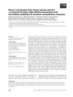
Expression and kinase activity of mutant ICP10 proteinsFigure 1
Expression and kinase activity of mutant ICP10 pro-
teins. (A). Schematic representation of the ICP10 and
mutant proteins. The wild type ICP10 protein expressed by
HSV-2 is a 140 kDa chimera that contains an amino-terminal
PK domain and a carboxy-terminal RR domain. In ∆RR, the
RR domain was replaced with the β-galactosidase gene
(LacZ) which was fused in frame to the PK domain, giving rise
to a 175 kDa protein (p175). In ∆PK, the PK domain of
ICP10 was deleted giving rise to a 95 kDa protein (p95). All
three proteins (ICP10, p175 and p95) retain the transmem-
brane (TM) and extracellular (EC) domains and amino acids
13–26, which are recognized by the ICP10 antibody. (B). SK-
NSH cells infected with HSV-2, ∆RR, ∆PK or PBS (mock-
infected) were collected at 18 hrs after infection and cell
extracts were assayed for protein expression (western) and
ICP10 kinase activity (PK) using immunoblotting and immu-
nocomplex kinase assays with ICP10 antibody.
Genetic Vaccines and Therapy 2008, 6:1 />Page 4 of 14
(page number not for citation purposes)
and visualized as before. To determine expression of
ICP10PK (p175), EOC2, EOC20, and Vero cells were
infected with 5 pfu/cell of ∆RR and the infection was syn-
chronized by adsorption (1 hr) at 4°C followed by culture
shift to 37°C (0 hrs p.i). Live cells that express the p175
protein were identified by staining with the green fluores-
cent β-galactosidase substrate, C
12
-fluorescein di-β-D-
galactopyranoside (C
12
FDG; Molecular Probes) according
to the manufacturer's instructions. Because ICP10PK is
fused in frame with LacZ, C
12
FDG staining reflects
ICP10PK expression [13]. Visualization was done with a
Nikon E4100 fluorescent microscope utilizing FITC (330–
380 nM), UV (for DAPI) (465–495 nM) and Texas Red
(540–580 nM) cubes. Each experiment was done in tripli-
cate and the % staining cells was determined by counting
5 randomly selected fields, (at least 250 cells each, in a 3
mm
2
area) and results are expressed as % positive cells/
total number of cells determined by DAPI staining
[13,14,17,21].
Collection of microglia culture supernatants (CM) and
ELISA
Culture supernatants (conditioned media, CM) were
obtained from infected or mock infected EOC2 and
EOC20 cultures (moi = 5; 48 hrs) and cleared of cell
debris by centrifugation at 14,000 × g for 30 min.
Although they were virus-free by plaque assay, the CM
were exposed to ultraviolet light using a Sylvania G15 T8
bulb at a distance of 17 cm (30 min; room temperature)
in order to insure virus inactivation, as previously
described [18]. They were assayed by ELISA for TNF-α,
RANTES (R&D Systems, Minneapolis, MN) and IL-10
(eBioscience, San Diego, CA), according to manufacturer's
instructions.
CM-mediated neuroprotection in culture
Hippocampal cultures were treated (or not) with NMDA
(50 µM; 3 hrs), extensively washed and grown in a 1:1
mixture of Neurobasal medium supplemented with B27
and CM. CM in which IL-10 was neutralized by incuba-
tion (1 hr; 37°C) with 20 µg/ml of IL-10 antibody (R&D
Systems) were studied in parallel. Neuroprotection was
calculated according to the formula: % neuroprotection =
[NMDA-(CM-B)/NMDA] × 100, where NMDA is the %
caspase3-p20+ cells in cultures given NMDA alone, CM is
the % caspase3-p20+cells in cultures incubated with CM,
and B is the %caspase3-p20+ cells in untreated cultures
(background).
∆
RR vaccination and neuroprotection
Sprague Dawley male rats (8–10 weeks old) were
obtained from Charles River Laboratories (Wilmington,
MA, USA). Animals were housed on a 12 h light/dark
cycle with water and food supplied ad libitum. All proce-
dures were performed in accordance with the University
of Maryland, Baltimore Institutional Animal Care and Use
Committee. They were vaccinated with ∆RR [50 µl (2.5 ×
10
6
pfu)] by intranasal instillation, using ∆PK or PBS as
controls. Delivery was over 15 minutes with 1 min breaks
between instillation into each naris. Three inoculations
were given at 24 hour intervals, with the last instillation
considered day 0 p.i. KA (A.G. Scientific, San Diego, CA)
was administered 24 hrs later (day 1) by i.p. injection. The
route and dose (15 mg/kg) of KA administration were pre-
viously shown to elicit a well-characterized seizure activity
followed by cell loss in the hippocampus [25,26]. Clinical
response was scored as an average behavioral score for
each animal every hour using the previously defined scale:
0, normal; 1, catatonic staring and immobilization; 2,
'wet-dog shakes', abnormal ambulation, stretching of
limbs; 3, rearing and falling behavior; 4, tonic-clonic sei-
zure activity; 5, death [27]. Results are expressed as the
mean behavioral score/hour for each treatment group ±
SEM. In addition, the % animals in each treatment group
that experienced tonic-clonic seizure activity (score = 4)
was recorded for each hour. To asses neuronal cell loss in
the hippocampus, brain sections were fixed with 4% PAF
in PBS (30 min; RT) and stained with thionin (J.T. Baker,
Phillipsburg, NJ, USA) for 30 min. Sections were dehy-
drated and mounted in Permount (Fisher Scientific, Fair
Lawn, NJ, USA). The numbers of neurons were counted in
3 randomly selected CA1 fields of 29 µm
2
(at least 250
cells) from 5 serial sections for all animals and the data are
expressed as % neuronal loss ± SEM relative to untreated
brains.
Statistical analyses
Analysis of variance (ANOVA) was performed with Sigma
Stat version 3.1 for Windows (Systat Software, Point Rich-
mond, CA)
Results
∆
RR and
∆
PK express the mutant ICP10 proteins p175 and
p95 but only p175 has kinase activity
ICP10 is a 140 kDa protein that consists of an amino-ter-
minal domain, which has protein kinase (PK) activity and
a carboxy-terminal domain, which has RR activity. The PK
domain is preceded by a transmembrane (TM) domain
and a short extracellular (EC) domain that retains amino
acids 13–26, which are recognized by the ICP10 antibody
[20]. In ∆RR, the RR domain of ICP10 was replaced with
LacZ, which was fused in frame with ICP10PK, giving rise
to a 175 kDa protein (p175). p175 retains the TM and EC
domains of the wild type ICP10 protein and it is under the
direction of the authentic ICP10 promoter. In ∆PK, the PK
domain of ICP10 was deleted, giving rise to a 95 kDa pro-
tein (p95), which also retains the authentic EC and TM
domains and is driven by the same wild type ICP10 pro-
moter [20] (Fig. 1A).
Genetic Vaccines and Therapy 2008, 6:1 />Page 5 of 14
(page number not for citation purposes)
SK-NSH cells (derived from neuroblastoma) were infected
with ∆RR, ∆PK or HSV-2 and cell extracts obtained at 18
hrs post infection (p.i) were immunoblotted with ICP10
antibody. A 140-kDa protein, consistent with the wild
type ICP10 [20], was seen in HSV-2 infected cells (Fig. 1B,
lane 1). In cells infected with ∆PK, the antibody recog-
nized a 95-kDa protein (p95) (Fig. 1B, lane 2) and in cells
infected with ∆RR, it recognized a 175-kDa protein
(p175) (Fig. 1A, lane 3). Mock-infected cells were negative
(Fig. 1B, lane 4). Immunocomplex PK assays with ICP10
antibody identified a 140-kDa phosphorylated protein
consistent with the autophosphorylated ICP10 in HSV-2
infected cells (Fig. 1B, lane 5). Kinase activity was retained
by p175, which was also autophosphorylated (Fig. 1B,
lane 7). p95 was kinase negative, as evidenced by the
absence of phosphorylated proteins in the ∆PK-infected
cells (Fig. 1B, lane 6). Phosphorylated proteins were not
seen in immunocomplex PK assays of extracts from mock-
infected cells (Fig. 1B, lane 8). The data support previous
conclusions that the PK and RR domains of ICP10 func-
tion independently of each other [20], and confirm that
the p175 protein expressed by ∆RR retains the ICP10
kinase activity.
Microglia are non-permissive for virus growth
In a first series of experiments to examine the effect of ∆RR
on microglia, we asked whether: (i) microglial cells are
permissive for virus growth, and (ii) permissiveness is
affected by prior cell activation. We used EOC2 and
EOC20 cells that differ in the levels of MHCII expression,
with high levels constitutively expressed by EOC20, but
not EOC2 cells [19]. Excessive activation was confirmed
for EOC20 cells by their rounded morphology and high
intensity staining with CD11b antibody (Fig. 2A). EOC2
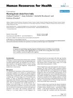
EOC2 and EOC20 Microglia cultures are non-permissive for HSV replicationFigure 2
EOC2 and EOC20 Microglia cultures are non-permissive for HSV replication. (A). EOC2 and EOC20 cells differ in
morphology and the intensity of staining with CD11b antibody before, but not after virus infection. (B). EOC2 and EOC20
cells were infected with ∆RR or ∆PK or HSV-2 (1 × 10
6
pfu) and examined for virus growth by plaque assay as described in
Materials and Methods.
Genetic Vaccines and Therapy 2008, 6:1 />Page 6 of 14
(page number not for citation purposes)
cells had lower CD11b staining intensity and retained
some morphologic ramification. However, high intensity
staining and rounded morphology were seen after virus
infection (Fig. 2A), indicative of virus-induced activation
[10,28]. EOC2 and EOC20 cells were non-permissive for
growth of ∆RR, ∆PK or HSV-2, as determined by plaque
assay. Virus titers decreased at similar rates during the first
4 hrs p.i For HSV-2, the titers remained at this reduced
level until 96 hrs p.i. For ∆RR and ∆PK the titers continued
to decrease until 12 hrs p.i. and remained stable at this
reduced level until 120 hrs p.i. During 4 – 96 hrs p.i., the
titers of ∆RR and ∆PK were approximately 10-fold lower
than those of HSV-2, but virus clearance after 120 hrs was
similar for all viruses, with lowest titers (almost complete
clearance) seen at 14 days p.i. (Fig. 2B). Infectious center
assays done up to 96 hrs p.i., indicated that approximately
90% of the cells formed plaques on Vero cells. Collec-
tively, the data indicate that: (i) microglia are non-permis-
sive for virus growth unrelated to their activation status
prior to infection, and (ii) the clearance of ∆RR and ∆PK
is somewhat more efficient than that of wild type virus.
ICP10PK is expressed in
∆
RR-infected EOC2 and EOC20
cells
ICP10PK expression is regulated with IE kinetics and is
independent of other viral proteins [22-24]. To verify that
it is expressed in ∆RR-infected microglia. EOC2 and
EOC20 cells were infected with 5 pfu/cell of ∆RR and the
infection was synchronized as described in Materials and
Methods. Vero cells, which are routinely used for virus
growth, were studied in parallel as control for the effect of
virus replication on ICP10PK expression. ICP10PK expres-
sion was determined by staining with the Lac-Z substrate
C
12
FDG, as described [13]. In both EOC2 and EOC20 cul-
tures, C
12
FDG staining was seen in most (90–95%) cells
at 2–96 hrs pi. In Vero cells, C
12
FDG staining was seen in
80–97% of the infected cells at 2–24 hrs p.i., but expres-
sion was lost by the end of the replicative cycle (Fig. 3).
The data indicate that ICP10PK expression is sustained in
∆RR-infected microglial cells for a relatively long time,
and it is independent of the cell activation state.
∆
RR does not trigger apoptosis in EOC2 and EOC20 cells
Having seen that ICP10PK is expressed in microglia, we
wanted to know whether it inhibits virus-induced apopto-
sis. EOC2 and EOC20 cells were infected with ∆RR or ∆PK
(moi = 5) or mock-infected with PBS and examined for
apoptosis by TUNEL at 24 hrs p.i. The % TUNEL+ (apop-
totic) cells were minimal in mock-infected EOC2 and
EOC20 cells (9.3 ± 1.5 and 8.7 ± 1.9%, respectively).
Infection with ∆PK caused a significant (p < 0.001)
increase in the % TUNEL+ cells (32.6 ± 5.2 and 21.8 ±
3.3% for EOC20 and EOC2, respectively), but this
increase was not seen in ∆RR-infected cells (13.8 ± 1.9 and
10.2 ± 1.4% for EOC2 or EOC20, respectively) (Fig 4A).
The data indicate that ICP10PK overrides virus-induced
microglial cell apoptosis independent of the state of cell
activation prior to infection.
CM from
∆
RR infected EOC2/EOC20 cells protect
hippocampal neurons from excitotoxin-induced apoptosis
In response to injury and neuronal stress/apoptosis,
microglia in the surrounding area are activated and release
inflammatory cytokines, which perpetuate cell death [29].
However, signals released by apoptotic neurons can also
potentiate the anti-apoptotic activity of microglia [10,30],
suggesting that their neurotoxic activity can be modulated
by the judicious choice of modulating strategies. In gen-
eral, classical pro-inflammatory cytokines (TNF-α and IL-
1β) seem to be neurotoxic, whereas anti-inflammatory
cytokines (IL-10) are neuroprotective [31]. Having seen
that ∆RR inhibits virus-induced apoptosis in infected
microglia, we wanted to know whether it also induces the
production of neuroprotective cytokines. E0C and EOC20
cells were infected with ∆RR or ∆PK (moi = 5) or mock-
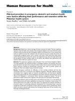
∆RR infected cells express ICP10PKFigure 3
∆RR infected cells express ICP10PK. (A) EOC2 and
EOC20 cells infected with ∆RR (moi = 5) were stained with
the LacZ substrate C
12
FDG at 24 hrs p.i. to visualize
ICP10PK expression (Lac-Z). (B) EOC2, EOC20 and Vero
cells were stained with C
12
FDG and the % staining cells at 4–
96 hrs p.i. was determined by counting 5 randomly selected
fields, (at least 250 cells each, in a 3 mm
2
area). Results are
expressed as % positive cells/total number of cells deter-
mined by DAPI staining. The mean % ICP10PK (Lac-Z) ± SD
are shown.
Genetic Vaccines and Therapy 2008, 6:1 />Page 7 of 14
(page number not for citation purposes)
infected with PBS and culture supernatants (conditioned
media, CM) were collected at 48 hrs p.i. and UV-treated,
as described in Materials and Methods, in order to inacti-
vate any potentially remaining virus that may have
escaped detection.
Primary hippocampal neurons that had been treated (or
not) with NMDA (50 µM; 3 hrs) were extensively washed
and the medium was replaced with a mixture of Neuroba-
sal medium with B27 supplement and CM (1:1 ratio).
Twenty-four hours later, the hippocampal neurons were
assayed for apoptosis by TUNEL. The % TUNEL+ (apop-
totic) cells was significantly increased in NMDA-treated
than untreated hippocampal cultures (p < 0.001) and this
percentage was not reduced by culture with CM from
mock-infected (61 ± 2.7%) or ∆PK-infected EOC2 or
EOC20 cells (45.8 ± 3.3 and 53.3 ± 4.2% respectively).
CM from the ∆RR-infected EOC2 or EOC20 cells caused a
significant (p < 0.001) decrease in the % TUNEL+ cells,
but the decrease was significantly (p < 0.01) better for
EOC20 than EOC2 cells (15.3 ± 2.7 and 25 ± 2.3 %
TUNEL+ cells, respectively) (Fig. 4B). The data indicate
that microglia activation by conditions other than virus
infection, potentiates the ability of ICP10PK to stimulate
neuroprotective modulation.
∆
RR inhibits TNF-a production by microglia
Having seen that CM from ∆RR- (but not ∆PK)-infected
microglia protect hippocampal neurons from NMDA-
induced apoptosis, we wanted to know whether neuro-
protection is associated with decreased production of pro-
inflammatory cytokines. We focused on TNF-α, which is a
known contributor to excitotoxicity-induced neuronal
cell death [5,10]. CM were collected from EOC2 and
EOC20 cells infected with ∆RR or ∆PK (moi = 5) or mock-
infected with PBS, at various times pi and assayed for TNF-
α by ELISA. ∆PK triggered a time-dependent production of
TNF-α in both EOC2 and EOC20 cells, with maximal lev-
els seen at 72 hrs p.i. The levels of TNF-α were significantly
higher for EOC20 than EOC2 cells, reaching approxi-
mately 3-fold higher concentrations at 72 hrs p.i. (315.7 ±
37.1 and 154.5 ± 12.4 pg/ml, respectively). By contrast,
TNF-α was not produced in ∆RR-infected EOC20 cells,
and low level production was seen in EOC2 cells (120.7 ±
12.3 pg/ml at 72 hrs p.i.) (Fig. 5). Collectively, the data
indicate that ∆RR-delivered ICP10PK inhibits TNF-α pro-
duction in virus-infected microglia. Inhibition appears to
depend on the state of cell activation, being somewhat
more potent in EOC20 than EOC2 cells.
∆
RR inhibits RANTES production in infected EOC2 or
EOC20 cells
RANTES/CCL5 is a member of the C-C (β) chemokine
family, which is believed to contribute to the recruitment
of T cells and monocytes from the periphery into the CNS.
ICP10PK inhibits apoptosis in ∆RR-infected microglia and CM from the infected microglia have neuroprotective activityFigure 4
ICP10PK inhibits apoptosis in ∆RR-infected microglia
and CM from the infected microglia have neuropro-
tective activity. (A). EOC2 and EOC20 cells were infected
with ∆RR or ∆PK (moi = 5) or mock infected with PBS, and
assayed for apoptosis by TUNEL at 24 hrs p.i. Each experi-
ment was done in triplicate and the % staining cells was
determined by counting 5 randomly selected fields, (at least
250 cells each, in a 3 mm
2
area). Results are expressed as %
TUNEL+ cells/total number of cells determined by DAPI
staining. The mean TUNEL+ cells ± SD are shown (***p <
0.001 relative to mock). (B). EOC2 and EOC20 cells were
mock infected with PBS or infected with ∆RR or ∆PK (moi =
5) and culture supernatants (CM) were collected at 48 hrs
p.i. and UV-treated as described in Materials and Methods.
Primary hippocampal neurons treated (3 hrs) with NMDA
(50 µM) or PBS, were extensively washed with MEM and re-
incubated with a mixture (1:1) of Neurobasal medium con-
taining B27 and CM from the infected microglia. They were
fixed 24 h later and assayed for cell death by TUNEL. Each
experiment was done in triplicate and the % staining cells was
determined by counting 5 randomly selected fields, (at least
250 cells each, in a 3 mm
2
area). Results are expressed as %
TUNEL+ cells/total number of cells determined by DAPI
staining. The mean TUNEL+ cells ± SD are shown (**p <
0.01).
Genetic Vaccines and Therapy 2008, 6:1 />Page 8 of 14
(page number not for citation purposes)
RANTES is produced by microglia in response to pro-
inflammatory stimuli [32]. Having seen that TNF-α pro-
duction is inhibited in ∆RR-, but not ∆PK-infected EOC20
cells, we wanted to know whether this is also true for
RANTES. Duplicate samples of the CM from the mock- or
virus-infected EOC2 and EOC20 cells were assayed for
RANTES by ELISA. RANTES was produced in both EOC2
and EOC20 cells infected with ∆PK. Its levels were signif-
icantly (2-fold) higher in EOC2 than EOC20 cells (Fig. 6),
suggesting that its regulation is distinct from that of TNF-
α. Significantly, however, RANTES was not seen in CM
from ∆RR-infected EOC2 or EOC20 cells (Fig. 6), indicat-
ing that ICP10PK inhibits its production, independent of
the cell activation state.
IL-10 is produced in
∆
RR-infected EOC2 and EOC20 cells
To examine whether ∆RR infection induces the produc-
tion of neuroprotective factors and verify the effect of the
cell activation state on their production, EOC2 and
EOC20 cells were infected with ∆RR or ∆PK (moi = 5) or
mock-infected with PBS and the CM were assayed for IL-
10 production by ELISA. We focused on IL-10, because: (i)
it is a pleiotropic cytokine with neuroprotective activity
[31], (ii) IL-10 inhibits the transcription and translation
of TNF-α and RANTES in macrophages [33], and (iii)
ICP10PK upregulates IL-10 production in T cells [34].
∆RR induced IL-10 production in both EOC2 and EOC20
cells. The kinetics of IL-10 production appeared to be
somewhat different for the two cell lines, but the maximal
levels at 72 hrs p.i. were similar (Fig. 7). In EOC2 cells, IL-
10 was first seen at 4 hrs p.i and production increased with
time, reaching maximal levels at 48–72 hrs p.i. In EOC20
cells, IL-10 was also first seen at 4 hrs p.i., but production
seemed to reflect a two-phase kinetics, reaching a plateau
at 24–48 hrs p.i. and increasing again, with maximal lev-
els apparently not yet reached at 72 hrs pi. IL-10 was not
seen in CM from ∆PK infected EOC2 or EOC20 cells (Fig.
7), indicating that its production is induced by ICP10PK.
This is consistent with previous reports that IL-10 is not
produced in microglia infected with HSV-1 [35], which
does not conserve a functional ICP10PK [17,36].
IL-10 contributes to the neuroprotective activity of the CM
from
∆
RR-infected EOC2 and E0C20 cells
To examine the effect of IL-10 on the neuroprotective
capacity of the CM from ∆RR-infected EOC2 and EOC20
cells, we asked whether neuroprotection was lost upon IL-
10 neutralization. CM obtained at 48 hrs p.i. were incu-
bated (1 hr; 37°C) with IL-10 neutralizing antibody (20
µg/ml) and examined for: (i) IL-10 levels and (ii) neuro-
ICP10PK induces IL-10 expression in ∆RR-infected microgliaFigure 7
ICP10PK induces IL-10 expression in ∆RR-infected
microglia. EOC2 and EOC20 cells were mock-infected with
PBS, or infected with ∆RR, ∆PK (moi = 5) or mock-infected
with PBS and culture supernatants collected 1–72 hrs p.i.
were assayed for IL-10 by ELISA, as described in Materials
and Methods. Results are the mean of three independent
experiments ± SD. (***p < 0.001 relative to ∆RR-infected).
RANTES production is inhibited in ∆RR-infected microgliaFigure 6
RANTES production is inhibited in ∆RR-infected
microglia. EOC2 and EOC20 cells were mock-infected with
PBS, or infected with ∆RR, ∆PK (moi = 5) or mock-infected
with PBS and culture supernatants collected 1–72 hrs p.i.
were assayed for RANTES by ELISA, as described in Materi-
als and Methods. Results are the mean of three independent
experiments ± SD. (***p < 0.001 relative to ∆RR-infected).
TNF-α production is inhibited in ∆RR-infected microgliaFigure 5
TNF-α production is inhibited in ∆RR-infected micro-
glia. EOC2 and EOC20 cells were mock-infected with PBS,
or infected with ∆RR, ∆PK (moi = 5) or mock-infected with
PBS and culture supernatants collected 1–72 hrs p.i. were
assayed for TNF-α by ELISA, as described in Materials and
Methods. Results are the mean of three independent experi-
ments ± SD. (***p < 0.001 relative to ∆RR-infected).
Genetic Vaccines and Therapy 2008, 6:1 />Page 9 of 14
(page number not for citation purposes)
protective potential in NMDA-treated hippocampal neu-
rons, as determined by double immunofluorescent
staining with antibodies to activated caspase-3 (caspase-
3p20) and β III tubulin. As shown in Fig. 8 for E0C20
cells, the levels of IL-10 were significantly higher in the
CM from ∆RR- than ∆PK- or mock-infected cells (127 ±
3.2, 2.8 ± 2.1 and 2.1 ± 1.5 pg/ml, respectively). IL-10 was
virtually lost by neutralization (8.2 ± 6.9 pg/ml) (Fig. 8A)
but its levels were not reduced by treatment with TNF-α
neutralizing antibody, used as control (data not shown).
The CM from ∆RR, but not ∆PK, infected cells significantly
decreased NMDA-induced caspase-3 activation in hippoc-
ampal cultures. Thus, the % caspase-3p20+ hippocampal
neurons (β III tubulin+) were (p < 0.001) increased by
NMDA, but this increase was not seen in hippocampal
cultures treated with NMDA in the presence of the CM
from ∆RR-infected microglia (55.6% ± 3.0 and 14.7% ±
3.0 for mock and ∆RR, respectively). Protection was not
seen in hippocampal cultures treated with NMDA
together with the ∆PK CM (Fig. 8B,C). Neuroprotection,
calculated as described in Materials and Methods, was
76.9 ± 5.3% for the ∆RR CM and it was reduced to 31.5 ±
7.9% by IL-10 neutralization. Neuroprotection by the
mock- or ∆PK-infected CM was 3.5 ± 5.4 and -5.8 ± 6.8%,
respectively. Similar results were obtained in E0C2 cells.
Thus, while IL-10 contributes to neuroprotection,
ICP10PK induces production of additional, as yet uniden-
tified, neuroprotective factors and is consequently a more
potent therapeutic regimen than IL-10 alone.
∆
RR vaccination prevents KA-induced seizures and
neuronal loss
Systemic KA injection causes epileptiform seizures, which
propagate from the CA3 to the CA1 field and other limbic
structures. These are followed by a pattern of neuronal cell
loss, which is similar to that seen in patients with tempo-
ral lobe epilepsy [37] and is associated with microglia-
related inflammatory responses [38]. We used this animal
model to examine whether vaccination with ∆RR can pre-
vent KA-induced seizures and neuronal loss. Sprague
Dawley rats were given ∆RR, ∆PK or PBS intranasally and
challenged with KA 24 hrs later, as described in Materials
and Methods. Mock or ∆PK treated rats evidenced sus-
tained tonic-clonic seizure activity and an increase in the
associated behavioral symptoms caused by KA adminis-
tration. 75% exhibited tonic-clonic seizure activity
(behavioral scale = 4) at 3 hrs after KA. By contrast, ∆RR-
treated animals did not progress beyond a score of 1–1.5
on the clinical scale. In the ∆RR-treated rats, symptoms
completely resolved at 2 – 3 hrs after KA administration,
as compared to 12 hrs in the ∆PK treated animals. While
the groups averaged a score of 2 on the clinical scale, clin-
ical response was variable, with individual animals show-
ing severe seizures. Tonic-clonic activity was seen in 20%
of the ∆PK treated rats and 40% of the PBS treated rats.
IL-10 contributes to neuroprotection by ∆RR-infected microgliaFigure 8
IL-10 contributes to neuroprotection by ∆RR-
infected microglia. (A). EOC20 cells were mock infected
with PBS or infected with ∆RR or ∆PK (moi = 5) and CM
were collected at 48 hrs p.i. The CM were UV-treated, as
described in Materials and Methods, incubated (1 hr; 37°C)
with IL-10 neutralizing antibody (20 µg/ml) and assayed for
IL-10 by ELISA. (B). Primary hippocampal cultures treated (3
hrs) with NMDA (50 µM) or PBS were extensively washed
with MEM and re-incubated with a mixture (1:1) of Neuroba-
sal medium containing B27 supplement and CM from infected
microglia that had been treated or not with 20 µg/ml of IL-10
neutralizing antibody. They were fixed 24 h later and co-
stained with AlexaFluor-546 conjugated antibody to active
caspase-3 (caspase-3p20) and FITC-conjugated antibody to
β
III
Tubulin (neuronal marker). Each experiment was done in
triplicate and the % staining cells was determined by counting
5 randomly selected fields (at least 250 cells each, in a 3 mm
2
area). Results are expressed as % caspase-3p20+ cells/total
number of cells determined by DAPI staining (C). ***p <
0.001, **p < 0.01 as compared to NMDA + Mock CM + IL-
10 antibody.
Genetic Vaccines and Therapy 2008, 6:1 />Page 10 of 14
(page number not for citation purposes)
Sustained tonic-clonic seizure activity and an increase in
the associated behavioral symptoms were seen with time
post KA administration, with 100% of the rats exhibiting
tonic-clonic seizure activity (behavioral scale = 4) at 3 hrs
after KA. Symptoms began to abate after 5 hours and all
animals were symptom-free by 12 hrs after treatment. By
contrast, ∆RR treated animals never progressed beyond a
score of 1 on the clinical scale, and the symptoms com-
pletely resolved between 2 and 3 hours after KA adminis-
tration. Not one of the ∆RR-treated animals displayed
tonic-clonic seizure activity (Fig. 9A).
Thionin staining (recognizes the Nissl substance in live
neurons) was done on the brains from the PBS- or ∆PK-
treated animals that had experienced seizures with clinical
scores of at least 3 and their ∆RR-treated matched pairs
(clinical scores = 1 or less). Staining cells were counted in
the CA1 hippocampal field, which is the recognized
lesion site [26], as described in Materials and Methods.
Significant neuronal loss (p < 0.001) was seen in the
mock- (54 ± 1.3%) and ∆PK- (51 ± 1.9%) treated animals
at 2 days after treatment with KA, but neuronal loss was
not seen in the ∆RR vaccinated animals (12 ± 3.1%). Rep-
resentative fields are shown in Fig. 9B.
∆RR vaccination protects from KA-induced seizures and neuronal lossFigure 9
∆RR vaccination protects from KA-induced seizures and neuronal loss. (A) Sprague Dawley rats were given 3 intra-
nasal doses of ∆RR or ∆PK (5 × 10
6
pfu) or PBS, and given of KA (15 mg/kg) 24 hrs later by i.p. injection. They were examined
for behavioral changes for 5 hours and rated on a scale of: 0, normal; 1, catatonic staring and immobilization; 2, 'wet-dog
shakes', abnormal ambulation, stretching of limbs; 3, rearing and falling behavior; 4, tonic-clonic seizure activity; 5, death. Aver-
age behavioral score ± SEM is presented for each hour of observation. The % animals in each treatment group experiencing a
behavioral score = 4 at any time during the observation period is shown. (B) Coronal sections of brains collected 2 days later
were stained with thionin. The numbers of neurons were counted in 3 randomly selected fields of 29 µm
2
(at least 250 cells)
from 5 serial sections for all animals and the data are expressed as % neuronal loss ± SEM relative to untreated brains.
Genetic Vaccines and Therapy 2008, 6:1 />Page 11 of 14
(page number not for citation purposes)
∆
RR-mediated neuroprotection is associated with
microglial IL-10 expression
To examine whether ∆RR-mediated neuroprotection is
associated with IL-10 production, duplicate brain sections
were stained in double immunofluorescene with antibod-
ies to IL-10 and CD11b. Replicate sections were stained
with ICP10 or TNFα antibody. ICP10PK and p95 were
respectively expressed in the CA1 fields from ∆RR and
∆PK treated animals. IL-10 staining was only seen in the
CA1 hippocampal fields from rats given KA and ∆RR and
it primarily co-localized with CD11b (Fig. 10). Of the
total CD11b+ cells in the CA1 fields from ∆RR-treated ani-
mals, 60 ± 5% also stained with IL-10 antibody. IL-10
staining was also seen in 16 ± 4% CD11b- cells, indicating
that IL-10 is also produced by other cells, potentially neu-
rons [39]. This compares to 5 ± 2% and 2 ± 1% IL-10+/
CD11b+ (and no IL-10+/CD11b- cells) in the CA1 fields
from rats given KA and respectively treated with ∆PK or
PBS. Consistent with our results for cultured cells, TNF-α
staining was barely detectable in the brains from the ∆RR
treated animals (5 ± 3% TNF-α + cells), while staining was
seen in the CA1 fields from animals given ∆PK (35 ± 5%)
or PBS (40 ± 6%) (Fig. 10). The data indicate that
ICP10PK-mediated neuroprotection is associated with
microglial IL-10 production and TNF-α inhibition, as well
as the production of additional, as yet unidentified, neu-
roprotective factors.
Discussion
Microglia are considered the CNS resident professional
macrophages. They function as the principal immune
effector cells of the CNS, responding to any pathological
event. Their excessive activation and the dysregulated
overproduction of inflammatory cytokines are the hall-
mark of many neurodegenerative diseases and ischemic
brain injury, emphasizing the importance of the neuro-
nal-microglial axis [4-6,10,11]. Most of the available liter-
ature indicates that the pro-inflammatory cytokine TNF-α
released by microglia activated in response to excitotoxic
injury, contributes to neuronal degeneration
[5,6,10,11,38,40,41]. However, microglia also produce
neuroprotective factors [31], suggesting that appropriate
modulation of the microglial-neuronal axis through inhi-
bition of pro-inflammatory cytokine production and the
induction of neuroprotective factors is a desirable thera-
peutic approach. However, identification of the target
∆RR vaccination is associated with IL-10 production by microglia and inhibition of TNF-αFigure 10
∆RR vaccination is associated with IL-10 production by microglia and inhibition of TNF-α. Sprague Dawley rats
were mock treated with PBS or treated with ∆RR or ∆PK [50 µl (2.5 × 10
6
pfu)] by intranasal delivery as described in Materials
and Methods. They were given KA by i.p. injection (24 hrs later) and the brains were collected 2 days later. Serial sections
were stained with FITC-labeled ICP10 antibody, Texas Red-labeled IL-10 + FITC-labeled CD11b antibodies, or Texas Red-
labeled TNF-α antibody. Blue staining is DAPI. The numbers of staining cells were counted in 3 randomly selected fields of 29
µm
2
(at least 250 cells) from 5 serial sections for all animals and the data are expressed as % staining cells ± SEM relative to
total DAPI stained cells.
Genetic Vaccines and Therapy 2008, 6:1 />Page 12 of 14
(page number not for citation purposes)
required for such microglial cell modulation and its rela-
tionship to neuronal life/death decisions, is a major clin-
ical challenge. The salient feature of the data presented in
this report is that in addition to its ability to induce sur-
vival pathways in neurons [13,17,42], ICP10PK modu-
lates microglial responses in favor of neuroprotection, by
inducing neuroprotective factors and inhibiting the pro-
duction of inflammatory (neurotoxic) cytokines. The fol-
lowing comments seem pertinent with respect to these
findings.
The construction and properties of the growth-compro-
mised ICP10PK vector ∆RR and its ICP10PK-deleted con-
trol ∆PK, were previously described. ∆RR retains the
ICP10PK gene that has anti-apoptotic activity in neurons
through activation of redundant survival pathways,
including MEK/ERK, PI3-K/Akt and AC/PKA [13,17,42].
∆PK is a particularly stringent control for ∆RR because: (i)
both viruses were constructed from the same HSV-2 strain
and are growth-compromised in the CNS, (ii) the two
viruses have no genetic differences other than the mutated
ICP10 protein, as evidenced by the study of revertant
viruses, (iii) the PK-deleted ICP10 protein p95 is driven by
the same authentic ICP10 promoter as the mutant protein
in ∆RR (p175) and both are expressed in the absence of
virus replication [20], and (iv) although p175 and p95 are
expressed equally well, only p175 retains kinase activity
(Fig. 1). To control for the possible contribution of inde-
pendent microglial cell activation (notably by excitotoxic
injury) to the ∆RR neuroprotective potential, we used two
cell lines (EOC2 and EOC20) that differ in their activation
state. EOC20 cells constitutively express high levels of
MHCII [19] and display a rounded morphology and high
levels of CD11b expression before infection. This is not
the case for EOC2 cells. Both cell lines were non-permis-
sive for virus growth, but p175 and p95 (respectively
encoded by ∆RR and ∆PK) were expressed, consistent with
the IE regulation of the ICP10 promoter [22,23]. ICP10PK
had anti-apoptotic activity, also in virus-infected micro-
glia, and similar results were obtained in EOC2 and
EOC20 cells, indicating that these properties were not
affected by independent microglial cell activation.
Significantly, ICP10PK modulats the neuronal-microglial
crosss-talk in favor of neuroprotection, as evidenced by
the finding that CM from ∆RR-infected EOC2 and EOC20
cells protected hippocampal neurons (β
III
Tubulin+) from
NMDA-induced apoptosis (determined by TUNEL and
caspase-3 activation). We conclude that neuroprotection
was through ICP10PK, because apoptosis was not inhib-
ited by the CM from the ∆PK-infected EOC2 and EOC20
cells. We focused on the contribution of IL-10, because it
is a pleiotropic cytokine with a strong suppressive effect
on the production of pro-inflammatory cytokines by mac-
rophages and dendritic cells [33,43], it is induced by
ICP10PK in T cells from popliteal lymph nodes of virus-
infected animals [34] and it has neuroprotective activity
in glutamate-induced cell death or hypoxic ischemia [31].
IL-10 contributed to the ∆RR-mediated neuroprotection,
as evidenced by the findings that: (i) CM from ∆RR, but
not ∆PK-infected EOC2 and EOC20 cells contained rela-
tively high levels of IL-10, and (ii) the % β
III
Tubulin+ cells
(neurons) that expressed the caspase-3 cleavage product
(caspase-3p20) was significantly increased by IL-10 neu-
tralization. The effect of the IL-10 antibody was specific as
evidenced by the failure to neutralize IL-10 with TNF-α,
antibody (data not shown), and the failure of the IL-10
antibody to reduce the % caspase-3p20+ cells in hippoc-
ampal cultures grown with CM from mock- or ∆PK-
infected microglia. However, ∆RR also induced additional
neuroprotective factors, as evidenced by the finding that
IL-10 neutralization did not abrogate neuroprotection
and it inhibited the production of the pro-inflammatroy
cytokine TNF-α and the chemokine RANTES, presumably
contributing to neuroprotection through inhibition of
inflammation and the recruitment of inflammatory cells
to the CNS. The multiplicity of microglial effects induced
by ICP10PK causes it to be a highly superior therapeutic
when compared to the single use of neuroprotective
cytokines, such as IL-10.
Systemic KA injection causes epileptiform seizures which
propagate from the CA3 to the CA1 field and other limbic
structures, and are followed by a pattern of neuronal cell
loss which is similar to that seen in patients with temporal
lobe epilepsy [37]. We used this model to examine the
ability of ∆RR to prevent neurodegeneration, because epi-
lepsy is a chronic disease in which periodic therapeutic
dosing could prevent recurrent seizure episodes. We chose
the non-invasive intranasal delivery route, because HSV
gains access to the temporal lobes by the olfactory route,
presumably by axonal transport [44-46], and we have pre-
viously shown that ICP10PK gains rapid (2 days) access to
the hippocampus after ∆RR intranasal delivery, appar-
ently through the lateral olfactory bulb tract [27]. How-
ever, our data did not exclude extracellular diffusion along
the open intercellular clefts in the olfactory epithelium
with subsequent diffusion to the olfactory bulb and CSF
circulation, bypassing the blood-brain barrier [47]. Virus
titers could also be minimally amplified through one
round of replication in the nasal epithelial cells or other
non-neuronal support cells, also causing infection of the
microglia. We found that ∆RR prevented KA-induced sei-
zures and neuronal loss in the hippocampal CA1 fields,
which were associated with IL-10 production by microglia
from these fields, as well as TNF-α inhibition. Unresolved
questions are the mechanisms whereby: (i) ICP10PK
induces IL-10 production, (ii) IL-10 protects neurons
from apoptosis induced by excitotoxic injury, and (iii)
ICP10PK prevents virus-induced apoptosis in microglia.
Genetic Vaccines and Therapy 2008, 6:1 />Page 13 of 14
(page number not for citation purposes)
Ongoing studies are designed to examine the mechanism
whereby ICP10PK modulates the microglial responses. IL-
10 upregulation could be related to the ability of ICP10PK
to activate transcription factors, notably AP-1
[13,14,17,18,20,22,27,48] or NF-kB, and different factors
could be involved in the apparent two-phase kinetics of
IL-10 production in excessively activated microglial cells,
such as EOC20. Similarly, ICP10PK could inhibit TNF-α
and RANTES production at the transcriptional level, or
inhibition could be mediated by the generation of IL-10,
which is known to suppress their production in macro-
phages [33,49]. HSV-1, which does not retain a functional
ICP10PK [17,36], does not induce IL-10 production in
microglia, while triggering a vigorous cascade of pro-
inflammatory responses that failed to protect susceptible
mice from HSV-1-induced brain lesions [35]. HSV-1
induced TNF-α production was inhibited by exogenously
supplied IL-10 [50].
Previous studies had shown that IL-10 can counteract the
effect of endotoxin on cerebral metabolism in the perina-
tal brain [51]. However, instead of delivering IL-10 to the
brain extracellular space, we can directly activate its effect
as well as that of additional neuroprotective factors by
using ∆RR. ∆RR has the additional therapeutic advantage
that it inhibits the production of pro-inflammatory
cytokines/chemokines and also functions directly in neu-
rons, where it activates neuronal survival pathways that
override apoptotic cascades [13-15,17,18,21,42]. Collec-
tively, these functions identify ∆RR as a most promising
genetic vaccine/therapy platform for neurodegenerative
diseases.
Competing interests
The author(s) declare that they have no competing inter-
ests.
Authors' contributions
JML carried out the experiments, participated in the
design of the study and the drafting of this manuscript. LA
designed the vectors, conceived the experiments and
drafted the manuscript. Both authors read and approved
the final manuscript.
Acknowledgements
These studies were supported by Public Health Service grant NS45169.
J.M.L. is supported by the NIEHS, NIH Training Grant, ES07263.
References
1. American Heart Association: Heart Disease and Stroke Statis-
tics - 2005 Update. .
2. Hargis ER: Targeting Epilepsy One of the Nation’s Most Com-
mon Disabling Neurological Conditions. At A Glance 2007,
National Center for Chronic Disease Prevention and Health
Promotion:1-4.
3. Thornberry NA, Lazebnik Y: Caspases: Enemies Within. Science
1998, 281(5381):1312-1316.
4. Carson MJ, Doose JM, Melchior B, Schmid CD, Ploix CC: CNS
immune privilege: hiding in plain sight. Immunol Rev 2006,
213:48-65.
5. Minghetti L, Ajmone-Cat MA, De Berardinis MA, De Simone R:
Microglial activation in chronic neurodegenerative diseases:
roles of apoptotic neurons and chronic stimulation. Brain
Research Reviews 2005, 48(2):251-256.
6. Viviani B, Bartesaghi S, Corsini E, Galli CL, Marinovich M: Cytokines
role in neurodegenerative events. Toxicology Letters 2004,
149(1-3):85-89.
7. Harvey BK, Chang CF, Chiang YH, Bowers WJ, Morales M, Hoffer BJ,
Wang Y, Federoff HJ: HSV amplicon delivery of glial cell line-
derived neurotrophic factor is neuroprotective against
ischemic injury. Experimental Neurology 2003, 183(1):47-55.
8. Spencer B, Agarwala S, Gentry L, Brandt CR: HSV-1 vector-deliv-
ered FGF2 to the retina is neuroprotective but does not pre-
serve functional responses. Mol Ther 2001, 3(5 pt1):746-756.
9. Dumas TC, McLaughlin JR, Ho DY, Lawrence MS, Sapolsky RM: Gene
Therapies That Enhance Hippocampal Neuron Survival
after an Excitotoxic Insult Are Not Equivalent in Their Abil-
ity to Maintain Synaptic Transmission. Experimental Neurology
2000, 166(1):180-189.
10. Rock RB, Gekker G, Hu S, Sheng WS, Cheeran M, Lokensgard JR,
Peterson PK: Role of microglia in central nervous system
infections. Clin Microbiol Rev 2004, 17(4):942-964.
11. Streit WJ: Microglial senescence: does the brain's immune sys-
tem have an expiration date? Trends in Neurosciences 2006,
29(9):506-510.
12. Aurelian L: HSV-Induced Apoptosis in Herpes Encephalitis.
Curr Top Microbiol Immunol 2005, 289:79-1122.
13. Gober MD, Laing JM, Thompson SM, Aurelian L: The growth com-
promised HSV-2 mutant DeltaRR prevents kainic acid-
induced apoptosis and loss of function in organotypic hippoc-
ampal cultures. Brain Res 2006, 1119(1):26-39.
14. Golembewski EK, Wales SQ, Aurelian L, Yarowsky PJ: The HSV-2
protein ICP10PK prevents neuronal apoptosis and loss of
function in an in vivo model of neurodegeneration associated
with glutamate excitotoxicity. Experimental Neurology 2007,
203(2):381-393.
15. Laing JM, Golembewski EK, Wales SQ, Liu J, Jafri MS, Yarowsky PJ,
Aurelian L: Growth-compromised HSV2 vector [delta]RR
protects from N-methyl-D-Aspartate- induced neuronal
degeneration through redundant activation of the MEK/ERK
and PI3K/ Akt survival pathways, either one of which over-
rides apoptotic cascades. J Neuro Res 2007 in press.
16. Perkins D, Gyure KA, Pereira EFR, Aurelian L: Herpes Simplex
Virus Type 1-Induced Encephalitis has an Apoptotic Compo-
nent Associated with Activation of c-Jun N-Terminal Kinase.
J Neurovirol 2003, 9(1):101-111.
17. Perkins D, Pereira EFR, Aurelian L: The Herpes Simplex Virus
Type 2 R1 Protein Kinase (ICP10 PK) Functions as a Domi-
nant Regulator of Apoptosis in Hippocampal Neurons
Involving Activation of the ERK Survival Pathway and
Upregulation of the Antiapoptotic Protein Bag-1. J Virol 2003,
77(2):1292-1305.
18. Perkins D, Pereira EFR, Gober M, Yarowsky PJ, Aurelian L: The Her-
pes Simplex Virus Type 2 R1 Protein Kinase (ICP10 PK)
Blocks Apoptosis in Hippocampal Neurons, Involving Activa-
tion of the MEK/MAPK Survival Pathway. J Virol 2002,
76(3):1435-1449.
19. Walker WS, Gatewood J, Olivas E, Askew D, Havenith CE: Mouse
microglial cell lines differing in constitutive and interferon-
gamma-inducible antigen-presenting activities for naive and
memory CD4+ and CD8+ T cells. J Neuroimmunol 1995,
63(2):163-174.
20. Smith CC, Peng T, Kulka M, Aurelian L: The PK Domain of the
Large Subunit of Herpes Simplex Virus Type
2 Ribonucleotide Reductase (ICP10) Is Required for Imme-
diate-Early Gene Expression and Virus Growth. J Virol 1998,
72(11):9131-9141.
21. Perkins D, Yu Y, Bambrick LL, Yarowsky PJ, Aurelian L: Expression
of herpes simplex virus type 2 protein ICP10 PK rescues neu-
rons from apoptosis due to serum deprivation or genetic
defects. Exp Neurol 2002, 174(1):118-122.
22. Gober MD, Wales SQ, Hunter JC, Sharma BK, Aurelian L: Stress up-
regulates neuronal expression of the herpes simplex virus
Publish with BioMed Central and every
scientist can read your work free of charge
"BioMed Central will be the most significant development for
disseminating the results of biomedical research in our lifetime."
Sir Paul Nurse, Cancer Research UK
Your research papers will be:
available free of charge to the entire biomedical community
peer reviewed and published immediately upon acceptance
cited in PubMed and archived on PubMed Central
yours — you keep the copyright
Submit your manuscript here:
/>BioMedcentral
Genetic Vaccines and Therapy 2008, 6:1 />Page 14 of 14
(page number not for citation purposes)
type 2 large subunit of ribonucleotide reductase (R1; ICP10)
by activating activator protein 1. Journal of NeuroVirology 2005,
11(4):329 -3336.
23. Wymer JP, Chung TD, Chang YN, Hayward GS, Aurelian L: Identifi-
cation of immediate-early-type cis-response elements in the
promoter for the ribonucleotide reductase large subunit
from herpes simplex virus type 2. J Virol 1989, 63(6):2773-2784.
24. Zhu J, Aurelian L: AP-1 cis-response elements are involved in
basal expression and Vmw110 transactivation of the large
subunit of herpes simplex virus type 2 ribonucleotide reduct-
ase (ICP10). Virology 1997, 231(2):301-312.
25. Kalwy SA, Akbar MT, Coffin RS, de Belleroche J, Latchman DS: Heat
shock protein 27 delivered via a herpes simplex virus vector
can protect neurons of the hippocampus against kainic-acid-
induced cell loss. Brain Res Mol Brain Res 2003, 111:91-103.
26. Sperk G, Lassmann H, Baran H, Kish SJ, Seitelberger F, Hornykiewicz
O: Kainic acid induced seizures: Neurochemical and his-
topathological changes. Neuroscience 1983, 10(4):1301-1315.
27. Laing JM, Gober MD, Golembewski EK, Thompson SM, Gyure KA,
Yarowsky PJ, Aurelian L: Intranasal administration of the
growth-compromised HSV-2 vector DeltaRR prevents kain-
ate-induced seizures and neuronal loss in rats and mice. Mol
Ther 2006, 13(5):870-881.
28. Kettenmann H: Triggering the brain's pathology sensor. Nat
Neurosci 2006, 9(12):1463-1464.
29. Kim YS, Kim SS, Cho JJ, Choi DH, Hwang O, Shin DH, Chun HS, Beal
MF, Joh TH: Matrix metalloproteinase-3: a novel signaling pro-
teinase from apoptotic neuronal cells that activates micro-
glia. J Neurosci 2005, 25:3701-3711.
30. Polazzi E, Gianni T, Contestabile A: Microglial cells protect cere-
bellar granule neurons from apoptosis: evidence for recipro-
cal signaling. Glia 2001, 36:271-280.
31. Bachis A, Colangelo AM, Vicini S, Doe PP, De Bernardi MA, Brooker
G, Mocchetti I: Interleukin-10 Prevents Glutamate-Mediated
Cerebellar Granule Cell Death by Blocking Caspase-3-Like
Activity. J Neurosci 2001, 21(9):3104-3112.
32. Ransohoff RM, Wei T, Pavelko KD, Lee JC, Murray PD, Rodriguez M:
Chemokine expression in the central nervous system of
mice with a viral disease resembling multiple sclerosis: roles
of CD4+ and CD8+ T cells and viral persistence. J Virol 2000,
76(5):2217-2224.
33. Denys A, Udalova IA, Smith C, Williams LM, Ciesielski CJ, Campbell
J, Andrews C, Kwaitkowski D, Foxwell BMJ: Evidence for a Dual
Mechanism for IL-10 Suppression of TNF-{alpha} Production
That Does Not Involve Inhibition of p38 Mitogen-Activated
Protein Kinase or NF-{kappa}B in Primary Human Macro-
phages. J Immunol 2002, 168(10):4837-4845.
34. Gyotoku T, Ono F, Aurelian L: Development of HSV-specific
CD4+ Th1 responses and CD8+ cytotoxic T lymphocytes
with activity by vaccination with the HSV-2 mutant
ICP10DPK. Vaccine 2002, 20:2796-2807.
35. Lokensgard JR, Hu S, Sheng W, vanOijen M, Cox D, Cheeran MC,
Peterson PK: Robust expression of TNF-alpha, IL-1beta,
RANTES, and IP-10 by human microglial cells during non-
productive infection with herpes simplex virus. J Neurovirol
2001, 7(3):208-219.
36. Nikas I, McLauchlan J, Davison AJ, Taylor WR, Clements JB: Struc-
tural features of ribonucleotide reductase. Proteins 1986,
1(4):376-384.
37. Ben-Ari Y, Cossart R: Kainate, a double agent that generates
seizures: two decades of progress. Trends Neurosci 2000,
22:580-587.
38. Wang Q, Yu S, Simonyi A, Sun GY, Sun AY: Kainic acid-mediated
excitotoxicity as a model for neurodegeneration. J Neurobiol
2005, 31:3-16.
39. Qian L, Block ML, Wei SJ, Lin CF, Reece J, Pang H, Wilson B, Hong JS,
Flood PM: Interleukin-10 protects lipopolysaccharide-induced
neurotoxicity in primary midbrain cultures by inhibiting the
function of NADPH oxidase. J Pharmacol Exp Ther 2006,
319(1):44-52.
40. Guo Z, Iyun T, Fu W, Zhang P, Mattson MP: Bone marrow trans-
plantation reveals roles for brain macrophage/microglia
TNF signaling and nitric oxide production in excitotoxic neu-
ronal death. Neuromolecular Med 2004, 5(3):219-234.
41. Ranaivo HR, Craft JM, Hu W, Guo L, Wing LK, Van Eldik LJ, Watter-
son DM: Glia as a Therapeutic Target: Selective Suppression
of Human Amyloid-beta-Induced Upregulation of Brain
Proinflammatory Cytokine Production Attenuates Neuro-
degeneration. J Neurosci 2006, 26(2):662-670.
42. Wales SQ, Li B, Laing JM, Aurelian L: The herpes simplex virus
type 2 gene ICP10PK protects from apoptosis caused by
nerve growth deprevation through inhibtion of caspase-3
activation and XIAP-upregulation. J Neurochem 2007:Epub 30
June 07.
43. Fiorentino DF, Zlotnik A, Mosmann TR, Howard M, O'Garra A: IL-
10 inhibits cytokine production by activated macrophages. J
Immunol 1991, 147(11):3815-3822.
44. Kandel ER: Learning and memory. In Principles of Neural Science,
4th ed Edited by: Kandel ER, Schwartz JH, Jessell TM. New York ,
McGraw-Hill; 2000:1227-1246.
45. Sewards TV, Sewards MA: Input and output stations of the
entorhinal cortex: superficial vs. deep layers or lateral vs.
medial divisions? Brain Res Brain Res Rev 2003, 42:243-251.
46. Tomlinson AH, Esiri MM: Herpes simplex encephalitis. Immu-
nolohistological demonstration of spread of virus via olfac-
tory plathways in mice. J Neurol Sci 1983, 60(3):473-484.
47. Thorne RG, Pronk GJ, Padmanabhan V, Frey WH: Delivery of insu-
lin-like growth factor-I to the rat brain and spinal cord along
olfactory and trigeminal pathways following intranasal
administration. Neurosci 2004, 127:481-496.
48. Smith CC, Nelson J, Aurelian L, Gober MD, Goswami BB: Ras-GAP
Binding and Phosphorylation by Herpes Simplex Virus Type
2 RR1 PK (ICP10) and Activation of the Ras/MEK/MAPK
Mitogenic Pathway Are Required for Timely Onset of Virus
Growth. J Virol 2000, 74(22):10417-10429.
49. Hu S, Chao CC, Ehrlich LC, Sheng WS, Sutton RL, Rockswold GL,
Peterson PK: Inhibition of microglial cell RANTES production
by IL-10 and TGF-beta. J Leukoc Biol 1999, 65(6):815-821.
50. Marques C, Shuxian H, Wen S, Maxim CJ, Cheeran DC, Lokensgard
JR: Interleukin-10 attenuates production of HSV-induced
inflammatory mediators by human microglia. Glia 2004,
47(4):358-366.
51. Kremlev SG, Palmer C: Interleukin-10 inhibits endotoxin-
induced pro-inflammatory cytokines in microglial cell cul-
tures. Journal of Neuroimmunology 2005, 162(1-2):71-80.