Báo cáo sinh học: "Therapeutic dendritic cell vaccine preparation using tumor RNA transfection: A promising approach for the treatment of prostate cancer" pps
Bạn đang xem bản rút gọn của tài liệu. Xem và tải ngay bản đầy đủ của tài liệu tại đây (366.3 KB, 7 trang )
BioMed Central
Page 1 of 7
(page number not for citation purposes)
Genetic Vaccines and Therapy
Open Access
Research
Therapeutic dendritic cell vaccine preparation using tumor RNA
transfection: A promising approach for the treatment of prostate
cancer
Juliana M Sousa-Canavez*
1
, Flavio C Canavez
1
, Kátia RM Leite
1,2
and
Luiz H Camara-Lopes
1
Address:
1
Oncocell Division, Genoa Biotechnology SA, Alameda Ministro Rocha Azevedo, 346, 1st floor, 01410-000, São Paulo, SP, Brazil and
2
Laboratório de Investigação Médica da Disciplina de Urologia da Faculdade de Medicina de USP-LIM 55, São Paulo, SP, Brazil
Email: Juliana M Sousa-Canavez* - ; Flavio C Canavez - ;
Kátia RM Leite - ; Luiz H Camara-Lopes -
* Corresponding author
Abstract
Background: Early prostate adenocarcinoma can be diagnosed through seric prostate-specific
antigen (PSA) screenings. However, a fraction of patients progress to an incurable metastatic
disease. Therefore, novel therapies for treating these patients are extremely desirable. Therapeutic
vaccines based on Dendritic Cells (DCs) carrying tumor antigens have emerged as a promising
strategy to initiate an immune response against tumor cells. These vaccines can be prepared using
different methodologies, such as the application of tumor mRNA described in this work.
Methods: Mature and immature DCs were obtained in vitro by adding specific cytokines to
monocyte cell cultures. RNA extracted from prostate tumor lineage (LNCAP) was introduced into
these cells by electroporation and co-incubation. Transfection success was measured by
immunocytochemistry of the PSA expression level in DCs.
Results: Cell surface markers, including CD14, CD80, CD86, CCR7, CD11c, and CD1a,
confirmed mature and immature DC phenotypes. Both cell maturation stages were successfully
used for RNA introduction as shown by PSA characterization.
Conclusion: Our data support the use of mature and immature DCs for vaccine preparation with
either RNA electroporation or RNA co-incubation. The highest efficiency, however, was observed
when RNA was delivered by electroporation into mature DCs. Due to in vitro RNA transcription,
this method allows small tumors to be used for DC vaccine preparation; it is therefore a promising
approach for the treatment of metastatic prostate cancer.
Background
Prostate adenocarcinoma is the most common malig-
nancy diagnosed in males and the second most common
cause of cancer death. In its latest report, the Brazilian
National Cancer Institute (INCA) estimated that 47,280
new cases of prostate cancer, or 51 new prostate cancer
cases for each 100,000 men, arise in Brazil each year [1].
Because of their population screenings using prostate-spe-
Published: 18 January 2008
Genetic Vaccines and Therapy 2008, 6:2 doi:10.1186/1479-0556-6-2
Received: 22 October 2007
Accepted: 18 January 2008
This article is available from: />© 2008 Sousa-Canavez et al; licensee BioMed Central Ltd.
This is an Open Access article distributed under the terms of the Creative Commons Attribution License ( />),
which permits unrestricted use, distribution, and reproduction in any medium, provided the original work is properly cited.
Genetic Vaccines and Therapy 2008, 6:2 />Page 2 of 7
(page number not for citation purposes)
cific antigens (PSA), the south and southeastern regions of
Brazil are expected to exhibit the highest incidence.
Seric PSA level has aided in the diagnosis of very small
prostate tumors. This, in association with radical prostate-
ctomy and radiation therapies, has contributed to increas-
ing the curative indexes [2]. After several years of primary
therapy, however, PSA levels can rise even in patients with
good outcomes predicted by tumor histological parame-
ters. Roughly one-third of these patients progress to incur-
able metastatic disease, for which there are few treatment
options. The chemotherapy currently used has limited
efficacy [3,4]. This lack of treatment has led scientists
around the world to search for new therapeutic options
[5]; one of these options involves the use of Dendritic Cell
(DCs) immunotherapy.
Clinical trails based on DCs have been conducted for the
treatment of patients with a variety of tumor types [6-8].
Two of these trials were performed by our group, which
developed individualized therapeutic vaccines by fusing
allogeneic DCs and neoplastic cells isolated from surgi-
cally-removed tumors. We have observed only minor side
effects but significant clinical benefits like disease stability
in 80% of patients after 2–3 doses of vaccine. The mean
survival rate was 13 months for melanoma patients and 6
months for renal cell carcinoma patients [9].
DC immunotherapy based on cell fusion depends on
large size tumors (>1 gram) and cannot be applied in
most cases of prostate cancer. This barrier does not pre-
vent its use, since different methods of vaccine prepara-
tion have been described [6-8]. For prostate cancer, DCs
are most commonly primed with entire or partial tissue-
specific antigens or tumor-associated antigens. Alterna-
tively, mRNA molecules can be transfected into DCs so
that entire proteins will be translated, processed, and pre-
sented by MHC on the cell surface. Both methods have
already been used in clinical trials and were able to initiate
immune response; for a review, see [10].
Here, we compare two methods, electroporation and co-
incubation, for transfecting RNA into DCs. The amount of
RNA and the DC maturation stage for transfection were
also analyzed. The prostate tumor cell line (LNCaP) was
chosen for this study because it overexpresses PSA and
permits easy DC characterization via immunocytochemis-
try using anti-PSA antibodies.
Methods
Reagents
Culture medium consisted of AIM-V or RPMI-1640 sup-
plemented with fetal bovine serum (FBS), streptomycin,
and penicillin (all from GIBCO, Rockville, MD, USA).
Recombinant human cytokines like granulocyte macro-
phage-colony stimulating factor (GM-CSF), interleukin 4
(IL-4), and tumor necrosis factor-α (TNF-α) were pur-
chased from Peprotech Inc. (Rocky Hill, NJ, USA). Ficoll-
Hypaque used in cell separation was purchased from
Amersham (Piscataway, NJ, USA).
Dendritic cell culture
As described in Barbuto et al. [9], peripheral blood mono-
nuclear cells (PBMC) were collected from informed, con-
senting, healthy donors through apheresis performed in a
Cobe Spectra Blood Cell Separator 7.0 (Cobe, Lakewood,
CO, USA) that had been programmed for mononuclear
cell collection. Acid citrate dextrose was used as a blood
anticoagulant (ratio of 1:8–1:11). Mononuclear cells were
separated by density gradient centrifugation (Ficoll-Paque
1,077 g/dl). After three washes with RPMI 1640 medium,
mononuclear cells were resuspended in AIM-V at a density
of 1.3 × 10
7
cells/mL and allowed to adhere to culture
flasks for 2 h at 37°C in a humidified incubator. Floating
cells were gently removed, and AIM-V containing GM-CSF
(50 ng/mL) and IL-4 (50 ng/mL) were added. Flasks were
maintained at 37°C in a 5% CO2 humidified incubator
for 5 days. For immature DC recovery, cultured cells were
harvested on the 5
th
day. For mature DCs, TNF-α (50 ng/
mL) was added to the medium on the 5
th
day and cultured
cells were harvested on the 7
th
day.
Cell surface phenotype by flow-cytometric analysis
Determination of phenotype was performed by two-color
immunostaining using combinations of FITC- and PE-
labeled mAbs directed to human CD14, CD80, CD86,
CCR7, CD11c, and CD1a (all purchased from BD-
Pharmingen, CA, USA). Corresponding isotype-matched
mAbs were used as controls. Cells (1 × 10
6
) were resus-
pended in PBS containing bovine serum albumin 0.1%
and then incubated for 20 min at 4°C with optimal con-
centrations of monoclonal antibodies. Membrane mark-
ers were determined by flow cytometry (FACScalibur,
Becton Dickinson Immunocytometry Systems, CA, USA),
and data from 10,000 events were analyzed by Cell Quest
Pro software (Becton Dickinson Immunocytometry Sys-
tems). Results are expressed as the percentage of positive
cells.
Tumor cell culture
The LNCaP cells were obtained from American Type Cul-
ture Collection (Rockville, MD) and cultivated in RPMI
1640 medium supplemented with 10% FBS, streptomycin
100 mg/mL, and penicillin 100 U/mL at 37°C in a 5%
CO
2
humidified incubator.
Tumor RNA preparation
LNCaP cells cultivated in RPMI-1640 with 10% FBS were
harvested and maintained at -80°C until RNA extraction.
The RNA was extracted with Trizol (Invitrogen, Carlsbad,
Genetic Vaccines and Therapy 2008, 6:2 />Page 3 of 7
(page number not for citation purposes)
USA) following the supplier's recommendations. RNA
concentration and purity were estimated in a 260/280 nm
spectrophotometer. RNA integrity was verified using an
Agilent 2100 Bioanalyzer (Agilent technologies, CA,
USA).
Dendritic cell transfection with LNCaP tumor cell RNA
Boczkowski et al. [11] suggested that RNA introduction
alone activates DCs and causes them to mature. Based on
this knowledge, we tested mature and immature DC trans-
fection. Suspensions of DCs (4 × 10
6
cells/mL) were har-
vested at the 5
th
(immature cells) and 7
th
(mature DC) day
of culture after TNF-α addition. Different conditions of
transfection were used. For mature DCs, 4 µg and 5 µg
RNA were electroporated with 300, 400, and 500 V pulses;
additionally, we incubated cells with 5 µg RNA. For
immature DCs, 4 µg and 16 µg were used for transfection
with 300 and 400 V pulses. The electroporation was per-
formed in a 4-mm curvette at 25 µF capacitance.
Kinetics of tumor antigen expression
The expression of the tumoral antigen PSA was examined
24 h after transfection via immunocytochemistry with
antibodies to PSA and the androgen receptor (Dako, Glos-
trup, Denmark). Briefly, DCs were recovered from culture
plates using a cell scraper. Cells were fixed in 70% alcohol
and submitted to 900 g centrifugation for 5 min at room
temperature. The cytocentrifugate were impressed on
adhesive coated slides and incubated overnight at 4°C
with monoclonal antibodies to PSA or the androgen
receptor at a dilution of 1:50 in PBS. Then, biotinylated
anti-mouse immunoglobulin G was applied at a 1:200
dilution for 60 minutes at room temperature. Slides were
rinsed with PBS for 30 minutes, incubated with peroxi-
dase-conjugated streptavidin (streptABC Kit, Dako) at a
1:400 dilution in PBS for 45 minutes at room tempera-
ture, and rinsed with PBS for 30 minutes. Color was devel-
oped by incubating the slides in 0.06% diaminobenzidine
in PBS for 15 minutes. Slides were then rinsed in tapwater,
counterstained with Harris hematoxylin, dehydrated, cov-
erslipped, and reviewed under a light microscope.
Results
No single marker is exclusively present on the DC surface.
Therefore, DC characterization requires the investigation
of several cell surface markers [12]. In this study, these
markers included CD80, CD86, CD14, CD11c, and
CD1a; expression levels were measured on the 5th and
7th days of monocyte cell culture. All observed values are
displayed at Table 1.
Monocyte cell cultures were obtained after apheresis and
cultivated with cytokines (GM-CSF, IL-4, and TNF-α). We
observed high levels of adhesion molecule CD11c (a mye-
loid blood DC marker) and low expression of monocyte/
macrophage marker CD14 at both the 5
th
and 7
th
days,
before and after TNF-α addition, respectively (Table 1).
These expression patterns confirm a low percentage of
monocytes at both cell culture stages.
DC maturation was characterized by an augmentation in
the expression of the costimulatory molecules CD80 and
CD86 (Table 1). At the 5
th
day of differentiation, cells pre-
sented low levels of CD80 (a maturation marker) and
CD86 (an early maturation marker). Furthermore, the
percentage of cells expressing CD1a and CCR7 was also
low. These expression levels are in accordance with the
pattern observed for immature DCs. At this point, TNF-α
was added to the culture medium and caused DC matura-
tion as indicated by the increased expression of CD80,
CD86, CD1a, and CCR7 at the 7
th
day. Immature (5
th
day)
and mature (7
th
day) DCs were used for RNA transfec-
tions.
The efficiency of RNA transfection into DCs was investi-
gated by immunocytochemistry using an anti-PSA anti-
body. All but one condition used for DC vaccine
preparations resulted in PSA expression, as shown in Fig-
ures 1 (immature DCs) and 2 (mature DCs). A negative
PSA assay was observed for one co-incubation condition
(Figure 2-IIIB). Overall, the best transfection efficiency
was observed for mature DC transfected via 400 V pulse
electroporation with 5 µg total RNA (Figure 2-IIB). Expres-
sion of the androgen receptor was also positive, confirm-
ing the success of this strategy (Figure 3).
Discussion
Immune responses depend on a variety of cellular proc-
esses, including transport of the MHC complex to the anti-
gen presenting cell surface and its recognition by T-cell
receptors as non-self. The core of these activities, however,
is the MHC-peptide ligation. This ligation occurs in a
restricted manner, in which each peptide binds only to its
Table 1: Phenotypic characterization of immature and mature
DCs by flow-cytometry.
Membrane markers Immature DC Mature DC
CD80 11.5% 48.4%
CD86 65.2% 82.4%
CD11c 84.9% 88.1%
CD14 0.59% 0.48%
CD1a 49.9% 97.9%
CCR7 1.75% 50.5%
Phenotype determination was performed by two color immunostaining
using combinations of FITC- and PE-labeled mAbs. Cells (1 × 10
6
) were
resuspended in PBS containing bovine serum albumin 0.1% and then
incubated for 20 min at 4°C with optimal concentrations of the
monoclonal antibodies. Membrane markers were determined by flow
cytometry, and data from 10,000 events in mononuclear cell gates
were collected and analyzed by Cell Quest Pro software. Results are
expressed as the percentage of positive cells.
Genetic Vaccines and Therapy 2008, 6:2 />Page 4 of 7
(page number not for citation purposes)
appropriate MHC. Thus, the use of selected peptides to
pulse DC might fail because of the absence of specific
MHCs. In Caucasians, HLA-A2 is frequent and motivates
scientists to focus on HLA-A2-restricted peptides [13,14].
For other populations, this approach might not be an
option because of the high genetic diversity observed. This
is the case for the Brazilian population, which consists of
three main ethnic groups (Caucasian, Black, and Amerin-
dians). Furthermore, the Brazilian racial pattern is charac-
terized by extensive miscegenation [15,16]. For this
reason, we believe that mRNA molecules are the best
choice for preparing therapeutic DC vaccines.
Different strategies have been proposed to transfect RNA
into monocyte derived DCs, including electroporation
and lipofection and others are still in development.
Improvement of DC loading with RNA is an important
issue for DC vaccine. RNA has the advantages of an effi-
cient cytoplasmic expression allowing the use of total
tumor antigens repertoire, and safe, because of its tran-
sient expression allied to non integrative properties into
the host genome (for a review see [17]).
In our experiments, RNA molecules were introduced into
immature and mature DCs, allowing both to be used in
trials. Previous studies in prostate patients have shown
that immune responses were initiated by vaccines with
immature [18] and mature [19] DCs. However, a compar-
ison study shows that the latter were superior to the
former in the induction of immunological responses in
melanoma patients [20]. The same conclusion was pre-
sented by McIlroy and Gregoire [21], who showed corre-
lation between TNF and favorable responses in meta-
analyses of ten clinical trials in melanoma patients (167
patients total). These data support mature DCs as the best
choice for conducting clinical trials.
We prepared DC vaccines using allogeneic DCs trans-
fected with cell culture RNA. Since MHC is highly poly-
morphic, there is a considerable possibility of MHC-
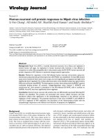
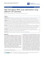
PSA antigen expression of immature DCs transfected with total RNA from LNCaP cellsFigure 1
PSA antigen expression of immature DCs transfected with total RNA from LNCaP cells. Immature DCs were
transfected by electroporation with 4 µg (IA and IB) and 16 µg (IIA and IIB) of RNA from LNCaP cells. The electroporation
conditions were 300 (IA and IIA) or 400 V (IB and IIB). In both cases, the capacitance was 25 µF. IIIA is the negative transfec-
tion control, and IIIB is the positive control represented by LNCaP. The panel of photographs represents PSA expression
detected by immunocytochemistry using an anti-PSA antibody.
Genetic Vaccines and Therapy 2008, 6:2 />Page 5 of 7
(page number not for citation purposes)
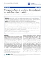
PSA antigen expression of mature DCs transfected with total RNA from LNCaP cellsFigure 2
PSA antigen expression of mature DCs transfected with total RNA from LNCaP cells. Mature DCs were trans-
fected by electroporation with 4 µg (IA, B, and C) and 5 µg (IIA, B, and C) or coincubated with 5 µg (IIIA, B, and C) of RNA
from LNCaP cells. The electroporation conditions were 300 (IA and IIA), 400 (IB and IIB), or 500 V (IC and IIC). In all cases,
the capacitance was 25 µF. The transfection by coincubation was done without preincubation (IIIA) and with 30 minutes (IIIB)
or 2 hours (IIIC) of preincubation at 22°C. Photos IVA and IVB are negative transfection controls, and IVC is the positive con-
trol represented by LNCaP cells. The panel of photographs represents PSA expression detected by immunocytochemistry
using an anti-PSA antibody.
IIA IIB IIC
IA IB IC
IIIA IIIB
IVC
IIIC
IVBIVA
Genetic Vaccines and Therapy 2008, 6:2 />Page 6 of 7
(page number not for citation purposes)
mismatch among patients and leukapheresis donors. This
difference can provoke immune responses against donor
DCs instead of the tumor proteins that they carry. This
problem could be solved by using patient peripheric
blood to generate DCs. Most cancer patients, however, are
already immunocompromised and debilitated due to pre-
vious treatment. Moreover, one tumor escape mechanism
involves the inactivation of DC function. Thus, the use of
patients' blood to obtain DCs may not be an option in all
cases. Clinical observations, on the other hand, have
shown that autologous DCs might be unnecessary; three
patients negative for HLA-A2 were able to initiate immune
responses after vaccination with HLA-A2 DCs loaded with
PSMA peptide [22]. We have also observed a recovery of
patients' immunological functions after two to three
doses of DC vaccination [23]. Therefore, a viable possibil-
ity involves using allogeneic DCs for only the initial doses
and then changing to more efficient vaccines prepared
with autologous DCs.
Conclusion
We believe that the treatment of patients with prostate
metastatic disease using immunotherapy based on DCs is
feasible in highly genetically polymorphic populations.
For a more efficient immune response, entire mRNA mol-
ecules should be used instead of small peptides because of
the diversity of the HLA molecules found in these popula-
tions. Furthermore, vaccines for at least the first two doses
should be prepared with allogeneic DCs since patient
immune systems may be suppressed by the tumor. Then,
periferic blood of patients could be collected to prepare
vaccines containing patients' own HLA molecules.
Abbreviations
DC, Dendritic Cell, PSA, prostate-specific antigen.
Competing interests
The author(s) declare that they have no competing inter-
ests.
Authors' contributions
JMSC carried out the dendritic cell culture and transfec-
tion experiments and drafted the manuscript. FCC partic-
ipated in the design of the study and helped to draft the
manuscript. KRML analyzed the immunocytochemistry
results and together with LHCL, coordinated the study. All
authors read and approved the final manuscript.
Acknowledgements
We are grateful to Dr. Cristina O. Massoco for helpful assistance in cytom-
etry. This work was funded by Genoa Biotechnology SA.
References
1. Instituto Nacional do Câncer [ />tiva/2006/]
2. Ali AS, Hamdy FC: The spectrum of prostate cancer care: from
curative intent to palliation. Curr Urol Rep 2007, 8(3):245-52.
3. Taplin ME: Biochemical (Prostate-Specific Antigen) Relapse:
An Oncologist's Perspective. Rev Urol 2003, 5(Suppl 2):S3-S13.
4. Pound CR, Partin AW, Eisenberger MA, Chan DW, Pearson JD,
Walsh PC: Natural history of progression after PSA elevation
following radical prostatectomy. JAMA 1999, 281(17):1642-5.
5. Hadaschik BA, Gleave ME: Therapeutic options for hormone-
refractory prostate cancer in 2007. Urol Oncol 2007, 25:413-9.
6. Mosca PJ, Lyerly HK, Clay TM, Morse MA, Lyerly HK: Dendritic cell
vaccines. Front Biosci 2007, 12:4050-60.
7. Osada T, Clay TM, Woo CY, Morse MA, Lyerly HK: Dendritic cell-
based immunotherapy. Int Rev Immunol 2006, 25(5–6):377-413.
8. Gilboa E: DC-based cancer vaccines. J Clin Invest 2007,
117(5):1195-203.
9. Barbuto JAM, Ensina LFC, Neves AR, Bergamini-Santos PC, Leite
KRM, Marques R, Costa F, Martins SC, Câmara-Lopes LH, Buzaid AC:
Dendritic cell-tumor hybrid vaccination for metastatic can-
cer. Cancer Immunol Immunother 2007, 53:1111-18.
10. Thomas-Kaskel AK, Waller CF, Schultze-Seemann W, Veelken H:
Immunotherapy with dendritic cells for prostate cancer. Int
J Cancer 2007, 121(3):467-73.
11. Boczkowski D, Nair SK, Nam JH, Lyerly HK, Gilboa E: Dendritic
cells pulsed with RNA are potent antigen-presenting cells in
vitro and in vivo. J Exp Med 1996, 184:465-72.
12. Banchereau J, Steinman RM: Dendritic cells and the control of
immunity. Nature 1998, 392:245-52.
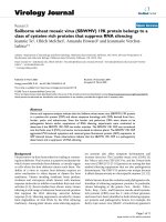
Androgen receptor expression after mature DC transfection with total RNA from LNCaP cellsFigure 3
Androgen receptor expression after mature DC transfection with total RNA from LNCaP cells. (A) Mature DCs
were transfected with 5 µg of LNCaP total RNA using 400 V electroporation and a capacitance of 25 µF. (B) Negative control
of transfection represented by DCs, and (C) Positive control represented by LNCaP cells.
A B C
Publish with BioMed Central and every
scientist can read your work free of charge
"BioMed Central will be the most significant development for
disseminating the results of biomedical research in our lifetime."
Sir Paul Nurse, Cancer Research UK
Your research papers will be:
available free of charge to the entire biomedical community
peer reviewed and published immediately upon acceptance
cited in PubMed and archived on PubMed Central
yours — you keep the copyright
Submit your manuscript here:
/>BioMedcentral
Genetic Vaccines and Therapy 2008, 6:2 />Page 7 of 7
(page number not for citation purposes)
13. Salgaller ML, Lodge PA, McLean JG, Tjoa BA, Loftus DJ, Ragde H,
Kenny GM, Rogers M, Boynton AL, Murphy GP: Report of immune
monitoring of prostate cancer patients undergoing T-cell
therapy using dendritic cells pulsed with HLA-A2-specific
peptides from prostate-specific membrane antigen (PSMA).
Prostate 1998, 35:144-51.
14. Murphy GP, Tjoa BA, Simmons SJ, Jarisch J, Bowes VA, Ragde H, Rog-
ers M, Elgamal A, Kenny GM, Cobb OE, Ireton RC, Troychak MJ, Sal-
galler ML, Boynton AL: Infusion of dendritic cells pulsed with
HLA-A2-specific prostate-specific membrane antigen pep-
tides: a phase II prostate cancer vaccine trial involving
patients with hormone-refractory metastatic disease. Pros-
tate 1999, 38:73-8.
15. Callegari-Jacques SM, Grattapaglia D, Salzano FM, Salamoni SP, Cros-
setti SG, Ferreira ME, Hutz MH: Historical genetics: spatiotem-
poral analysis of the formation of the Brazilian population.
Am J Hum Biol 2003, 15:824-34.
16. Abe-Sandes K, Silva WA Jr, Zago MA: Heterogeneity of the Y
chromosome in Afro-Brazilian populations. Hum Biol 2004,
76:77-86.
17. Van Tendeloo VF, Ponsaerts P, Berneman ZN: mRNA-based gene
transfer as a tool for gene and cell therapy. Curr Opin Mol Ther
2007, 9:423-31.
18. Heiser A, Coleman D, Dannull J, Yancey D, Maurice MA, Lallas CD,
Dahm P, Niedzwiecki D, Gilboa E, Vieweg J: Autologous dendritic
cells transfected with prostate-specific antigen RNA stimu-
late CTL responses against metastatic prostate tumors. J
Clin Invest 2002, 109:409-17.
19. Su Z, Dannull J, Yang BK, Dahm P, Coleman D, Yancey D, Sichi S,
Niedzwiecki D, Boczkowski D, Gilboa E, Vieweg J: Telomerase
mRNA-transfected Dendritic Cells stimulate antigen-spe-
cific CD8+ and CD4+ T cell responses in patients with meta-
static prostate cancer. J Immunol 2005, 174(6):3798-807.
20. de Vries IJ, Lesterhuis WJ, Scharenborg NM, Engelen LP, Ruiter DJ,
Gerritsen MJ, Croockewit S, Britten CM, Torensma R, Adema GJ, Fig-
dor CG, Punt CJ: Maturation of dendritic cells is a prerequisite
for inducing immune responses in advanced melanoma
patients. Clin Cancer Res 2003, 9(14):5091-100.
21. McIlroy D, Gregoire M: Optimizing dendritic cell-based anti-
cancer immunotherapy: maturation state does have clinical
impact. Cancer Immunol Immunother 2003, 52:583-91.
22. Tjoa BA, Simmons SJ, Bowes VA, Ragde H, Rogers M, Elgamal A,
Kenny GM, Cobb OE, Ireton RC, Troychak MJ, Salgaller ML, Boynton
AL, Murphy GP: Evaluation of phase I/II clinical trials in pros-
tate cancer with dendritic cells and PSMA peptides. Prostate
1998, 36(1):39-44.
23. Neves AR, Ensina LFC, Anselmo LB, Leite KRM, Buzaid AC, Camara-
Lopes LH, Barbuto JAM: Dendritic cells derived from meta-
static cancer patients vaccinated with allogeneic dendritic
cell-autologous tumor cell hybrids express more CD86 and
induce higher levels of interferon-gamma in mixed lym-
phocyte reactions. Cancer Immunol Immunother 2005, 54:61-66.