Báo cáo sinh học: " A strategy of tumor treatment in mice with doxorubicin-cyclophosphamide combination based on dendritic cell activation by human double-stranded DNA preparation" doc
Bạn đang xem bản rút gọn của tài liệu. Xem và tải ngay bản đầy đủ của tài liệu tại đây (1018.61 KB, 10 trang )
RESEARC H Open Access
A strategy of tumor treatment in mice with
doxorubicin-cyclophosphamide combination
based on dendritic cell activation by human
double-stranded DNA preparation
Ekaterina A Alyamkina
1,2
, Valeriy P Nikolin
2
, Nelly A Popova
1,2
, Evgenia V Dolgova
1,2
, Anastasia S Proskurina
2
,
Konstantin E Orishchenko
2
, Yaroslav R Efremov
2
, Elena R Chernykh
3
, Alexandr A Ostanin
3
, Sergey V Sidorov
4
,
Dmitriy M Ponomarenko
5
, Stanislav N Zagrebelniy
1
, Sergey S Bogachev
2*
, Mikhail A Shurdov
6
Abstract
Background: Immunization of mice with tumor homogenate after combined treatment with cyclophosphamide
(CP) and double-stranded DNA (dsDNA) preparation is effective at inhibition of growth of tumor challenged after
the treatment. It was assumed that this inhibition might be due to activation of the antigen-presenting cells. The
purpose was to develop improved antitumor strategy using mice. We studied the combined action of cytostatics
doxorubicin (Dox) plus CP with subsequent dsDNA preparation on tumor growth.
Methods: Three-month old CBA/Lac mice were used in the experiments. Mice were injected with CP and human
dsDNA preparation. The percentage of mature dendritic cells (DCs) was estimated by staining of mononuclear cells
isolated from spleen and bone marrow 3, 6, and 9 days later with monoclonal antibodies CD34, CD80, and CD86.
In the next set of experiments, mice were given intramuscularly injections of 1-3 × 10
5
tumor cells. Four days later,
they were injected intravenously with 6-6.7 mg/kg Dox and intraperitoneally with 100-200 mg/kg CP; 200 mkg
human DNA was injected intraperitoneally after CP administration. Differences in tumor size between groups were
analyzed for statistical significance by Student’s t-test. The MTT-test was done to determine the cytotoxic index of
mouse leucocytes from treated groups.
Results: The conducted experiments showed that combined treatment with CP and dsDNA preparation produce
an increase in the total amount of mature DCs in vivo. Treatment of tumor bearers with preparation of fragmented
dsDNA on the background of pretreatment with Dox plus CP demonstrated a strong suppression of tumor growth
in two models. RLS, a weakly immunogenic, resistant to alkalyting cytostatics tumor, grew 3.4-fold slower when
compared with the control (p < 0.001). In experiment with Krebs-2 tumor, only 2 of the 10 mice in the Dox+CP+DNA
group had a palpable tumor on day 16. Th e cytotoxic index of leucocytes was 86.5% in the Dox+CP+DNA group, but
it was 0% in the Dox+CP group.
Conclusions: Thus, the set of experiments we performed showed that exogenous dsDNA, when administered on
the background of pretreatment with Dox plus CP, has an antitumor effect possibly due to DC activation.
* Correspondence:
2
Institute of Cytology and Genetics, Siberian Branch, Russian Academy of
Sciences, Novosibirsk, Russia
Full list of author information is available at the end of the article
Alyamkina et al. Genetic Vaccines and Therapy 2010, 8:7
/>GENETIC VACCINES
AND THERAPY
© 2010 Alyamkina et al; licen see BioMed Central Ltd. This is an Open Access article distributed under the terms of the Creative
Commons Attribution License ( which perm its unrestricted u se, distribution, and
reproduction in any medium, provided the original work is properly cited.
Background
The most effective antitumor treatment is currently
achieved by chemotherapeutic agents that abrogate
tumor c ells [1]. Despite this, chemotherapy is virtually
without influence on life expectancy of patients with
certain cancers. With this in mind, novel strategies for
treating malignancies are being developed in experi-
ments and applied in clinical setting. These are targeted
towards potentiation of immune mechanisms of antitu-
mor defense [2, 3]. The co nventional vaccines a re uti-
lized, also those based on the pathogen-associated
molecular patterns (PAMPs) of bacteria, including endo/
exotoxins of bacterial origin, and CpG DNA prepara-
tions [4-12].
Dendritic cells (DCs), which are capable of activating
T-lymphocytes, including naive T-cells, have an impor-
tant role in triggering and development of the adaptive
immunity [9,13,14]. Mature DCs that express MHC
antigens of class I and class II, also the various costimu-
latory molecules CD40, CD54, CD80, and CD86 are
capable of only presenting foreign antigens within the
MHC complex [15-21].
Search of novel inducers of antitu mor immunity h as
been intense over the past years. It has been revealed
that mammalian double-stranded DNA (dsDNA)
induces both humoral and adaptive immune responses
[15,22,23]. This induction is provided by the action o f
dsDNA preparations primarily on professional antigen-
presenting cells. This process enfolds via the TLR-inde-
pendent pathway and is mainly due to activation of
TANK-binding kinase-1, TBK1 [22-27]. As a result of
internalization of exogenous DNA, DCs up-regulate
expression and secretion of type I interferon-beta (INF-b)
[22,25]. In addition, dsDNA induces complete DC matura-
tion, by stimulating expression of cofactor molecules
on cell wall needed for development of the adaptive
immunity [15].
Cyclophosphamide (CP) is a drug widely applied in
the c linic to treat cancers. The effect is predominantly
based on direct cytotoxic action on tumor cells resulting
in their lysis. CP has an influence on CD4+CD25+FoxP3
regulatory T cells. Regulatory T cells accumulate predo-
minantly in the tumor microenviro nment and l ymphoid
organs [28] where they suppress activation and prolif-
eration of the other immune cells [28-32]. When admi-
nistered at moderate doses, CP not only induces a
reduction in numbers of regulatory T cells [33-35], also
diminishes their functionality [32,34], thereby allowing
to reduce the intensity of the immunosuppressive back-
ground in tumor microenvironment and to activate the
antitumor immune response [31,32,35]. The effect of CP
on various DC subsets was manifest as enhancement of
antitumor immunity [36-38].
It has been amply demonstrated that under the com-
bined effect of CP and dsDNA preparation (CpG DNA,
for example), the immune system is stimulated and
tumor growth is suppressed [for reference, see 9]. The
therapeutic effect is synergic in that cytostatic prefe ren-
tially decreases the amount of regulatory T cells in the
tumor microenvironment and/or directly kills tumor
cells, while dsDNA preparation stimulate maturation
and activity of cells of the adaptive immunity [9,39].
There are chemotherapeutic agents capable of poten-
tiating immunogenicity of tumor cells directly at the
level of the organism. Doxorubicin (Dox), idarubucin,
and mitoxanthrone, cytostatics of the antracycline series,
are of this kind. A relevant observation was that induc-
tion of exposure of the protein calreticulin on cell
surface of dying cells is required for activation of
the antitumor immune system [40,41]. Calreticulin is a
calcium-binding lectin chaperone, mainly represented
on endoplasmic membrane. Its exposure on cell sur-
face of dying tumor cells acts as an “eat me” signal for
removal by neighboring phagocytic cells [40,42] and
facilitates thereby their almost instantaneous capture
[41]. The combinatio n of Dox with cytostatic drugs
(CP plus pacli taxel) and whole-cell vaccines was highly
effective in enhancing antitumor response in transgenic
mice [43].
Here, we demonstrate that human exogenous dsDNA
preparation induces maturation o f mouse spleen and
bone marrow DCs in vivo. To evaluate the efficacy of
vaccination with human dsDNA preparation, we chose a
strategy whereby mice were treated with preparation of
fragmented dsDNA on the background of pretreatment
with Dox plus CP. This strategy provided the presence
of tumor antigens thanks to the in vivo abrogation of
tumor by the combined action of cytostatics. The subse-
quently injected dsDNA preparation induced effective
DC maturation. This strategy demonstrated a consider-
able delay in tumor growth. Cytotoxic test provided evi -
dence indicating that in the blood there appeared a cell
population with high, up to 86.5%, cytotoxi c activity
against cells of the challenged tumor.
Methods
Laboratory animals and tumor models
Three-month old CBA/Lac mice (henceforth designated
as CBA) that were bred at the animal fac ility of the
Institute of Cytology and Genetics (IC&G), the Siberian
Branch o f the Russian A cademy of Science s, were used
in experiments. Mice in groups of 10 were housed in
plastic cages in a well-il luminated room. They had free
access to food and water. All experiments were per-
formed in accordance with protocols approved by the
Animal Care and Use Committee of the IC&G.
Alyamkina et al. Genetic Vaccines and Therapy 2010, 8:7
/>Page 2 of 10
Krebs 2 a scitic carcinoma is a st rain-nonspecific
tumor derived from epithelial cells; all inbred mouse
strains can be challenged with Krebs 2 tumor cells.
When challenged subcutaneously (s.c.) or intramuscu-
larly (i.m.), it grows as solid nodes. It is weakly immuno-
genic for mice of all s trains. It does not give rise to
metastases [44].
Lymphosarcoma LS is strain-specific to CBA mice; it
was induced in them by nitrosomethylurea, passages in
ascitic form. When challenged i.m., it grows as solid
nodes. It develops in 100% of challenged mice, d oes not
regress spontaneously. It is subjected to apoptosis under
the effect of alkylating antitumor agents. It metastasizes
to liver, kidneys, lungs. Lymphosarcoma RLS-40 is a ver-
sion of LS tumor. It is resistant to alkylating compounds
[45,46].
Mice were injected i.m. into the right hind limb with
tumor cells at a dose of 1-3 × 10
5
cells/mouse. The
tumors were allowed to grow to solid nodes. As soon as
tumor became palpable, about 7 days after challenge, it
size was measured with calipers every 1-2 days. Tumor
size was calculated by multiplying the three perpendicu-
lar diameters. Differences in tumor size between groups
were analyzed for statistical significance by Student’s
t-test.
DNA preparation
Human DNA preparation was isolated from the placen-
tas of healthy women using a phenol-free method. It
was fragmented in an ultrasonic disintegrator at a fre-
quency of 22 kHz to obtain a mixture of DNA frag-
ments with a size 200-6,000 bp. The human DNA was a
pharmacopeian preparation “Panagen” (Registration cer-
tificate Medical Drugs of Russia No. 004429/08 of
09.06.2008). This preparation does not contain steroid
hormones and RNA. It gives negative PCR results for
hepatitisBvirusDNA,hepatitisCvirusRNA,
HIV DNA, H IV RNA. The DNA preparation does not
contain histones and polysaccharides; it is also endo-
toxin-free.
Estimation of DC maturity in vivo
Mice were injected with CP (Veropharm, Russia) at 200
mg/kg and 200 mkg of human dsDNA preparation 1
day ( on the day of CP injection), 3, 4, and 5 days after
CP treatment. Three, 6, and 9 days later, the fraction of
mononuclear cells (MNCs) was isolated from spleen and
bone marrow. MNCs were isolate d also from untreated
mice. Every group consisted of 4-6 mice. The experi-
ment was repeated twice.
Mice were anesthetized and sacrificed by cervical dis-
location. Femurs and tibias were removed a nd bone
marrow cells were flushed from them b y RPMI-1640
(Sigma-Aldrich) medium. Washed bone marrow cells
(DC precursors) were suspended in RPMI-1640. Spleen
content s were scraped out with pincers into Petri dishes
and resuspended in PBS. The obtained cell suspension
was applied onto 3 ml ficoll 400 (Farmaceg) - urografin
(Schering) gradient, centrifuged (5810R, Eppendorf) at
1,500 rpm for 30 min. MNCs were col lected, washed and
precipitated. Cell residue was suspended in RPMI-1640,
the number of cells was counted and diluted to 2 × 1 0
5
in 200 μl of medium.
The percentage of mature spleen and bone marrow
DCs was estimated by staining with monoclonal antibo-
dies CD34-PerCP, CD80-FITC, and CD86-PE (Santa
Cruz). Cells were analyzed on a flow cytofluorometer
BD FACSAria (BD Biosciences). Additional file 1 is a
dot plot figure of the event gating for CP+DNA group.
Statistics was based on estimates of the number of
mature DCs relative to the total number of isolated
MNCs.
Schedule for treatment with exogenous dsDNA
preparation after administration of cytostatics Dox plus
CP
CBA mice were given an i.m. challenge with 10
5
RLS-40
tumor cells. Four days later, they were injected intrave-
nously (i.v.) with 6.7 mg/kg Dox (Veropharm, Russia)
and i.p. wit h 100 mg/kg CP; 200 mkg human DNA was
injected i.p. after 30 min, then 2 and 3 days after CP
administration. Mice were assigned to three groups
(n = 10) according to treatment schedule: 1) challenged
tumor + PBS injections (control) ; 2) Dox + CP; 3) Do x +
CP + DNA.
CBA mice were given 3 × 10
5
Krebs-2 tumor cells
injected i.m. Four days later, they were administered i.v.
6 mg/kg Dox and i.p. 200 mg/kg CP; 200 mkg human
DNA was administered i.p. 30 min after CP, also 2, 3,
and 5 days after it. Assignment of mice to groups, with 10
in each, was as follows: 1) challenged tumor + PBS injec-
tions (control); 2) Dox + CP; 3) DNA; 4) Dox + CP +
DNA. The experiment was done in triplicate.
The dosages of Dox and CP were the c onventionally
used for chemoth erapy in the clinic, 100-200 mg/kg for
CP and 6-7 mg /kg for Dox. The DNA preparation was
used at 200 mkg/mouse/injection. This amount has
been defined in experiments [39].
MTT test
Mice of all the 4 groups and one untreated mouse were
sacrificed by decapitation on day 16 after tumor Krebs-2
challenge. Blood (200-500 μl) was drawn into tubes con-
taining 800 μl PBS with 50 mM EDTA. Blood cells were
precipitated by centrifugation (5810R, E ppendorf) at
1,500 rpm for 5 min at room temperature; erythrocytes
from cell residue were lysed with 0.15 M ammonium
chloride.
Alyamkina et al. Genetic Vaccines and Therapy 2010, 8:7
/>Page 3 of 10
In in vitro cytotoxicity study, Krebs-2 cells were plated
in 96-well plates (3 × 10
4
cells/well), and mouse leuco-
cytes were added at a 1:1 ratio. Cells were incubated in
RPMI-1640 medium supplemented with gentamycin sul-
fate (100 mkg/ml) and main tained at 37°C for 18 h in
5% CO
2
atmosphere. After incubation, MTT (Sigma)
was added to a final concentration of 0.5 mg/ml and
cell s were cultured for additional 3 h. Cells were centri-
fuged (5810 R, Eppendorf) at 4,00 0 rpm for 10 min.
Medium was collected, precipitated blue formasan crys-
tals were dissolved in 100 μl DMSO. Optical density
was determined on a Multiscan RC at 570 nm, back-
ground was subtracted at 620 nm. Measurements were
done for three samples. The MTT-test was repeated
twice for different experiments.
The standard formula was applied to calculate the
percentage of dead cells:
%/,
()
=
()
⎡
⎣
⎤
⎦
×
+
1D D D 1
et e t
−− 00
D
e+t
, the optical density value in wells with cells from
mice of the treated groups incubated with tumor cells;
D
e
, the optical density value in wells with effectors
(leucocytes);
D
t
, the optical density value in wells with targets
(tumor cells).
The cytotoxic index (CI) w as expressed as the differ-
ence between the percentage of dead cells in the treated
groups and the untreated mouse.
Results
Our previous study ha s demonstrated that a preparation
of human fragmented dsDNA stimulated maturation of
mous e DCs in culture [47]. The salient finding was that
the dsDNA preparation was just as effective at induction
of DC maturation as the standard inducer TNF-a.The
obtained mature DCs loaded with antigen during
maturation were used in the comparative test. A marked
antitumor effect was observed after vacc ination with
DCs irrespective of the type of maturation inducer [47].
Previous experimental sets with Krebs-2 tumor demon-
strated that immunization of mice w ith tumor homoge-
nate after combined treatment with CP and dsDNA
preparation is e ffective at inhibition of growth of tumor
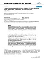
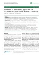
challenged after the treatment (Figure 1) [39]. Proceeding
on reported observations [14,39,47], we assumed that this
inhibition may be due to the inducer effect of dsDNA on
DC maturation in vivo that causes effective presentation
of antigens o f tumor lysate and activates antitumor
mechanisms of the adaptive immunity.
The results provided evidence indicating that the
described antitumor activity was not related to natural
Figure 1 Time course of Krebs-2 tumor growth in mice (mean ± SEM). Time course of Krebs-2 tumor growth in mice (mean ± SEM). Mice
received 200 mg/kg CP and human DNA at a total dose 4.5-6 mg. After this treatment, one group of mice was pre-immunized with Krebs-2
tumor antigens by a s.c. injection of 20 × 10
6
repeatedly thawed-frozen tumor cells. The control group was injected with saline. Every group
consisted of 10 mice. 10
6
Krebs-2 tumor cells were challenged i.m. after the treatment. Immunization enhanced the suppressive effect on tumor
growth [31].
Alyamkina et al. Genetic Vaccines and Therapy 2010, 8:7
/>Page 4 of 10
killer cells [39]. This appeared plausible, because, to our
knowledge, NK-cells neither displayed nor enhanced
antigen-specific cytotoxicity associated with tumor
homogenate immunization [48,49].
Effect of dsDNA preparation on maturation of spleen and
bone marrow DCs in vivo
To obtain assurance that dsDNA has an inducer e ffect
on DCs in vivo, a se t of exp eriments was undertaken.
Mice wer e treated with CP 200 mg/kg followed by 200
mkg human dsDNA preparation a dministra tion 1, 3, 4,
and 5 days after C P injection. The number of mature
CD34-CD80+CD86+ DCs a mong spleen and bone mar-
row cells was e stimated 3, 6, and 9 days after CP h ad
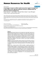
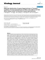
been injected (Figure 2).
The peak of spleen DC maturation was 3 days after
combined DNA+CP treatment. This peak was followed
by a decrease in the number of mature DCs presumably
Figure 2 Time course of maturation of mouse DCs from spleen (A) and bone marrow (B) after treatment with CP and dsDNA
preparation (mean ± SEM). Time course of maturation of mouse DCs from spleen (A) and bone marrow (B) after treatment with CP and
dsDNA preparation (mean ± SEM). 0 represents the number of mature DCs in untreated mice. Mice were injected with CP 200 mg/kg and 200
mkg of human dsDNA preparation 1 day (on the day of CP injection), 3, 4, and 5 days after CP treatment. Three, 6, and 9 days later, the fraction
of MNCs was isolated from spleen and bone marrow. Every group consisted of 4-6 mice. The experiment was repeated twice.
Alyamkina et al. Genetic Vaccines and Therapy 2010, 8:7
/>Page 5 of 10
due to migration of cells to lymph nodes and other sites
of their specific locali zation. Mouse groups treated with
an agent alone, CP or dsDNA preparation, showed no
marked increase in the number of mature DCs.
The peak of bone marrow DC maturation in the DNA
and DNA+CP groups was also on day 3. In the case of
DNA+CP treatment, the interval during which DCs
retained mature phenotype and were able to effectively
present antigen was longer, several days. DNA alone
caused a transient rise in level of mature DCs. In the
CP group, the number of mature DCs in bone marrow
reached the maximum by day 6 only, thereafter it
decreased to the initial level.
Thus, the conducted experiments showed that com-
bined t reatment with CP and dsDNA preparation pro-
duces an increase in the total amount of mature DCs.
This was assoc iated with an increase in the time during
which mature DCs persisted at high levels.
Effect of inhibition of tumor growth induced by Dox+CP
+DNA treatment
Our previous s tudies have demonstrated that the CP
+DNA combination was statistically superior to each
treatment modality alone [39,50]. From comparisons of
schedules, the standard with additional immunization
with tumor homogenate, it followed that the presence of
specific antigens further enhanced the suppression effect
on tumor growth. There were reasons for suggesting
that the integration of cytostatics with dsDNA prepara-
tion may be a treatment modality for enhancing regres-
sion of established tumors.
According to the data in the literature a combination
of cytostatics is superior to ea ch modality alone [51,52].
Two-three potent drugs are u sually combined in the
cli nic. In the current stud y, we did not strive to control
the effectiveness o f a drug as monotherapeutic agent.
We were rather interested in the antitumor action of
DNA preparation when used in combination with cyto-
statics Dox and CP.
Proceeding on the combined cytotoxic action of Dox
and CP, also on the course of changes in DC maturation
in vivo , a set of experiments was designed. The idea was
to superimpose the effects of released tumor antigens
and of their capture by DCs. Mice bearing established
tumors were treated on day 4 with Dox and CP, there-
after they were injected with human dsDNA prepara-
tion. As known [41,53], Dox provides the exposure of
the cell surface endoplasmic protein calreticulin that
acts as an “eat me” signal and mediates the phagocytosis
of tumor cells by DCs. CP abrogates tumor cells,
thereby increasing the amount of free tumor antigens
that,thankstothe“ eat me” signal, are uptaken
promptly, and presented by DCs. The induction of DC
maturation is the necessary condition for antigen
presentation on the surface o f DCs. In the following
experiments, we chose dsDNA preparation as a matura-
tion stimulus.
Using this schedule, a strong suppression of tumor
growth was observed in two murine models. The size of
RLS, a weakly immunogenic, resist ant to alkalyting cyto-
statics tumor, on day 14 was 3.4-fold smaller (p < 0.001)
in the Dox+CP+DNA group compared with the control
(Figure 3). The difference in RLS size on day 14 between
the groups Dox+CP and Dox+CP+DNA was 1.5-fold
(p < 0.1).
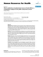
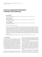
Krebs-2 tumor growth was effectively suppressed as
compared to the control in both Dox+CP and Dox+CP+
DNA groups (p < 0.001) (Figure 4). A tumor burden was
of measurable size 16 days after treatment in 9 of the 10
mice in the Dox+CP group, but only in 2 of the 10 mice
tumor was palpable on day 16 in Dox+CP+DNA group.
There was a 14-fold significan t difference (p < 0.005) in
tumor size on day 14 between the Dox+CP and Dox+CP
+DNA groups. Injection of dsDNA preparation alone
slightly suppressed Krebs-2 tumor growth, the difference
from the control being significant, however (p < 0.05).
The schedule for DNA preparation administra tion dif-
fered slightly from the one we applied to estimate the
efficacy of DC maturation in vivo. DNA was injected at
the time when the number of mature DCs was
maximum.
We used CP at high doses since evaluation of thera-
peutic combined action of CP and dsDNA did not
demon strate enhancement of antitumor effect with low-
dose CP (data not shown).
Estimation of cytotoxic activity of blood cells in mice with
Krebs-2 tumor after combined treatment with Dox+CP+
dsDNA preparation
The experimental results provided evidence for activa-
tion of the antitumor immune response in vivo.Sup-
porting data of the MTT test were required. For this
purpose, treated mice bearers of Krebs-2 tumor w ere
sacrificed 16 days after treatment. A ll the mouse groups
coul d be monitored at the same time, on day 16 for the
presence of cytotoxic cells. This became feasible because
tumors r eached the size that led to lethal development
in the control group. Tumor size in the treated groups
attained a statistically significant difference from the
control by this time.
Peripheral blood was monitored for the appear ance of
cells showing antitumor cytotoxic activity. Krebs-2 cells
derived from ascitic version of tumor served as targets
(Table 1).
The cytotoxic index (CI) was expressed as the percen-
tage of dead cells relative to their number in an
untreated mouse. It was 86.5% in the Dox+CP+DNA
group, consistent with the time course of tumor growth
Alyamkina et al. Genetic Vaccines and Therapy 2010, 8:7
/>Page 6 of 10
(Figure 4). It was 0% in the Dox+CP group, although
there w as a considerable suppr ession of tumor growth.
This may be attributed to the direct cytostatic effect on
tumor growth of the kind t hat does not enhance
cytotoxic activity of circulating leucocytes. Such was the
case, because there was no DNA stimulus for DC
maturation and ultimate development of antigen-specific
immune responses. dsDNA preparation itself raised cell
Figure 4 Time course of Krebs-2 tumor growth in mice treated with Dox, C P, and dsDNA preparation (mean ± SE).Timecourseof
Krebs-2 tumor growth in mice treated with Dox, CP, and dsDNA preparation (mean ± SE). Mice were given 3 × 10
5
Krebs-2 tumor cells injected
i.m. Four days later, they were administered i.v. 6 mg/kg Dox and i.p. 200 mg/kg CP; 200 mkg human DNA was administered i.p. 30 min after
CP, also 2, 3, and 5 days after it. The control group was injected with PBS. Every group consisted of 10 mice. The experiment was done in
triplicate.
Figure 3 Time course of RLS tumor growth in mice that received combined treatment with Dox, CP, and dsDNA preparation (mean ±
SE). Time course of RLS tumor growth in mice that received combined treatment with Dox, CP, and dsDNA preparation (mean ± SE). Mice were
given i.m. injections of 10
5
RLS-40 tumor cells. Four days later, they were injected i.v. with 6.7 mg/kg Dox and i.p. with 100 mg/kg CP; 200 mkg
human DNA was injected i.p. after 30 min, then 2 and 3 days after CP administration. The control group was injected with PBS. Every group
consisted of 10 mice.
Alyamkina et al. Genetic Vaccines and Therapy 2010, 8:7
/>Page 7 of 10
cytotoxic activity to 44.2%, but t umor growth was sup-
pressed just slightly.
Discussion
Tumors have unique properties allowing them to elude
immune defense. To begin with, they are genetically flex-
ible owing to the inces santly activated repair-recombina-
tion system of tumor cells [32]. Second, tumor tissue
takes advantage of the properties of regulatory T lympho-
cytes. Third, a tumor is, as a rule, weakly immunogenic
and this makes the more difficul t for the immune system
to reveal malignized cells and to eradicate them. Modula-
tion or elimination of these three properties of tumors
would create conditions favorable for the immune system
to eliminate neotransformed cells [1,13,54,55].
The current increasing trend is to affect tumor tissue
by using in a defined sequence two modalities, a che-
motherapeutic (a cytostatic, most commonly CP) fol-
lowed by an immunotherapeutic [21,56-58]. This
strategy is fully consistent with the idea how tumor tis-
sue may be affected. To recapitulate, CP directly attacks
tumor cells, it also causes a decrease in the numbers of
regulatory T cells and reduces their functionality
[21,32,34,35,58,59], thereby improves the efficacy of
immune-based therapies directed at stimulation/
enhancement of antitumor immune responses.
Recent studies on the chemotherapeutic effects of
antracyclines have established that Dox, for example,
transposes calreticulin to the cell surface. This protein
may play t he role of surveillance “eat me” signal and
mediate the phagocytosis of tumor cells by DCs. As a
result, tumor immunogenicity is enhanced [41,53].
Cytostatics (CP and Dox) in combination with immu-
notherapeutics (DNA activators) allow to develop
improved antitumor strategy. CP directly injures tumor
cells, concomitantly switches regulatory T cells off. Dox
also abrogates tumor and renders tumor cell debris
immunogenic. The D NA activated immune system kills
the remaining ne otransformed cells at the time when
the regulatory T-lymphocytes are inactive and tumor is
defenseless.
In the current experiments, we relied on the ability of
dsDNA to induce complete DC maturation ex vivo rea-
sonably expecting that this would augment their stimu-
latory activity in an allogenic mixed lymphocyte culture
[14,22,47]. It was a reasonable assumption that dsDNA
would manifest its stimulatory action o n DCs at the
level of the whole organism. The suggestion that antitu-
mor dsDNA activity [39,50,60] is due to precisely endo-
genous DC activation and development of the adaptive
immune response lent credibili ty to our line o f
reasoning.
We determined the extent to which spleen and bone
marrow derived DCs were mature and followed the time
course of changes in their quantitative accumulation
after different treatments. Given the results, a schedule
for combined Dox plus CP, which form apoptotic/
necrotic debris, plus dsDNA preparation was developed.
Strongest suppression of tumor growth was achieved
with this schedule and an optimal sequence of adminis-
tration of each modality. Its effectiveness was confirmed
by the MTT test estimates. The suppression effect on
tumor growth was, indeed, due to both damaging action
of cytost atics and formation of a pool of cytoto xic cells.
Importantly, challenged tumors virtually stopped grow-
ing when chemotherapeutic agents were combined with
dsDNA preparation.
Conclusions
Thus, the set of experiments we performed showed that
exogenous dsDNA, when administered on the back-
ground of pretreatment with Dox plus CP, has an anti-
tumor effect possibly due to DC activation. The effect
may be also explained by DC-mediated activation of
cytotoxic T-lymphocytes [37,38]. Crucial here are the
mature phenotype of DCs, i.e. their antigen-presenting
ability, and the real presence of tumor antigens achieved
by combined treatment with Dox and CP.
The described approach to therapy of cancers appears
promising. Injections of dsDNA preparation may be well
integrated into classical schedules of chemotherapy.
Additional material
Additional file 1: Dot plot figure. Dot plot figure of the event gating
for CP+DNA group.
Acknowledgements
The work was funded by federal target p rogram “Scientific and educational
manpower of innovative Russia (2009-2013)” No. 2009-1.1-203-020-010_0091
and LLC Panagen. The authors are grateful to Anna Fadeeva for translating
the manuscript from Russian to English.
The authors express their gratitude to Vladimir Rogachev for production and
purification of the preparation “Panagen” substance.
Table 1 Cytotoxic activity of leucocytes in MTT test
Absorption Dead cells, % CI
Tumor cells (targets) 1.363
Leucocytes (effectors) 0.34
Untreated mouse 1.42 20.9
Control (tumor only) 1.706 -0.2 -21.1
Dox+CP 1.424 20.5 -0.4
DNA 0.815 65.1 44.2
Dox+CP+DNA 0.239 107.4 86.5
Alyamkina et al. Genetic Vaccines and Therapy 2010, 8:7
/>Page 8 of 10
Author details
1
Novosibirsk State University, Novosibirsk, Russia.
2
Institute of Cytology and
Genetics, Siberian Branch, Russian Academy of Sciences, Novosibirsk, Russia.
3
Institute of Clinical Immunology, Siberian Branch, Russian Academy of
Medical Sciences, Novosibirsk, Russia.
4
Municipal Hospital, Oncology
Department, Novosibirsk, Russia.
5
Regional Oncologic Dispensary, Irkutsk,
Russia.
6
LLC Panagen, Gorno-Altaisk, Russia.
Authors’ contributions
EAA carried out the mice experiments and performed the statistical analysis.
VPN carried out the mice experiments, performed the analysis, and
interpreted the data. NAP participated in the design of the study and
performed the statistical analysis. EVD carried out the mice experiments and
performed the statistical analysis. ASP carried out the mice experiments and
drafted the manuscript. KEO participated in the design of the study. YRE
performed the analysis. ERC performed the analysis and interpreted the data.
AAO participated in the design of the study and helped with drafting the
manuscript. SVS helped in the data interpretation. DMP participated in the
study design. SNZ participated in the study design and helped with the data
interpretation. SSB conceived the study, participated in its design, and
coordinated and drafted the manuscript. MAS participated in the study
design and coordination. All authors read and approved the final
manuscript.
Competing interests
The authors declare that they have no competing interests.
Received: 13 August 2010 Accepted: 1 November 2010
Published: 1 November 2010
References
1. Granov AM, Molchanov OE: Carcinogenesis and tumor immunobiology.
Basic and clinical aspects. Vopr Onkol 2008, 54:401-409, In Russian.
2. Pulendran B: Variegation of the immune response with dendritic cells
and pathogen recognition receptors. J Immunol 2005, 174:2457-2465.
3. Olishevsky SV, Kozak VV, Yanish YuV, Rybalko SL, Shliakhovenko VA:
Immunostimulatory CpG DNA: prospects for clinical use in oncology.
Oncologia 2006, 8:209-217, In Russian.
4. Baldueva IA: Antitumor vaccines. Applied oncology 2003, 4:157-166, In
Russian.
5. Ishii KJ, Kawakami K, Gursel I, Conover J, Joshi BH, Klinman DM, Puri RK:
Antitumor therapy with bacterial DNA and toxin: complete regression of
established tumor induced by liposomal CpG oligodeoxynucleotides
plus interleukin-13 cytotoxin. Clin Cancer Res 2003, 9:6516-6522.
6. Weigel BJ, Rodeberg DA, Krieg AM, Blazar BR: CpG oligodeoxynucleotides
potentiate the antitumor effects of chemotherapy or tumor resection in
an orthotopic murine model of rhabdomyosarcoma. Clin Cancer Res 2003,
9:3105-3114.
7. Klinman DM: Immunotherapeutic uses of CpG oligodeoxynucleotides.
Nat Rev Immunol 2004, 4:249-258.
8. Krieg AM: Therapeutic potential of Toll-like receptor 9 activation. Nat Rev
Drug Discov 2006, 5:471-484.
9. Krieg AM: Development of TLR9 agonists for cancer therapy. J Clin Invest
2007, 117:1184-1194.
10. Lee MS, Kim YJ: Signaling pathways downstream of pattern-recognition
receptors and their cross talk. Annu Rev Biochem 2007, 76:447-480.
11. Medzhitov R: Recognition of microorganisms and activation of the
immune response. Nature 2007, 449:819-826.
12. Rakoff-Naum S, Medzhitov R: Role of Toll-like receptors in tissue repair
and carcinogenesis. Biokhimia 2008, 73:690-698, In Russian.
13. Berejnoy AE, Gnuchev NV, Georgiev GP, Kozlov AM, Larin SS: Molecular
mechanisms of interaction between tumor and the immune system.
Vopr Onkol 2008, 54:669-683, In Russian.
14. Alyamkina EA, Dolgova EV, Likhacheva AS, Rogachev VA, Sebeleva TE,
Nikolin VP, Popova NA, Kiseleva EV, Orishchenko KE, Sakhno LV, Gel’fgat EL,
Ostanin AA, Chernykh ER, Zagrebelniy SN, Bogachev SS, Shurdov MA:
Exogenous allogenic fragmented double-stranded DNA is internalized
into human dendritic cells and enhances their allostimulatory activity.
Cell Immunol 2010, 262:120-126.
15. Ishii KJ, Suzuki K, Coban C, Takeshita F, Itoh Y, Matoba H, Kohn LD,
Klinman DM: Genomic DNA released by dying cells induces the
maturation of APCs.
J Immunol 2001, 167:2602-2607.
16. Boczkowski D, Nair SK, Snyder D, Gilboa E: Dendritic cells pulsed with RNA
are potent antigen-presenting cells in vitro and in vivo. J Exp Med 1996,
184:465-472.
17. Esslinger C, Chapatte L, Finke D, Miconnet I, Guillaume P, Levy F,
MacDonald HR: In vivo administration of a lentiviral vaccine targets DCs
and induces efficient CD8(+) T cell responses. J Clin Invest 2003,
111:1673-1681.
18. Zhang S, Li WF, Zhang HJ, Wang Q: Antitumor reactivity of splenocytes
primed in vivo with dendritic-cell-based vaccine and secondarily
activated with a cocktail of cytokines in vitro. Exp Oncol 2004, 26:243-245.
19. Dullaers M, Van Meirvenne S, Heirman C, Straetman L, Bonehill A, Aerts JL,
Thielemans K, Breckpot K: Induction of effective therapeutic antitumor
immunity by direct in vivo administration of lentiviral vectors. Gene Ther
2006, 13:630-640.
20. Kim JH, Majumder N, Lin H, Watkins S, Falo LD Jr, You Z: Induction of
therapeutic antitumor immunity by in vivo administration of a lentiviral
vaccine. Hum Gene Ther 2005, 16:1255-1266.
21. Taieb J, Chaput N, Schartz N, Roux S, Novault S, Menard C, Ghiringhelli F,
Terme M, Carpentier AF, Darrasse-Jeze G, Lemonnier F, Zitvogel L:
Chemoimmunotherapy of tumors: cyclophosphamide synergizes with
exosome based vaccines. J Immunol 2006, 176:2722-2729.
22. Coban C, Koyama S, Takeshita F, Akira S, Ishii KJ: Molecular and cellular
mechanisms of DNA vaccines. Hum Vaccin 2008, 4:453-456.
23. Martin DA, Elkon KB: Intracellular mammalian DNA stimulates myeloid
dendritic cells to produce type I interferons predominantly through a
toll-like receptor 9-independent pathway. Arthritis Rheum 2006,
54:951-962.
24. Ishii KJ, Akira S: Innate immune recognition of, and regulation by, DNA.
Trends Immunol 2006, 27:525-532.
25. Shirota H, Ishii KJ, Takakuwa H, Klinman DM: Contribution of interferon-
beta to the immune activation induced by double-stranded DNA.
Immunology 2006, 118:302-310.
26. Ishii KJ, Kawagoe T, Koyama S, Matsui K, Kumar H, Kawai T, Uematsu S,
Takeuchi O, Takeshita F, Coban C, Akira S: TANK-binding kinase-1
delineates innate and adaptive immune responses to DNA vaccines.
Nature 2008, 451:725-729.
27. Takeshita F, Ishii KJ: Intracellular DNA sensors in immunity. Curr Opin
Immunol 2008, 20:383-388.
28. Curiel TJ, Coukos G, Zou L, Alvarez X, Cheng P, Mottram P, Evdemon-
Hogan M, Conejo-Garcia JR, Zhang L, Burow M, Zhu Y, Wei S, Kryczek I,
Daniel B, Gordon A, Myers L, Lackner A, Disis ML, Knutson KL, Chen L,
Zou W: Specific recruitment of regulatory T cells in ovarian carcinoma
fosters immune privilege and predicts reduced survival. Nat Med 2004,
10:942-949.
29. Onizuka S, Tawara I, Shimizu J, Sakaguchi S, Fujita T, Nakayama E: Tumor
rejection by in vivo administration of anti-CD25 (interleukin-2 receptor
alpha) monoclonal antibody. Cancer Res 1999, 59:3128-3133.
30. Shimizu J, Yamazaki S, Sakaguchi S: Induction of tumor immunity by
removing CD25+CD4+ T cells: a common basis between tumor
immunity by removing. J Immunol 1999, 163:5211-5218.
31. Casares N, Arribillaga L, Sarobe P, Dotor J, Lopez-Diaz de Cerio A, Melero I,
Prieto J, Borras-Cuesta F, Lasarte JJ: CD4+/CD25+ regulatory cells inhibit
activation of tumor-primed CD4+ T cells with IFN-gamma-dependent
antiangiogenic activity, as well as long-lasting tumor immunity elicited
by peptide vaccination. J Immunol 2003, 171:5931-5939.
32. Lutsiak ME, Semnani RT, De Pascalis R, Kashmiri SV, Schlom J, Sabzevari H:
Inhibition of CD4(+)25+ T regulatory cell function implicated in
enhanced immune response by low-dose cyclophosphamide. Blood
2005, 105:2862-2868.
33. Ercolini AM, Ladle BH, Manning EA, Pfannenstiel LW, Armstrong TD,
Machiels JP, Bieler JG, Emens LA, Reilly RT, Jaffee EM: Recruitment of latent
pools of high-avidity CD8(+) T cells to the antitumor immune response.
J Exp Med 2005, 201:1591-1602.
34. Ikezawa Y, Nakazawa M, Tamura C, Takahashi K, Minami M, Ikezawa Z:
Cyclophosphamide decreases the number, percentage and the function
of CD25+ CD4+ regulatory T cells, which suppress induction of contact
hypersensitivity. J Dermatol Sci 2005, 39:105-112.
Alyamkina et al. Genetic Vaccines and Therapy 2010, 8:7
/>Page 9 of 10
35. Motoyoshi Y, Kaminoda K, Saitoh O, Hamasaki K, Nakao K, Ishii N,
Nagayama Y, Eguchi K: Different mechanisms for anti-tumor effects of
low- and high-dose cyclophosphamide. Oncol Rep 2006, 16:141-146.
36. Darrasse-Jeze G, Deroubaix S, Mouquet H, Victora GD, Eisenreich T, Yao KH,
Masilamani RF, Dustin ML, Rudensky A, Liu K, Nussenzweig MC: Feedback
control of regulatory T cell homeostasis by dendritic cells in vivo. J Exp
Med 2009, 206:1853-1862.
37. Nakahara T, Uchi H, Lesokhin AM, Avogadri F, Rizzuto GA, Hirschhorn-
Cymerman D, Panageas KS, Merghoub T, Wolchok JD, Houghton AN:
Cyclophosphamide enhances immunity by modulating the balance of
dendritic cell subsets in lymphoid organs. Blood 2010, 115:4384-4392.
38. Salem ML, El-Naggar SA, Cole DJ: Cyclophosphamide induces bone
marrow to yield higher numbers of precursor dendritic cells in vitro
capable of functional antigen presentation to T cells in vivo. Cell
Immunol 2010, 261:134-143.
39. Alyamkina EA, Dolgova EV, Likhacheva AS, Rogachev VA, Sebeleva TE,
Nikolin VP, Popova NA, Orishchenko KE, Strunkin DN, Chernykh ER,
Zagrebelniy SN, Bogachev SS, Shurdov MA: Combined therapy with
cyclophosphamide and DNA preparation inhibits the tumor growth in
mice. Genet Vaccines Ther 2009, 7 :12.
40. Gardai SJ, McPhillips KA, Frasch SC, Janssen WJ, Starefeldt A, Murphy-
Ullrich JE, Bratton DL, Oldenborg PA, Michalak M, Henson PM: Cell-surface
calreticulin initiates clearance of viable or apoptotic cells through trans-
activation of LRP on the phagocyte. Cell 2005, 123:321-334.
41. Obeid M, Panaretakis T, Tesniere A, Joza N, Tufi R, Apetoh L, Ghiringhelli F,
Zitvogel L, Kroemer G: Leveraging the immune system during
chemotherapy: moving calreticulin to the cell surface converts apoptotic
death from “silent” to immunogenic. Cancer Res 2007, 67:7941-7944.
42. Henson PM, Hume DA: Apoptotic cell removal in development and tissue
homeostasis. Trends Immunol 2006, 27:244-250.
43. Machiels JP, Reilly RT, Emens LA, Ercolini AM, Lei RY, Weintraub D, Okoye FI,
Jaffee EM: Cyclophosphamide, doxorubicin, and paclitaxel enhance the
antitumor immune response of granulocyte/macrophage-colony
stimulating factor-secreting whole-cell vaccines in HER-2/neu tolerized
mice. Cancer Res 2001, 61:3689-3697.
44. Yushok WD, Mallalieu LJ, Batt WG: Properties of Krebs 2 ascites carcinoma
cells: Weight, size, specific gravity, and protein content. Journal of the
Franklin Institute 1956, 262:507-509.
45. Poteryaeva ON, Falameeva OV, Zhanaeva SY, Svechnikova IG, Korolenko TA,
Kaledin VI: Role of cystatin C and cysteine proteinases in the
development of mouse LS-lymphosarcoma. Bull Exp Biol Med 2001,
132:675-677.
46. Nikolin VP, Kaledin VI, Baimak TIu, Galiamova MR, Popova NA, Voitsitskii VE:
Apoptosis-inducing and anti-tumor effect of cyclophosphamide, cisplatin
and adriamycin used separately or combined in murine lymphosarcoma
LS. Vopr Onkol 2002, 48:211-215, In Russian.
47. Alyamkina EA, Leplina OY, Sakhno LV, Chernykh ER, Ostanin AA, Efremov YR,
Shilov AG, Proskurina AS, Orishchenko KE, Dolgova EV, Rogachev VA,
Nikolin VP, Popova NA, Zagrebelniy SN, Bogachev SS, Shurdov MA: Effect of
double-stranded DNA on maturation of dendritic cells in vitro. Cell
Immunol 2010, PMID: 20863487.
48. Groh V, Bahram S, Bauer S, Herman A, Beauchamp M, Spies T: Cell stress-
regulated human major histocompatibility complex class I gene
expressed in gastrointestinal epithelium. Proc Natl Acad Sci USA 1996,
93:12445-12450.
49. Girardi M, Oppenheim DE, Steele CR, Lewis JM, Glusac E, Filler R, Hobby P,
Sutton B, Tigelaar RE, Hayday AC: Regulation of cutaneous malignancy by
gammadelta T cells. Science 2001, 294:605-609.
50. Nikolin VP, Popova NA, Sebeleva TE, Strunkin DN, Rogachev VA,
Semenov DV, Bogachev SS, Yakubov LA, Shurdov MA: Effect of exogenous
DNA injection on leukopoietic repair and antitumor action of
cyclophosphamide. Vopr Onkol 2006, 52:336-340, In Russian.
51. Chabner B, Longo DL: Cancer Chemotherapy and Biotherapy: Principles
and Practice. Philadelphia, Lippincott Willians & Wilkins 2006.
52. Mayer RJ: Targeted therapy for advanced colorectal cancer–more is not
always better. N Engl J Med 2009, 360:623-625.
53. Obeid M: ERP57 membrane translocation dictates the immunogenicity of
tumor cell death by controlling the membrane translocation of
calreticulin. J Immunol 2008, 181:2533-2543.
54. Khochenkov DM: Biology of dendritic cells. Biological membranes 2008,
25:403-419, In Russian.
55. Lutsiak ME, Tagaya Y, Adams AJ, Schlom J, Sabzevari H: Tumor-induced
impairment of TCR signaling results in compromised functionality of
tumor-infiltrating regulatory T cells. J Immunol 2008, 180:5871-5881.
56. Correale P, Cusi MG, Tsang KY, Del Vecchio MT, Marsili S, Placa ML,
Intrivici C, Aquino A, Micheli L, Nencini C, Ferrari F, Giorgi G, Bonmassar E,
Francini G: Chemo-immunotherapy of metastatic colorectal carcinoma
with gemcitabine plus FOLFOX 4 followed by subcutaneous granulocyte
macrophage colony-stimulating factor and interleukin-2 induces strong
immunologic and antitumor activity in metastatic colon cancer patients.
J Clin Oncol 2005, 23:8950-8958.
57. Pratesi G, Petrangolini G, Tortoreto M, Addis A, Belluco S, Rossini A, Selleri S,
Rumio C, Menard S, Balsari A: Therapeutic synergism of gemcitabine and
CpG-oligodeoxynucleotides in an orthotopic human pancreatic
carcinoma xenograft. Cancer Res 2005, 65:6388-6393.
58. Bopp T, Radsak M, Schmitt E, Schild H: New strategies for the
manipulation of adaptive immune responses. Cancer Immunol
Immunother 2010, 59:1443-1448.
59. Ghiringhelli F, Larmonier N, Schmitt E, Parcellier A, Cathelin D, Garrido C,
Chauffert B, Solary E, Bonnotte B, Martin F: CD4+CD25+ regulatory T cells
suppress tumor immunity but are sensitive to cyclophosphamide which
allows immunotherapy of established tumors to be curative. Eur J
Immunol 2004, 34:336-344.
60. Nikolin VP, Popova NA, Sebeleva TE, Strunkin DN, Rogachev VA,
Semenov DV, Bogachev SS, Yakubov LA, Shurdov MA: Effect of exogenous
DNA on the growth of transplantable tumors. Vopr Onkol
2006, 52:66-69,
In Russian.
doi:10.1186/1479-0556-8-7
Cite this article as: Alyamkina et al.: A strategy of tumor treatment in
mice with doxorubicin-cyclophosphamide combination based on
dendritic cell activation by human double-stranded DNA preparation.
Genetic Vaccines and Therapy 2010 8:7.
Submit your next manuscript to BioMed Central
and take full advantage of:
• Convenient online submission
• Thorough peer review
• No space constraints or color figure charges
• Immediate publication on acceptance
• Inclusion in PubMed, CAS, Scopus and Google Scholar
• Research which is freely available for redistribution
Submit your manuscript at
www.biomedcentral.com/submit
Alyamkina et al. Genetic Vaccines and Therapy 2010, 8:7
/>Page 10 of 10