Báo cáo sinh học: "Comparative analysis of macrophage associated vectors for use in genetic vaccine" ppt
Bạn đang xem bản rút gọn của tài liệu. Xem và tải ngay bản đầy đủ của tài liệu tại đây (991.17 KB, 12 trang )
RESEARC H Open Access
Comparative analysis of macrophage associated
vectors for use in genetic vaccine
Mohammad Feraz Ahsan and Milind M Gore
*
Abstract
Background: Antigen presentation by non professional antigen presenting cells (APC) can lead to anergy. In
genetic vaccines, targeting the macrophages and APC for efficient antigen presentation might lead to balanced
immune response. One such approach is to incorporate APC specific promoter in the vector to be used.
Methods: Three promoters known to be active in macrophage were selected and cloned in mammalian
expressing vector (pAcGFP1-N1) to reconstruct (pAcGFP-MS), (pAcGFP-EMR) and (pAcGFP-B5I) with macrosialin,
EmrI and Beta-5 Integrin promoters respectively. As a positive control (pAcGFP-CMV) was used with CMV pro moter
and promoterless vector (pAcGFP-NIX) which served as a negative control. GFP gene was used as readout under
the control of each of the promoter. The expression of GFP was analyzed on macrophage and non-macrophage
cell lines using Flow cytometry and qRT-PCR with TaqMan probe chemistries.
Results: All the promoters in question were dominant to macrophage lineage cell lines as observed by
fluorescence, Western blot and quantitative RT-PCR. The activity of macrosialin was significantly higher than other
macrophage promoters. CMV promoter showed 1.83 times higher activity in macrophage cell lines. The expression
of GFP driven by macrosialin promoter after 24 hours was 4.40 times hi gher in macrophage derived cell lines in
comparison with non macrophage cell lines.
Conclusions: Based on this study, macrosialin promoter can be utilized for targeting macrophage dominant
expression. In vivo study needs to be carried out for its utility as a vaccine candidate.
Background
DNA vaccination, wherein plasmid DNA encoding the
desired antigen is inocul ated in the host is thought to be
one of the best approaches to comba t several challenging
diseases. The DNA thus elicits both the arms of immune
response following in vivo expression of the antigen [1].
It has been endeavoured for the treatment of autoimmu-
nity [2], canc er [3], allergic diseases [4] bacterial infec-
tions [5] and viral diseases [6]. Several strategies have
been proposed to improve t he efficacy of DNA vaccine,
such as the use of liposomes [7], inclusion of CpG motif
[8], administration of plasmid expressing costimulatory
molecules and cytokines [9], exploring different routes of
administration o f vaccine [10-12] and targeting the vac-
cine to specific cells [13]. Targeting of DNA to endoso-
mal/lysosomal compartment has also been explored to
enhance the immune response [14].
Successful immune response requires engagement of
T cell receptor with M HC-peptide on professional anti-
gen presenting cell (APC) as a first signal. Simultaneously
second signal in the form of various costimulatory mole-
cule engagement is necessary for sustained immune
response. Failure to have this second signal may lead to
reduced immune response or even anergy [15]. In DNA
vaccines, expression of antigen in non APC cells might
lead to such an outcome. In order to achieve the APC
specific expression is to target the antigen expression in
professional APC. For the treatment of HIV-1, APC have
been targeted through ex vivo priming by expressed anti-
gen and reinoculation [16]. Another approach is to target
the expression to APC without expression in non APC
cells, which could be achieved by using promoters active
only in APC [17]. Dendritic cell as an APC has gained
major attention over macrophage and B cells as a potent
cell in priming and stimulating naïve T cel ls. Langerhans
cells have been targeted by Dectin-2 promoter [18]. Len-
tiviral vectors were also studied to deliver the gene into
* Correspondence:
National Institute of Virology, Pashan Campus, 130/1, Sus Road, Pashan,
Pune, 411021, India
Ahsan and Gore Genetic Vaccines and Therapy 2011, 9:10
/>GENETIC VACCINES
AND THERAPY
© 2011 Ahsan and Gore; licensee BioMed Central Ltd. This is an Open Acce ss article distri buted under the terms of the Creative
Commons Attr ibution Lice nse ( /by/ 2.0), which permits unrestricted use, distribution, and
reproduction in any medium, provided the original work is properly cited.
APCs [19]. CD11c promoter was widely studied as a DC
selective promoter [20].
Though DC specific promoter has shown promising
results, it also has some inconsistencies. In an immuniza-
tion study, DC restricted DNA vaccine could not generate
either humoral or cellular response and the role of B cell
in cross presentation of antigen was thought to be respon-
sible [21]. Moreover, a study has reported that targeting of
DC was insufficient to opti mally induce T cell immunity
and the role of non-DC needs to be explored for sustained
effector functions during DNA vaccination [22]. Hence
the role of other professional APC (Macrophage and
B-cells) as a target cell for DNA vaccine could not be
ignored. It has been shown that macrophages are potent
enough to stimulate naïve CD8 T cells to proliferate and
mature [23]. In vitro studies have shown that macrophages
are as good as DC in cross presentation of antigen [24], B
cells have been shown to prime naïve CD4 T cells [25].
Thus there is a need to explore promoters which could be
active also in other cells of APC and just not a single
population.
The curren t study is aimed at ex vi vo evaluation with a
comparative account of macrophage dominant promoters
in reference to widely used CMV promoter. Such promo-
ters were selected on the basis of their expression profiles
and association with activation following antigen encoun-
ter. GFP based reporter system wa s exploited due to its
comparable sensitivity as the luciferase system and can
be used to monitor expression of cells with low transfec-
tion efficiency [26]. Such expression studies of DNA vac-
cine to limited cells could also help us to improve the
safety in clinical implication.
Methods
Cloning
Plasmid used in the study was pAcGFP1-N1 (Clonetech,
Takara, USA) which has CMV as an immediate early pro-
moter and GFP as a reporter (pAcGFP-CMV). Promoters
were selected based on the published data. For the con-
struction of various promoter constructs, RAW 264.7 cell
line was used for genomic DNA isolation (Tri-reagent,
MRC) and subsequently used to amplify promoters from
sequences [GenBank: AF039399], [GenBank: AJ295275]
and [GenBank: AF022111] for macrosialin, Emr I a nd
Beta -5 Integrin respectively usin g primers (Table 1). The
restriction sites for insertion in the plasmid were
included in primers as indicated. Respective amplicons
were cloned in StrataClone™ PCR Cloning kit ( Strata-
gene,USA) and digested with the sets of restriction
enzymes (Table 1).
pAcGFP-CMV was digested with VspI/Eco47III
restriction enzymes to excise CMV promoter. The
digested products were cloned to reconstruct the respec-
tive vector. Promoterless vector (pAcGFP-NIX) for
negative control was c reated by excision of CMV pro-
moter using VspI/Eco47III sites and self ligated after
klenow treatment. All reconstructed clones were con-
firmed through restriction analysis and sequencing.
Plasmids were prepared using Qiagen Maxiprep,
according to manufacturer’sprotocol.Thequantityand
quality of plasmids was assessed using nanodrop by
light absorption at 260/280 nm ratio and by 1% agarose
gel electrophoresis. All the plasmids were dissolved in
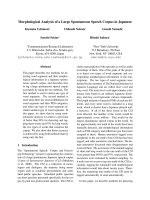
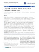
nuclease free water. The overall strategy of cloning and
construction of plasmids with specific promoters is
shown in Figure 1.
Cell culture
RAW 264.7 (National Center for Cell Sciences, Pune,
India) was maintained in high glucose DMEM with 10%
fetal bovine serum (FBS) (Gibco, USA). This cell line
was selected as a model to study expression in mouse
macrophage cell lines [27]. L929 was obtained from the
American Type Culture Collection (Rockville, MD) and
maintained in MEM (Sigma) with 10% F BS. This cell
line served as a modality to study expression in non-
macrophage cell [28]. All cultures were incubated at 37°
C, 5% CO
2
in humidified environment. Antibiotic free
mediawereusedduringtransfectionandforregular
maintenance of cells.
Table 1 Primers used for cloning to amplify promoters with underlined restriction sites.
Promoter
(Constructs)
Primer Sequence (5’ ® 3’) Restriction site
Macrosialin
(pAcGFP-MS)
F-T
ATTAATGACCAAATCTACAGGGAGAACCC VspI/Eco47III
R-
AGCGCTAGATGCTCAGACCAGCTA
EMR-1
(pAcGFP-EMR)
F-T
CATATGGAATTCTTTGTTTAGGTCTGTATGC NdeI/Eco47III
R-T
AGCGCTTACTGTGGCAGTCATTCA
Beta-5 Integrin
(pAcGFP-B5I)
F-CCG
ATTAATATTCAAACGCCTTAGGTAGGTTT VspI/Eco47III
R-
AGCGCTTCTACTCTCGGAGACCCT
F: Forward Primer, R: Reverse Primer, Underlined sequences are the restriction enzyme sites
Ahsan and Gore Genetic Vaccines and Therapy 2011, 9:10
/>Page 2 of 12
Transfection
Newly constructed pAcGFP-CMV, pAcGFP-MS, pAcGFP-
EMR, pAcGFP-B5I, pAcGFP-NIX plasmids were used for
transient transfection experiment. Transfections were per-
formed using Lipofecatmine ™ 2000 (Invitrogen, USA).
RAW264.7 and L929 cells were harvested and seeded in 6
well plates (3 × 10
5
cells/well). The plate was then incu-
bated for 16 hours and after reaching confluence wa s
transfected using 2 μg of each plasmid with 2 μlLipofecta-
mine 2000 as per manufacturer’s protocol. Opti-MEM
®
I
reduced serum media (Invitrogen, USA) was used as a
medium for transfection. Negative control was used both
for Lipofectamine 2000 and plasmid DNA.
Western blot
Expression of GFP protein was analysed by Western blot
using standard protocols. Briefly, 2 4 hours after trans-
fection with different DNA constructs encoding GFP,
RAW 264.7 cells were harvested, washed twice with
PBS, mixed with an equal volume of 2 ×loading buffer
and boiled for 10 min. Proteins form 50 μg of cell lysate
were separated onto a discontinuous SDS-polyarylamide
gel with 5% s tacking gel and 12% separating gel and
transferred to a nitrocellulose membrane (Amersham
Biosciences, USA). The membrane was blocked by 5%
skimmed milk powder in PBS and then incubated with
anti-GFP Ab (1:1000, Clontech) followed by goat anti-
mouse IgG-HRP conjugate (1:5000, Sigma). Bands were
visualized with substrate solution containing diamino-
benzidine tetrahydrochloride and H
2
O
2
solution.
Fluorescent Microscopy
Both RAW 264.7 and L929 cells were monitored for
GFP fluorescence at 6, 12, 24, 36 and 48 hours post
transfection under UV microscope (Nikon eclipse Ti).
The setting of microscope and camera was constant
throughout, so as t o get the semi-quantitative analysis.
The photograph was captured with following settings:
Resolution- Fast; Focus-640 × 480; Quality-2560 × 1920;
Mode-manual exposure; Exposure-800 ms; Gain-1.20×;
Objective-20×; Contrast- high. T he software used for
the analysis was: NIS-Elements BR version 3.1
pAcGFP-NIX
T4 DNA Ligase
VspI/Eco47III digested to excis
e
CMV promoter, end repaired &
self ligated
GFP Reporter
CMV Promoter
VspI
Eco47III
pAcGFP1-N1
VspI/Eco47III digested to
excise CMV promoter
pAcGFP1-N1
Digested
pAcGFP-CMV
Unmodified
MS
VspI
Eco47III
EMR
NdeI
Eco47III
B5I
VspI
Eco47III
pAcGFP-MS
pAcGFP-EMR
pAcGFP-B5I
T4 DNA Li
g
ase
T4 DNA Ligase
Figure 1 Schematic representation of reconstructed promoters constructs with GFP as a reporter gene.
Ahsan and Gore Genetic Vaccines and Therapy 2011, 9:10
/>Page 3 of 12
Flow cytometry
After transfection at different time points, cells were
harvested by trypsinization, washed twice with PBS and
suspended in FACS buffer (PBS + 2% FBS and 0.1%
sodium azide). All samples w ere analysed using FACS
Calibur (Becton Dickinson) and data were analysed
using CellQuest Pro (Becton Dickinson) software.
10,000 events were used for analysis. GFP was excited
through argon LASER and fluorescence was captured in
FL1 channel by using 530/30 nm bandpass filter. The
debr is and dead cells were excluded using FSC and SSC
param eters. Mean fluorescence was used to evaluate the
level of GFP expression above the threshold level of
autofluorescence of non-transfected control cells. For
each assay three independent transfections were per-
formed and mean fluorescence with ± SEM was used
for analysis.
Standardization of quantitative RT-PCR for detection of
GFP mRNA
Primer and probe design
Selected GFP sequences available in the GenBank were
aligned using MEGA4 software [29]. Primers and probe
were designed using Primer Express software™
3.0 (Applied B iosystems International, Foster City, CA)
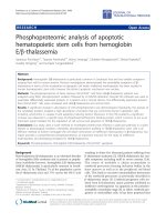
(Figure 2). Primers and probe were picked from GFP
sequence [GenBank: AY233272] nt. 196-295 with ampli-
con size of 100 bp. The probe was labelled with FAM
(5-carboxyfluorescein) at t he 5’en d and BHQ-1 (Black
hole quencher 1) at 3’end.
Preparation of RNA standard for the qRT-PCR
The 187 bp region was amplified using primer sets
(Table 2, Cloning) from vector pAcGFP1-N1 and cloned
into the pGEM
®
-T Easy cloning vector (Promega
Corporation, Madison, USA). The orientation of the
insert was confirmed by sequencing. Plasmid was line-
arised by SpeI re striction enzyme. Target sequence wa s
transcribed in vitro, DNAase treated and purified by
MEGAscript
®
kit (Ambion, USA) as per manufacturer’s
instructions. The RNA was quantified by spectrophoto-
metry. The c opy numbers of the RNA was calculated
based on the concentration and its molecular weight.
Ten fold serial dilutions of RNA from 10
2
to 10
10
copies
per reaction were used as standard in all qRT-PCRs.
qRT-PCR
After the desired period of post transfection, total RNA
was extracted from the cell pellet of RAW 264.7 and
L929 cells using RNEasy kit (Qiagen, Valencia, CA) and
DNAse t reated as per the manufacturer’sprotocol.RNA
was eluted i n 50 μl RNAse-free water and stored at -8 0°
C. 5 μl (300 ng) of total RNA was used for all qRT-PCR
for transfected cells. All reactions were carried out along
with standards. The assay was run in triplicates in Rotor-
Gene 3000 ™ (Corbett Researc h, Sydney, Australia) with
the following thermal steps, RT at 50°C for 15 min, initial
denaturation at 95°C for 2 min, 45 cycles of denaturation
at 95°C for 15 sec and annealing with extension at 60°C
for 30 sec. Fluorescence data were collected at the end of
each cycle. Each reaction comprise d no templ ate control
(NTC), cell control and cells treated with plasmid with-
out transfectant. Primers and probe were used from a
range of 100 to 600 nM for optimum concentration. CT
values were re corded each time. 200 nM of forward and
reverse primer with 100 nM of probe were found to be
optimal for one step qRT-PCR in 25 μL final reaction
volume. Optimised concentr ation of primer and probes
were used to detect the copy number of in vitro tran-
scribed RNA (IVT-RNA).
Forwar
d
pr
i
mer Pro
b
e
A
Y233272.1 CTACGGCGTG CAGTGCTTCT CACGCTACCC CGATCACATG AAGCAGCACG ACTTCTTCAA
A
B255038.1
A
Y533824.1
E
F441290.1 G .C C
X
83959.1 T T T T A A.A A T A T. T
A
F302837.1 G T TC.T A TG .GA.A A T A T. T
Reverse primer
A
Y233272.1 GAGCGCCATG CCTGAGGGCT ACATCCAGGA GCGCACCATC TT
A
B255038.1
A
Y533824.1
E
F441290.1 .TC C A G
X
83959.1 T C A T. .TG.A AA.A T A
A
F302837.1 T C A T. .TG.A AA.A T A
Figure 2 Sequence alignment of GFP variants in GenBank showing the location of primers and probe. GFP sequences were selected
from data bank and aligned using MEGA4 software. The references of sequences are mentioned with the Accession number of GenBank. The
sequence used for the primer and probe design was: Accession number-AY233272, GI-34421677.
Ahsan and Gore Genetic Vaccines and Therapy 2011, 9:10
/>Page 4 of 12
Statistics
All the data obtained thr ough Flow cytomet ry or qRT-
PCR was analysed for statistical significance using Gen-
eral Linear model, Tukey’s comparison test. Analysis
was performed using SPSS version 11 software.
Results
Selection of promoter
Following promoters were selected for the studies based
on their known expression profiles. Macrosialin is a glyco-
protein expressed specifically in murine monocytes and
macrophages, and to a lesser extent by DC [30-32].
Macrosialin is murine homologue of CD 68 sharing 80%
similarity [32]. Emr-1 (EMR) promoter is reported to con-
trol its gene expression mainly in macrophages [33-35].
The human orthologue of EmrI is EMRI. The promoter of
EmrI and EMRI share 60% identity and is with purine rich
conserved region. Its gene product has also served as a
marker for macrophage population in many immunohisto-
logical studies [36]. Beta-5 Integrin promoter is expressed
in macrophages and osteoblasts [37,38]. Integrin belongs
tothefamilyoftypeItransmembraneglycoprotein.It
helps in cell migration, proliferation and differentiation.
As a positive control we chose immediate early promoter
of cytomegalovirus (CMV) which is widely used and is
strong enough to drive constitutive expression in all cell
types. As a negative control promoterless vector was con-
structed. This vector though has GFP as a reporter gene
but is devoid of any promoter. All the selected promoters
except CMV are TATA-less promoters and have PU.1 as
a transcription factor which assembles the transcription
machinery on myeloid promoters.
Promoter amplification from genomic DNA and
expression studies of various promoter constructs
Promoter sequences were amplified from RAW264.7
cells using Tri-reagent (MRC) and PCR. Amplicons
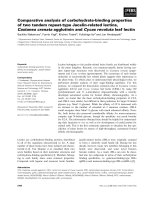
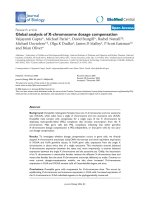
obtained are shown in Figure 3. These were further
used for cloning after sequence confirmation. Expression
of GFP with different promoter constructs was analysed
by fluorescent microscopy. Strong GFP expression was
detected with pAcGFP-CMV in RAW264.7 and L929
cells, in contrast no GFP expression was observed with
pAcGFP-NIX or Untransfected cells at any time point
Table 2 Nucleotide sequence of primers and probe used in pGEM-T Easy cloning and qRT-PCR assay
Assay Primer/Probe Sequence (5’® 3’) Nucleotide positions
qRT-PCR Forward Primer TACGGCGTGCAGTGCTTCT 196-214
Reverse Primer AGATGGTGCGCTCCTGGAT 277-295
TaqMan Probe CTACCCCGATCACATGAAGCAGCACG 219-244
Cloning Forward Primer AAGTTCATCTGCACCAC 133-149
Reverse Primer TGTAGTTGCCGTCATCCT 302-319
1
M 2 3
M
4
2063 bp
1035 bp 973 bp
1000 bp
1000 bp
Figure 3 PCR analysis of amplified promoters. M: 1 Kb+ Ladder (Invitrogen); 2: Macrosialin; 3: Beta-5 Integrin; 4: EMR1 are the respective
amplicons of promoters documented in 1% Agarose gel in TAE buffer.
Ahsan and Gore Genetic Vaccines and Therapy 2011, 9:10
/>Page 5 of 12
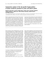
of studies (Figure 4). Figure shows representative pic-
tures taken at different time point for each cell type
(06-48 hrs following transfection) (Figure 4, A, B, C, D
and 4E). Fluoresce nce of cells transfected with pAcGFP-
MS was significantly higher than other modified
constructs expressing GFP. The difference in fluores-
cence intensities were observed when the same con-
structs were used for RAW 264.7 and L929 cells. As
expected non macrophage celllineL929showedlesser
expression of GFP driven by APC promoters.
C
MV M
S
EMR B5I NIX
CC
A
B
C
D
E
R
L
R
R
R
R
L
L
L
L
Figure 4 Fluorescent Microscopy pictures of cells transfected with respective plasmid. Expression of GFP in transfected RAW 264.7 (R) and
L929 (L) cells at different time points as: A-6 hrs, B-12 hrs, C-24 hrs, D-36 hrs and E-48 hrs. The constructs for the transfected cells are mentioned
at the top which follows throughout the respective column followed by (pAcGFP-). CC represents cell control.
Ahsan and Gore Genetic Vaccines and Therapy 2011, 9:10
/>Page 6 of 12
Western blot
The transfected RAW 264.7 cell lysates prepared after
24 hours post transfection were subjected to Western
blot analysis. The anti-GFP monoclonal antibody reacted
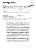
specifically with GFP protein of ~2 7 kDa. Negative con-
trol did not show detectable levels of GFP. Strong
expression of GFP under CMV promoter, se rved as a
positive control (Figure 5, A and 5B).
Flow cytometry analysis of GFP with different promoter
constructs
Preliminary screening was performed using fluorescent
microscope, gre en fluorescence was ob served in cells
transfected with respective constructs, confirming the
successful protein expression. Precise specificity and
strength of the promoter constructs were evaluated by
Flow cytometry through transient transfectio n in
RAW264.7 and L929 cells. MFI of pAcGFP-CMV con-
struct after 24 hours was 11 fold in RAW 264.7 and 8.8
fold in L929 cells over that of Untransfected cells,
whereas 6 fold and 2 fold in RAW264.7 and L929 cells
respectively for pAcGFP-MS (Figure 6, A and 6B). The
MFI of cells transfected with diffe rent constructs was sig-
nificantly higher (p <0.05) when compared with Untrans-
fected cells. No significant difference was observed
between pAcGFP-NIX and Untran sfected cells at any
time point of studies. The differential level of expression
of pAcGFP-MS when compared in RAW 264.7 and L929
cells, was found to be highly significant up to 36 hours.
Similarly it was significant for pAcGFP-B5I up to 48
hours and non-significant for pAcGFP-EMR at all time
points. For the comparative accoun t of promot er specifi -
city we have also used ratio of promoter activity in
macrophage to that of non macrophage cells (Figure 6C).
Among the promoters under stud y macrosialin promoter
drove the high expression of reporter gene and conferred
the highest myeloid specificity. This ratio could not be
taken as absolute values due to variance in transfection
efficiency in both the cell lines, rather it rendered a useful
index of specificity.
Studies were also carried out using P388D1 and Vero
cells as a macrophage and non macrophag e cells respec-
tively. Fluorescent microscopy showed the same trend of
expression with different con str ucts (dat a not shown). It
was difficult to transfect P388D1 cell line when the proto-
col mentioned above for the other c ells were followed.
The efficiency of transfection was very low. Increasing the
Lipofectamine 2000 concentration increased the efficiency
slightly. The expression levels directed by the promoters
were highest after 24 hours. Intensity of GFP expression
through CMV promoter was the highest followed by
macrosialin and the ot her two promoters, following the
same trend of expression as that of RAW264.7. Similarly,
expression level in Vero cells was same as L929 cells, how-
ever, they got transfected with ease. Hence we carried out
our further study on RAW264.7 and L929 cells.
M 1 2 3 4 5
2
8kDa
36kDa
A
B
M 1 2 3 4 5
Figure 5 PAGE/Western blot Analysis. (A) 12% SDS-PAGE gel (B) Western blot analysis of the total cell lysates of the RAW 264.7 cells. M :
PageRuler™ (Fermentas); 1: pAcGFP-CMV; 2: pAcGFP-MS; 3: pAcGFP-EMR; 4: pAcGFP-B5I; 5: pAcGFP-NIX. The blot shows expressed GFP protein
from different constructs after 24 hours of transfection.
Ahsan and Gore Genetic Vaccines and Therapy 2011, 9:10
/>Page 7 of 12
A
B
C
Figure 6 Flow cytome try analysis. Mean fluorescence of cells of different cons tructs transfected in (A) RAW264.7, (B) L929 cells. (C) Ratio of
(RAW264.7/L929) were determined as an expression of macrophage specificity. The activity was measured at various time points. The average
and SEM shown are from three independent assays. For ststistical analysis, General Linear Model (GLM), Tukey’s comparison test was performed
to compare the significance difference on fluorescence level amongst transfected plasmid.
Ahsan and Gore Genetic Vaccines and Therapy 2011, 9:10
/>Page 8 of 12
Quantification of GFP in transfected cells
The assay was sensitive enough to detect <100 copies of
IVT-RNA (CT = 38.59 ). Linear correlation value in CT
values obtained over the range of IVT-RNA per reaction
was (R
2
= 0.99), when 10
2
to 10
10
copies were used. The
assaydidnotamplifyanynonspecificsequencefrom
cellular RNA of cells used. There were clean bands of
amplicons when observed in agarose gel electrophoresis.
To check the reproducibility of the assay the standards
were run on six different days and similar CT values
were found for the given inputs of IVT-RNA. The data
is the representative of the test (Figure 7).
RNA was quantified post transfection after 12, 24 and
48 hours. It wa s observed that GFP in construct with
CMV promoter was highly expressed in both RAW 264.7
and L929 cells (5.07^5 vs 8.94^6). The construct with
macrosialin promoter showed >36 fold copy numbers in
RAW264.7 cells in comparison to L929 cells at the end
of 48 hrs. Data represented here is from analysis of three
independent transfection assays with ±SEM. O ne way
ANOVA, Tukey’ s comparison test was performed to
compare the GFP transcripts in cells transfected with dif-
ferent construct. pAcGFP-CMV and pAcGFP-MS has a
significantly higher number of GFP transcripts compared
with Untransfected or pAcGFP-NIX construct (P <0.05).
We get no amplification in Untransfected and pAcGFP-
NIX (Figure 8, A and 8B)
Discussion
The promoters of viruses are widely used in many mam-
malian expression vectors due to their strong activity in
large variety of cells. CMV promoter has been of choice
because of high level of constitutive expression in
several mammalian cell lines [39]. Constitutive expres-
sion of gene could be suitable for gene therapy or cer-
tain applications [40]. However importance of using
lineage specific promoter in DNA vaccine to limit gene
expression to the target cells is of paramount impor-
tance,notonlyasanadjuvant[41]butalsoasasafety
concern [42].
In the present study, we have compared the activity of
promoters mainly active in macrophages, delineated as a
macrophage expressing promoters. GFP gene as a quan-
titative reporter was used to evaluate the strength of
promoters. Vectors were engineered with different pro-
moters coding GFP readout for the study. pAcGFP-
CMV has a strong CMV immediate-early promoter and
wasusedasapositivecontrol.Three(pAcGFP-MS,
pAcGFP-EMR, pAcGFP-B5I) aforesaid promoter con-
structs with GFP reporter were compared. pAcGFP-NIX
without promoter bu t with GFP gene was constructed
as a negative control.
RAW264.7 cells (macrophage) and L929 (fibroblast)
cells were selected for the study. These cell lines were
selected to evaluate the behaviour of promoters in
macrophage and non-macrophage cells respectively.
Comparison of GFP expression through CMV promoter
simultaneously in both the cells also helped us to ana-
lyze the difference in expression level due to difference
in transfection efficiency.
To evaluate the activity of promoters under study, fluor-
escent microscopic analysi s of GFP expre ssing cells were
carried out. Fluorescence of GFP increased based on the
expression which correlates to the activity of respective
promoter. Besides the visual confirmation, functionality of
GFP gene standard curve
R
2
=
0.997
Figure 7 Standard curve plot of log10 diluted in vitro transcribed RNA for GFP.
Ahsan and Gore Genetic Vaccines and Therapy 2011, 9:10
/>Page 9 of 12
all the promoter constructs was confirmed by Western
blot of GFP which agreed to the microscopic analysis.
In order to assess the expression over large population
of cells and achieve more sensitive data, flow cytometry
was carried out for such differential expression. Mean
fluorescent intensity (MFI) which was used for data acqui-
sition is the average of certain number of cells obtained
from individual cells in the population; such analysis pro-
vides the reproducible method to quantitate changes in
repo rter gene expression from a populati on. The expres -
sion of GFP by CMV promoter was robust in both the
cells at all time points in comparison to other promoters
(Figure 4). Among the macrophage specific promoter
expression in RAW264.7, macrosialin showed higher
expression followed by the other two constru cts. Kinetics
of promoter activity was assessed by evaluating reporter
expression at various time points after transfection. All the
constructs exhibited gradual increase in activity up to
A
B
Figure 8 Transcri pt profiling of GFP. Transcript profiling of RAW264.7 (A) and L929 (B) ce lls transfected with different promoter constructs at
the given time interval.
Ahsan and Gore Genetic Vaccines and Therapy 2011, 9:10
/>Page 10 of 12
24 hours, which decreased further. The expression of
macrosialin promoter was significantly higher in macro-
phage cell line in comparison to non-macrophage cells.
The ratio of macrophage/non-macrophage evaluation was
the highest in macrosalin as an indicator of macrophage
specificity. After 24 hours of analysis there was a decreas-
ing trend in expression. The probable reason could be due
to cells reaching confluence and underwent death, more-
over the effect of toxicity of transfectant also increased
over time. GFP fluorescence in creased a s per increase in
protein concentration which was well depicted by Flow
cytometry and fluorescent microscopy as reported earlier
[43].
In order to understand the transcriptional activity of
promoter, GFP mRNA levels were quantitated by one step
qRT-PCR with TaqMan based probe chemistry developed
in house. It supported the data obtained by Flow cytome-
try. Highest expression at all time point through CMV
promoter was observed, followed by macrosialin. There
was increasing trend in mRNA expression as per time, till
48 hours, however, after 72 h ours no transcripts were
detected. This difference may be attributed due to several
reasons i.e. increase in level of toxicity over the time, there
was no tight control over mRNA expression hence rigor-
ous c ontrol over translation, all the mRNA were not
turned into protein. The correlation at RNA and protein
level depends upon the balance of transcriptional and
translational regulatory elements [44]. The cessation of the
expression of gene after limited expression could be bene-
ficial for in vivo studies to avoid continued sustenance of
antigen, since prolonged Ag exposure might lead to lower
affinity Ab. Thus among the promoters under study,
macrosialin directed the macrophage dominant expression
in terms of both transcription and translation. Macrosialin
governed the highest expression when compared with
either EmrI or Beta-5 Integrin promoters. Regardless of
the observed difference between mRNA or protein level,
our finding clearly shows that macrosialin dominantly gov-
ern the expression in macrophage derive d cells. It might
be poss ible to use this promoter for directing expression
of desired protein dominantly in APC.
Successful demonstration of APC dominant expression
of GFP has opened an avenue to construct plasmids
with virus encoded proteins. Use of these plasmids to
evaluate the effect of cell dominant expression on the
immune response and indication of protective ability
would be interesting. In addition, targeting macrophage
for various applications including immunotherapy might
also be explored.
Conclusions
To determine whether APC expressing promoters could
be useful in terms of its specificity and activity, we com-
pared with the CMV immediate early promoter in
macrophage and non-macrophage derived cells. The activ-
ity of macrosialin was significantly higher in macrophage
cells in comparison to EmrI and Beta-5 Integrin, whereas
CMV showed the highest activity in both the cell types.
Our work presents a systemat ic ex vivo study at the level
of protein expression and mRNA transcription. This indi-
cates that macrosialin promoter might prove beneficial for
targeting expression majorly in APC, however in vivo
potential needs to be carried out for its suitable
application.
Acknowledgements
Ahsan MF acknowledges Indian Council of Medical Research, Government of
India, for providing Senior Research Fellowship (SRF) and National Institute
of Virology, Pune, for the extended fellowship. We thankfully acknowledge
Dr. Bondre VP and Dr. Sapkal GN for their support and Mr. Walimbe AM for
his help in statistical analysis of the data. We gratefully appreciate the
intellectual discussion and suggestions by Dr. Satyendra K and his kind help.
We thank Dr. Gurukumar KR, Mr. Devhare P, Mr. Fulmali P for the discussion
and Mr. Subhashis C for his help in Fluorescent Microscopy. We also thank
Mr. Ayachit, Mr. Naidu J, Ms. Reshma, Ms. Daya, Mr. Roopesh and Ms. Harini.
Authors’ contributions
MFA has planned, designed and carried out all the experiments. MMG
envisioned and supervised all the studies. Both the authors read and
approved the final manuscript.
Competing interests
The authors declare that they have no competing interests.
Received: 28 February 2011 Accepted: 18 June 2011
Published: 18 June 2011
References
1. Chikhlikar P, Barros de Arruda L, Maciel M, Silvera P, Lewis MG, August JT,
Marques ET: DNA encoding an HIV-1 Gag/human lysosome-associated
membrane protein-1 chimera elicits a broad cellular and humoral
immune response in Rhesus macaques. PLoS One 2006, 1:e135.
2. Okura Y, Miyakoshi A, Kohyama K, Park IK, Staufenbiel M, Matsumoto Y:
Nonviral Abeta DNA vaccine therapy against Alzheimer’s disease: Long-
term effects and safety. Proc Natl Acad Sci USA 2006, 103(25):9619-9624.
3. McKinney KA, Al-Rawi N, Maciag PC, Banyard DA, Sewell DA: Effect of a
novel DNA vaccine on angiogenesis and tumor growth in vivo. Arch
Otolaryngol Head Neck Surg 2010, 136(9):859-864.
4. Li G, Liu Z, Zhong N, Liao B, Xiong Y: Therapeutic effects of DNA vaccine
on allergen-induced allergic airway inflammation in mouse model. Cell
Mol Immunol 2006, 3(5):379-384.
5. Shiau JW, Tang TK, Shih YL, Tai C, Sung YY, Huang JL, Yang HL: Mice
immunized with DNA encoding a modified Pseudomonas aeruginosa
exotoxin A develop protective immunity against exotoxin intoxication.
Vaccine 2000, 19(9-10):1106-1112.
6. Tung WS, Bakar SA, Sekawi Z, Rosli R: DNA vaccine constructs against
enterovirus 71 elicit immune response in mice. Genet Vaccines Ther 2007,
5:6.
7. Ishii N, Fukushima J, Kaneko T, Okada E, Tani K, Tanaka SI, Hamajima K,
Xin KQ, Kawamoto S, Koff W, Nishioka K, Yasuda T, Okuda K: Cationic
liposomes are a strong adjuvant for a DNA vaccine of human
immunodeficiency virus type 1. AIDS Res Hum Retroviruses 1997,
13(16):1421-1428.
8. Sato Y, Roman M, Tighe H, Lee D, Corr M, Nguyen MD, Silverman GJ,
Lotz M, Carson DA, Raz E: Immunostimulatory DNA sequences necessary
for effective intradermal gene immunization. Science 1996,
273(5273):352-354.
9. Iwasaki A, Stiernholm BJ, Chan AK, Berinstein NL, Barber BH: Enhanced CTL
responses mediated by plasmid DNA immunogens encoding
costimulatory molecules and cytokines. J Immunol 1997,
158(10):4591-4601.
Ahsan and Gore Genetic Vaccines and Therapy 2011, 9:10
/>Page 11 of 12
10. Okada E, Sasaki S, Ishii N, Aoki I, Yasuda T, Nishioka K, Fukushima J,
Miyazaki J, Wahren B, Okuda K: Intranasal immunization of a DNA vaccine
with IL-12- and granulocyte-macrophage colony-stimulating factor
[GMCSF]-expressing plasmids in liposomes induces strong mucosal and
cell-mediated immune responses against HIV-1 antigens. J Immunol
1997, 159(7):3638-3647.
11. Sha Z, Vincent MJ, Compans RW: Enhancement of mucosal immune
responses to the influenza virus HA protein by alternative approaches to
DNA immunization. Immunobiology 1999, 200(1):21-30.
12. Livingston JB, Lu S, Robinson H, Anderson DJ: Immunization of the female
genital tract with a DNA-based vaccine. Infect Immun 1998, 66(1):322-329.
13. Tuting T, Storkus WJ, Falo LD Jr: DNA immunization targeting the skin:
Molecular control of adaptive immunity. J Invest Dermatol 1998,
111(2):183-188.
14. Ji H, Wang TL, Chen CH, Pai SI, Hung CF, Lin KY, Kurman RJ, Pardoll DM,
Wu TC: Targeting human papillomavirus type 16 E7 to the endosomal/
lysosomal compartment enhances the antitumor immunity of DNA
vaccines against murine human papillomavirus type 16 E7-expressing
tumors. Hum Gene Ther 1999, 10(17):2727-40.
15. Felix NJ, Suri A, Salter-Cid L, Nadler SG, Gujrathi S, Corbo M, Aranda R:
Targeting lymphocyte co-stimulation: from bench to bedside.
Autoimmunity 2010, 43(7):514-525.
16. Lori F, Kelly LM, Lisziewicz J: APC-targeted immunization for the
treatment of HIV-1. Expert Rev Vaccines 2004, 3(4 Suppl):S189-198.
17. Ni J, Nolte B, Arnold A, Fournier P, Schirrmacher V: Targeting anti-tumor
DNA vaccines to dendritic cells via a short CD11c promoter sequence.
Vaccine 2009, 27(40):5480-5487.
18. Bonkobara M, Zukas PK, Shikano S, Nakamura S, Cruz PD Jr, Ariizumi K:
Epidermal Langerhans cell-targeted gene expression by a dectin-2
promoter. J Immunol 2001, 167(12):6893-6900.
19. Kimura T, Koya RC, Anselmi L, Sternini C, Wang HJ, Comin-Anduix B,
Prins RM, Faure-Kumar E, Rozengurt N, Cui Y, Kasahara N, Stripecke R:
Lentiviral vectors with CMV or MHCII promoters administered in vivo:
immune reactivity versus persistence of expression. Mol Ther 2007,
15(7):1390-1399.
20. Brocker T, Riedinger M, Karjalainen K: Driving gene expression specifically
in dendritic cells. Adv Exp Med Biol 1997, 417:55-57.
21. Hon H, Oran A, Brocker T, Jacob J: B lymphocytes participate in cross-
presentation of antigen following gene gun vaccination. J Immunol 2005,
174(9):5233-5242.
22. Lauterbach H, Gruber A, Ried C, Cheminay C, Brocker T: Insufficient APC
capacities of dendritic cells in gene gun-mediated DNA vaccination. J
Immunol 2006, 176(8):4600-4607.
23. Pozzi LA, Maciaszek JW, Rock KL: Both dendritic cells and macrophages
can stimulate naive CD8 T cells in vivo to proliferate, develop effector
function, and differentiate into memory cells. J Immunol 2005,
175(4):2071-2081.
24. Kovacsovics-Bankowski M, Clark K, Benacerraf B, Rock KL: Efficient major
histocompatibility complex class I presentation of exogenous antigen
upon phagocytosis by macrophages. Proc Natl Acad Sci USA 1993,
90(11):4942-4946.
25. Constant S, Schweitzer N, West J, Ranney P, Bottomly K: B lymphocytes
can be competent antigen-presenting cells for priming CD4+ T cells to
protein antigens in vivo. J Immunol 1995, 155(8):3734-3741.
26. Ducrest AL, Amacker M, Lingner J, Nabholz M: Detection of promoter
activity by flow cytometric analysis of GFP reporter expression. Nucleic
Acids Res 2002, 30(14):e65.
27. Weeratna RD, Wu T, Efler SM, Zhang L, Davis HL: Designing gene therapy
vectors: avoiding immune responses by using tissue-specific promoters.
Gene Ther 2001, 8(24):1872-1878.
28. Billingsley KG, Fraker DL, Strassmann G, Loeser C, Fliot HM, Alexander HR:
Macrophage-derived tumor necrosis factor and tumor-derived of
leukemia inhibitory factor and interleukin-6: possible cellular
mechanisms of cancer cachexia. Ann Surg Oncol 1996, 3(1):29-35.
29. Tamura K, Dudley J, Nei M, Kumar S: Molecular Evolutionary Genetics
Analysis [MEGA] software version 4.0. Mol Biol Evol 2007, 24(8):1596-1599.
30. Li AC, Guidez FR, Collier JG, Glass CK: The macrosialin promoter directs
high levels of transcriptional activity in macrophages dependent on
combinatorial interactions between PU.1 and c-Jun. J Biol Chem 1998,
273(9):5389-5399.
31. Rabinowitz SS, Gordon S: Macrosialin, a macrophage-restricted
membrane sialoprotein differentially glycosylated in response to
inflammatory stimuli. J Exp Med 1991, 174(4):827-836.
32. Holness CL, da Silva RP, Fawcett J, Gordon S, Simmons DL: Macrosialin, a
mouse macrophage-restricted glycoprotein, is a member of the lamp/
lgp family. J Biol Chem 1993, 268(13):9661-9666.
33. O’Reilly D, Addley M, Quinn C, MacFarlane AJ, Gordon S, McKnight AJ,
Greaves DR: Functional analysis of the murine Emr1 promoter identifies
a novel purine-rich regulatory motif required for high-level gene
expression in macrophages. Genomics 2004, 84(6):1030-1040.
34. Schaller E, Macfarlane AJ, Rupec RA, Gordon S, McKnight AJ, Pfeffer K:
Inactivation of the F4/80 glycoprotein in the mouse germ line. Mol Cell
Biol 2002, 22(22):8035-8043.
35. McKnight AJ, Macfarlane AJ, Dri P, Turley L, Willis AC, Gordon S: Molecular
cloning of F4/80, a murine macrophage-restricted cell surface
glycoprotein with homology to the G-protein-linked transmembrane 7
hormone receptor family. J Biol Chem 1996, 271(1):486-489.
36. Hirsch S, Austyn JM, Gordon S: Expression of the macrophage-specific
antigen F4/80 during differentiation of mouse bone marrow cells in
culture. J Exp Med 1981, 154(3):713-25.
37. Feng X, Teitelbaum SL, Quiroz ME, Cheng SL, Lai CF, Avioli LV, Ross FP:
Sp1/Sp3 and PU.1 differentially regulate beta[5] integrin gene
expression in macrophages and osteoblasts. J Biol Chem 2000,
275(12):8331-8340.
38. Feng X, Teitelbaum SL, Quiroz ME, Towler DA, Ross FP: Cloning of the
murine beta5 integrin subunit promoter. Identification of a novel
sequence mediating granulocyte-macrophage colony-stimulating factor-
dependent repression of beta5 integrin gene transcription. J Biol Chem
1999, 274(3):1366-1374.
39. Boshart M, Weber F, Jahn G, Dorsch-Häsler K, Fleckenstein B, Schaffner W: A
very strong enhancer is located upstream of an immediate early gene
of human cytomegalovirus. Cell 1985, 41(2):521-530.
40. Mizuguchi H, Xu ZL, Sakurai F, Mayumi T, Hayakawa T: Tight positive
regulation of transgene expression by a single adenovirus vector
containing the rtTA and tTS expression cassettes in separate genome
regions. Hum Gene Ther 2003, 14(13):1265-1277.
41. Ross R, Sudowe S, Beisner J, Ross XL, Ludwig-Portugall I, Steitz J, Tüting T,
Knop J, Reske-Kunz AB: Transcriptional targeting of dendritic cells for
gene therapy using the promoter of the cytoskeletal protein fascin. Gene
Ther 2003, 10(12):1035-1040.
42. Glenting J, Wessels S: Ensuring safety of DNA vaccines. Microb Cell Fact
2005, 4:26.
43. Furtado A, Henry R: Measurement of green fluorescent protein
concentration in single cells by image analysis. Anal Biochem 2002,
310(1):84-92.
44. Shen Y, Iqbal J, Huang JZ, Zhou G, Chan WC: BCL2 protein expression
parallels its mRNA level in normal and malignant B cells. Blood 2004,
104(9):2936-2939.
doi:10.1186/1479-0556-9-10
Cite this article as: Ahsan and Gore: Comparative analysis of
macrophage associated vectors for use in genetic vaccine. Genetic
Vaccines and Therapy 2011 9:10.
Submit your next manuscript to BioMed Central
and take full advantage of:
• Convenient online submission
• Thorough peer review
• No space constraints or color figure charges
• Immediate publication on acceptance
• Inclusion in PubMed, CAS, Scopus and Google Scholar
• Research which is freely available for redistribution
Submit your manuscript at
www.biomedcentral.com/submit
Ahsan and Gore Genetic Vaccines and Therapy 2011, 9:10
/>Page 12 of 12