Báo cáo y học: "Repetitive DNA is associated with centromeric domains in Trypanosoma brucei but not Trypanosoma cruzi" pot
Bạn đang xem bản rút gọn của tài liệu. Xem và tải ngay bản đầy đủ của tài liệu tại đây (863.71 KB, 14 trang )
Genome Biology 2007, 8:R37
comment reviews reports deposited research refereed research interactions information
Open Access
2007Obadoet al.Volume 8, Issue 3, Article R37
Research
Repetitive DNA is associated with centromeric domains in
Trypanosoma brucei but not Trypanosoma cruzi
Samson O Obado
*
, Christopher Bot
*
, Daniel Nilsson
†
, Bjorn Andersson
†
and
John M Kelly
*
Addresses:
*
Department of Infectious and Tropical Diseases, London School of Hygiene and Tropical Medicine, Keppel Street, London WC1E
7HT, UK.
†
Center for Genomics and Bioinformatics, Karolinska Institutet, Berzelius vag, S-171 77 Stockholm, Sweden.
Correspondence: John M Kelly. Email:
© 2007 Obado et al.; licensee BioMed Central Ltd.
This is an open access article distributed under the terms of the Creative Commons Attribution License ( which
permits unrestricted use, distribution, and reproduction in any medium, provided the original work is properly cited.
Repetitive DNA and centromeres in trypanosomes<p>Centromeres in <it>Trypanosoma cruzi </it>and <it>Trypanosoma brucei </it>can be localised to regions between directional gene clusters that contain degenerate retroelements, and in the case of <it>T. brucei</it>, repetitive DNA.</p>
Abstract
Background: Trypanosomes are parasitic protozoa that diverged early from the main eukaryotic
lineage. Their genomes display several unusual characteristics and, despite completion of the
trypanosome genome projects, the location of centromeric DNA has not been identified.
Results: We report evidence on the location and nature of centromeric DNA in Trypanosoma cruzi
and Trypanosoma brucei. In T. cruzi, we used telomere-associated chromosome fragmentation and
found that GC-rich transcriptional 'strand-switch' domains composed predominantly of degenerate
retrotranposons are a shared feature of regions that confer mitotic stability. Consistent with this,
etoposide-mediated topoisomerase-II cleavage, a biochemical marker for active centromeres, is
concentrated at these domains. In the 'megabase-sized' chromosomes of T. brucei, topoisomerase-
II activity is also focused at single loci that encompass regions between directional gene clusters
that contain transposable elements. Unlike T. cruzi, however, these loci also contain arrays of AT-
rich repeats stretching over several kilobases. The sites of topoisomerase-II activity on T. brucei
chromosome 1 and T. cruzi chromosome 3 are syntenic, suggesting that centromere location has
been conserved for more than 200 million years. The T. brucei intermediate and minichromosomes,
which lack housekeeping genes, do not exhibit site-specific accumulation of topoisomerase-II,
suggesting that segregation of these atypical chromosomes might involve a centromere-
independent mechanism.
Conclusion: The localization of centromeric DNA in trypanosomes fills a major gap in our
understanding of genome organization in these important human pathogens. These data are a
significant step towards identifying and functionally characterizing other determinants of
centromere function and provide a framework for dissecting the mechanisms of chromosome
segregation.
Published: 12 March 2007
Genome Biology 2007, 8:R37 (doi:10.1186/gb-2007-8-3-r37)
Received: 3 November 2006
Revised: 16 January 2007
Accepted: 12 March 2007
The electronic version of this article is the complete one and can be
found online at />R37.2 Genome Biology 2007, Volume 8, Issue 3, Article R37 Obado et al. />Genome Biology 2007, 8:R37
Background
Centromeres are the chromosomal loci where kinetochores
are assembled. The centromere/kinetochore complex is the
anchor for attachment of the microtubule spindles that facil-
itate segregation. Two main classes of centromere have been
identified. In most eukaryotes, centromeres are 'regional' and
can encompass large regions of chromosomal DNA, ranging
from 0.3-15 Mb in species as diverse as plants, insects and
mammals [1]. In microorganisms, regional centromeres are
also extensive; in Schizosaccharomyces pombe they cover 35-
110 kb [2]. Less common are 'point' centromeres, such as
those in Saccharomyces cerevisiae, where specific 125 base-
pair (bp) elements are sufficient for spindle attachment [3]. A
few organisms, including Caenorhabditis elegans, lack spe-
cific centromeric domains and have holocentric chromo-
somes, where microtubules bind along the entire length of the
chromosome [4].
Regional centromeres generally contain long stretches of
repetitive DNA, often interrupted by retrotransposons. For
example, the human × chromosome has a conserved core of
α-satellite repeats (approximately 170 bp) stretching over 2-4
Mb and flanked by long regions with multiple retrotranspo-
son insertions [5]. In S. pombe, centromeres are structured as
chromosome-specific core elements, flanked by inverted
repeats of approximately 3-7 kb, which in turn are flanked by
more extensive outer repeats [2]. Some features of centro-
mere organization are widespread, although there is little
conservation at the level of DNA sequence [6]. The observa-
tion that inheritable neocentromeres in human cells can form
at loci lacking α-satellite repeats suggests that epigenetic fac-
tors must be major determinants of centromere identity [7].
Neocentromeres have also been observed in other species,
including insects and plants.
Topoisomerase-II (Topo-II) is thought to have an important
role in centromere function [8-10]. During metaphase the
enzyme accumulates specifically at active centromeres, where
it has been implicated in maintaining kinetochore/centro-
mere structure and decatenation of sister chromatids [9,11-
13]. Decatenation involves double-stranded DNA cleavage,
passage of the unbroken helix of the duplex through the gap,
and re-ligation to repair the lesion [14]. This activity can be
blocked by etoposide, which inhibits the re-ligation step,
thereby promoting double-stranded DNA breaks in the chro-
mosome at sites specified by Topo-II binding. As a result,
etoposide has been used to map active centromeres and as a
tool to explore the key role of Topo-II in centromere function
[15-18]. In Plasmodium falciparum, Topo-II activity concen-
trates at single chromosomal loci that encompass 2 kb AT-
rich domains previously identified as candidate centromeres
[19].
Protozoan parasites of the family Trypanosomatidae are the
causative agents of African sleeping sickness (Trypanosoma
brucei), American trypanosomiasis (Trypanosoma cruzi)
and leishmaniasis (Leishmania spp.), diseases that affect
more than 30 million people, mainly in the developing world.
Trypanosomatids are early diverging eukaryotes and share
several unusual genetic traits [20]. Protein coding genes lack
RNA polymerase II-mediated promoters, transcription is
polycistronic and all mRNAs are post-transcriptionally mod-
ified by addition of a 5'-spliced leader RNA. Directional gene
clusters often stretch over hundreds of kilobases. Trypano-
somes exhibit significant intra-strain variation in chromo-
some size, and although generally diploid, chromosome
homologues can differ considerably in length. T. cruzi has a
haploid genome size of 55 Mb and approximately 30 chromo-
somes. The precise number has been difficult to determine
because of size heterogeneity, recombination and, in some
instances, triploidy [21,22]. In T. brucei (haploid genome size
25 Mb), unusually there are 3 chromosome classes; 11 homol-
ogous pairs (1-6 Mb) that contain the actively expressed
genes, 3-5 intermediate-sized chromosomes (0.2-0.7 Mb)
that contain some variable surface glycoprotein (VSG)
expression sites, but lack housekeeping genes, and approxi-
mately 100 minichromosomes (approximately 0.1 Mb) which
may be a reservoir for VSG sequences. The trypanosome
sequencing projects have been completed [21,23]. A striking
feature of genome organization is the high level of synteny.
However, sequence elements that could have a role in chro-
mosome segregation were not recognized. Furthermore,
there are no obvious homologues of the core proteins that dis-
play constitutive centromere location in other eukaryotes
[1,23].
To identify T. cruzi sequence elements with centromeric
properties, we previously used telomere-associated chromo-
some fragmentation to delineate a region of chromosome 3
required for mitotic stability [22]. A major feature of this
locus is a 16 kb GC-rich transcriptional 'strand-switch'
domain composed predominantly of degenerate retroele-
ments that separates two large directional gene clusters that
are transcribed towards the telomeres. We proposed that this
type of organization could serve as a model for centromeric
DNA. The fragmented nature of the T. cruzi genome dataset
[21] has negated testing of this hypothesis. Here, we demon-
strate that an analogous region of chromosome 1 is required
for mitotic stability and that the locations of these putative
centromeres on both chromosomes coincide with sites of
etoposide-mediated Topo-II cleavage. Furthermore, we show
that Topo-II activity on T. brucei chromosomes also localizes
to regions between directional gene clusters that contain
degenerate retroelements, and additionally a domain of
repetitive DNA.
Results
Similarity between the regions of T. cruzi
chromosomes 1 and 3 required for mitotic stability
Chromosome 1 occurs as 0.51 Mb and 1.2 Mb homologues in
the T. cruzi genome reference clone CL Brener. Because of the
Genome Biology 2007, Volume 8, Issue 3, Article R37 Obado et al. R37.3
comment reviews reports refereed researchdeposited research interactions information
Genome Biology 2007, 8:R37
hybrid origin of this clone and the presence of extensive het-
erozygosity, it has not been possible to assemble fully contig-
uous sequences for the chromosomes of this parasite [21].
However, we have now identified a 300 kb contig, derived
from chromosome 1, that contains an 11 kb GC-rich strand-
switch domain composed mainly of degenerate retroele-
ments, including a composite vestigial interposed repetitive
retroelement/short interspersed repetitive element (VIPER/
SIRE) and degenerate non-long terminal repeat (non-LTR)
retrotransposon sequences (Figure 1a). This has remarkable
organizational similarity to the putative centromeric region of
chromosome 3 [22]. To determine if this domain is also
required for mitotic stability, we used telomere-associated
chromosome fragmentation to generate a series of cloned cell
lines containing truncated versions of chromosome 1 (see
Additional data files 1-3 for more details on these proce-
dures). CL Brener displays allelic variation of 3% to 5% [24].
Primers used to amplify targeting fragments were based on
sequence from the 0.51 Mb chromosome and most of the
truncations arose from integration into this homologue.
Analysis of mitotic stability focused on these (Figure 1b).
Clones containing truncations of the 0.51 Mb homologue
were cultured in the absence of G418 (truncated chromo-
somes contain a neo
r
gene and associated plasmid DNA back-
bone [22]). Genomic DNA was assessed at various time points
to determine the level of each truncated chromosome (Figure
1b). All four shortened chromosomes that retained the GC-
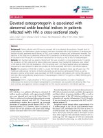
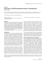
Functional mapping of the putative centromere on T. cruzi chromosome 1Figure 1
Functional mapping of the putative centromere on T. cruzi chromosome 1. (a) Organization of the GC-rich strand-switch region. Green arrows identify
ORFs in the polycistronic gene clusters and the implied direction of transcription. The degenerate retrotransposon-like VIPER/SIRE element (black) and
L1Tc autonomous retroelements (red) are indicated. The %GC content was determined by the Artemis 7 program [38]. (b) Mitotic stability of truncated
chromosomes. Sequences used for fragmentation (Tc1-Tc4) are indicated by yellow arrows. Vectors were targeted in both directions (+/-), with black
arrowheads representing the positions and orientations of de novo telomeres after fragmentation (see Additional data files 1-3 for further details). Clones
with truncated chromosomes were grown in the absence of G418 for the generations indicated above or below the corresponding track. Genomic DNA
was ScaI digested, Southern blotted, probed with plasmid DNA, then re-hybridized with β-tubulin as a loading control.
VIPER/SIRE L1Tc L1Tc
11 kb
76%
48%
6
%
GC-rich region
Tc1(+)
Tc2(+)
Tc3(+)
Tc4(+)
11kb
Tc1(-)
Tc2(-)
Tc3(-)
Tc4(-)
0 50 100 0 50 100
Plasmid
Tubulin
0 50 100
0 50 100
0 10 25
0 10 25
0 10 25 0 10 25
Plasmid
Tubulin
Plasmid
Tubulin
Plasmid
Tubulin
(a)
(b)
%GC
R37.4 Genome Biology 2007, Volume 8, Issue 3, Article R37 Obado et al. />Genome Biology 2007, 8:R37
rich strand-switch domain (+) were found to be maintained
for more than 100 generations (5 months) in the absence of
the selective drug. In contrast, chromosomes lacking this
domain (-) were unstable and disappeared in 10-25 genera-
tions. Therefore, in both chromosome 1 and 3 of T. cruzi, we
have now shown that the region required for mitotic stability
centers on a GC-rich strand-switch domain composed pre-
dominantly of degenerate retrotransposons.
Etoposide-mediated Topo-II cleavage sites in T. cruzi
chromosomes are associated with regions required for
mitotic stability
In CL Brener, chromosome 3 occurs as homologues of 0.65
and 1.1 Mb (Figure 2). Most of this difference is due to a 0.40
Mb insertion in the right arm of the larger homologue,
although the left arm is also 30 kb longer. Previously, we
delineated the determinants of mitotic stability on chromo-
some 3 [22]. To provide independent evidence that this
region has centromeric properties, we have now used Topo-II
activity as a biochemical marker for active centromeres [15-
18]. The procedure involved etoposide treatment of epimas-
tigotes to promote double-stranded cleavage at the sites of
Topo-II accumulation, isolation of chromosomal DNA and
Southern analysis following fractionation by contour-
clamped homogenous electric field gel electrophoresis
(CHEFE).
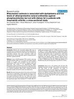
We identified two major etoposide-mediated cleavage sites in
the vicinity of the GC-rich strand-switch domain of T. cruzi
chromosome 3 (Figure 2). Bands of 0.39, 0.34 and 0.31 Mb
were detected with probes Tc7 and Tc8, sequences from the
left arm of the chromosome, 20 and 10 kb respectively, from
the strand-switch domain. The Tc11 probe, from a gene array
closer to the left telomere, identified products of 0.34, 0.31
and 0.28 Mb. The 0.28 Mb fragment (band 1), which was not
detected with probes Tc7 and Tc8, allows tentative location of
one cleavage site to a region 30 kb from the strand-switch
domain on the smaller chromosome (Figure 2). Cleavage at
the corresponding site on the 1.1 Mb chromosome should
generate a 0.31 Mb product (band 5), since the left arm of this
homologue is 30 kb longer. The 0.34 Mb product (band 6) in
the Tc11 autoradiograph can be inferred to arise from a sec-
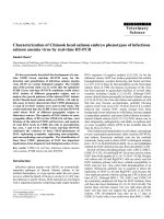
Etoposide-mediated cleavage sites in T. cruzi chromosome 3Figure 2
Etoposide-mediated cleavage sites in T. cruzi chromosome 3. Epimastigotes were treated with 1 mM etoposide for 6 h and chromosomal DNA
fractionated by CHEFE and assessed by Southern analysis. Probe Tc12 is specific to the larger homologue (see Materials and methods). Lane N, non-
treated parasites; lane E, etoposide-treated. The schematic shows both chromosome 3 homologues, location of the 16 kb GC-rich strand-switch domain
(GC), positions of the probes and predicted locations of the major Topo-II cleavage sites (large black arrowheads). The fragments generated (1-7), and
their sizes and inferred positions on the chromosomes are shown in red, green and blue. With the exception of probe Tc12, fragments derived from the
right arm of the 1.1 Mb homologue (blue) cannot be detected, due to co-migration with the cross-hybridizing 0.65 Mb homologue.
Tc11
Tc11
Tc7 8
0.65 Mb
1.1 Mb
N E
N E N E
N E
N E
N E
Tc11
Tc7
Tc8
Tc12
Tc9
Tc10
910
GC
3 (0.39 Mb)
4 (0.36 Mb)
3
3
6
6
(0.31 Mb) 2
(0.28 Mb) 1
(0.34 Mb) 6
(0.31 Mb) 5
6
2+5
1
6
3
4
3
4
22
GC
Tc12
1.1 Mb
0.65 Mb
7 (0.75 Mb)
7
Genome Biology 2007, Volume 8, Issue 3, Article R37 Obado et al. R37.5
comment reviews reports refereed researchdeposited research interactions information
Genome Biology 2007, 8:R37
ond cleavage site located within the strand-switch domain of
the 1.1 Mb chromosome. This band hybridizes to probe Tc12,
a sequence unique to the larger homologue. Cleavage at this
site in the 0.65 Mb chromosome should generate a 0.31 Mb
fragment (band 2, Tc7, Tc8 and Tc11). In the Tc11 autoradio-
graph, this fragment co-migrates with band 5, a cleavage
product derived from the larger homologue. Probes Tc9 and
Tc10, from the right arm of chromosome 3 (Figure 2), hybrid-
ized to cleavage products of 0.39 and 0.36 Mb (bands 3 and
4). Cleavage of the smaller homologue, at the sites predicted
above, should generate these products. The corresponding
cleavage of the 1.1 Mb chromosome would produce a band
masked by the hybridization signal of the intact smaller
homologue. A product of this size was detected with homo-
logue-specific probe Tc12 (band 7).
Together, these data indicate the presence of two major sites
of Topo-II accumulation on chromosome 3, one located
within the strand-switch region and one approximately 30 kb
downstream. Therefore, functional [22] and biochemical
mapping now provide independent evidence of an active cen-
tromere at this locus. We also investigated if etoposide treat-
ment resulted in lesions close to the GC-rich strand-switch
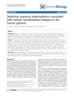
region of chromosome 1 (Figure 3). The data confirm that the
presence of two major sites of Topo-II activity on chromo-
some 1, situated close to the strand-switch domain, within the
region required for mitotic stability.
Synteny in the location of Topo-II activity on T. brucei
chromosome 1 and T. cruzi chromosome 3
Despite a completed genome sequence, there are no experi-
mental data on centromere location in T. brucei. To address
Mapping of etoposide-mediated Topo-II cleavage sites in T. cruzi chromosome 1Figure 3
Mapping of etoposide-mediated Topo-II cleavage sites in T. cruzi chromosome 1. Epimastigotes were treated with 1 mM etoposide for 6 h and
chromosomal DNA fractionated by CHEFE and assessed by Southern hybridization. Probes Tc1 and Tc4 were used (Additional data file 5). Large black
arrowheads identify the predicted locations of Topo-II activity adjacent to the GC-rich strand-switch domain (yellow oval). The cleavage fragments are
identified in red and green. Lane N, non-treated parasites; lane E, etoposide-treated.
GC
GC
Probe:
Tc1 Tc4
N E N E
Tc1 Tc4
1.2 Mb
0.38 Mb
0.31 Mb
1.2 Mb
0.51 Mb
0.23 Mb
0.17 Mb
0.51 Mb
1.2 Mb
~0.9 Mb
0.51 Mb
(~0.9 Mb)
(~0.9 Mb)
(0.31 Mb)
(0.38 Mb)
(0.31 Mb)
(0.38 Mb)
(0.23 Mb)
(0.17 Mb)
R37.6 Genome Biology 2007, Volume 8, Issue 3, Article R37 Obado et al. />Genome Biology 2007, 8:R37
this, we treated procyclic parasites with etoposide and
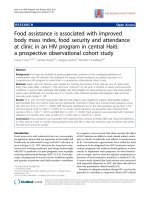
mapped the sites of Topo-II activity. With chromosome 1
(homologues of 1.15 and 1.2 Mb), probes from the left arm of
the chromosome (Tb1 and Tb2) hybridized to a major cleav-
age product of 0.8 Mb, whereas those from the right arm (Tb3
and Tb4) identified a smear ranging from 0.3-0.45 Mb (Fig-
ure 4a). These data localize etoposide-mediated cleavage of
chromosome 1 to the region between probes Tb2 and Tb3.
The more extensive smearing of products from the right arm
of the chromosome further suggests that the main size differ-
ences between the homologues result from additional
sequences in this arm of the chromosome. In T. brucei, most
differences between homologues are restricted to the subtelo-
meric regions.
To assess the extent of the Topo-II 'footprint' on this chromo-
some, we analyzed restriction digested genomic DNA. With
DNA from non-treated parasites, NotI digestion generated
two bands detectable with probe Tb2, indicative of differ-
ences in the lengths of the corresponding regions on each
homologue (Figure 4b). Furthermore, these regions were
larger than inferred from the genome sequence [23]. Bands of
Etoposide-mediated cleavage sites on T. brucei chromosome 1Figure 4
Etoposide-mediated cleavage sites on T. brucei chromosome 1. (a) Procyclics were treated with 500 µM etoposide for 1 h and chromosomal DNA
fractionated by CHEFE. Hybridization was carried out with probes Tb1-Tb4. Their positions and the location of the strand-switch domain (yellow oval) are
shown. Lane N, non-treated parasites; lane E, etoposide-treated. (b) Fine-mapping of cleavage sites. Chromosomal DNA from treated/non-treated
parasites was immobilized in agar blocks, restriction digested and fractionated by CHEFE. Fragment sizes are shown above the schematic, with their
predicted sizes (GeneDB) in parentheses. Black triangles identify the fragments and cleavage products on the relevant autoradiographs. As control, blots
were re-hybridized with probe Tb4, from a gene 150 kb upstream of the putative centromere. (c) Comparison of the T. cruzi chromosome 3 centromeric
domain with the syntenic region of T. brucei chromosome 1. In the T. cruzi chromosome, the degenerate VIPER/SIRE element and L1Tc retroelements are
indicated, together with a truncated cruzipain pseudogene (
ψ
CZP) and an U2snRNA gene. The corresponding region in T. brucei chromosome 1 contains 2
INGI retrotransposons and 1 DIRE. The locations of a leucine rich repeat protein gene (LRRP), a rRNA gene array and a 5.5 kb array of approximately 30
bp repeats are shown. Green arrows indicate putative ORFs and the implied direction of transcription. The dashed lines between the T. cruzi and T. brucei
maps identify the equivalent positions of the first ORFs of the conserved directional gene clusters.
Tb1 Tb2 Tb3 Tb4
Tb1 Tb2 Tb3 Tb4
N E N E N E N E
1.15/1.20 Mb
1.15/1.2 Mb
0.8 Mb
0.45 Mb
0.30 Mb
(a)
C
50 kb
35 kb
130 kb
90 kb
N E
80 kb
N E
Swa I
Not I
Tb2
Tb3
Tb4 Tb3
10 kb
(b)
Not I
Swa I/Not I
N E
50 kb
190 kb
150 kb
110 kb
Probe:
Tb2
Probe:
Not I
150/190 kb (120 kb)
90 kb/130 kb (56 kb)
T. cruzi chr 3
VIPER/SIRE
U2sn RNA
ΨCZP
L1Tc
L1Tc
78%
~30 bp repeats
DIRE
INGI
rRNA array
T. brucei chr 1
INGI
66%
LRRP
48%
9%
46%
33%
(c)
5 kb
%GC
%GC
Genome Biology 2007, Volume 8, Issue 3, Article R37 Obado et al. R37.7
comment reviews reports refereed researchdeposited research interactions information
Genome Biology 2007, 8:R37
150 and 190 kb were generated rather than the predicted 120
kb. Similarly, SwaI/NotI digestion produced fragments of 90
and 130 kb, instead of the expected 56 kb. With DNA from
etoposide-treated parasites, we observed a series of major
cleavage products and a smear that stretched >60 kb (Figure
4b). Cleavage sites could not be accurately mapped onto the
chromosome because of the heterogeneity between homo-
logues, and possible gaps in sequence assembly. Neverthe-
less, it is implicit that Topo-II activity is regional, confined to
the sequence between probes Tb2 and Tb3 (approximately 85
kb/120 kb, depending on the homologue), and exhibits signif-
icant site-specificity within this region. When blots were
hybridized with probe Tb4, from an open reading frame
(ORF) 150 kb upstream, there was minimal etoposide-medi-
ated cleavage.
Intriguingly, the location of Topo-II activity on T. brucei
chromosome 1 is syntenic with the region of etoposide-medi-
ated cleavage on T. cruzi chromosome 3 (Figure 2), which is
also required for mitotic stability [22]. This suggests that cen-
tromere location on these chromosomes has been conserved
since species divergence, more than 200 million years ago. In
T. brucei, this region encompasses a transcriptional strand-
switch domain containing two full-length INGI retrotranspo-
son-like elements closely linked to a degenerated INGI/L1Tc-
related element (DIRE), and a short array of ribosomal RNA
genes (Figure 4c). A major difference between the domains is
the presence in the T. brucei chromosome of a 5.5 kb element
of degenerate AT-rich repeats of approximately 30 bp. The
analogous region of T. cruzi chromosome 3 lacks any kind of
repetitive array. In the case of the putative centromere of T.
cruzi chromosome 1 (Figure 1), the corresponding region is
located on T. brucei chromosome 11, associated with a break
in synteny.
Repetitive arrays are a feature of Topo-II cleavage sites
in T. brucei chromosomes
With T. brucei chromosome 4 (homologues of 1.9 and 2 Mb),
the major products generated by etoposide treatment were
doublets of 1.3/1.4 Mb (probe Tb9) and 0.65/0.85 Mb (probe
Tb10) (Figure 5a). The ends of chromosome 4 have not been
fully assembled [23] and it was not possible to accurately map
the cleavage sites on the basis of product size. However, it can
be inferred from the hybridization patterns that the sites are
located between the ORFs from which probes Tb9 and Tb10
were derived (Figure 5a). This sequence contains a domain
that separates directional gene clusters, with head-to-head
DIREs located either side of a 3.5 kb AT-rich element made
up of 149 bp repeats. There are no other repetitive arrays else-
where on chromosome 4.
The extent of Topo-II activity in this region was investigated
further by Southern analysis of NotI digested DNA. Based on
the genome sequence, the major cleavage sites were expected
to be within a 95 kb fragment. However, NotI digestion gen-
erated a doublet of 145/155 kb that covers this region (probe
Tb10; Figure 5). As with chromosome 1, this could reflect het-
erogeneity between chromosome homologues and possible
gaps in sequence assembly. In the track containing DNA from
etoposide-treated cells, four major products of 40-80 kb were
identified on a background smear. Precise localization of the
corresponding sites on the genome map is complicated by the
issues discussed above. Nevertheless, it is implicit from the
data (Figure 5a, b) that Topo-II activity on chromosome 4 is
concentrated in this region, which contains an array of AT-
rich repeats, similar to the putative centromeric region of
chromosome 1. Using a probe 500 kb distant from this
domain (Tb18), we detected minimal etoposide-mediated
cleavage.
To assess if repeat arrays are a conserved feature of Topo-II
accumulation sites, we delineated these regions in chromo-
somes 1-8 (the results are shown in Additional data file 4 and
summarized in Figure 6). Etoposide-mediated cleavage sites
could be mapped to regions between specific gene probes, or
inferred from the sizes of the cleavage products. In each case,
Topo-II binding was closely associated with regions that sep-
arate directional gene clusters and contain at least one DIRE
or INGI retroelementand an array of AT-rich repeats. The
arrays are restricted to a single site on each chromosome.
They typically range from 2-8 kb, although on chromosome 3
the estimate is 30 kb, and others (chromosomes 6 and 8)
remain to be fully sequenced (GeneDB). Contiguous
sequences for chromosomes 9, 10 and 11 have yet to be assem-
bled and we did not attempt their analysis. However,
sequences similar to the AT-rich repeats have been assigned
to these chromosomes. The consensus repeat sequences for
each chromosome and a summary of their properties are
given in Figure 7 and Table 1, respectively.
Based on available sequence (GeneDB), the AT-rich repeats
fall into four classes. In the largest, chromosomes 4, 5, 8, 9, 10
and 11, they are organized in units of approximately 147 bp
and share >90% identity. These units have a complex struc-
ture built from degenerate sub-repeats of approximately 48
and 30 bp. Where these arrays have been fully sequenced,
they do not display a gradient of divergence moving from the
centre towards the edge, unlike the α-satellite repeats in
human centromeric DNA [5]. In two of the other groupings,
chromosomes 2/7 and chromosomes 1/6, the arrays are made
up of repeats of approximately 30 bp, which share 83% and
76% identity, respectively. The array in chromosome 3 is
distinctive; it is organized in units of 120 bp, with an AT con-
tent of only 49%. Despite this, it is related to the other repeats,
for example, sharing 53% identity with that of chromosome 4.
We also noted that the repeat regions were adjacent to arrays
of rRNA genes in chromosomes 1, 2, 3, 6 and 7, although the
significance of this is unknown.
R37.8 Genome Biology 2007, Volume 8, Issue 3, Article R37 Obado et al. />Genome Biology 2007, 8:R37
T. brucei intermediate and minichromosomes are
refractory to etoposide-mediated cleavage
In addition to 11 'conventional' chromosomes, T. brucei also
contains approximately 100 minichromosomes and several
intermediate-sized chromosomes [25]. We investigated if
these were susceptible to etoposide-mediated cleavage. For
the minichromosomes, we used the 177 bp repeat as a probe.
This element is also present on intermediate-sized chromo-
somes, but in considerably fewer copies (Figure 8a). In
contrast to chromosome 1, which was analyzed in parallel
using an α-tubulin probe, we could detect no evidence of
minichromosome cleavage, even when etoposide treatment
was extended for three hours. To assess the intermediate
chromosomes, we used bloodstream forms of the T. brucei
Etoposide-mediated cleavage sites on T. brucei chromosome 4Figure 5
Etoposide-mediated cleavage sites on T. brucei chromosome 4. (a) Procyclics were treated with 500 µM etoposide for 1 h and chromosomal DNA
fractionated by CHEFE and Southern blotted. Red arrows identify DIREs and green arrows indicate putative ORFs and the implied direction of
transcription. The location of the AT-rich repeat array is highlighted (striped box). The positions of probes and location of the putative centromeric region
(yellow oval) are indicated. Lane N, non-treated parasites; lane E, etoposide-treated. (b) Fine mapping of cleavage sites. NotI digested DNA was
fractionated by CHEFE as in Figure 4b and Southern blotted. Fragment sizes are shown above the schematic, with their predicted sizes (GeneDB) in
parentheses. Black triangles identify these fragments on the autoradiograph and show the major cleavage products. As control, the membrane was
hybridized with probe Tb18, from a gene 500 kb downstream of the putative centromere.
N E
Tb10
N E
Tb9
2.0 Mb
1.9 Mb
1.3 Mb
1.4 Mb
2.0 Mb
1.9 Mb
0.85 Mb
0.65 Mb
Tb9
C
Tb10
47%
DIRE
71%
13%
(a)
DIRE
1.9/2.0 Mb
%GC
N EN E
80 kb
60 kb
40 kb
155 kb
145 kb
47 kb
Tb10
Tb18 Tb10
Not I
Not I
145/155 kb (95 kb)
10 kb
(b)
Tb9
Tb18
Genome Biology 2007, Volume 8, Issue 3, Article R37 Obado et al. R37.9
comment reviews reports refereed researchdeposited research interactions information
Genome Biology 2007, 8:R37
427 strain and the T3 VSG probe. This fragment hybridizes to
a 0.32 Mb intermediate-sized chromosome in strain 427 (D
Horn, personal communication). Generally, we observed that
cleavage of the mega-based sized chromosomes in blood-
stream form parasites required lower drug concentrations
and shorter incubation periods than in procyclics. Under con-
ditions in which there was significant site-specific cleavage of
chromosome 1, we could detect no evidence for etoposide-
mediated cleavage of the 0.32 Mb chromosome (Figure 8b). It
can be inferred, therefore, that Topo-II does not undergo site-
specific accumulation on the intermediate and
minichromosomes.
Discussion
With the completion of the trypanosome genome projects, the
lack of information on the location of centromeric DNA is the
major remaining gap in our understanding of chromosome
organization. Here, we have exploited the genome sequence
data and a combination of genetic and biochemical tech-
niques to address this question. In the case of T. cruzi,
centromere location was mapped using two independent
approaches. The first, telomere-associated fragmentation,
delineated the region required for the mitotic stability of
chromosome 1 to a 40 kb sequence with striking organiza-
tional similarity to the putative centromeric region of chro-
mosome 3 [22]. Consistent with this, we mapped Topo-II
activity to these same regions on both chromosomes. In
mammalian cells, etoposide-mediated cleavage sites are bio-
chemical markers of centromeric DNA [12,15-18]. As cells
enter mitosis, sister chromatids remain attached, partly
through strand catenation at centromeres [8]. Centromeric
accumulation of Topo-II at this stage of the cell cycle acts to
regulate sister chromatid cohesion and enzyme activity is
essential for ordered segregation [9].
On the basis of these two independent approaches applied to
two separate chromosomes, we now suggest a paradigm for T.
cruzi centromeric DNA; GC-rich strand-switch domains com-
posed predominantly of degenerate retrotransposons. This
latter feature is shared to an extent with centromeres of
higher eukaryotes, where transposable elements have been
suggested to have important roles in centromere function,
including mediating heterochromatin formation [26]. In
human cells, functionally active and genetically stable neo-
centromeres can also form in euchromatic regions of chromo-
somes that lack repetitive arrays but are rich in LINE non-
LTR retrotransposons [27]. By analogy, retrotransposons
could have a role in some aspect of centromere function in T.
cruzi. Alternatively, centromeric domains may simply
provide a genomic niche that favors their integration and
retention [28]. The regions that separate directional gene
clusters are one of the few areas on T. cruzi chromosomes not
transcribed constitutively. However, this itself cannot be a
determinant of centromere location, since parasite chromo-
somes typically contain several such regions [21]. Only a sub-
set of the T. cruzi strand-switch domains so far assembled
have an organization similar to those identified on chromo-
somes 1 and 3. We therefore propose that only one strand-
switch domain per chromosome will be found to have this
type of configuration, a hypothesis that will be testable when
a more complete version of the T. cruzi genome becomes
available.
In T. cruzi chromosome 3, we detected two major Topo-II
cleavage sites, one within the strand-switch region and the
other 30 kb distant (Figure 2). Two cleavage sites were also
detected in the region required for mitotic stability of chro-
mosome 1 (Figure 1). These patterns could correlate with the
functional boundary of a 'centromeric domain' or merely
reflect other aspects of higher-order chromatin structure
formed in the neighborhood of an active centromere. In
human chromosomes, although etoposide-mediated Topo-II
cleavage is confined to the arrays of α-satellite DNA, enzyme
binding appears to be dependent on structural features asso-
ciated with chromatin, rather than DNA sequence [17].
Interest in trypanosomatid Topo-II has stemmed mainly
from its potential as a drug target [29] and its role in replica-
tion of kinetoplast DNA minicircles [30,31]. Trypanosomes
have distinct classes of mitochondrial and nuclear Topo-II. In
T. brucei, there are two genes for nuclear isoforms, TbTOP2
α
and TbTOP2
β
, although the latter may be a pseudogene.
TbTOP2α is essential, with RNAi-mediated knockdown caus-
ing growth arrest and abnormalities in nuclear morphology
consistent with mis-segregation [32]. We found Topo-II
activity to be a regional phenomenon in T. brucei chromo-
somes, concentrated at single sites located between direc-
tional gene clusters. These zones contain at least one DIRE/
Etoposide-mediated Topo-II cleavage sites (red circles) on T. brucei (strain 927) chromosomes 1-8Figure 6
Etoposide-mediated Topo-II cleavage sites (red circles) on T. brucei (strain
927) chromosomes 1-8. The locations of the probes (Tb1-17) are
identified by black bars. Details on the AT-rich arrays are given in Figure 7
and Table 1 and the experimental results are shown in Additional data file
4.
Chr 7
Chr 6
Chr 5
Chr 4
Chr 3
Chr 2
Chr 1
2.7 Mb
1.8/2.0 Mb
2.0/2.5 Mb
1.3 Mb
1.15/1.2 Mb
1.9/2.0 Mb
2.0 Mb
1 2 3 4
5 6
7 8
9 10
11
12
13
14 15
Chr 8
2.7/2.9 Mb
16 17
R37.10 Genome Biology 2007, Volume 8, Issue 3, Article R37 Obado et al. />Genome Biology 2007, 8:R37
INGI retrotransposon and an array of AT-rich repeats, con-
served to varying degrees between chromosomes. We pro-
pose that this type of organization is a central feature of
centromeric DNA in T. brucei. The major difference in the
putative centromeric regions of T. cruzi and T. brucei is the
presence of repetitive arrays in the latter. Centromeric
repeats in other eukaryotes display considerable sequence
variation, even between closely related species. In T. brucei,
these arrays exhibit a range of intra-chromosomal divergence
and appear to be dynamic in terms of recombination, with
evidence that active processes operate to generate both
sequence homogenization and unequal crossover. Complete
sequencing of the arrays on each chromosome may reveal
more of the mechanisms involved. Similarly, resolution of
discrepancies between sequence data and restriction analysis,
such as those in the centromeric domains of chromosomes 1
and 4 (Figures 4 and 5), should facilitate more detailed func-
tional mapping of these regions.
In terms of centromere organization, a major question
remains: were the repeat elements lost by T. cruzi or did they
arise only in T. brucei? Leishmania diverged early in trypano-
somatid evolution, followed by a later split in the Trypano-
soma lineage. Evidence from genome analysis suggests that
the progenitor species had a karyotype more analogous to T.
cruzi and Leishmania major (approximately 30 and 36 chro-
mosome pairs, respectively) [21], both of which lack this class
of repeat element. Therefore, the repetitive DNA probably
became a feature of T. brucei centromeres after their diver-
gence from T. cruzi. Retrotransposons have been proposed as
templates for the evolution of centromeric repeats in higher
eukaryotes [26]. In this context, one explanation for the pres-
ence of repetitive DNA at T. brucei centromeres could be that
local chromatin provided an environment that supported its
expansion and propagation. It could be feasible, using
transfection-based approaches, to test if these arrays confer
Alignment of repeat array consensus sequences on each T. brucei chromosome, determined by the program Tandem Repeats Finder [39]Figure 7
Alignment of repeat array consensus sequences on each T. brucei chromosome, determined by the program Tandem Repeats Finder [39]. The nucleotides
marked in bold correspond to two copies of the array (approximately 30 bp) in chromosomes 1, 2, 6 and 7.
Tb10 ATGCAATATGTAAGGTGTTTT-GGTGTAAAACACGCATTCTTG-CATAACATGCACAATG 58
Tb11 ATGCAATATGTAAGGTGTTTT-GGTGTAAAACACGCATTCTTG-CATAACATGCACAATG 58
Tb4 ATGCAATATGTAAGGTGTTTT-GGTGCAAAACACGCATTCTTG-CATAACATGCACAATG 58
Tb9 ATGCAATATGTAAGGTGTTTT-GGTGCAAAACACGCATTCTTG-CATAACATGCACAATG 58
Tb5 ATGCAACATGTAAGGTGTTTTTGGTGTAAAACACGCATTCTTG-CACAACCTGCACAATG 59
Tb8 ATGCAACATGTAACGTGTTTT-GGCGTAAAACACGCATTCTTG-CACAACCTGCACAACG 58
Tb3 ATGCATTGGTGGCACATCATGGCCCATC 28
Tb2
Tb7
Tb1
Tb6
Tb10 TGGCATGTTTGT-GTGCAAATTGTGCACTATTGCGTATTTT-CACGTCAAATACGCGTTC 116
Tb11 TGGCATGTTTGT-GTGCAAATTGTGCACTATTGCGTATTTT-CACGTCAAATACGCGTTC 116
Tb4 TGGCATGTTTGT-GTGCAAATTGTGCACTATTGCGTATTTT-CACGTCAAATACGCGTTT 116
Tb9 TGGCATGTTTGT-GTGCAAATTGTGCACTATTGCGTATTTT-CACGTCAAATACGCGTTC 116
Tb5 TTGCATGTTTGT-TCACAAATTGTGCATTATTGCGTATTTT-CACGTCAAATACGCATTC 117
Tb8 TTGCATGTTTGT-GCACAAAATGTGCACTATTGCGTATTTT-CACGTAAAATACGCGTTC 116
Tb3 TTTCATGGTTATCGCCCCTACGGCGCATAATGGCGTGTTAT-CGCACAAAACCCTGTTAC 87
Tb2 ATGTCATTACGTGTTTTATGTGCAAAAGCATGTCAT 36
Tb7 GTGTTATTAAGTGTTTTATGTGAAAAAGCGTGTTAT 36
Tb1 ATGCGCAATAATACGCAATAATACGCAAT-AATGCGCAATAATGCACA 47
Tb6 ATGTGCAATAATGTGCAATTATATGCAAT-AATGTGCAAT TGCAAT 45
Tb10 -ATGCGTATGATTGCGCAAAAACAGTGTTGCA 147
Tb11 -ATGCGTATGATTGCGCAAAAACAGTGTTGCA 147
Tb4 -ATGCGTATGATTGCGCAAAAACAGTGTTGCA 147
Tb9 -ATGCGTATGATTGCGCAAAAACAGTGTTGCA 147
Tb5 -ATGCGTATGATTGTGCAAAAACAGTGTTGCA 148
Tb8 -ATACGTGTGTTTGTGCAAAAACAGTGTTGCA 147
Tb3 -A GTGTGATTGGGTAACGCCCTTCCACTGATCAC 120
Tb2 TACGTGTTTTATGT-GCAAAAGC 58
Tb7 TAAGTGTTTTATTTTGAAAAAGC 59
Tb1 CATATGCACAATTATGCAATA 68
Tb6 AATGTGCA-ATTTGTGCAATT 65
Genome Biology 2007, Volume 8, Issue 3, Article R37 Obado et al. R37.11
comment reviews reports refereed researchdeposited research interactions information
Genome Biology 2007, 8:R37
mitotic stability. However, in other eukaryotes, the fact that
centromere location is determined epigenetically has often
complicated the interpretation of such experiments.
We could detect no significant etoposide-mediated cleavage
of T. brucei intermediate or minichromosomes (Figure 8),
implying that site-specific Topo-II activity does not have a
role in their segregation. Any mechanism for minichromo-
some segregation must account for the high fidelity of the
process and the observation that the number of minichromo-
somes exceeds the number of microtubule spindles. The 'lat-
eral-stacking model' [25] proposes that replicated
minichromosomes attach laterally to anti-parallel microtu-
bules, with directional poleward movement following
chromatid separation. It is envisaged that several replicated
minichromosomes could attach to each microtubule pair.
This model is centromere-independent and lacks a specific
requirement for Topo-II accumulation at single sites on the
minichromosome, or the intermediate-sized chromosomes, if
an analogous mechanism is involved. Similar to the situation
with the intermediate and minichromosomes, we could not
detect site-specific etoposide-mediated cleavage of L. major
chromosomes (data not shown). In this instance, however, it
could be that the drug cannot access the parasite nucleus.
The evidence that we report here provides a basis for identi-
fying other determinants of centromere function. This is
important, since several observations hint that aspects of seg-
regation in trypanosomes differ from the mammalian host.
The number of kinetochores appears to be less than the
number of chromosomes [33,34], trypanosomes lack obvious
equivalents of the 'core' centromeric proteins [1] and few of
the other proteins involved in kinetochore assembly/function
have been conserved [23]. It may be that segregation in
trypanosomes is more streamlined than in other organisms,
or that the proteins involved are highly divergent. Whatever
the reason, resolution of these issues should provide new
insights into the evolution of this complex process and could
Table 1
Summary of information available (GeneDB) on the AT-rich repeat domains
Chromosome number Total size of repeat domain (kb) Unit repeat size (bp) AT content (%) AT-rich array position orientated by flanking
ORFs
1 5.5 ~30 66 Tb927.1.3670 - Tb927.1.3750
2 8.0 29 66 Tb927.2.1470 - Tb927.2.1560
3 Unknown* 120 49 Tb927.3.3410 - Tb927.3.3430
4 3.5 147 61 Tb927.4.3740 - Tb927.4.3750
5 2.3 148 62 Tb927.5.600 - Tb927.5.610
6 Unknown
†
~30 71 Tb927.6.180 - Tb927.6.190
7 3.0 29 75 Tb927.7.6860 - Tb927.7.6870
8 Unknown
‡
147 59 Tb927.8.7740 - Tb927.8.7750
9Unknown
§
147 60 Incomplete assembly
10 Unknown
¶
147 61 Incomplete assembly
11 Unknown
¥
147 61 Incomplete assembly
The AT-rich repeat domains are associated with the sites of etoposide-mediated Topo-II cleavage on T. brucei chromosomes 1-8 (Additional data file
4 and summarized in Figure 6). The flanking ORFs are those immediately adjacent to the repeat array. *BAC clone RPCI93-3K10 contains a large
number of 120 bp repeats that span up to 30 kb (GeneDB). This region has not yet been closed.
†
The repeat array contains a gap of unknown size.
‡
There are 1.5 copies of this repeat next to a gap in the chromosome 8 sequence at position 2,233,104 bp. The unresolved array is estimated to be
5-9 kb (GeneDB).
§
Several chromosome 9 reads (approximately 700 bp) contain these repeats (for example, tryp_IXb-344h12.p1c, GeneDB).
¶
A 10
kb contig tryp_X-275g09.p1c (Tb10.v4.0130) and several chromosome 10 reads (approximately 700 bp) contain these repeats (for example, tryp_X-
429d08.q1c, GeneDB).
¥
A 14 kb contig tryp_XI-974h12.q1k (Tb11.1750) and several chromosome 11 reads (approximately 900 bp) contain these
repeats (for example, tryp_XI-991b09.q1k, GeneDB).
T. brucei (a) minichromosomes and (b) intermediate chromosomes are resistant to etoposide-mediated cleavageFigure 8
T. brucei (a) minichromosomes and (b) intermediate chromosomes are
resistant to etoposide-mediated cleavage. (a) Procyclics (927 strain) were
treated with 500 µM etoposide for 3 h and chromosomal DNA separated
by CHEFE. Southern blots were hybridized with the 177 bp repeat probe
or α-tubulin. (b) Bloodstream form T. brucei (427 strain) were treated with
25 µM etoposide for 1 h and chromosomal DNA analyzed as above. Blots
were hybridized with the intermediate chromosome-specific probe T3. In
strain 427, chromosome 1 is larger than in strain 927, as is the major
cleavage product identified with the α-tubulin probe. Lane N, non-treated
parasites; lane E, etoposide-treated.
N E
N E
177bp repeat
Tubulin
1.6 Mb
1.1 Mb
0.32 Mb
1.2 Mb
0.82 Mb
0.10 Mb
N E
N E
Tubulin
VSG T3
(a)
(b)
R37.12 Genome Biology 2007, Volume 8, Issue 3, Article R37 Obado et al. />Genome Biology 2007, 8:R37
lead to the identification of novel parasite proteins that might
serve as targets for chemotherapy.
Conclusion
We report the first experimental evidence on the location of
centromeres in both T. cruzi and T. brucei. Centromeric DNA
in these primitive eukaryotes is associated with regions
between directional gene clusters that contain degenerate ret-
roelements. In T. brucei, but not T. cruzi, these loci also con-
tain arrays of AT-rich repeats. These findings provide a
framework for investigating chromosome segregation in a
more systematic manner. Until now, progress in this area has
been extremely limited.
Materials and methods
Parasites and DNA preparation
T. cruzi epimastigotes (CL Brener) were cultured as described
[22]. T. brucei procyclics (genome project TREU 927/4
strain) were grown at 27°C in SDM-79 medium [35] and
bloodstream forms (427 strain) at 37°C in a 5% CO
2
atmos-
phere in modified Iscove's medium [36]. Genomic DNA was
extracted using DNeasy tissue mini-kits (Qiagen, Venlo,
Netherlands). Intact chromosomes extracted by an agarose-
embedding technique [22] were separated with a CHEF Map-
per System (Bio-Rad, Hercules, CA, USA). For in situ restric-
tion digestion, agarose blocks were incubated in 1 mM
phenylmethanesulphonylfluoride (PMSF) at 25°C for 1 h,
washed in TE buffer (10 mM Tris-Cl, 1 mM EDTA, pH7.4) and
incubated with enzyme for 24 h.
Analysis of transformants
For targeted chromosome fragmentation, we used vector
pTEX-CF [22]. Briefly, targeting fragments were amplified
and inserted into the vector. Gel-purified linear DNA (1-2 µg)
was then introduced into T. cruzi epimastigotes by electropo-
ration [22]. Transformed cells were selected over 6-8 weeks
in 100 µg ml
-1
G418, and cloned by limiting dilution. Clones
were analyzed by Southern blotting of CHEFE-separated
chromosomal DNA and by sequencing of transformant-spe-
cific PCR fragments [22]. Transformants containing trun-
cated forms of chromosome 1 were continuously cultured in
the presence or absence of G418, and DNA isolated at various
time points. Typically, mitotic stability was assessed by
Southern hybridization using plasmid DNA as a probe for
truncated chromosomes. Results were confirmed by CHEFE.
Analysis of etoposide-treated cells
Logarithmically growing T. cruzi epimastigotes and T. brucei
procyclics were treated with DMSO-solubilized etoposide
(Sigma, St Louis, MO, USA). Drug dose and incubation times
optimal for generating specific cleavage fragments were
determined empirically for each parasite. Chromosomal DNA
was then isolated and fractionated by CHEFE, or genomic
DNA prepared. DNA probes were generated by PCR. For T.
cruzi chromosome 3, we amplified a 1.1 Mb homologue-spe-
cific 290 bp fragment (probe Tc12; Figure 2) using primers
ACCATGTCCAAATTGATCTGCAAATCA and TAGAAGCAT-
TAAATCCCAGCATCAGAC. For T. brucei chromosome 6, we
used an intergenic probe (Tb12), which was amplified with
primers TCTTGATAGGCGCATGTGCATGTAACC and CGAT-
GGCGCAAGCAGATAGTTGTTACC. In the case of the T. bru-
cei 177 bp repeat [37], a probe was generated using primers
ATTAAACAATGCGCAGTTAACG and GTGTATAATAGCGT-
TAACTGCG. Other probes are described in Additional data
file 5.
Additional data files
The following additional data are available with the online
version of this paper. Additional data file 1 outlines the strat-
egy for telomere-associated chromosome fragmentation of
the small homologue of T. cruzi chromosome 1. The sche-
matic of the 0.51 Mb chromosome shows the position of the 11
kb GC-rich strand-switch domain (yellow oval). The
expanded section (70 kb) contains the four ORFs used for
chromosome fragmentation (Tc1-Tc4; Additional data file 5).
The implied direction of polycistronic transcription is indi-
cated. A 0.9 kb DNA fragment from ORF Tc1 was cloned into
the pTEX-CF vector [22]. This was linearized so that the
targeting fragment was at one end and telomeric sequences at
the other. Plasmid DNA within the vector is marked in red.
Site-specific integration (crossed lines) results in the deletion
of approximately 100 kb of DNA between the target sequence
and the telomere (Additional data file 2). Telomeric
sequences supplied by the vector are shown as horizontal
arrowheads. Expression of neo
r
is under control of the rDNA
promoter (flagged). Additional data file 2 shows the genera-
tion of a truncated version of the small homologue of T. cruzi
chromosome 1. The 11 kb GC-rich strand-switch domains are
shown (yellow oval). Arrows (Tc1-Tc4) indicate positions of
the target sequences used for fragmentation (Additional data
file 5). Parasites were transfected with linearized vector DNA
and transformants selected with G418 [22] (see Materials and
methods). Chromosomal DNA from wild-type (WT) and
transfected (T) parasites was separated by CHEFE with S.
cerevisiae markers (M) and analyzed by Southern blotting
using plasmid, Tc5 and Tc6 radiolabelled probes.
Autoradiographs are shown, together with an ethidium bro-
mide (EtBr) stained gel, overexposed to show the truncated
chromosome. In the example shown, integration at the Tc1
locus of the 0.51 Mb chromosome, in the direction of polycis-
tronic transcription, results in deletion of approximately 100
kb of DNA between the target sequence and the telomere.
Hybridization identifies the larger (1.2 Mb) and smaller
homologues (0.51 Mb) and the truncated product (0.40 Mb).
Plasmid DNA is incorporated into truncated chromosomes as
part of the integration process (Additional data file 1). Addi-
tional data file 3 shows how chromosome fragmentation
demonstrates that the major size difference between the T.
cruzi chromosome 1 homologues is the result of the insertion/
Genome Biology 2007, Volume 8, Issue 3, Article R37 Obado et al. R37.13
comment reviews reports refereed researchdeposited research interactions information
Genome Biology 2007, 8:R37
deletion of 0.7 Mb of DNA into/from the left arm of the chro-
mosome. (a) Schematic showing the 0.51 Mb and 1.2 Mb
homologues and their respective truncated products C1 (0.2
Mb) and C2 (0.9 Mb), with the locations of probes Tc2-Tc5
(Additional data file 5). (b) Autoradiograph illustrating the
deletion of the right arms of each chromosome homologue
following integration of the fragmentation vector at ORF Tc4.
Two clones were isolated after transfection and chromosomal
DNA from wild-type (WT) and both clones (C1 and C2) were
separated by CHEFE and analyzed by Southern blotting using
radiolabeled probe Tc4. Hybridization identifies the larger
homologue (1.2 Mb), the smaller homologue (0.51 Mb) and
their respective truncated products (0.9 Mb and 0.2 Mb). We
had previously shown that probe Tc5 is located a similar dis-
tance (approximately 50 kb) from the end of both homo-
logues [22]. Additional data file 4 shows mapping of
etoposide-mediated Topo-II cleavage sites in T. brucei chro-
mosomes 1-8. T. brucei procyclic cultures were treated with
500 µM etoposide and chromosomal DNA isolated and frac-
tionated using a Bio-Rad CHEF Mapper system. Membranes
were hybridized with probes Tb1-Tb17, the GeneDB systemic
names of which are shown in Additional data file 5. The posi-
tions of probes (arrows) and locations of the putative centro-
meric regions (yellow ovals) are indicated. Red arrows
indicate INGI/DIRE retroelements and green arrows identify
putative ORFs and the implied direction of transcription. The
locations of the AT-rich repeat arrays are highlighted (striped
box) and the %GC contents across the regions are shown.
GeneDB systemic names of ORFs adjacent to the arrays are
given and allow the repeat arrays to be mapped onto the chro-
mosomal contigs. Lane N, non-treated parasites; lane E,
etoposide-treated. Chromosome sizes were calculated using
S. cerevisiae (0.2-2.2 Mb) and Hansenula wingei (1.0-3.1
Mb) chromosome markers (Bio-Rad). In most cases,
sequence contigs do not extend to the ends of chromosomes.
Gaps in the sequence of AT-rich arrays in chromosomes 3, 6
and 8 are indicated by asterixes and double slashes. We have
also found, using long-range restriction mapping (Figures 4b
and 5b), that the 'centromeric-domains' of chromosomes 1
and 4 are larger than predicted by the genome sequence.
Additional data file 5 is a list of T. cruzi (Tc1-12) and T. brucei
(Tb1-18) probes used in this study, together with their
GeneDB systemic names.
Additional data file 1Strategy for telomere-associated chromosome fragmentation of the small homologue of T. cruzi chromosome 1The schematic of the 0.51 Mb chromosome shows the position of the 11 kb GC-rich strand-switch domain (yellow oval). The expanded section (70 kb) contains the four ORFs used for chromo-some fragmentation (Tc1-Tc4; Additional data file 5). The implied direction of polycistronic transcription is indicated. A 0.9 kb DNA fragment from ORF Tc1 was cloned into the pTEX-CF vector [22]. This was linearized so that the targeting fragment was at one end and telomeric sequences at the other. Plasmid DNA within the vec-tor is marked in red. Site-specific integration (crossed lines) results in the deletion of approximately 100 kb of DNA between the target sequence and the telomere (Additional data file 2). Telomeric sequences supplied by the vector are shown as horizontal arrow-heads. Expression of neo
r
is under control of the rDNA promoter (flagged)Click here for fileAdditional data file 2Generating a truncated version of the small homologue of T. cruzi chromosome 1The 11 kb GC-rich strand-switch domains are shown (yellow oval). Arrows (Tc1-Tc4) indicate positions of the target sequences used for fragmentation (Additional data file 5). Parasites were trans-fected with linearized vector DNA and transformants selected with G418 [22] (see Materials and methods). Chromosomal DNA from wild-type (WT) and transfected (T) parasites was separated by CHEFE with S. cerevisiae markers (M) and analyzed by Southern blotting using plasmid, Tc5 and Tc6 radiolabelled probes. Autora-diographs are shown, together with an ethidium bromide (EtBr) stained gel, overexposed to show the truncated chromosome. In the example shown, integration at the Tc1 locus of the 0.51 Mb chromo-some, in the direction of polycistronic transcription, results in dele-tion of approximately 100 kb of DNA between the target sequence and the telomere. Hybridization identifies the larger (1.2 Mb) and smaller homologues (0.51 Mb) and the truncated product (0.40 Mb). Plasmid DNA is incorporated into truncated chromosomes as part of the integration process (Additional data file 1)Click here for fileAdditional data file 3Chromosome fragmentation demonstrates that the major size dif-ference between the T. cruzi chromosome 1 homologues is the result of the insertion/deletion of 0.7 Mb of DNA into/from the left arm of the chromosome(a) Schematic showing the 0.51 Mb and 1.2 Mb homologues and their respective truncated products C1 (0.2 Mb) and C2 (0.9 Mb), with the locations of probes Tc2-Tc5 (Additional data file 5). (b) Autoradiograph illustrating the deletion of the right arms of each chromosome homologue following integration of the fragmenta-tion vector at ORF Tc4. Two clones were isolated after transfection and chromosomal DNA from wild-type (WT) and both clones (C1 and C2) were separated by CHEFE and analyzed by Southern blot-ting using radiolabeled probe Tc4. Hybridization identifies the larger homologue (1.2 Mb), the smaller homologue (0.51 Mb) and their respective truncated products (0.9 Mb and 0.2 Mb). We had previously shown that probe Tc5 is located a similar distance (approximately 50 kb) from the end of both homologues [22]Click here for fileAdditional data file 4Mapping of etoposide-mediated Topo-II cleavage sites in T. brucei chromosomes 1-8T. brucei procyclic cultures were treated with 500 µM etoposide and chromosomal DNA isolated and fractionated using a Bio-Rad CHEF Mapper system. Membranes were hybridized with probes Tb1-Tb17, the GeneDB systemic names of which are shown in Addi-tional data file 5. The positions of probes (arrows) and locations of the putative centromeric regions (yellow ovals) are indicated. Red arrows indicate INGI/DIRE retroelements and green arrows iden-tify putative ORFs and the implied direction of transcription. The locations of the AT-rich repeat arrays are highlighted (striped box) and the %GC contents across the regions are shown. GeneDB sys-temic names of ORFs adjacent to the arrays are given and allow the repeat arrays to be mapped onto the chromosomal contigs. Lane N, non-treated parasites; lane E, etoposide-treated. Chromosome sizes were calculated using S. cerevisiae (0.2-2.2 Mb) and Hansenula wingei (1.0-3.1 Mb) chromosome markers (Bio-Rad). In most cases, sequence contigs do not extend to the ends of chro-mosomes. Gaps in the sequence of AT-rich arrays in chromosomes 3, 6 and 8 are indicated by asterixes and double slashes. We have also found, using long-range restriction mapping (Figures 4b and 5bB), that the 'centromeric-domains' of chromosomes 1 and 4 are larger than predicted by the genome sequenceClick here for fileAdditional data file 5T. cruzi (Tc1-12) and T. brucei (Tb1-18) probes used in this study, together with their GeneDB systemic namesT. cruzi (Tc1-12) and T. brucei (Tb1-18) probes used in this study, together with their GeneDB systemic namesClick here for file
Acknowledgements
We acknowledge the work of colleagues involved in the trypanosomatid
genome projects and thank Martin Taylor, David Horn and David Baker for
comments on the manuscript, and Eva Gluenz for drawing our attention to
the properties of probe Tc12. This work was funded by the BBSRC.
References
1. Fukagawa T: Centromere DNA, proteins and kinetochore
assembly in vertebrate cells. Chromosome Res 2004, 12:557-567.
2. Steiner NC, Hahnenberger KM, Clarke L: Centromeres of the fis-
sion yeast Schizosaccharomyces pombe are highly variable
genetic loci. Mol Cell Biol 1993, 13:4578-4587.
3. Pidoux AL, Allshire RC: Kinetochore and heterochromatin
domains of the fission yeast centromere. Chromosome Res
2004, 12:521-534.
4. Maddox PS, Oegema K, Desai A, Cheeseman IM: 'Holo'er than
thou: chromosome segregation and kinetochore function in
C. elegans. Chromosome Res 2004, 12:641-653.
5. Schueler MG, Higgins AW, Rudd MK, Gustashaw K, Willard HF:
Genomic and genetic definition of a functional human
centromere. Science 2001, 294:109-115.
6. Henikoff S, Ahmad K, Malik HS: The centromere paradox: stable
inheritance with rapidly evolving DNA. Science 2001,
293:1098-1102.
7. Warburton PE: Chromosomal dynamics of human neocentro-
mere formation. Chromosome Res 2004, 12:617-626.
8. Haering CH, Nasmyth K: Building and breaking bridges
between sister chromatids. Bioessays 2003, 25:1178-1191.
9. Carpenter AJ, Porter ACG: Construction, characterization, and
complementation of a conditional-lethal DNA topoisomer-
ase IIalpha mutant human cell line. Mol Biol Cell 2004,
15:5700-5711.
10. Toyoda Y, Yanagida M: Coordinated requirements of human
topo II and cohesin for metaphase centromere alignment
under Mad2-dependent spindle checkpoint surveillance. Mol
Biol Cell 2006, 17:2287-2302.
11. Rattner JB, Hendzel MJ, Furbee CS, Muller MT, Bazett-Jones DP:
Topoisomerase II alpha is associated with the mammalian
centromere in a cell cycle- and species-specific manner and
is required for proper centromere/kinetochore structure. J
Cell Biol 1996, 134:1097-1107.
12. Andersen CL, Wandall A, Kjeldsen E, Mielke C, Koch J: Active, but
not inactive, human centromeres display topoisomerase II
activity in vivo. Chromosome Res 2002, 10:305-312.
13. Bachant J, Alcasabas A, Blat Y, Kleckner N, Elledge SJ: The SUMO-
1 isopeptidase Smt4 is linked to centromeric cohesion
through SUMO-1 modification of DNA topoisomerase II.
Mol Cell 2002, 9:1169-1182.
14. Chen GL, Yang L, Rowe TC, Halligan BD, Tewey KM, Liu LF: Nonin-
tercalative antitumor drugs interfere with the breakage-
reunion reaction of mammalian DNA topoisomerase II. J Biol
Chem 1984, 259:13560-13566.
15. Floridia G, Zatterale A, Zuffardi O, Tyler-Smith C: Mapping of a
human centromere onto the DNA by topoisomerase II
cleavage. EMBO Rep 2000, 1:489-493.
16. Spence JM, Critcher R, Ebersole TA, Valdivia MM, Earnshaw WC,
Fukagawa T, Farr CJ: Co-localization of centromere activity,
proteins and topoisomerase II within a subdomain of the
major human X α-satellite array. EMBO J 2002, 21:5269-5280.
17. Porter ACG, Farr CJ: Topoisomerase II: untangling its contri-
bution at the centromere. Chromosome Res 2004, 12:569-583.
18. Spence JM, Fournier RE, Oshimura M, Regnier V, Farr CJ: Topoi-
somerase II cleavage activity within the human D11Z1 and
DXZ1 α-satellite arrays. Chromosome Res 2005, 13:637-648.
19. Kelly JM, McRobert L, Baker DA: Evidence on the chromosomal
location of centromeric DNA in Plasmodium falciparum from
etoposide-mediated topoisomerase-II cleavage. Proc Natl Acad
Sci USA 2006, 103:6706-6711.
20. Campbell DA, Thomas S, Sturm NR: Transcription in kineto-
plastid protozoa: why be normal? Microbes Infect 2003,
5:1231-1240.
21. El-Sayed NM, Myler PJ, Bartholomeu DC, Nilsson D, Aggarwal G,
Tran A-N, Ghedin E, Worthey EA, Delcher AL, Blandin G, et al.: The
genome sequence of Trypanosoma cruzi, etiologic agent of
Chagas disease. Science 2005, 309:409-415.
22. Obado SO, Taylor MC, Wilkinson SR, Bromley EV, Kelly JM: Func-
tional mapping of a trypanosome centromere by chromo-
some fragmentation identifies a 16 kb GC-rich
transcriptional 'strand-switch' domain as a major feature.
Genome Res 2005, 15:36-43.
23. Berriman M, Ghedin E, Hertz-Fowler C, Blandin G, Renauld H, Bar-
tholomeu DC, Lennard NJ, Caler E, Hamlin NE, Haas B, et al.: The
genome of the African trypanosome Trypanosoma brucei. Sci-
ence 2005, 309:416-422.
24. Machado CA, Ayala FJ: Nucleotide sequences provide evidence
of genetic exchange among distantly related lineages of
Trypanosoma cruzi. Proc Natl Acad Sci USA 2001, 98:7396-7401.
25. Gull K, Alsford S, Ersfield K: Segregation of minichromosomes
in trypanosomes: implications for mitotic mechanisms.
Trends Microbiol 1998, 6:319-323.
26. Wong LH, Choo KH: Evolutionary dynamics of transposable
R37.14 Genome Biology 2007, Volume 8, Issue 3, Article R37 Obado et al. />Genome Biology 2007, 8:R37
elements at the centromere. Trends Genet 2004, 20:611-616.
27. Lo AW, Craig JM, Saffery R, Kalitsis P, Irvine DV, Earle E, Magliano DJ,
Choo KH: A 330 kb CENP-A binding domain and altered rep-
lication timing at a human neocentromere. EMBO J 2001,
20:2087-2096.
28. Langdon T, Seago C, Mende M, Leggett M, Thomas H, Forster JW,
Jones RN, Jenkins G: Retrotransposon evolution in diverse
plant genomes. Genetics 2000, 156:313-325.
29. Das A, Dasgupta A, Sengupta T, Majumder HK: Topoisomerases of
kinetoplastid parasites as potential chemotherapeutic
targets. Trends Parasitol 2004, 20:381-387.
30. Shapiro TA, Klein VA, Englund PT: Drug-promoted cleavage of
kinetoplast DNA minicircles. Evidence for type II topoi-
somerase activity in trypanosome mitochondria. J Biol Chem
1989, 264:4173-4178.
31. Wang Z, Englund PT: RNA interference of a trypanosome
topoisomerase II causes progressive loss of mitochondrial
DNA. EMBO J 2001, 20:4674-4683.
32. Kulikowicz T, Shapiro TA: Distinct genes encode type II topoi-
somerases for the nucleus and mitochondrion in the proto-
zoan parasite Trypanosoma brucei. J Biol Chem 2006,
281:3048-3056.
33. Solari AJ: Mitosis and genome partitioning in trypanosomes.
Biocell 1995, 19:65-84.
34. Ogbadoyi E, Ersfeld K, Robinson D, Sherwin T, Gull K: Architecture
of the Trypanosoma brucei nucleus during interphase and
mitosis. Chromosoma 2000, 108:501-513.
35. Brun R, Schonenberger M: Cultivation and in vitro cloning of
procyclic culture forms of Trypanosoma brucei in a semi-
defined medium. Acta Tropica 1979, 36:289-292.
36. Hirumi H, Hirumi K: Continuous cultivation of Trypanosoma
brucei blood stream forms in a medium containing a low con-
centration of serum protein without feeder cell layers. J
Parasitol 1989, 75:985-989.
37. Wickstead B, Ersfeld K, Gull K: The small chromosomes of
Trypanosoma brucei involved in antigenic variation are con-
structed around repetitive palindromes. Genome Res 2004,
14:1014-1024.
38. Berriman M, Rutherford K: Viewing and annotating sequence
data with Artemis. Brief Bioinform 2003, 4:124-132.
39. Benson G: Tandem repeats finder: a program to analyze DNA
sequences. Nucleic Acid Res 1999, 27:573-580.