Báo cáo y học: " The WASP and WAVE family proteins Shusaku Kurisu and Tadaomi Takenawa" ppsx
Bạn đang xem bản rút gọn của tài liệu. Xem và tải ngay bản đầy đủ của tài liệu tại đây (406.31 KB, 9 trang )
Genome
BBiioollooggyy
2009,
1100::
226
Protein family review
TThhee WWAASSPP aanndd WWAAVVEE ffaammiillyy pprrootteeiinnss
Shusaku Kurisu and Tadaomi Takenawa
Address: Division of Lipid Biochemistry, Department of Biochemistry and Molecular Biology, Kobe University Graduate School of Medicine,
7-5-1 Kusunoki-cho, Chuo-ku, Kobe, Hyogo 650-0017, Japan.
Correspondence: Tadaomi Takenawa. Email:
SSuummmmaarryy
All eukaryotic cells need to reorganize their actin cytoskeleton to change shape, divide, move,
and take up nutrients for survival. The Wiskott-Aldrich syndrome protein (WASP) and WASP-
family verprolin-homologous protein (WAVE) family proteins are fundamental actin-cytoskeleton
reorganizers found throughout the eukaryotes. The conserved function across species is to
receive upstream signals from Rho-family small GTPases and send them to activate the Arp2/3
complex, leading to rapid actin polymerization, which is critical for cellular processes such as
endocytosis and cell motility. Molecular and cell biological studies have identified a wide array of
regulatory molecules that bind to the WASP and WAVE proteins and give them diversified roles
in distinct cellular locations. Genetic studies using model organisms have also improved our
understanding of how the WASP- and WAVE-family proteins act to shape complex tissue
architectures. Current efforts are focusing on integrating these pieces of molecular information
to draw a unified picture of how the actin cytoskeleton in a single cell works dynamically to build
multicellular organization.
Published: 15 June 2009
Genome
BBiioollooggyy
2009,
1100::
226 (doi:10.1186/gb-2009-10-6-226)
The electronic version of this article is the complete one and can be
found online at />© 2009 BioMed Central Ltd
GGeennee oorrggaanniizzaattiioonn aanndd eevvoolluuttiioonnaarryy hhiissttoorryy
The human Wiskott-Aldrich syndrome protein (WASP) gene
was the first of the WASP and WAVE family genes to be
isolated, in 1994, as a mutated gene associated with Wiskott-
Aldrich syndrome (WAS), an X-linked recessive disease
characterized by immunodeficiency, thrombocytopenia and
eczema, clinical features caused by complex defects in
lymphocyte and platelet function [1]. Another WASP family
member, neural (N-) WASP, was then identified from a
proteomic search for mammalian proteins that interact with
the Src homology 3 (SH3) domain of growth factor receptor
binding protein 2 (Grb2, also known as Ash) [2]. Although
expressed ubiquitously, N-WASP is most abundant in the
brain - hence its name. The first WAVE protein was identi-
fied in humans by our group and another group indepen-
dently as a WASP-like molecule and was named WAVE and
SCAR1, respectively [3,4]. Currently, it is agreed that mam-
mals possess five genes for the WASP and WAVE family,
WASP, N-WASP, WAVE1/SCAR1, WAVE2, and WAVE3
[5-9]. Human WASP and WAVE family genes are located on
different chromosomes, with each gene showing a unique
expression pattern (Figure 1). The human WASP gene is
carried on the X chromosome and is expressed exclusively in
hematopoietic cells, which explains the inheritance pattern
and the immunodeficiency and platelet deficiency charac-
teristic of WAS. WAVE1 and WAVE3 are strongly enriched
in the brain and are moderately expressed in some hemato-
poietic lineages, whereas WAVE2 appears to be ubiquitous.
Human WASP and WAVE proteins are between 498 and 559
amino acids long and are encoded by 9 to 12 exons. The
length of the genes is relatively similar, ranging from 67.1 kb
for N-WASP to 131.2 kb for WAVE3, with the exception of
WASP, which is a compact 7.6 kb. The restricted expression
of WASP in hematopoietic cells is dependent on a 137-bp
region upstream of the transcription start site [10]. It is
unclear how brain-specific expression of WAVE1 and
WAVE3 is regulated, but the proximal promoter region of
mouse WAVE1 retains potential recognition motifs for the
transcription factor hepatocyte nuclear factor 3β (HNF3β)
and putative E2-box sequences that can be recognized by
some basic helix-loop-helix transcription factors, such as
MyoD and Twist, upstream of the transcription start site [11].
The WASP and WAVE family proteins possess a carboxy-
terminal homologous sequence, the VCA region, consisting
of the verprolin homology (also known as WASP homology 2
(WH2)) domain, the cofilin homology (also known as
central) domain, and the acidic region, through which they
bind to and activate the Arp2/3 complex, a major actin
nucleator in cells (Figure 1). Besides the VCA region, the
WASP subfamily proteins are characterized by the
amino-terminal WH1 (WASP homology 1; also known as an
Ena-VASP homology 1, EVH1) domain, which functions as a
protein-protein interaction domain. In contrast, WAVE
subfamily proteins are characterized by the presence of the
WHD/SHD domain (WAVE homology domain/SCAR
homology domain), which is located at the amino terminus.
This domain is highly conserved between species, for even
the distantly related Arabidopsis WHD/SHD domain has
74% amino acid similarity to the WHD/SHD domain of
human WAVE1. This domain seems to be involved in the
formation of the WAVE complex (see later). Using these
sequence signatures together with genomic information
/>Genome
BBiioollooggyy
2009, Volume 10, Issue 6, Article 226 Kurisu and Takenawa 226.2
Genome
BBiioollooggyy
2009,
1100::
226
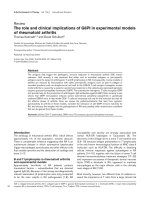
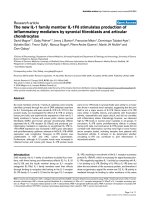
FFiigguurree 11
Comparison of the domain structures of the WASP and WAVE family proteins from different species. Color coding indicates conserved domains. The
percentage amino acid similarity of WH1/EVH1 domains or WHD/SHD domains is shown below each domain. For species abbreviations, see the legend
to Figure 2.
WASP family
WAVE family
At SCAR1
Hs WASP
Hs N-WASP
Dm WASP
Dw WASP
Sc Las17/Bee1
Ce WSP-1
Hs WAVE
Hs WAVE2
Hs WAVE3
Dm SCAR
Dd SCAR
Ce WVE-1
100%
87%
79%
68%
75%
70%
100%
96%
95%
90%
89%
74%
74%
100 amino
acids
Chromosomal
location
Tissue distribution
in mammals
Xp11.4-p11.27
Hematopoietic
7q31.3 Ubiquitous
6q21-q22
Brain/
hematopoietic
Brain/
hematopoietic
1p36.11-p34.3
13q12
Ubiquitous
Key:
WH1/EVH1 WHD/SHDCRIB/GBD BasicProline-rich V/WH2
C
A
from various organisms, WASP and WAVE homologs have
been discovered in a wide variety of eukaryotic species;
WASP and WAVE homologs (one of each) are found in
Dictyostelium discoideum (WASP and SCAR) [12,13],
Caenorhabditis elegans (WSP-1 and WVE-1) [14-16], and
Drosophila melanogaster (WASP and SCAR) [17,18].
Budding yeast has only one WASP homolog, Las17/Bee1
[19,20], and seems to lack WAVEs. In contrast, the plant
Arabidopsis thaliana appears to have four WAVE genes,
SCAR1-4 [21], but no WASPs.
Given that even plants have WAVE homologs, the evolu-
tionary history of the WASP and WAVE family is likely to
extend back to before the divergence of the eukaryotes. Along
with the evolution of the actin cytoskeleton, eukaryotic cells
must have needed means to control actin polymerization and
reorganize the actin cytoskeleton, which presumably led to the
development of the WASP/WAVE-Arp2/3 axis of actin-
polymerizing mechanisms. Although it is difficult to
determine whether the WASP and WAVE subfamilies evolved
from a common ancestral gene, Arabidopsis SCARs seem to
have evolved independently of the evolution of WASPs and
other fungal and metazoan WAVE/SCARs, which is suggested
by the alignment of conserved verprolin domain (V) and
cofilin homology domain (C) sequences (Figure 2a). More
detailed phylogenetic trees can be drawn from the alignment
/>Genome
BBiioollooggyy
2009, Volume 10, Issue 6, Article 226 Kurisu and Takenawa 226.3
Genome
BBiioollooggyy
2009,
1100::
226
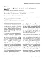
FFiigguurree 22
Evolutionary relationships between the WASP and WAVE family proteins. The phylogeny was inferred using the neighbor-joining method. ClustalW was
used to align sequences and perform phylogenetic analysis. Any position containing gaps was excluded from the dataset. Trees were drawn by NJplot
[89]. Bootstrap values were calculated over 1,000 iterations and values greater than 50% are shown as percentages next to branches. The bar in each
figure indicates the proportion of amino acid differences.
((aa))
The phylogenetic tree based on the alignment of combined sequences of V and C regions.
WASP and WAVE sequences were retrieved from the NCBI protein database and the V/WH2 domain for each protein was identified by homology
search over the Pfam-A database. C regions were identified according to the previously reported consensus sequence [29]. The sequence to be analyzed
was generated by joining the identified V sequence and C sequence.
((bb))
The phylogenic tree based on WH1/EVH1 domain alignment. WH1/EVH1
domains were identified by homology search over the PROSITE database.
((cc))
The phylogenetic tree based on WHD/SHD domain alignment. WHD/SHD
domains were identified following the consensus sequence described previously [90]. Species examined are
Homo sapiens
(Hs),
Mus musculus
(Mm),
Danio rerio
(Dr),
Drosophila melanogaster
(Dm),
Caenorhabditis elegans
(Ce),
Saccharomyces cerevisiae
(Sc),
Dictyostelium discoideum
(Dd) and
Arabidopsis thaliana
(At). Ensembl protein IDs for the zebrafish sequences used in the analysis are as follows: Dr WASP1, ENSDARP00000039217; Dr
WASP2, ENSDARP00000007963; Dr N-WASPa, ENSDARP00000094295; Dr N-WASPb, ENSDARP00000005823; Dr WAVE1, ENSDARP00000079387;
Dr WAVE2, ENSDARP00000093195; Dr WAVE3a, ENSDARP00000077123; Dr WAVE3b, ENSDARP00000085962. Two other homologous genes for
WAVE were identified in the zebrafish genome, but could not be assigned to homologs of mammalian WAVE1/2/3, so they were omitted from the
analysis. These proteins are ENSDARP00000047935 and ENSDARP00000102646.
(b) WH1/EVH1 phylogeny
Dd WASP (outgroup)
Sc Las17/Bee1
Sc Las17/Bee1
Ce WSP-1
Dm WASP
Dr WASP1
Dr WASP2
Dr WAVE1
Dr WAVE2
Mm WAVE2
Mm WAVE2
Hs WAVE2
Dr WAVE3a
Dr WAVE3b
Hs WAVE1
Mm WASP
Hs WASP
Dr N-WASPa
Dr N-WASPb
Mm N-WASP
Hs N-WASP
Vertebrate
WASP
Vertebrate
N-WASP
Vertebrate
WAVE1
Vertebrate
WAVE2
Vertebrate
WAVE3
Dd SCAR (outgroup)
Ce WVE-1
Ce WSP-1
Ce WVE-1
Dm SCAR
Dm SCAR
Mm WAVE3
Hs WAVE3
100
100
100
100
100
100
100
100
100
95
95
78
68
88
93
0.05
0.05
60
99
98
Dm SCAR
Hs WAVE1
Hs WAVE3
Hs WAVE2
WAVE
At SCAR1
At SCAR3
At SCAR2
At SCAR4
0.1
Dd WASP
Hs N-WASP
Hs WASP
Dm WASP
67
57
82
97
100
88
99
Plant SCAR
WASP
(a) V/WH2+C phylogeny
(c) WHD/SHD phylogeny
of highly conserved WH1/EVH1 domains of WASPs and the
alignment of WHD/SHD domains of WAVEs. Zebrafish
homologs of human WASP and N-WASP have been reported
recently [22], and a TBLAST search over the Ensembl
zebrafish genome (Zv8) revealed at least one homolog of
WAVE1, one of WAVE2 and two of WAVE3 (see the legend to
Figure 2 for the zebrafish gene accession numbers).
Phylogenetic analyses that include the zebrafish amino acid
sequences give us some interesting insights into the
evolution of these proteins in vertebrates. First, both
ancestral WASP and N-WASP seem to be present in a
common ancestor of fish and mammals (Figure 2b). This
means that WASP could have acquired its specialized
function in the adaptive immune system early in vertebrate
evolution, as the adaptive immune system is first seen in the
jawed fishes. Second, WAVE is split into three distinct
clades, WAVE1-3, as early as the emergence of the verte-
brates (Figure 2c). Considering that WAVE1 and probably
WAVE3 are involved in brain development in mammals
[23-27], WAVE1 and WAVE3 might be the basis for the
advent of the central nervous system (CNS).
CChhaarraacctteerriissttiicc ssttrruuccttuurraall ffeeaattuurreess
The WASP and WAVE family proteins share a common
domain architecture: a proline-rich stretch followed by the
VCA region located at the carboxyl terminus (Figure 1). The
VCA region simultaneously binds to two proteins to trigger
actin polymerization. The V domain binds to an actin
monomer (G-actin) and the CA domain binds to the Arp2/3
complex. The rate-limiting step to initiate actin polymeriza-
tion is the assembly of a trimeric actin nucleus. The Arp2/3
complex contains two actin-like proteins, Arp2 and Arp3,
serving as an actin pseudodimer. Therefore, the VCA region
can mimic the assembly of an actin trimer by providing a
platform that efficiently brings an actin monomer and the
Arp2/3 complex into close proximity, which leads to efficient
actin nucleation (Figure 3) [28]. The C domain, which con-
sists of approximately 20 amino acids, forms an amphi-
pathic α-helix whose hydrophobic surface interacts with and
activates the Arp2/3 complex [29]. Notably, there are two V
domains in tandem in mammalian N-WASP as well as in
Drosophila WASP and C. elegans WSP-1, a configuration
that is thought to increase their actin-nucleating activity
[30]. Recently, Co et al. [31] suggested a novel function for V
domains - that they capture elongating ends of actin
filaments (barbed ends) to ensure the dynamic attachment
of growing barbed ends to the membrane. Thus, the tandem
V domains of N-WASP would not only provide efficient actin
nucleation, but might also increase the ability of N-WASP to
localize and concentrate at the interface between the barbed
ends and the membrane.
The amino-terminal sequence of WASP subfamily proteins is
different from that of WAVEs. The amino terminus of
WASPs has the WH1/EVH1 domain following a basic region
and a GTPase-binding domain (GBD; also known as the
CDC42/Rac-interactive binding (CRIB) domain). The
WH1/EVH1 domain binds to WASP-interacting protein
(WIP) family proteins, which include WIP, CR16 (cortico-
steroids and regional expression-16), and WICH/WIRE
(WIP- and CR16-homologous protein/WIP-related) in
/>Genome
BBiioollooggyy
2009, Volume 10, Issue 6, Article 226 Kurisu and Takenawa 226.4
Genome
BBiioollooggyy
2009,
1100::
226
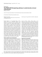
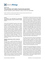
FFiigguurree 33
Multiple regulatory pathways for N-WASP and WAVE2 activation.
((aa))
N-WASP is autoinhibited in a basal state through the interaction
between the GBD/CRIB domain and the VCA region. PIP
2
and GTP-
loaded Cdc42 bind to the B and GBD/CRIB domains, respectively,
resulting in synergistic activation of N-WASP. Binding of SH3 domains to
N-WASP can independently compete with the autoinhibitory interaction,
and thus can activate N-WASP. SH3-domain-containing proteins that
interact and potentially activate N-WASP include cortactin, WISH, Nck,
Grb2, Crk, FBP17, CIP4, Toca1, Abi1, endophilin A, and sorting nexin 9
(not all shown on the diagram). Concurrently, the BAR-domain
superfamily proteins bend the membrane.
((bb))
WAVE proteins exist in cells
as a heteropentameric protein complex as indicated. WAVE2 has been
shown to translocate to the membrane via interactions with
phosphatidylinositol-(3,4,5)-triphosphate (PIP
3
) and IRSp53. The affinity of
WAVE2 for IRSp53 is enhanced when GTP-loaded Rac binds to the
RCB/MIM domain of IRSp53. IRSp53 is also able to enhance the ability of
WAVE2 to stimulate Arp2/3-mediated actin polymerization [91]. This
pathway via IRSp53 is an indirect activation by Rac, as it is suggested that
Rac can activate the WAVE complex through direct interaction with Sra1.
The direct pathway was shown in a recent paper but needs more
experimental evidence to be widely accepted (hence marked by a question
mark in the figure).
WH1/EVH1
B
CRIB
VV
CA
WIP
CR16
WICH
Cdc42
PIP
2
SH3
P
P
P
P
P
P
P
P
P
VVCA
G-actin
Arp2/3
‘Open VCA’
‘Closed N-WASP’
B
Rac
PIP
3
IRSp53
V
C
A
G-actin
Arp2/3
WHD/SHD
HSPC300
Sra1/PIR121
Nap1
Abi1/2/3
VC
A
P
P
P
P
P
P
P
PPP
‘Closed WAVE complex (?)’
Recruitment
only
Direct
pathway (?)
Indirect pathway
‘Open VCA (?)’
FBP17
CIP4
Toca1
Membrane
deformation
Membrane
deformation (?)
BAR domain
(a)
(b)
Actin polymerization
Actin polymerization
mammals [32-34]. In cells, most WASP proteins and
N-WASP proteins appear to form a stable one-to-one
complex with the WIP-family proteins, which seem to
protect WASP and N-WASP proteins from proteasomal
degradation [35-37]. NMR studies suggest that the WIP
ligands wrap around the N-WASP WH1/EVH1 domain and
that the interacting surface of WH1/EVH1 is a hotspot for
mutations in WAS patients, suggesting that disruption of
WASP-WIP binding and resulting WASP degradation
underlies the loss of WASP function and defective actin
cytoskeleton mophology of immune cells in WAS [38].
GBD/CRIB domains are critical for the control of WASP
and N-WASP activity because they bind to and inhibit the
VCA region. The hydrophobic cleft of GBD/CRIB domains
forms an intramolecular interaction with the hydrophobic
face of the amphipathic helix of the C domain, thereby
exerting an autoinhibitory control on VCA activity [39].
This autoinhibition is released by the competitive binding
of GTP-bound Cdc42 to the GBD/CRIB domain, leading to
activation of the Arp2/3 complex. Phosphatidylinositol-
(4,5)-bisphosphate (PIP
2
) binds to the basic region amino-
terminal to the GBD/CRIB domain, and synergizes with
Cdc42 to activate WASPs and N-WASPs.
The amino-terminal feature of WAVE is the presence of the
WHD/SHD domain followed by a stretch of basic residues
(Figure 1). In the cell, the WAVE proteins are constitutively
incorporated into a heteropentameric complex, the WAVE
complex, whose components seem to be conserved among
species ranging from plants to humans. The other members
of this complex are Sra1/CYFIP1 (and the homologous
PIR121/CYFIP2), Nap1 (also known as Kette in Drosophila),
Abi1/2/3 (Abelson-interactor), and HSPC300/Brick1
[40,41]. Lack of any of these components destabilizes the
WAVE complex, leading to proteasomal degradation of the
whole complex [42-44]. Biochemical studies suggest that
direct stoichiometric association of the WHD/SHD domain
with Abi and HSPC300 appears to contribute to the forma-
tion of the WAVE complex [45]. All the known WHD/SHD
domains contain conserved coiled-coil motifs spanning at
least 36 amino acids. These motifs are thought to associate
tightly with other coiled-coil motifs predicted to exist in Abi
and HSPC300.
LLooccaalliizzaattiioonn aanndd ffuunnccttiioonn
The localization of the WASP and WAVE family proteins has
been extensively studied in cultured cells, revealing that
both WASPs and WAVEs are closely associated with the cell
membrane through either direct or indirect binding to
membrane phosphoinositides. As the Arp2/3 complex with
which they interact intrinsically causes the rapid formation
of branched actin networks, the common feature of WASP
and WAVE function is coupling of the cell membrane to
Arp2/3-dependent actin polymerization to achieve
coordinated membrane-cytoskeleton dynamics.
Although N-WASP was originally proposed to be a
down-stream effector of Cdc42 in the formation of filopodia
[46], which are spiky actin-based motile structures protru-
ding from the cell periphery, its role in endocytosis is
currently the subject of intensive study. Whereas it remains
unclear whether N-WASP in endocytosis is also under the
control of Cdc42 activity, N-WASP is recruited to the site
where the clathrin-coated pit (CCP) forms. This recruitment
seems to be mediated through binding of the proline-rich
domain of N-WASP to the SH3 domains of EFC (extended
Fer-CIP4 homology)/F-BAR (FCH-Bin/Amphiphysin/Rvs)
domain-containing proteins, which are thought to be
involved in causing curvature of the membrane [47,48]. N-
WASP is thought to accelerate actin polymerization near the
invaginating CCPs, providing them with the energy to pinch
off from the plasma membrane. The idea that N-WASP may
be involved in endocytosis arose originally from the study of
Las17, the budding yeast homolog of WASP, which was first
identified in a screen for mutants defective in endocytosis
[20]. In yeast, Las17 and verprolin 1 (the yeast homolog of
WIP) are recruited to CCPs with the proteins Bzz1 and
Rvs167, which are now known to be members of the EFC/
F-BAR and BAR domain-containing proteins [49,50].
In contrast, mammalian WASP has been studied in relation
to the pathology of WAS. When a T cell is stimulated by
antigen on a target cell binding to the T-cell antigen receptor
(TCR), a stable contact between the two cells, called an
immunological synapse, is formed by the T-cell receptor
interaction and by adhesion molecules on both cells.
Dynamic filamentous actin (F-actin) rearrangement has
been shown to be necessary for the formation of a mature
immunological synapse. WASP seems to be involved in the
late stage of its formation, as WASP-deficient T cells are able
to form a stable immunological synapse in the initial contact
with antigen-presenting cells, but are unable to re-establish
it once the initial synapse is disturbed [51,52]. Upon T-cell
receptor activation, a signaling cascade is initiated by
interaction with cytoplasmic protein tyrosine kinases that
phosphorylate the receptor complex component CD3, and a
transmembrane protein LAT. Phosphorylated tyrosine resi-
dues of these proteins then recruit various adaptor proteins,
such as SLP-76, CrkL, Nck, and PSTPIP1, which in turn
recruit and concentrate WASP at the immunological synapse
to facilitate actin polymerization [53-55]. Apart from T-cell
activation, T lymphocytes from WAS patients have been
shown to display defects in cell migration in response to the
chemokine SDF1-α [56]. Thus, when WASP is defective and
actin polymerization fails, T cells are unable to carry out their
functions, resulting in immunodeficiency.
The activation of both WASP and N-WASP is tightly linked
to their recruitment to the membrane (Figure 3). GTP-
bound activated forms of Cdc42 localized at the membrane
bind to the GBD/CRIB domain. PI(4,5)P
2
is abundant in the
plasma membrane and binds to the basic region. The Src
/>Genome
BBiioollooggyy
2009, Volume 10, Issue 6, Article 226 Kurisu and Takenawa 226.5
Genome
BBiioollooggyy
2009,
1100::
226
family of tyrosine kinases phosphorylates tyrosine residues
near the GBD/CRIB domain. All these events are thought to
loosen the intramolecular interactions between the GBD and
VCA domains, thereby activating the WASPs [9]. The
EFC/F-BAR/BAR domain-containing proteins are anchored
on the membrane via their affinity for acidic phospholipids,
and many of them contain SH3 domains that can bind to the
proline-rich domains of WASP/N-WASP. This interaction
also seems to activate WASP/N-WASP, but as yet, the
mechanism is unclear (see the Figure 3 legend for examples
of proteins with N-WASP-activating SH3 domains).
WAVEs localize to the leading edges of lamellipodia, the flat
protrusions that cells extend in the direction of cell move-
ment [57]. Lamellipodia are filled with dense networks of
branched actin filaments. This actin architecture is
generated by the activity of the small GTPase Rac, and
WAVE was originally identified as a downstream effector for
Rac-mediated actin polymerization. Subsequently, WAVEs
were found to activate the Arp2/3 complex, and now WAVEs
are known to act downstream of Rac to trigger actin
polymerization by the Arp2/3 complex. In this regard,
WAVEs are essential for cell motility, as this is accomplished
by cycles of lamellipodial extension and substrate adhesion.
The localization of WAVEs to the edges of the lamellipodia is
regulated by a similar but not identical mechanism to N-
WASP localization (Figure 3). Through its basic domain,
WAVE2 preferentially binds to and is recruited to the
membrane by PI(3,4,5)P
3
rather than PI(4,5)P
2
[58]. Rac
seems to recruit WAVEs to the membrane by at least two
cooperative mechanisms. First, GTP-loaded forms of Rac
directly bind to the WAVE complex component Sra1 [59].
This interaction presumably recruits WAVEs to the
membrane in a Rac activity-dependent manner. Second, the
proline-rich domain of mammalian WAVEs binds to the SH3
domain of membrane-associated IRSp53, which belongs to
the RCB (Rac binding)/IMD (IRSp53-MIM homology
domain) domain-containing proteins, another class of
membrane-associated protein families with similar proper-
ties to the EFC/F-BAR proteins. The RCB/IMD domain
simultaneously binds to activated Rac, which contributes to
the Rac-dependent localization of WAVEs [60-63]. Interes-
tingly, WAVE2 has much stronger affinity for IRSp53 than
have WAVE1 and WAVE3 [60]. Therefore, the interaction
with IRSp53 is likely to contribute specifically to the
localization of WAVE2 at lamellipodial tips.
In a multicellular context, WAVEs also function in cell-cell
adhesion. In cultured epithelial cells, WAVEs localize at the
cell-cell boundaries and are necessary for maintaining the
integrity of the actin cytoskeleton at cell-cell junctions [64].
Genetic studies in multicellular organisms support this
observation in cultured cells. The developmental defects
observed in C. elegans embryos mutant for the WAVE
homolog wve-1 suggest that the protein WVE-1 is required for
epidermal cell-cell junction remodeling and for the
remodeling of intestinal epithelium to modulate apical
expansion of the gut lumen [16]. In Drosophila, SCAR/WAVE
is required for fusion of myoblasts to form muscle cells, which
is driven by remodeling of the actin cytoskeleton at cell-cell
junctions [65]. In Arabidopsis mutant for SCAR complex
genes and the Arp2/3 complex genes, the pavement cells of
the epidermis are abnormally shaped and show occasional
intercellular gaps [66,67]. These studies clearly demonstrate
the role of WAVEs in cell-cell junction formation and/or
maintenance, although the molecular mechanism of action of
WAVEs in cell adhesion is still not clearly understood.
The activating mechanism of the heteropentameric WAVE
complex remains controversial. Consistent with the notion
that WAVEs lack the GBD/CRIB domain by which the VCA
region would be autoinhibited, many studies have reported
that the WAVE complex reconstituted in vitro is con-
stitutively active [9]. However, the in vivo WAVE complex
biochemically purified from tissue homogenates appears to
be basically inhibited [40,68]. Recently, Ismail et al. [69]
accurately reconstituted the human WAVE1 complex with
purified components and showed that this reconstituted
complex is inhibited. They also demonstrated that a
similarly constructed Drosophila SCAR complex is inhibited,
suggesting that the inhibited state is likely to be the default
state. They then showed that these reconstituted complexes
could be activated by active Rac. Thus, our current
knowledge supports a model in which the WAVE complex is
normally inhibited in cells. Yet, the precise mechanism of
how Rac activates the WAVE complex is still unclear. There
are other levels of regulation as well. For example,
phosphorylation of WAVE1 by cyclin-dependent kinase 5
(Cdk5) suppresses Arp2/3-complex activation by WAVE1
during spine morphogenesis of neurons [26]. WAVE2 is also
phosphorylated by extracellular signal-regulated kinase 2
(ERK2) or by c-Abl or casein kinase 2 (CK2), and its actin-
polymerizing activity appears to be controlled by these
kinases [70-72]. Degradation of WAVEs appears to be
controlled by the vinexin family of adaptor proteins, but as
yet, the physiological significance of this is unknown [73,74].
FFrroonnttiieerrss
With a wealth of information now in hand about the
molecular interactions and biochemical activities of the
WASP and WAVE family proteins, one of the main issues to
be addressed is how WASPs and WAVEs and their
associated proteins work together to shape various and
complex actin architectures. For example, N-WASP is
essential for the formation of distinct cellular architectures
such as endocytic vesicles, filopodia and podosomes/
invadopodia [9]. How does N-WASP form these structures
separately yet with a similar molecular action? One of the
clues to solving this question exists in recently identified
classes of membrane-deforming proteins, which bind
directly to phospholipids and can deform membranes into
/>Genome
BBiioollooggyy
2009, Volume 10, Issue 6, Article 226 Kurisu and Takenawa 226.6
Genome
BBiioollooggyy
2009,
1100::
226
curved surfaces [75,76]. These proteins are classified into
three structural families: the BAR domain, the EFC/F-BAR
domain and the RCB/MIM domain. Most of these proteins
have SH3 domains that interact with WASP and WAVE
proteins. Thus, membrane-deforming proteins recruit
WASPs and WAVEs to the membrane and concurrently may
modulate the membrane curvature to shape unique
membrane-cytoskeleton architectures. The EFC/F-BAR-
containing protein FBP17, for instance, facilitates endo-
cytosis through coordination of membrane invagination and
N-WASP activation [48]. The linkage of WAVEs to
membrane deformation remains to be examined.
Another unanswered question is how WASP and WAVE
proteins function in tissue morphogenesis. To construct
multicellular organs, the actin cytoskeleton underlying the
adhesive junctions that connect neighboring cells must be
plastic and be able to be remodeled in response to morpho-
genetic factors during organ development. In Drosophila
epithelial cells, WASP is required for adherens junction
stability, probably through a role in mediating E-cadherin
endocytosis [77]. In mammalian cells, WAVEs are required
for the maintenance and remodeling of the junctional actin
cytoskeleton [64,78]. Interestingly, studies in C. elegans
embryos showed differential localization of WVE-1 in
different epithelial tissues undergoing morphogenesis [16].
Therefore, WASPs and WAVEs seem to play distinct roles in
the formation and modification of cell-cell contacts.
However, how the activity of WASPs and WAVEs at the sites
of cell-cell contact is regulated and coordinated by morpho-
genetic signals during development is largely unknown and
thus needs to be investigated.
Recently, novel classes of WASP/WAVE-like proteins were
identified by a database search based on similarity to the
characteristic VCA segment [79-81]. These include WHAMM
and WASH in humans, and JMY in mouse. Although their
physiological roles remain elusive, their existence clearly
indicates that there are expanding signaling networks
surrounding the WASP/WAVE-Arp2/3 complex in cells.
As the WASPs and WAVEs have an important role in cell
motility, their dysregulation results in aberrant cell-motility
phenotypes, such as those discussed above for WAS. In a
quite different context, cancer invasiveness and metastasis
are promoted by enhanced cell motility caused by aberrant
upregulation of WAVEs [82]. WAVE2 appears to be
associated with several types of human cancers, although
why and how WAVE2 could be a factor in cancer progression
is enigmatic [83-88]. Thus, better understanding of WAVE
functioning in cancer pathology as well as in normal cell
physiology could lead to novel cancer therapeutics.
AAcckknnoowwlleeddggeemmeennttss
The writing of this review was supported by grants-in-aid from MEXT/JST
to T Takenawa.
RReeffeerreenncceess
1. Derry JM, Ochs HD, Francke U:
IIssoollaattiioonn ooff aa nnoovveell ggeennee mmuuttaatteedd iinn
WWiisskkootttt AAllddrriicchh ssyynnddrroommee
Cell
1994,
7788::
635-644.
2. Miki H, Miura K, Takenawa T:
NN WWAASSPP,, aa nnoovveell aaccttiinn ddeeppoollyymmeerriizz
iinngg pprrootteeiinn,, rreegguullaatteess tthhee ccoorrttiiccaall ccyyttoosskkeelleettaall rreeaarrrraannggeemmeenntt iinn aa
PPIIPP22 ddeeppeennddeenntt mmaannnneerr ddoowwnnssttrreeaamm ooff ttyyrroossiinnee kkiinnaasseess
EMBO J
1996,
1155::
5326-5335.
3. Miki H, Suetsugu S, Takenawa T:
WWAAVVEE,, aa nnoovveell WWAASSPP ffaammiillyy
pprrootteeiinn iinnvvoollvveedd iinn aaccttiinn rreeoorrggaanniizzaattiioonn iinndduucceedd bbyy RRaacc
EMBO J
1998,
1177::
6932-6941.
4. Machesky LM, Insall RH:
SSccaarr11 aanndd tthhee rreellaatteedd WWiisskkootttt AAllddrriicchh ssyynn
ddrroommee pprrootteeiinn,, WWAASSPP,, rreegguullaattee tthhee aaccttiinn ccyyttoosskkeelleettoonn tthhrroouugghh tthhee
AArrpp22//33 ccoommpplleexx
Curr Biol
1998,
88::
1347-1356.
5. Vartiainen MK, Machesky LM:
TThhee WWAASSPP AArrpp22//33 ppaatthhwwaayy:: ggeenneettiicc
iinnssiigghhttss
Curr Opin Cell Biol
2004,
1166::
174-181.
6. Millard TH, Sharp SJ, Machesky LM:
SSiiggnnaalllliinngg ttoo aaccttiinn aasssseemmbbllyy vviiaa
tthhee WWAASSPP ((WWiisskkootttt AAllddrriicchh ssyynnddrroommee pprrootteeiinn)) ffaammiillyy pprrootteeiinnss aanndd
tthhee AArrpp22//33 ccoommpplleexx
Biochem J
2004,
338800::
1-17.
7. Deeks MJ, Hussey PJ:
AArrpp22//33 aanndd SSCCAARR:: ppllaannttss mmoovvee ttoo tthhee ffoorree
Nat Rev Mol Cell Biol
2005,
66::
954-964.
8. Stradal TE, Scita G:
PPrrootteeiinn ccoommpplleexxeess rreegguullaattiinngg AArrpp22//33 mmeeddiiaatteedd
aaccttiinn aasssseemmbbllyy
Curr Opin Cell Biol
2006,
1188::
4-10.
9. Takenawa T, Suetsugu S:
TThhee WWAASSPP WWAAVVEE pprrootteeiinn nneettwwoorrkk:: ccoonn
nneeccttiinngg tthhee mmeemmbbrraannee ttoo tthhee ccyyttoosskkeelleettoonn
Nat Rev Mol Cell Biol
2007,
88::
37-48.
10. Petrella A, Doti I, Agosti V, Giarrusso PC, Vitale D, Bond HM,
Cuomo C, Tassone P, Franco B, Ballabio A, Venuta S, Morrone G:
AA
55’’ rreegguullaattoorryy sseeqquueennccee ccoonnttaaiinniinngg ttwwoo EEttss mmoottiiffss ccoonnttrroollss tthhee
eexxpprreessssiioonn ooff tthhee WWiisskkootttt AAllddrriicchh ssyynnddrroommee pprrootteeiinn ((WWAASSPP)) ggeennee
iinn hhuummaann hheemmaattooppooiieettiicc cceellllss
Blood
1998,
9911::
4554-4560.
11. Benachenhou N, Massy I, Vacher J:
CChhaarraacctteerriizzaattiioonn aanndd eexxpprreessssiioonn
aannaallyysseess ooff tthhee mmoouussee WWiisskkootttt AAllddrriicchh ssyynnddrroommee pprrootteeiinn ((WWAASSPP))
ffaammiillyy mmeemmbbeerr WWaavvee11//SSccaarr
Gene
2002,
229900::
131-140.
12. Bear JE, Rawls JF, Saxe CL, 3rd:
SSCCAARR,, aa WWAASSPP rreellaatteedd pprrootteeiinn,, iissoo
llaatteedd aass aa ssuupppprreessssoorr ooff rreecceeppttoorr ddeeffeeccttss iinn llaattee
DDiiccttyyoosstteelliiuumm
ddeevveell
ooppmmeenntt
J Cell Biol
1998,
114422::
1325-1335.
13. Myers SA, Han JW, Lee Y, Firtel RA, Chung CY:
AA
DDiiccttyyoosstteelliiuumm
hhoommoolloogguuee ooff WWAASSPP iiss rreeqquuiirreedd ffoorr ppoollaarriizzeedd FF aaccttiinn aasssseemmbbllyy
dduurriinngg cchheemmoottaaxxiiss
Mol Biol Cell
2005,
1166::
2191-2206.
14. Sawa M, Suetsugu S, Sugimoto A, Miki H, Yamamoto M, Takenawa T:
EEsssseennttiiaall rroollee ooff tthhee
CC eelleeggaannss
AArrpp22//33 ccoommpplleexx iinn cceellll mmiiggrraattiioonn
dduurriinngg vveennttrraall eenncclloossuurree
J Cell Sci
2003,
111166::
1505-1518.
15. Shakir MA, Jiang K, Struckhoff EC, Demarco RS, Patel FB, Soto MC,
Lundquist EA:
TThhee AArrpp22//33 aaccttiivvaattoorrss WWAAVVEE aanndd WWAASSPP hhaavvee ddiissttiinncctt
ggeenneettiicc iinntteerraaccttiioonnss wwiitthh RRaacc GGTTPPaasseess iinn
CCaaeennoorrhhaabbddiittiiss eelleeggaannss
aaxxoonn gguuiiddaannccee
Genetics
2008,
117799::
1957-1971.
16. Patel FB, Bernadskaya YY, Chen E, Jobanputra A, Pooladi Z, Freeman
KL, Gally C, Mohler WA, Soto MC:
TThhee WWAAVVEE//SSCCAARR ccoommpplleexx pprroo
mmootteess ppoollaarriizzeedd cceellll mmoovveemmeennttss aanndd aaccttiinn eennrriicchhmmeenntt iinn eeppiitthheelliiaa
dduurriinngg
CC eelleeggaannss
eemmbbrryyooggeenneessiiss
Dev Biol
2008,
332244::
297-309.
17. Ben-Yaacov S, Le Borgne R, Abramson I, Schweisguth F, Schejter ED:
WWaasspp,, tthhee
DDrroossoopphhiillaa
WWiisskkootttt AAllddrriicchh ssyynnddrroommee ggeennee hhoommoolloogguuee,,
iiss rreeqquuiirreedd ffoorr cceellll ffaattee ddeecciissiioonnss mmeeddiiaatteedd bbyy NNoottcchh ssiiggnnaalliinngg
J Cell
Biol
2001,
115522::
1-13.
18. Zallen JA, Cohen Y, Hudson AM, Cooley L, Wieschaus E, Schejter
ED:
SSCCAARR iiss aa pprriimmaarryy rreegguullaattoorr ooff AArrpp22//33 ddeeppeennddeenntt mmoorrpphhoollooggiiccaall
eevveennttss iinn
DDrroossoopphhiillaa
J Cell Biol
2002,
115566::
689-701.
19. Li R:
BBeeee11,, aa yyeeaasstt pprrootteeiinn wwiitthh hhoommoollooggyy ttoo WWiissccootttt AAllddrriicchh ssyynn
ddrroommee pprrootteeiinn,, iiss ccrriittiiccaall ffoorr tthhee aasssseemmbbllyy ooff ccoorrttiiccaall aaccttiinn ccyyttoosskkeellee
ttoonn
J Cell Biol
1997,
113366::
649-658.
20. Naqvi SN, Zahn R, Mitchell DA, Stevenson BJ, Munn AL:
TThhee WWAASSpp
hhoommoolloogguuee LLaass1177pp ffuunnccttiioonnss wwiitthh tthhee WWIIPP hhoommoolloogguuee EEnndd55pp//vveerr
pprroolliinn aanndd iiss eesssseennttiia
all ffoorr eennddooccyyttoossiiss iinn yyeeaasstt
Curr Biol
1998,
88::
959-962.
21. Deeks MJ, Kaloriti D, Davies B, Malho R, Hussey PJ:
AArraabbiiddooppssiiss
NNAAPP11 iiss eesssseennttiiaall ffoorr AArrpp22//33 ddeeppeennddeenntt ttrriicchhoommee mmoorrpphhooggeenneessiiss
Curr Biol
2004,
1144::
1410-1414.
22. Cvejic A, Hall C, Bak-Maier M, Flores MV, Crosier P, Redd MJ,
Martin P:
AAnnaallyyssiiss ooff WWAASSpp ffuunnccttiioonn dduurriinngg tthhee wwoouunndd iinnffllaammmmaattoorryy
rreessppoonnssee——lliivvee iimmaaggiinngg ssttuuddiieess iinn zzeebbrraaffiisshh llaarrvvaaee
J Cell Sci
2008,
112211::
3196-3206.
23. Soderling SH, Langeberg LK, Soderling JA, Davee SM, Simerly R,
Raber J, Scott JD:
LLoossss ooff WWAAVVEE 11 ccaauusseess sseennssoorriimmoottoorr rreettaarrddaattiioonn
aanndd rreedduucceedd lleeaarrnniinngg aanndd mmeemmoorryy iinn mmiiccee
Proc Natl Acad Sci USA
2003,
110000::
1723-1728.
24. Dahl JP, Wang-Dunlop J, Gonzales C, Goad ME, Mark RJ, Kwak SP:
CChhaarraacctteerriizzaattiioonn ooff tthhee WWAAVVEE11 kknnoocckk oouutt mmoouussee:: iimmpplliiccaattiioonnss ffoorr
CCNNSS ddeevveellooppmmeenntt
J Neurosci
2003,
2233::
3343-3352.
/>Genome
BBiioollooggyy
2009, Volume 10, Issue 6, Article 226 Kurisu and Takenawa 226.7
Genome
BBiioollooggyy
2009,
1100::
226
25. Kim HJ, DiBernardo AB, Sloane JA, Rasband MN, Solomon D,
Kosaras B, Kwak SP, Vartanian TK:
WWAAVVEE11 iiss rreeqquuiirreedd ffoorr oolliiggooddeenn
ddrrooccyyttee mmoorrpphhooggeenneessiiss aanndd nnoorrmmaall CCNNSS mmyyeelliinnaattiioonn
J Neurosci
2006,
2266::
5849-5859.
26. Kim Y, Sung JY, Ceglia I, Lee KW, Ahn JH, Halford JM, Kim AM,
Kwak SP, Park JB, Ho Ryu S, Schenck A, Bardoni B, Scott JD, Nairn
AC, Greengard P:
PPhhoosspphhoorryyllaattiioonn ooff WWAAVVEE11 rreegguullaatteess aaccttiinn ppoollyy
mmeerriizzaattiioonn aanndd ddeennddrriittiicc ssppiinnee mmoorrpphhoollooggyy
Nature
2006,
444422::
814-817.
27. Soderling SH, Guire ES, Kaech S, White J, Zhang F, Schutz K, Lange-
berg LK, Banker G, Raber J, Scott JD:
AA WWAAVVEE 11 aanndd WWRRPP ssiiggnnaalliinngg
ccoommpplleexx rreegguullaatteess ssppiinnee ddeennssiittyy,, ssyynnaappttiicc ppllaassttiicciittyy,, aanndd mmeemmoorryy
J
Neurosci
2007,
2277::
355-365.
28. Pollard TD, Beltzner CC:
SSttrruuccttuurree aanndd ffuunnccttiioonn ooff tthhee AArrpp22//33
ccoommpplleexx
Curr Opin Struct Biol
2002,
1122::
768-774.
29. Panchal SC, Kaiser DA, Torres E, Pollard TD, Rosen MK:
AA ccoonnsseerrvveedd
aammpphhiippaatthhiicc hheelliixx iinn WWAASSPP//SSccaarr pprrootteeiinnss iiss eesssseennttiiaall ffoorr aaccttiivvaattiioonn ooff
AArrpp22//33 ccoommpplleexx
Nat Struct Biol
2003,
1100::
591-598.
30. Yamaguchi H, Miki H, Suetsugu S, Ma L, Kirschner MW, Takenawa T:
TTwwoo ttaannddeemm vveerrpprroolliinn hhoommoollooggyy ddoommaaiinnss aarree nneecceessssaarryy ffoorr aa ssttrroonngg
aaccttiivvaattiioonn ooff AArrpp22//33 ccoommpplleexx iinndduucceedd aaccttiinn ppoollyymmeerriizzaattiioonn aanndd
iinndduuccttiioonn ooff mmiiccrroossppiikkee ffoorrmmaattiioonn bbyy NN WWAASSPP
Proc Natl Acad Sci
USA
2000,
9977::
12631-12636.
31. Co C, Wong DT, Gierke S, Chang V, Taunton J:
MMeecchhaanniissmm ooff aaccttiinn
nneettwwoorrkk aattttaacchhmmeenntt ttoo mmoovviinngg mmeemmbbrraanneess:: bbaarrbbeedd eenndd ccaappttuurree bbyy
NN WWAASSPP WWHH22 ddoommaaiinnss
Cell
2007,
112288::
901-913.
32. Anton IM, Jones GE, Wandosell F, Geha R, Ramesh N:
WWAASSPP iinntteerr
aaccttiinngg pprrootteeiinn ((WWIIPP)):: wwoorrkkiinngg iinn ppoollyymmeerriissaattiioonn aanndd mmuucchh mmoorree
Trends Cell Biol
2007,
1177::
555-562.
33. Ho HY, Rohatgi R, Ma L, Kirschner MW:
CCRR1166 ffoorrmmss aa ccoommpplleexx
wwiitthh NN WWAASSPP iinn bbrraaiinn aanndd iiss aa nnoovveell mmeemmbbeerr ooff aa ccoonnsseerrvveedd
pprroolliinnee rriicchh aaccttiinn bbiinnddiinngg pprrootteeiinn ffaammiillyy
Proc Natl Acad Sci USA
2001,
9988::
11306-11311.
34. Kato M, Miki H, Kurita S, Endo T, Nakagawa H, Miyamoto S, Take-
nawa T:
WWIICCHH,, aa nnoovveell vveerrpprroolliinn hhoommoollooggyy ddoommaaiinn ccoonnttaaiinniinngg
pprrootteeiinn tthhaatt ffuunnccttiioonnss ccooooppeerraattiivveellyy wwiitthh NN WWAASSPP iinn aaccttiinn
mmiiccrroossppiikkee ffoorrmmaattiioonn
Biochem Biophys Res Commun
2002,
229911::
41-47.
35. de la Fuente MA, Sasahara Y, Calamito M, Antón IM, Elkhal A,
Gallego MD, Suresh K, Siminovitch K, Ochs HD, Anderson KC,
Rosen FS, Geha RS, Ramesh N:
WWIIPP iiss aa cchhaappeerroonnee ffoorr WWiisskkootttt
AAllddrriicchh ssyynnddrroommee pprrootteeiinn ((WWAASSPP))
Proc Natl Acad Sci USA
2007,
110044::
926-931.
36. Ho HY, Rohatgi R, Lebensohn AM, Le M, Li J, Gygi SP, Kirschner
MW:
TTooccaa 11 mmeeddiiaatteess CCddcc4422 ddeeppeennddeenntt aaccttiinn nnuucclleeaattiioonn bbyy aaccttiivvaatt
iinngg tthhee NN WWAASSPP WWIIPP ccoommpplleexx
Cell
2004,
111188::
203-216.
37. Suetsugu S, Banzai Y, Kato M, Fukami K, Kataoka Y, Takai Y, Yoshida
N, Takenawa T:
MMaallee ssppeecciiffiicc sstteerriilliittyy ccaauusseedd bbyy tthhee lloossss ooff CCRR1166
Genes Cells
2007,
1122::
721-733.
38. Volkman BF, Prehoda KE, Scott JA, Peterson FC, Lim WA:
SSttrruuccttuurree
ooff tthhee NN WWAASSPP EEVVHH11 ddoommaaiinn WWIIPP ccoommpplleexx:: iinnssiigghhtt iinnttoo tthhee mmoolleecc
uullaarr bbaassiiss ooff WWiisskkootttt AAllddrriicchh SSyynnddrroommee
Cell
2002,
111111::
565-576.
39. Kim AS, Kakalis LT, Abdul-Manan N, Liu GA, Rosen MK:
AAuuttooiinnhhiibbii
ttiioonn aanndd aaccttiivvaattiioonn mmeecchhaanniissmmss ooff tthhee WWiisskkootttt AAllddrriicchh ssyynnddrroommee
pprrootteeiinn
Nature
2000,
440044::
151-158.
40. Eden S, Rohatgi R, Podtelejnikov AV, Mann M, Kirschner MW:
MMeecchh
aanniissmm ooff rreegguullaattiioonn ooff WWAAVVEE11 iinndduucceedd aaccttiinn nnuucclleeaattiioonn bbyy RRaacc11 aanndd
NNcckk
Nature
2002,
441188::
790-793.
41. Innocenti M, Zucconi A, Disanza A, Frittoli E, Areces LB, Steffen A,
Stradal TE, Di Fiore PP, Carlier MF, Scita G:
AAbbii11 iiss eesssseennttiiaall ffoorr tthhee
ffoorrmmaattiioonn aanndd aaccttiivvaattiioonn ooff aa WWAAVVEE22 ssiiggnnaalllliinngg ccoommpplleexx
Nat Cell
Biol
2004,
66::
319-327.
42. Blagg SL, Stewart M, Sambles C, Insall RH:
PPIIRR112211 rreegguullaatteess ppsseeuuddoo
ppoodd ddyynnaammiiccss aanndd SSCCAARR aaccttiivviittyy iinn
DDiiccttyyoosstteelliiuumm
Curr Biol
2003,
1133::
1480-1487.
43. Rogers SL, Wiedemann U, Stuurman N, Vale RD:
MMoolleeccuullaarr rreeqquuiirree
mmeennttss ffoorr aaccttiinn bbaasseedd llaammeellllaa ffoorrmmaattiioonn iinn DDrroossoopphhiillaa SS22 cceellllss
J Cell
Biol
2003,
116622::
1079-1088.
44. Kunda P, Craig G, Dominguez V, Baum B:
AAbbii,, SSrraa11,, aanndd KKeettttee
ccoonnttrrooll tthhee ssttaabbiilliittyy aanndd llooccaalliizzaattiioonn ooff SSCCAARR//WWAAVVEE ttoo rreegguullaattee tthhee
ffoorrmmaattiioonn ooff aaccttiinn bbaasseedd pprroottrruussiioonnss
Curr Biol
2003,
1133::
1867-1875.
45. Gautreau A, Ho HY, Li J, Steen H, Gygi SP, Kirschner MW:
PPuurriiffiiccaa
ttiioonn aanndd aarrcchhiitteeccttuurree ooff tthhee uubbiiqquuiittoouuss WWaavvee ccoommpplleexx
Proc Natl
Acad Sci USA
2004,
110011::
4379-4383.
46. Miki H, Sasaki T, Takai Y, Takenawa T:
IInndduuccttiioonn ooff ffiillooppooddiiuumm ffoorr
mmaattiioonn bbyy aa WWAASSPP rreellaatteedd aaccttiinn ddeeppoollyymmeerriizziinngg pprrootteeiinn NN WWAASSPP
Nature
1998,
339911::
93-96.
47. Tsujita K, Suetsugu S, Sasaki N, Furutani M, Oikawa T, Takenawa T:
CCoooorrddiinnaattiioonn bbeettwweeeenn tthhee aaccttiinn ccyyttoosskkeelleettoonn aanndd mmeemmbbrraannee ddeeffoorr
mmaattiioonn bbyy aa nnoovveell mmeemmbbrraannee ttuubbuullaattiioonn ddoommaaiinn ooff PPCCHH pprrootteeiinnss iiss
iinnvvoollvveedd iinn eennddooccyyttoossiiss
J Cell Biol
2006,
117722::
269-279.
48. Shimada A, Niwa H, Tsujita K, Suetsugu S, Nitta K, Hanawa-Suetsugu
K, Akasaka R, Nishino Y, Toyama M, Chen L, Liu ZJ, Wang BC,
Yamamoto M, Terada T, Miyazawa A, Tanaka A, Sugano S, Shirouzu
M, Nagayama K, Takenawa T, Yokoyama S:
CCuurrvveedd EEFFCC//FF BBAARR
ddoommaaiinn ddiimmeerrss aarree jjooiinneedd eenndd ttoo eenndd iinnttoo aa ffiillaammeenntt ffoorr mmeemmbbrraannee
iinnvvaaggiinnaattiioonn iinn eennddooccyyttoossiiss
Cell
2007,
112299::
761-772.
49. Soulard A, Lechler T, Spiridonov V, Shevchenko A, Li R, Winsor B:
SSaacccchhaarroommyycceess cceerreevviissiiaaee
BBzzzz11pp iiss iimmpplliiccaatteedd wwiitthh ttyyppee II mmyyoossiinnss iinn
aaccttiinn ppaattcchh ppoollaarriizzaattiioonn aanndd iiss aabbllee ttoo rreeccrruuiitt aaccttiinn ppoollyymmeerriizziinngg
mmaacchhiinneerryy
iinn vviittrroo
Mol Cell Biol
2002,
2222::
7889-7906.
50. Kaksonen M, Toret CP, Drubin DG:
AA mmoodduullaarr ddeessiiggnn ffoorr tthhee
ccllaatthhrriinn aanndd aaccttiinn mmeeddiiaatteedd eennddooccyyttoossiiss mmaacchhiinneerryy
Cell
2005,
112233::
305-320.
51. Sims TN, Soos TJ, Xenias HS, Dubin-Thaler B, Hofman JM, Waite JC,
Cameron TO, Thomas VK, Varma R, Wiggins CH, Sheetz MP,
Littman DR, Dustin ML:
OOppppoossiinngg eeffffeeccttss ooff PPKKCCtthheettaa aanndd WWAASSpp oonn
ssyymmmmeettrryy bbrreeaakkiinngg aanndd rreellooccaattiioonn ooff tthhee iimmmmuunnoollooggiiccaall ssyynnaappssee
Cell
2007,
112299::
773-785.
52. Ramesh N, Geha R:
RReecceenntt aaddvvaanncceess iinn tthhee bbiioollooggyy ooff WWAASSPP aanndd
WWIIPP
Immunol Res
2009,
4444::
99-111.
53. Sasahara Y, Rachid R, Byrne MJ, de la Fuente MA, Abraham RT,
Ramesh N, Geha RS:
MMeecchhaanniissmm ooff rreeccrruuiittmmeenntt ooff WWAASSPP ttoo tthhee
iimmmmuunnoollooggiiccaall ssyynnaappssee aanndd ooff iittss aaccttiivvaattiioonn ffoolllloowwiinngg TTCCRR lliiggaattiioonn
Mol Cell
2002,
1100::
1269-1281.
54. Billadeau DD, Burkhardt JK:
RReegguullaattiioonn ooff ccyyttoosskkeelleettaall ddyynnaammiiccss aatt
tthhee iimmmmuunnee ssyynnaappssee:: nneeww ssttaarrss jjooiinn tthhee aaccttiinn ttrroouuppee
Traffic
2006,
77::
1451-1460.
55. Badour K, Zhang J, Shi F, McGavin MK, Rampersad V, Hardy LA,
Field D, Siminovitch KA:
TThhee WWiisskkootttt AAllddrriicchh ssyynnddrroommee pprrootteeiinn aaccttss
ddoowwnnssttrreeaamm ooff CCDD22 aanndd tthhee CCDD22AAPP aanndd PPSSTTPPIIPP11 aaddaappttoorrss ttoo
pprroommoottee ffoorrmmaattiioonn ooff tthhee iimmmmuunnoollooggiiccaall ssyynnaappssee
Immunity
2003,
1188::
141-154.
56. Haddad E, Zugaza JL, Louache F, Debili N, Crouin C, Schwarz K,
Fischer A, Vainchenker W, Bertoglio J:
TThhee iinntteerraaccttiioonn bbeettwweeeenn
CCddcc4422 aanndd WWAASSPP iiss rreeqquuiirreedd ffoorr SSDDFF 11 iinndduucceedd TT llyymmpphhooccyyttee
cchheemmoottaaxxiiss
Blood
2001,
9977::
33-38.
57. Nozumi M, Nakagawa H, Miki H, Takenawa T, Miyamoto S:
DDiiffffeerreenn
ttiiaall llooccaalliizzaattiioonn ooff WWAAVVEE iissooffoorrmmss iinn ffiillooppooddiiaa aanndd llaammeelllliippooddiiaa ooff tthhee
nneeuurroonnaall ggrroowwtthh ccoonnee
J Cell Sci
2003,
111166::
239-246.
58. Oikawa T, Yamaguchi H, Itoh T, Kato M, Ijuin T, Yamazaki D, Suet-
sugu S, Takenawa T:
PPttddIInnss((33,,44,,55))PP33 bbiinnddiinngg iiss nneecceessssaarryy ffoorr
WWAAVVEE22 iinndduucceedd ffoorrmmaattiioonn ooff llaammeelllliippooddiiaa
Nat Cell Biol
2004,
66::
420-426.
59. Soto MC, Qadota H, Kasuya K, Inoue M, Tsuboi D, Mello CC,
Kaibuchi K:
TThhee GGEEXX 22 aanndd GGEEXX 33 pprrootteeiinnss aarree rreeqquuiirreedd ffoorr ttiissssuuee
mmoorrpphhooggeenneessiiss aanndd cceellll mmiiggrraattiioonnss iinn
CC eelleeggaannss
Genes Dev
2002,
1166::
620-632.
60. Miki H, Yamaguchi H, Suetsugu S, Takenawa T:
IIRRSSpp5533 iiss aann eesssseennttiiaall
iinntteerrmmeeddiiaattee bbeettwweeeenn RRaacc aanndd WWAAVVEE iinn tthhee rreegguullaattiioonn ooff mmeemmbbrraannee
rruufffflliinngg
Nature
2000,
440088::
732-735.
61. Scita G, Confalonieri S, Lappalainen P, Suetsugu S:
IIRRSSpp5533:: ccrroossssiinngg
tthhee rrooaadd ooff mmeemmbbrraannee aanndd aaccttiinn ddyynnaammiiccss iinn tthhee ffoorrmmaattiioonn ooff mmeemm
bbrraannee pprroottrruussiioonnss
Trends Cell Biol
2008,
1188::
52-60.
62. Suetsugu S, Kurisu S, Oikawa T, Yamazaki D, Oda A, Takenawa T:
OOppttiimmiizzaattiioonn ooff WWAAVVEE22 ccoommpplleexx iinndduucceedd aaccttiinn ppoollyymmeerriizzaattiioonn bbyy
mmeemmbbrraannee bboouunndd IIRRSSpp5533,, PPIIPP((33)),, aanndd RRaacc
J Cell Biol
2006,
117733::
571-585.
63. Abou-Kheir W, Isaac B, Yamaguchi H, Cox D:
MMeemmbbrraannee ttaarrggeettiinngg
ooff WWAAVVEE22 iiss nnoott ssuuffffiicciieenntt ffoorr WWAAVVEE22 ddeeppeennddeenntt aaccttiinn ppoollyymmeerriizzaa
ttiioonn:: aa rroollee ffoorr IIRRSSpp5533 iinn mmeeddiiaattiinngg tthhee iinntteerraaccttiioonn bbeettwweeeenn RRaacc aanndd
WWAAVVEE22
J Cell Sci
2008,
112211::
379-390.
64. Yamazaki D, Oikawa T, Takenawa T:
RRaacc WWAAVVEE mmeeddiiaatteedd aaccttiinn
rreeoorrggaanniizzaattiioonn iiss rreeqquuiirreedd ffoorr oorrggaanniizzaattiioonn aanndd mmaaiinntteennaannccee ooff cceellll
cceellll aaddhheessiioonn
J Cell Sci
2007,
112200::
86-100.
65. Richardson BE, Beckett K, Nowak SJ, Baylies MK:
SSCCAARR//WWAAVVEE aanndd
AArrpp22//33 aarree ccrruucciiaall ffoorr ccyyttoosskkeelleettaall rreemmooddeelliinngg aatt tthhee ssiittee ooff mmyyoobbllaasstt
ffuussiioonn
Development
2007,
113344::
4357-4367.
66. Zhang C, Mallery EL, Schlueter J, Huang S, Fan Y, Brankle S, Staiger
CJ, Szymanski DB:
AArraabbiiddooppssiiss
SSCCAARRss ffuunnccttiioonn iinntteerrcchhaannggeeaabbllyy ttoo
mmeeeett aaccttiinn rreellaatteedd pprrootteeiinn 22//33 aaccttiivvaattiioonn tthhrreesshhoollddss dduurriinngg mmoorrpphhoo
ggeenneessiiss
Plant Cell
2008,
2200::
995-1011.
67. Djakovic S, Dyachok J, Burke M, Frank MJ, Smith LG:
BBRRIICCKK11//HHSSPPCC330000 ffuunnccttiioonnss wwiitthh SSCCAARR aanndd tthhee AARRPP22//33 ccoommpplleexx ttoo
/>Genome
BBiioollooggyy
2009, Volume 10, Issue 6, Article 226 Kurisu and Takenawa 226.8
Genome
BBiioollooggyy
2009,
1100::
226
rreegguullaattee eeppiiddeerrmmaall cceellll sshhaappee iinn
AArraabbiiddooppssiiss
Development
2006,
113333::
1091-1100.
68. Derivery E, Lombard B, Loew D, Gautreau A:
TThhee WWaavvee ccoommpplleexx iiss
iinnttrriinnssiiccaallllyy iinnaaccttiivvee
Cell Motil Cytoskeleton
2009, [Epub ahead of
print]
69. Ismail AM, Padrick SB, Chen B, Umetani J, Rosen MK:
TThhee WWAAVVEE
rreegguullaattoorryy ccoommpplleexx iiss iinnhhiibbiitteedd
Nat Struct Mol Biol
2009,
1166::
561-
563.
70. Nakanishi O, Suetsugu S, Yamazaki D, Takenawa T:
EEffffeecctt ooff WWAAVVEE22
pphhoosspphhoorryyllaattiioonn oonn aaccttiivvaattiioonn ooff tthhee AArrpp22//33 ccoommpplleexx
J Biochem
2007,
114411::
319-325.
71. Leng Y, Zhang J, Badour K, Arpaia E, Freeman S, Cheung P, Siu M,
Siminovitch K:
AAbbeellssoonn iinntteerraaccttoorr 11 pprroommootteess WWAAVVEE22 mmeemmbbrraannee
ttrraannssllooccaattiioonn aanndd AAbbeellssoonn mmeeddiiaatteedd ttyyrroossiinnee pphhoosspphhoorryyllaattiioonn
rreeqquuiirreedd ffoorr WWAAVVEE22 aaccttiivvaattiioonn
Proc Natl Acad Sci USA
2005,
110022::
1098-1103.
72. Pocha SM, Cory GO:
WWAAVVEE22 iiss rreegguullaatteedd bbyy mmuullttiippllee pphhoosspphhoorryyllaa
ttiioonn eevveennttss wwiitthhiinn iittss VVCCAA ddoommaaiinn
Cell Motil Cytoskeleton
2009,
6666::
36-47.
73. Cestra G, Toomre D, Chang S, De Camilli P:
TThhee AAbbll//AArrgg ssuubbssttrraattee
AArrggBBPP22//nnAArrggBBPP22 ccoooorrddiinnaatteess tthhee ffuunnccttiioonn ooff mmuullttiippllee rreegguullaattoorryy
mmeecchhaanniissmmss ccoonnvveerrggiinngg oonn tthhee aaccttiinn ccyyttoosskkeelleettoonn
Proc Natl Acad
Sci USA
2005,
110022::
1731-1736.
74. Mitsushima M, Sezaki T, Akahane R, Ueda K, Suetsugu S, Takenawa
T, Kioka N:
PPrrootteeiinn kkiinnaassee AA ddeeppeennddeenntt iinnccrreeaassee iinn WWAAVVEE22 eexxpprreess
ssiioonn iinndduucceedd bbyy tthhee ffooccaall aaddhheessiioonn pprrootteeiinn vviinneexxiinn
Genes Cells
2006,
1111::
281-292.
75. Frost A, Unger VM, De Camilli P:
TThhee BBAARR ddoommaaiinn ssuuppeerrffaammiillyy::
mmeemmbbrraannee mmoollddiinngg mmaaccrroommoolleeccuulleess
Cell
2009,
113377::
191-196.
76. Itoh T, De Camilli P:
BBAARR,, FF BBAARR ((EEFFCC)) aanndd EENNTTHH//AANNTTHH ddoommaaiinnss
iinn tthhee rreegguullaattiioonn ooff mmeemmbbrraannee ccyyttoossooll iinntteerrffaacceess aanndd mmeemmbbrraannee ccuurr
vvaattuurree
Biochim Biophys Acta
2006,
11776611::
897-912.
77. Georgiou M, Marinari E, Burden J, Baum B:
CCddcc4422,, PPaarr66,, aanndd aaPPKKCC
rreegguullaattee AArrpp22//33 mmeeddiiaatteedd eennddooccyyttoossiiss ttoo ccoonnttrrooll llooccaall aaddhheerreennss
jjuunnccttiioonn ssttaabbiilliittyy
Curr Biol
2008,
1188::
1631-1638.
78. Nakao S, Platek A, Hirano S, Takeichi M:
CCoonnttaacctt ddeeppeennddeenntt pprroommoo
ttiioonn ooff cceellll mmiiggrraattiioonn bbyy tthhee OOLL pprroottooccaaddhheerriinn NNaapp11 iinntteerraaccttiioonn
J
Cell Biol
2008,
118822::
395-410.
79. Linardopoulou EV, Parghi SS, Friedman C, Osborn GE, Parkhurst SM,
Trask BJ:
HHuummaann ssuubbtteelloommeerriicc WWAASSHH ggeenneess eennccooddee aa nneeww ssuubbccllaassss
ooff tthhee WWAASSPP ffaammiillyy
PLoS Genet
2007,
33::
e237.
80. Campellone KG, Webb NJ, Znameroski EA, Welch MD:
WWHHAAMMMM iiss
aann AArrpp22//33 ccoommpplleexx aaccttiivvaattoorr tthhaatt bbiinnddss mmiiccrroottuubbuulleess aanndd ffuunnccttiioonnss
iinn EERR ttoo GGoollggii ttrraannssppoorrtt
Cell
2008,
113344::
148-161.
81. Zuchero JB, Coutts AS, Quinlan ME, Thangue NB, Mullins RD:
pp5533
ccooffaaccttoorr JJMMYY iiss aa mmuullttiiffuunnccttiioonnaall aaccttiinn nnuucclleeaattiioonn ffaaccttoorr
Nat Cell
Biol
2009,
1111::
451-459.
82. Kurisu S, Suetsugu S, Yamazaki D, Yamaguchi H, Takenawa T:
RRaacc
WWAAVVEE22 ssiiggnnaalliinngg iiss iinnvvoollvveedd iinn tthhee iinnvvaassiivvee aanndd mmeettaassttaattiicc pphheennoo
ttyyppeess ooff mmuurriinnee mmeellaannoommaa cceellllss
Oncogene
2005,
2244::
1309-1319.
83. Semba S, Iwaya K, Matsubayashi J, Serizawa H, Kataba H, Hirano T,
Kato H, Matsuoka T, Mukai K:
CCooeexxpprreessssiioonn ooff aaccttiinn rreellaatteedd pprrootteeiinn
22 aanndd WWiisskkootttt AAllddrriicchh ssyynnddrroommee ffaammiillyy vveerrpprroolliinnee hhoommoollooggoouuss
pprrootteeiinn 22 iinn aaddeennooccaarrcciinnoommaa ooff tthhee lluunngg
Clin Cancer Res
2006,
1122::
2449-2454.
84. Iwaya K, Oikawa K, Semba S, Tsuchiya B, Mukai Y, Otsubo T, Nagao
T, Izumi M, Kuroda M, Domoto H, Mukai K:
CCoorrrreellaattiioonn bbeettwweeeenn
lliivveerr mmeettaassttaassiiss ooff tthhee ccoollooccaalliizzaattiioonn ooff aaccttiinn rreellaatteedd pprrootteeiinn 22 aanndd 33
ccoommpplleexx aanndd WWAAVVEE22 iinn ccoolloorreeccttaall ccaarrcciinnoommaa
Cancer Sci
2007,
9988::
992-999.
85. Sanz-Moreno V, Gadea G, Ahn J, Paterson H, Marra P, Pinner S,
Sahai E, Marshall CJ:
RRaacc aaccttiivvaattiioonn aanndd iinnaaccttiivvaattiioonn ccoonnttrrooll ppllaassttiicciittyy
ooff ttuummoorr cceellll mmoovveemmeenntt
Cell
2008,
113355::
510-523.
86. Yamazaki D, Kurisu S, Takenawa T:
IInnvvoollvveemmeenntt ooff RRaacc aanndd RRhhoo ssiigg
nnaalliinngg iinn ccaanncceerr cceellll mmoottiilliittyy iinn 33DD ssuubbssttrraatteess
Oncogene
2009,
2288::
1570-1583.
87. Fernando HS, Davies SR, Chhabra A, Watkins G, Douglas-Jones A,
Kynaston H, Mansel RE, Jiang WG:
EExxpprreessssiioonn ooff tthhee WWAASSPP vveerrpprroo
lliinn hhoommoolloogguueess ((WWAAVVEE mmeemmbbeerrss)) iinn hhuummaann bbrreeaasstt ccaanncceerr
Oncol-
ogy
2007,
7733::
376-383.
88. Yang LY, Tao YM, Ou DP, Wang W, Chang ZG, Wu F:
IInnccrreeaasseedd
eexxpprreessssiioonn ooff WWiisskkootttt AAllddrriicchh ssyynnddrroommee pprrootteeiinn ffaammiillyy vveerrpprroolliinn
hhoommoollooggoouuss pprrootteeiinn 22 ccoorrrreellaatteedd wwiitthh ppoooorr pprrooggnnoossiiss ooff hheeppaattoocceell
lluullaarr ccaarrcciinnoommaa
Clin Cancer Res
2006,
1122::
5673-5679.
89. Perriere G, Gouy M:
WWWWWW qquueerryy:: aann oonn lliinnee rreettrriieevvaall ssyysstteemm ffoorr
bbiioollooggiiccaall sseeqquueennccee bbaannkkss
Biochimie
1996,
7788::
364-369.
90. Suetsugu S, Miki H, Takenawa T:
IIddeennttiiffiiccaattiioonn ooff ttwwoo hhuummaann
WWAAVVEE//SSCCAARR hhoommoolloogguueess aass ggeenneerraall aaccttiinn rreegguullaattoorryy mmoolleeccuulleess
wwhhiicchh aassssoocciiaattee wwiitthh tthhee AArrpp22//33 ccoommpplleexx
Biochem Biophys Res
Commun
1999,
226600::
296-302.
91. Miki H, Takenawa T:
WWAAVVEE22 sseerrvveess aa ffuunnccttiioonnaall ppaarrttnneerr ooff IIRRSSpp5533
bbyy rreegguullaattiinngg iittss iinntteerraaccttiioonn wwiitthh RRaacc
Biochem Biophys Res Commun
2002,
229933::
93-99.
/>Genome
BBiioollooggyy
2009, Volume 10, Issue 6, Article 226 Kurisu and Takenawa 226.9
Genome
BBiioollooggyy
2009,
1100::
226