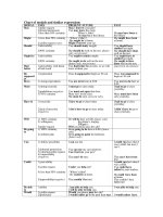
Radiographic atlas of skull and brain anatomy
Bạn đang xem bản rút gọn của tài liệu. Xem và tải ngay bản đầy đủ của tài liệu tại đây (38.27 MB, 371 trang )
M.
Gallucci
•
S.
Capoccia
•
A.
Catalucci, Radiographic Atlas of Skull and Brain Anatomy
Massimo Gallucci
•
Silvia Capoccia
•
Alessia Catalucci
Radiographic Atlas
of
Skull
and Brain
Anatomy
With 794 Figures
Springer
MASSIMO GALLUCCI
MD,
Professor and Chairman, Department of Neuroradiology, University of U Aquila, Italy
SILVIA CAPOCCIA
MD,
Senior Staff Radiologist at the S. Maria della Stella Hospital, Orvieto, Italy
ALESSIA CATALUCCI
MD,
Senior Staff Radiologist at the S. Salvatore Hospital, L'Aquila, Italy
This edition of Radiographic Atlas of Skull an Brain Anatomy by Gallucci - Capoccia - Catalucci is published by arrange-
ment with Idelson-Gnocchi srl, Naples, Italy
© 2005 CASA EDITRICE IDELSON-GNOCCHI Srl-Editori dal 1908
ISBN-10 3-540-34190-0 Springer-Verlag, Berlin Heidelberg New York
ISBN-13 978-3-540-34190-1
Library of Congress Control Number: 2006926503
This work is subject to copyright.
All
rights are reserved, whether the whole or part of the material is concerned, specifically the rights of
translation, reprinting, reuse of illustrations, recitation, broadcasting, reproduction on microfilm or in any other
way,
and storage in data
banks.
Duplication of this publication or parts thereof
is
permitted only under the provisions of the German Copyright Law of September
9,1965, in its current version, and permission for use must always be obtained from Springer-Verlag. Violations are liable for prosecution
under the German Copyright Law.
Springer is a part of Springer Science+Business Media
© Springer-Verlag Berlin Heidelberg 2007
Printed in Germany
The use of general descriptive names, registered names, trademarks,
etc.
in this publication does not imply, even in the absence of
a
speci-
fic statement, that such names are exempt from the relevant protective laws and regulations and therefore free for general use.
Product hability: The pubUshers cannot guarantee the accuracy of any information about the application of operative techniques and
medications contained in this book. In every individual case the user must check such information by consulting the relevant literature.
Editor: Dr. Ute Heilmann
Desk Editor: Meike Stoeck
Production Editor: Joachim
W.
Schmidt
Cover design: Frido Steinen-Broo, eStudio Calamar, Spain
Typesetting: FotoSatz Pfeifer GmbH, D-82166 Grafelfing
Printed on acid-free paper - 24/3150 - 5 4
3
2
1
0
Preface
The English Edition contains a few differences from the first ItaHan Edition, which require an explanation. Firstly, some imag-
es,
especially some 3D reconstructions, have been modified in order to make them clearer. Secondly, in agreement with the
Publisher, we have disowned one of our statements in the preface to the Italian Edition. Namely, we have now added a brief
introductory text for each
section,
by way
of explanation to the anatomical and physiological
notes.
This should make it easier
for the reader to understand and refer to this Atlas.
These differences derive from our experience with the previous edition and are meant to be an improvement thereof
Hopefully, there will be more editions to follow, so that we may further improve our work and keep ourselves busy on lone-
some evenings.
Finally, the improvements in this edition are a reminder to the reader that one should never purchase the first edition of a
work.
UAquila, January 2006
The Authors
Preface to the Italian Edition
I have been meaning to publish an atlas of neuroradiologic cranio-encephaHc anatomy for at least the last decade. Normal
anatomy has always been of great and charming interest to me. Over the years, while preparing lectures for my students, I have
always enjoyed lingering on anatomical details that today are rendered with astonishing realism by routine diagnostic ima-
ging.
To the allure of the images we should add the necessary teamwork with our colleagues and associates, and as I finally found
a go-ahead and open-minded publisher in Guido Gnocchi, I decided to pursue this idea.
The project was fulfilled thanks to the constant, friendly willingness of Silvia Capoccia and Alessia Catalucci, without whom
the idea would have remained an idea, Mr Gnocchi, whom I had alerted about the idea of an anatomical atlas at least four
years ago, would have been disappointed, the atlas would have been shelved, and I would have been credited with something
I did not do.
I am, therefore, grateful to Alessia and Silvia (in inverse alphabetical order of surname on the front cover) for carrying out
much of the work. I am equally grateful to Massimo Caulo for providing a number of the images of functional studies, and to
architect and graphic wizard Mauro Trappolino for solving problems we had with the more complex images.
To
myself,
besides my own share of this work, remained the most difficult and challenging part: writing the preface and the
dedication.
The preface: the following pages contain images but no text, as is fitting for an atlas. We specifically chose not to focus on
technical notes or physiological explanations because we believe that the atlas can be referred to for comparison, checking,
or for the location of pathologies, and not for an understanding of their functional meaning or clinical expression. This atlas,
therefore, is not intended for those who are already experienced in navigating the anatomy and physiopathology of the ner-
vous system. Rather, it is hoped the less experienced will benefit from it. Should this not be the case, we will not return their
money, which in any case will be donated to health projects in the Third World.
The dedication: you may have noticed that, in recent years, medical books have appeared without a bibliography, index, or
even whole chapters, and yet one element is always present: a dedication. I gather it represents a crucial element and, thus, I
must do my best to provide one. Upon perusing available dedications, I noticed that they nearly always involve family mem-
bers or professional masters. To whom, then, should I dedicate this work? To a great master of mine. Professor AgnoH, who
passed away over a decade ago, I have already dedicated a previous work. As for my family, I am not sure that a dedication
would be appropriate. They, too, were the recipients of a previous dedication and, to be honest, this kind of gift might not seem
totally unselfish: it is less an authentic dedication than an attempt to quench feelings of guilt for depriving one's family of qua-
lity time in order to achieve a "higher" goal. Yet at other times, when the goal is not that high, a dedication is an awkward
attempt to involve the neglected family in a form of narcissistic care which, as such, does not leave any room for others.
Therefore, if my daughters should someday ask me: "Why on earth did you dedicate to us a book on skulls?" I would be
hard-pressed to find an answer. The best reply would be "because I never wrote a book of poetry". That is why, in the hope of
succeeding in writing a book of poetry within the next half century to dedicate to my dearest ones, I do not think it is out of
place if I dedicate this work to the job of artisans like
myself,
Alessia, Silvia, and most of those who will consult it.
VIII
Tibetans do not eat meat. Only rarely do they do so, when forced by famine. Folco Maraini wrote (Segreto Tibet, Leonardo da
Vinci Publisher, Bari, 1951) that in this case, before they kill the animal, they explain to it the necessity of
its
sacrifice, and that
this will benefit its soul. The animal is also told that the body of its killer will in turn become a meal for other creatures, after
its death.
Ce qualcuno sulla cui pelle ho imparato
Ce qualcuno per la cui pelle non ho dormito.
A questo gioco inevitabile di dare e avere
Che caratterizza il nostro mestiere
E la nostra vita
(There are some on whose skin
1
learnt
There are some for whose skin I did not sleep.
To this unavoidable game of give and take
That characterizes our job
And our lives)
M.G.
Contents
1 - Surface Anatomy 1
A Skull: Plain Film 3
B Skull: Three-Dimensional Computed Tomography 11
C Brain: Three-Dimensional Magnetic Resonance 25
2 - Sectional Anatomy of the Telencephalon 29
A Sagittal Sections 31
B Coronal Sections 45
C Axial Sections 63
3 - Brainstem and Cerebellum 85
A Sagittal Sections 87
B Coronal Sections 93
C Axial Sections 101
4 - Cranial Nerves and Related Systems 113
A Olfactory Nerve (I) and Olfactory System 115
B Optic Nerve (II), Visual Pathway and Orbit
I Optic Nerve
(II),
Visual Pathway 119
II Orbit 127
C Oculomotor Nerve (III), Trochlear Nerve (IV), Abducens Nerve (VI) 141
D Trigeminal Nerve (V) 147
E Facial Nerve (VII) and Vestibulo-Cochlear Nerve (VIII), Acoustic and Vestibular Systems, Petrous Pyramid
I Facial Nerve (VII) and Vestibulo-Cochlear Nerve (VIII), Acoustic and Vestibular Systems 157
II Petrous Pyramid: Computed Tomography 171
F Glossopharyngeal Nerve
(IX),
Vagus Nerve
(X),
Accessory Nerve (XI), Hypoglossal Nerve (XII) 199
5 - Functional Systems 203
A Motor Systems
I Pyramidal System 205
II Extrapyramidal System: Basal Ganglia; Thalamus
A
Sagittal Sections 217
B Coronal Sections 225
C Axial Sections 235
B Sensory Systems
I Somatosensory System 243
II Gustatory System 249
C Speech System 253
D Brainstem Pathways and Nuclei 257
E Limbic System and Hippocampus 263
F Commissural and Associative Pathways 275
G Neuroendocrine System 277
H Cerebrospinal Fluid System 289
6
-
Vascular Anatomy 297
A Intracranial Arteries 299
B Arterial Vascular Territories 319
C Intracranial Veins 341
Bibiography 353
Subject
Index 355
1 Surface Anatomy
2 Short Introduction
pp.
3-23.
Surfoce Anatomy
The Skull
The skull can be divided into two portions: neural and facial.
The neural skull is made up of
6
bones: frontal, parietal, tem-
poral, occipital, sphenoid and ethmoid. The facial skull com-
prises 8 different bones. In this chapter, they will be briefly
described. Synthetic descriptions of sutures between them
and of the skull base foramina will follow.
I.
Skull
Bones
• frontal bone
The frontal bone resembles a cockleshell, and consists of two
portions: a vertical portion, the squama, corresponding to the
forehead region; and an orbital or horizontal portion, which
extends to form the roofs of the orbital and nasal cavities.
• parietal bone
The parietal bones form the sides and roof of the cranium.
Each bone is irregularly quadrilateral in form.
• temporal bone
The temporal bones are situated at the base of the skull. Each
consists of five parts: squama, petrous, mastoid, and tym-
panic parts, and styloid process.
• occipital bone
The occipital bone is trapezoid-shaped and situated at the
back and lower part of the cranium. It contains a large oval
aperture, the foramen magnum, through which the cranial
cavity communicates with the vertebral canal. The curved,
expanded plate behind the foramen magnum is termed the
squama, while the thick part in front of the foramen is called
the basilar part.
• sphenoid bone
The sphenoid bone is a bone situated at the base of the skull
in front of the temporal and basilar parts of the occipital
bone. It resembles a bat with open wings. It is divided into a
median portion or body, two greater and two lesser wings ex-
tending outwards from the sides of the body, and two ptery-
goid processes which project from it downwards.
• ethmoid bone
The ethmoid bone is located at the roof of the nose and sepa-
rates the nasal cavity from the brain. It is lightweight due to a
spongy construction. The ethmoid bone consists of four parts:
- the horizontal "cribriform" plate (lamina cribrosa), part
of the cranial base
- the vertical "perpendicular" plate (lamina perpendicu-
laris),
which is part of the nasal septum
- the two lateral masses (labyrinths)
• mandible
The mandible forms the lower jaw (inferior maxillary bone).
It is the largest bone of the face. The mandible consists of a
curved, horizontal portion, the body, and two perpendicular
portions, the rami, connected with the ends of the body at al-
most right angles.
• maxilla
The maxillae join together to form the whole of the upper
jaw. They hold the upper teeth, and are connected to the zy-
gomatic bones on the left and right. They assist in forming
the roof of the mouth, the floor and lateral wall of the nose,
and the floor of the orbit. They contribute to the formation
of two fossae, the infratemporal and pterygopalatine, and
two fissures, the inferior orbital and pterygomaxillary. Each
bone consists of a body and four processes - zygomatic,
frontal, alveolar, and palatine.
• palatine bone
The palatine bone is situated at the back part of the nasal
cavity between the maxilla and the pterygoid process of the
sphenoid bone. It contributes to the walls of three cavities:
the floor and lateral wall of the nasal cavity, the roof of the
mouth, and the floor of the orbit; it contributes to the for-
mation of the pterygopalatine and pterygoid fossae, and the
inferior orbital fissure. The palatine bone consists of a hori-
zontal and a vertical part.
• zygomatic bone
The zygomatic bone (zygoma; malar bone) is a paired bone
articulated with the maxilla, the temporal bone, and the
sphenoid bone. It presents a malar and a temporal surface;
four processes, the frontosphenoidal, orbital, maxillary, and
temporal; and four borders.
• nasal bone
The nasal bones are two small oblong bones, varying in size
and form; they are placed side by side at the middle and up-
per part of the face, and join to form the nose.
• lacrimal bone
The smallest and most fragile bone of the face, the lacrimal
bone is situated at the front part of the medial wall of the or-
bit.
• vomer bone
The vomer bone is located in the midsagittal line, forms the
hind and lower part of the nasal septum, and touches the
sphenoid, the ethmoid, the left and right palatine bones, and
the left and right maxillary bones.
• inferior nasal concha
The inferior nasal concha is a lamina of spongy bone (con-
cha nasahs inferior; inferior turbinated bone) that extends
horizontally along the lateral wall of the nasal cavity.
II.
Sutures
- Sagittal - along the midline, between parietal bones
- Coronal - between the frontal and parietal bones
- Lambdoid - between the parietal and occipital bones
- Squamosal - between the parietal and temporal bones
- Metopic - between the two frontal bones, prior to the fu-
sion of the two into a single bone
III.
Skull Base Foramina
The skull base is crossed by several foramina. The following
is a list of them and their contents:
- foramen caecum: emissary vein to superior sagittal sinus
- foramina of cribriform plate: olfactory nerve bundles
- posterior ethmoidal foramen: posterior ethmoidal ar-
tery, vein and nerve
- optic canal: optic nerve [II], ophthalmic artery
- superior orbital fissure:
oculomotor nerve [III]
trochlear nerve [IV]
lacrimal, frontal and nasociliary branches of ophthalmic
nerve [VI]
A
Skull:
Plain
Film 3
abducens nerve [VI]
superior ophthalmic vein
foramen rotundum: maxillary nerve [V2]
foramen ovale:
mandibular nerve [V3]
accessory meningeal artery
lesser petrosal nerve (occasionally)
foramen spinosum:
middle meningeal artery and vein
meningeal branch of mandibular nerve
foramen lacerum:
internal carotid artery
internal carotid nerve plexus
canal of lesser petrosal nerve
canal of greater petrosal nerve
internal acoustic canal:
facial nerve [VII]
vestibulocochlear nerve [VIII]
labyrinthine artery
jugular foramen:
inferior petrosal sinus
glossopharyngeal nerve [IX]
vagus nerve [X]
accessory nerve [XI]
sigmoid sinus
posterior meningeal artery
internal jugular vein
hypoglossal canal: hypoglossal nerve [XII]
foramen magnum:
medulla oblongata
vertebral arteries
meningeal branches of vertebral arteries
spinal roots of accessory nerves.
Lesser wing of sphenoid bone
Innominate line
Orbital border
Ethnnoidal cells
or ethmoidal labyrinth
Foramen rotundum
Mandibular condyle
Mastoid process
Odontoid process
of axis
Vertical ramus of mandible
Atlanto-axial joint
Horizontal ramus of mandible
Sagittal suture
Frontal sinus
Superior border
of petrous pyramid
Nasal septum
Atlanto-occipital joint
Lateral mass of atlas
Axis (body)
Angle of mandible
4 1
Surface Anatomy
Frontal sinus
interior clinoid process
Sphenoidal sinus
Floor of sella turcica
Anterior nasal spine
Hard palate
Outer table
Diploe
Inner table
Vasal imprints
Lambdoid suture
External occipital protuberance
Posterior clinoid process
A
Skull:
Plain
Film 5
Frontal sinus
Nasal bones
Malar bone
Anterior nasal spine
Alveolar processes -
Mandible
Sphenoethmoidal planum
Maxillary sinus
Hard palate
Hyoid bone
6 1
Surface Anatomy
Sagittal suture
Superior orbital border
(roof of the orbit)
Fronto-zygomatic suture
Orbital floor
Zygomatic bone (body)
Maxillary sinus -^
Petrous pyramid of temporal bone -^
Occipital foramen -^
Angle of mandible —
Frontal sinus
Ethmoidal cells
Orbit
Nasal septum
Foramen rotundum
Superior margin
of petrous pyramid
Odontoid process
of axis
k
A
Skull:
Plain
Film 7
Mastoid process
Vertical ramus of mandible-
Odontoid process of axis
Peduncle and lateral mass of atlas
Spinous process of axis
Angle of mandible
Horizontal ramus of mandible
^m
Atlanto-axial articular space
Lateral mass of axis
Lower border of the posterior arch of atlas
8 1
Surface Anatomy
Bregma Vertex
Pterion
Glabella
Anterior nasal spine
External acoustic meatus
Gonion
Lambda
_ Asterion
Porion
Inion or external occipital protuberance
A
Skull:
Plain Film 9
Glabella
Nasion
Anterior nasal spine
Pogonion —
Gnation
^- _,,,^'
10 1
Surface Anatomy
Superior horizontal line
Orbitomeatal line -
V
'""^Jtu^^/-
'ISf'
1 w^^
;V
^i: ;
Auricular vertical line
Frankfurt plane
B
Skull:
Three-Dlmenslonal Computed
Tomography
11
iiit'iWVjr*
Frontal bone
Sphenoid bone
Parietal bone
Lacrimal bone
Ethmoid bone
Nasal bones
Temporal bone
t ll^lfePHlli
IP Zygomatic bone
^^^^^^^
*
^^ Maxillary bone
Mandible
12
1
Surface Anatomy
|SH
:P :
#
^B Orbital process of the palatine bone
^^ Frontal bone
^^ Sphenoid bone
' Parietal bone
^^ Lacrimal bone
Ethmoid bone
Nasal bones
^P Temporal bone
^P Zygomatic bone
^P Maxillary bone
^P Mandible
B
Skull:
Three-Dlmensional
Computed
Tomography 13
^*.lt
iiis
;*
B Frontal bone
^ Sphenoid bone
I Parietal bone
^ Lacrimal bone
Ethmoid bone
^ Occipital bone
i Nasal bones
B Temporal bone
B Zygomatic bone
A Maxillary bone
A Mandible
14 1
Surface Anatomy
:^t2 l
^i r^^£j
Parietal bone
Temporal bone
Zygomatic bone
Occipital bone
Mandible
^ '^•••^^ r-
B
Skull:
Three-Dimensional Computed Tomography
15
N
M
Frontal bone
^ Sphenoid bone
I I Parietal bone
A Palatine bone
Ethmoid bone
^ Nasal bones
^ Vomer bone
^^ Occipital bone
^ Maxillary bone
^ Mandible
^p Temporal bone
16 1
Surface
Anatomy
/
Frontal bone
Sphenoid bone
Parietal bone
Ethmoid bone
Temporal bone
Occipital bone