THE EFFECTS OF CHROMIUM ON SKELETAL MUSCLE MEMBRANE/CYTOSKELETAL PARAMETERS AND INSULIN SENSITIVITY
Bạn đang xem bản rút gọn của tài liệu. Xem và tải ngay bản đầy đủ của tài liệu tại đây (2.94 MB, 198 trang )
THE EFFECTS OF CHROMIUM ON SKELETAL MUSCLE
MEMBRANE/CYTOSKELETAL PARAMETERS AND INSULIN
SENSITIVITY
Nolan John Hoffman
Submitted to the faculty of the University Graduate School
in partial fulfillment of the requirements
for the degree
Doctor of Philosophy
in the Department of Cellular and Integrative Physiology,
Indiana University
February 2012
ii
Accepted by the Faculty of Indiana University, in partial
fulfillment of the requirements for the degree of Doctor of Philosophy.
________________________________
Jeffrey S. Elmendorf, Ph.D., Chair
________________________________
Robert V. Considine, Ph.D.
Doctoral Committee
________________________________
Nuria Morral, Ph.D.
December 13, 2011
________________________________
Fredrick M. Pavalko, Ph.D.
iii
Dedication
This dissertation is dedicated to my family. I would not be where I am today
without the continued love, support and guidance I have received from my family.
I would like to thank my mother and father for always being there for me and
encouraging me to work up to my potential. They have been wonderful role
models who have taught me to have a strong work ethic, integrity, compassion
for others and a passion for my career. I would also like to thank my brother,
sister and extended family for always being there for me and being great sources
of advice, encouragement and support throughout the years.
iv
Acknowledgements
First, I especially thank my mentor at Indiana University School of Medicine,
Dr. Jeff Elmendorf, for being a great role model in both science and life. I would
like to thank Jeff for allowing me the freedom to explore my own ideas while
keeping me focused on the goals of my research projects. I also thank Jeff for
providing a wonderful graduate training experience in which I obtained a strong
skill set in experimental design, scientific techniques, scientific writing and oral
data presentation.
Next, I thank the members of my graduate research committee, Drs. Robert
Considine, Nuria Morral and Fredrick Pavalko for their continued support and
guidance throughout my thesis research. I thank my research committee for
useful advice about my research and teaching me to always be critical in my
experimental design and data interpretation. In addition, I thank my research
collaborators, Drs. Joseph Brozinick, Richard Day and Madhu Dhar for their
contributions and advice related to my thesis research and collaborative projects.
I am grateful for my undergraduate mentor at Butler University, Dr. Stephen
Perrill, who gave me the opportunity to become involved with Butler’s
undergraduate research program, sparked my interest in pursuing a career in
scientific research and encouraged me to pursue my passion for scientific
research by enrolling in an international exchange program and graduate school.
I thank past and present members of Dr. Jeff Elmendorf’s laboratory for being
great friends, scientific colleagues and for providing such an enjoyable
experience in the laboratory during my graduate training. I especially thank Drs.
v
Lauren Nicole Bell, Kirk Habegger, Guruprasad Pattar and Whitney Sealls for
training me in the laboratory and for always being there for advice and to discuss
my research projects. I also thank fellow lab members Brent Penque, Colin
Ridenour and Lixuan Tackett for their continued friendship, support and
assistance with experiments related to my thesis research. In addition, I thank
the faculty and staff of the Department of Cellular and Integrative Physiology for
all of their assistance throughout my graduate training. I also thank Dr. Simon
Rhodes and Monica Henry of the Indiana Biomedical Gateway Program for
providing me with a wonderful graduate school experience and numerous
leadership opportunities within the Indiana University Graduate School, including
the opportunity to serve as the student representative on the Indiana University
School of Medicine Graduate Committee.
Finally, I thank the Indiana University Center for Diabetes Research and the
Diabetes and Obesity Research Training Program for their generous financial
support of my thesis research through the DeVault Diabetes Fellowship and a
T32 Grant, T32-DK064466. I also thank the IUPUI Graduate and Professional
Student Government for an Educational Enhancement Grant and the IUPUI
Center for Membrane Biosciences for a travel fellowship that provided financial
support for travel to professional conferences. I am grateful to Drs. Amira Klip
and Steve Waters for generously providing the GLUT4myc expressing L6
myotubes and L6 myotube protocols.
vi
Abstract
Nolan John Hoffman
THE EFFECTS OF CHROMIUM ON SKELETAL MUSCLE
MEMBRANE/CYTOSKELETAL PARAMETERS AND INSULIN SENSITIVITY
A recent review of randomized controlled trials found that trivalent chromium
(Cr
3+
) supplementation significantly improved glycemia among patients with
diabetes, consistent with a long-standing appreciation that this micronutrient
optimizes carbohydrate metabolism. Nevertheless, a clear limitation in the
current evidence is a lack of understanding of Cr
3+
action. We tested if increased
AMP-activated protein kinase (AMPK) activity, previously observed in Cr
3+
-
treated cells or tissues from Cr
3+
-supplemented animals, mediates improved
glucose transport regulation under insulin-resistant hyperinsulinemic conditions.
In L6 myotubes stably expressing the glucose transporter GLUT4 carrying an
exofacial myc-epitope tag, acute insulin stimulation increased GLUT4myc
translocation by 69% and glucose uptake by 97%. In contrast, the
hyperinsulinemic state impaired insulin stimulation of these processes.
Consistent with Cr
3+
’s beneficial effect on glycemic status, chromium picolinate
(CrPic) restored insulin’s ability to fully regulate GLUT4myc translocation and
glucose transport. Insulin-resistant myotubes did not display impaired insulin
signaling, nor did CrPic amplify insulin signaling. However, CrPic normalized
elevated membrane cholesterol that impaired cortical filamentous actin (F-actin)
vii
structure. Mechanistically, data support that CrPic lowered membrane cholesterol
via AMPK. Consistent with this data, siRNA-mediated AMPK silencing blocked
CrPic’s beneficial effects on GLUT4 and glucose transport regulation.
Furthermore, the AMPK agonist 5-aminoimidazole-4-carboxamide-1-ß-D-
ribonucleoside (AICAR) protected against hyperinsulinemia-induced
membrane/cytoskeletal defects and GLUT4 dysregulation. To next test Cr
3+
action in vivo, we utilized obesity-prone C57Bl/6J mice fed a low fat (LF) or high
fat (HF) diet for eight weeks without or with CrPic supplementation administered
in the drinking water (8 µg/kg/day). HF feeding increased body weight beginning
four weeks after diet intervention regardless of CrPic supplementation and was
independent of changes in food consumption. Early CrPic supplementation
during a five week acclimation period protected against glucose intolerance
induced by the subsequent eight weeks of HF feeding. As observed in other
insulin-resistant animal models, skeletal muscle from HF-fed mice displayed
membrane cholesterol accrual and loss of F-actin. Skeletal muscle from CrPic-
supplemented HF-fed mice showed increased AMPK activity and protection
against membrane cholesterol accrual and F-actin loss. Together these data
suggest a mechanism by which Cr
3+
may positively impact glycemic status,
thereby stressing a plausible beneficial action of Cr
3+
in glucose homeostasis.
Jeffrey S. Elmendorf, Ph.D., Chair
viii
Table of Contents
List of Figures……………………………………………………………………… x
Abbreviations……………………………………………………………………… xii
I. Introduction………………………………………………………… 1
A. Insulin-Regulated Glucose Homeostasis
B. Signaling, Cytoskeletal and Membrane-Based GLUT4
Regulation
C. Obesity, Insulin Resistance and GLUT4 Dysregulation
D. Chromium: History and Effects on Glucose/Lipid Metabolism
E. AMPK Regulation of Glucose Transport and Cholesterol
Synthesis
F. Thesis Hypothesis and Specific Aims
II. Results………………………………………………………… 56
A. AMPK is Involved in a Membrane/Cytoskeletal Pathway of
Chromium Action that Improves Glucose Transport
Regulation in Insulin-Resistant Skeletal Muscle Cells
B. AMPK Enhances Insulin-Stimulated GLUT4 Regulation via
Lowering Membrane Cholesterol: Evidence for AMPK
Activity Countering Membrane Cholesterol-Induced Insulin
Resistance
C. Chromium Improves Skeletal Muscle Membrane/
Cytoskeletal Parameters and Insulin Sensitivity in High
Fat-Fed C57Bl/6J Mice
ix
III. Perspectives……… ……………………………………………… 109
IV. Experimental Procedures………………………………………… 127
V. References……………………………………………………… 137
VI. Curriculum Vitae
x
List of Figures
Figure 1……………………………………………………………………… 8
Figure 2……………………………………………………………………… 58
Figure 3……………………………………………………………………… 61
Figure 4……………………………………………………………………… 62
Figure 5……………………………………………………………………… 64
Figure 6……………………………………………………………………… 65
Figure 7……………………………………………………………………… 67
Figure 8……………………………………………………………………… 68
Figure 9……………………………………………………………………… 70
Figure 10……………………………………………………………………… 74
Figure 11……………………………………………………………………… 75
Figure 12……………………………………………………………………… 77
Figure 13……………………………………………………………………… 78
Figure 14……………………………………………………………………… 80
Figure 15……………………………………………………………………… 82
Figure 16……………………………………………………………………… 83
Figure 17……………………………………………………………………… 85
Figure 18……………………………………………………………………… 87
Figure 19……………………………………………………………………… 91
Figure 20……………………………………………………………………… 93
Figure 21……………………………………………………………………… 95
Figure 22……………………………………………………………………… 96
xi
Figure 23……………………………………………………………………… 97
Figure 24……………………………………………………………………… 99
Figure 25……………………………………………………………………… 101
Figure 26……………………………………………………………………… 102
Figure 27……………………………………………………………………… 104
Figure 28……………………………………………………………………… 106
Figure 29……………………………………………………………………… 108
Figure 30……………………………………………………………………… 115
xii
Abbreviations
2-DG 2-deoxy-D-glucose
ABC ATP-binding cassette transporter
ACAT Acyl-coenzyme A:cholesterol acyltransferase
ACC Acetyl-CoA carboxylase
ACTN4 Alpha-actinin-4
AICAR 5-aminoimidazole-4-carboxamide-1-beta-D-ribonucleoside
AMP 5’ adenosine monophosphate
AMPK 5’ AMP-activated protein kinase
APS Adaptor protein containing PH and SH domains
Arp2/3 Actin-related proteins 2/3
AS160 Akt substrate of 160-kDa
ATP 5’ adenosine triphosphate
ATV Atorvastatin
AUC Area under the curve
CaMKKβ Calmodulin-dependent protein kinase kinase β
CAP Cbl-associated protein
Cav-actin Caveolin-associated filamentous actin
CBS Cystathionine-β-synthase
CDK5 Cyclin-dependent kinase-5
Chol Cholesterol
CoA Coenzyme A
CPT-1 Carnitine palmitoyltransferase 1
xiii
Cr
3+
Trivalent chromium
Cr
6+
Hexavalent chromium
CrCIT Chromium citrate
CrCl
3
Chromium chloride
Cr(D-Phe)
3
Chromium (D-phenylalanine)
3
CrN Chromium nicotinate/niacin
CRP C-reactive protein
CrPic Chromium picolinate
CrY Chromium yeast
DAG Diacylglycerol
DMEM Dulbecco’s modified Eagle’s medium
DNP 2,4-dinitrophenol
ER Endoplasmic reticulum
FA Fatty acid
FAK Focal adhesion kinase
FBS Fetal bovine serum
F-actin Filamentous actin
GAP GTPase activating domain
GEF GLUT4 enhancer factor
GFAT Glutamine:fructose-6-phosphate amidotransferase
GLUT Glucose transporter
GSV GLUT4 storage vesicle
HBP Hexosamine biosynthesis pathway
xiv
HDL High density lipoprotein
HF High fat
HMG-CoA 3-hydroxymethyl-3-glutaryl coenzyme A
HMGR HMG-CoA reductase
IL-6 Interleukin-6
INSIG Insulin-induced protein
IPGTT Intraperitoneal glucose tolerance test
IPITT Intraperitoneal insulin tolerance test
IR Insulin receptor
IRAP Insulin-responsive amino peptidase
IRS Insulin receptor substrate
IVGTT Intravenous glucose tolerance test
kDa Kilodalton
LDL Low density lipoprotein
LF Low fat
LXR Liver X receptor
L6-GLUT4myc L6 muscle cells stably expressing GLUT4 that carries an
exofacial myc-epitope tag
MEF-2 Myocyte enhancer factor-2
NMR Nuclear magnetic resonance
NO Nitric oxide
NRF1 Nuclear respiratory factor 1
OGA O-GlcNAcase
xv
OGT O-linked N-acetylglucosamine transferase
PA Phosphatidic acid
PAS Phosho-Akt substrate
PBS Phosphate buffered saline
PGC-1α Peroxisome proliferator-activated receptor gamma,
coactivator 1 alpha
PI3K Phosphatidylinositol 3-kinase
PIP
2
Phosphatidylinositol 4,5 bisphosphate
PIP
3
Phosphatidylinositol 3,4,5 triphosphate
PLD1 Phospholipase D1
PM Plasma membrane
PP2A Protein phosphatase 2A
RXR Retinoic X receptor
SCAP SREBP cleavage-activating protein
SDS-PAGE Sodium dodecyl sulfate polyacrylamide gel electrophoresis
siRNA Small interfering RNA
SNAP23 Synaptosomal-associated protein 23
SNARE Soluble N-ethylmaleimide sensitive factor attachment protein
receptor
SRE Sterol response element
SREBP Sterol response element binding protein
T2D Type 2 diabetes
TBC1D Tre-2 BUB2 CDC16, 1 domain family member
xvi
TIRFM Total internal reflection microscopy
TNFα Tumor necrosis factor alpha
t-SNARE Target SNARE
TUG Tether containing UBX domain for GLUT4
Ubc9 Ubiquitin-conjugating enzyme 9
UDP-GlcNAc Uridine diphosphate-N-acetylglucosamine
v-SNARE Vesicle SNARE
VAMP2 Vesicle-associated membrane protein 2
ZA Zaragozic acid A
α-MEM Alpha minimum essential medium
βCD Methyl-beta-cyclodextrin
βCD:Chol Methyl-beta-cyclodextrin preloaded with cholesterol
1
Chapter I. Introduction
Humans have evolved to fight starvation. It is ironic that food has become
modern man’s foe contributing to the increasing worldwide prevalence of obesity
and type 2 diabetes (T2D). Despite enormous medical progress in infectious
diseases leading to a dramatic increase in human life expectancy, for the first
time we are witnessing a reversal of this trend. This is mainly being driven by
non-infectious diseases including diabetes, cardiovascular disease and cancer.
Overnutrition and lack of physical activity in the developed world have
contributed to the growing incidence of obesity and T2D, which have now
reached epidemic proportions. According to the United States Centers for
Disease Control and Prevention 2011 National Diabetes Fact Sheet (1), 25.8
million Americans and 79 million American adults now have diabetes and pre-
diabetes, respectively. T2D accounts for over 90% of those afflicted with
diabetes. Diabetes has become a major healthcare and economic burden in the
United States and worldwide with an estimated total annual cost of $174 billion in
the United States in 2007. As one of the world’s fastest growing chronic
diseases, diabetes is a leading cause of heart failure, kidney failure, stroke, lower
limb amputations and blindness in adults (1). Importantly, there is also a hidden
burden whereby diabetes can lead to other chronic diseases such as cancer,
heart disease and Alzheimer’s disease.
Insulin resistance is a well-recognized pathophysiological feature of pre-
diabetes and T2D. Insulin resistance is known to drive the progression of T2D
and has been found to be highly correlative with cardiovascular risk factors that
2
often contribute to morbidity in T2D patients (2). A comprehensive understanding
of the cellular and molecular mechanisms contributing to insulin resistance has
not been deciphered. However, it is well-appreciated that nutrient excess and
obesity due to overeating and lack of physical activity predispose individuals for
insulin resistance and T2D. While new drugs continue to be introduced and have
shown some promise for patients, these strategies as a whole are not effectively
curbing the worldwide epidemic. This is mainly due to the complexity of insulin
resistance and adaptable nature of obesity. For example, when a person goes
on a diet and reduces energy intake, this can be accompanied by a
compensatory reduction in whole body energy expenditure (3). The shift towards
a positive energy balance during the progression of obesity not only involves an
increased energy intake, but also a concomitant lack of physical activity to utilize
this excess energy (3). Achieving patient compliance with recommended
programs involving increased physical activity and reduced energy intake
remains a major obstacle in curbing the obesity and T2D epidemics. Therefore, it
is crucial to continue dissecting the cellular and molecular mechanisms involved
in insulin resistance to identify new drug targets of therapeutic interest and
develop novel strategies for the treatment and/or prevention of obesity and T2D.
While the complex links between obesity and insulin resistance are still
incompletely understood, increased levels of glucose, insulin and fatty acids
(FAs) have been shown to negatively impact insulin sensitivity in both in vitro and
in vivo experimental models (4-15). For example, high levels of glucose and
lipids have been shown to prevent the ability of insulin to activate key signaling
3
intermediates resulting in insulin resistance (5, 6, 9). Interestingly, studies have
demonstrated that pathophysiologically-relevant nutrient toxicity can result in
insulin resistance without altering key insulin signaling intermediates (7, 8, 13,
16). Collectively, several studies have established membrane and cytoskeletal
derangements (i.e. alterations in the plasma membrane (PM) lipid environment
and/or cellular cytoskeletal structure) as key distal aspects of insulin resistance
that can impair insulin sensitivity independent of insulin signaling abnormalities
(7, 8, 10, 17-21). Interestingly, trivalent chromium (Cr
3+
) has been shown to
positively impact GLUT4 regulation by lowering PM cholesterol (22-24). Cr
3+
is a
micronutrient that has been appreciated to be beneficial for optimal glucose and
lipid metabolism since the 1950s. However, whether Cr
3+
can protect against
membrane cholesterol accrual and whether this prevents cortical filamentous
actin (F-actin) loss and GLUT4 dysregulation remains unknown.
Building upon fundamental findings in the field presented next, my thesis
research focused on determining the effects of Cr
3+
on skeletal muscle
membrane/cytoskeletal parameters and insulin sensitivity. The following
introductory sections will highlight insulin-regulated glucose homeostasis, GLUT4
regulation by insulin, insulin resistance, Cr
3+
, and AMP-activated protein kinase
(AMPK).
I.A. Insulin-Regulated Glucose Homeostasis
Insulin is a pancreatic hormone produced by β-cells in the pancreatic islets of
Langerhans. Insulin regulates a plethora of cellular functions in many tissues
4
throughout the body. A primary function of insulin entails the regulation of post-
prandial glucose homeostasis. In the post-prandial state, an elevation of blood
glucose triggers release of insulin from β-cells. Once released into the
bloodstream, insulin acts on the adipose tissue, skeletal muscle and liver to clear
excess circulating glucose and restore glucose homeostasis. Insulin acts on the
liver to inhibit hepatic glucose output by turning off glycogenolysis and
gluconeogenesis. In adipose tissue and striated muscle (i.e. skeletal and cardiac
muscle) insulin signals to stimulate glucose transport out of the bloodstream and
into these target tissues. The combined effects of insulin on suppressing hepatic
glucose output by the liver and stimulation of glucose uptake into adipose tissue
and skeletal muscle are essential in maintaining whole body glucose
homeostasis.
In adipose and striated muscle tissues, insulin-mediated glucose transport is
achieved by the ability of insulin to stimulate the redistribution of the insulin-
responsive glucose transporter GLUT4 from intracellular pools to the PM (17, 25-
27). In the absence of insulin, GLUT4 primarily resides in intracellular membrane
pools. Upon insulin binding to the insulin receptor on the surface of muscle and
fat cells, insulin triggers a signaling cascade that stimulates an increase in the
exocytosis rate of GLUT4-containing vesicles. These complex trafficking events
orchestrated by insulin populates the PM with GLUT4 to allow glucose transport.
The inability of insulin to properly stimulate glucose transport into muscle/fat and
inhibit hepatic glucose output, termed insulin resistance, is a central feature of
obesity, pre-diabetes and T2D. Insulin resistance initially results in glucose
5
intolerance, and the hyperinsulinemic response from the pancreatic β-cells
immediately compensates to account for the reduced peripheral sensitivity to
insulin action. While increased insulin secretion from the β-cells can be effective
in maintaining blood glucose levels over time, this hyperinsulinemia and insulin
resistance can lead to β-cell expansion and eventual exhaustion/death. Once the
remaining β-cells can no longer secrete sufficient insulin to maintain glucose
homeostasis, blood glucose levels increase indicating progression of insulin
resistance to frank T2D.
At the cellular and molecular levels, insulin resistance is very complicated and
involves many different mechanisms varying between different tissues. In
adipose tissue and skeletal muscle, one certain definition of insulin resistance is
the failure of insulin to properly recruit, mobilize and insert GLUT4 into the PM to
stimulate glucose transport in the setting of normal GLUT4 protein expression.
Together these two tissues account for over 90% of post-prandial glucose
disposal (28). The complex derangements observed in insulin resistance stress
the importance for efforts to dissect these mechanisms of GLUT4 dysregulation
to develop new drug targets and therapeutic strategies for the treatment and/or
prevention of insulin resistance and T2D. While a complete understanding of how
insulin regulates GLUT4 translocation to the PM and glucose transport does not
yet exist, significant advances have been made to help us understand the actions
of insulin how these processes become deranged in insulin resistance.
The focus of this thesis research was to dissect membrane/cytoskeletal
parameters of skeletal muscle insulin sensitivity altered in insulin resistance and
6
determine the mechanisms amendable to Cr
3+
action. This research is primarily
focused on skeletal muscle, as this tissue is responsible for a large majority of
post-prandial glucose disposal (29) and is regarded as a major site of insulin
resistance (28). Therefore, the following sections and subsections will provide a
pertinent outline and analysis of our current state of knowledge regarding insulin
regulation of glucose transport and insulin resistance, primarily in skeletal
muscle. Expanded information on hepatic and/or adipocyte insulin action can be
found in several detailed reviews on these topics (30-32). While skeletal muscle
will be the major focus of the background and research outlined in this thesis, it is
important to note that skeletal muscle is by no means the only tissue involved
with maintenance of glucose homeostasis and development of insulin resistance.
I.B. Signaling, Cytoskeletal and Membrane-Based GLUT4 Regulation
Solving how insulin regulates glucose transport into skeletal muscle and
adipose tissue remains a fundamental challenge in biology and a significant
issue in medicine. A central feature of this process is the coordinated
accumulation of the glucose transporter GLUT4 into the plasma membrane. New
signaling and cytoskeletal mechanisms of insulin-stimulated GLUT4 exocytosis
are of emerging interest, particularly those at or just beneath the plasma
membrane. The following subsections examine signals that functionally engage
GLUT4 exocytosis, consider cytoskeletal regulation of the stimulated GLUT4
itinerary, and appraise involvement of plasma membrane parameters in GLUT4
control. Explored further are how these newly defined signaling, cytoskeletal, and
7
membrane mechanisms may be of therapeutic interest in the treatment and/or
prevention of GLUT4 dysregulation in disease.
I.B.1. Recruiting GLUT4
Under normal insulin responsiveness, insulin promotes the removal of excess
glucose from the circulation by stimulating the exocytic recruitment of intracellular
GLUT4 storage vesicles (GSVs) to the plasma membrane (PM) of skeletal
muscle and fat cells (25, 27). This stimulated redistribution of intracellular GSVs
results in PM GLUT4 accrual that facilitates cellular glucose uptake (Fig. 1).
Activation of GSVs by insulin requires a phosphatidylinositol 3-kinase (PI3K)
signal involving the upstream insulin receptor (IR) and insulin receptor substrate
(IRS) activators and the downstream Akt2 target enzyme (25, 27, 33).
Until the discovery of AS160 (Akt substrate of 160-kilodaltons (kDa)) in 2002
(34), how the IR/IRS1/PI3K/Akt2 signal coupled to GSVs remained unclear. This
protein, also known as TBC1D4 (Tre-2 BUB2 CDC16, 1 domain family member
4), contains a GTPase activating domain (GAP) for Rabs, small G proteins
implicated in vesicle trafficking (35, 36). In the basal state, the Rab-GAP function
of TBC1D4 is thought to contribute to the intracellular retention of GSVs by
promoting the inactive GDP-bound state of Rabs; whereas insulin-stimulated
Akt2 suppresses the Rab-GAP activity of the TBC1D4 and thus increases the
active GTP-bound form of Rabs on GSVs to promote exocytosis (Fig. 1A).
Consistent with this localized functionality, TBC1D4 associates with GSVs via
binding to the insulin-responsive amino peptidase (IRAP), a GSV cargo
8
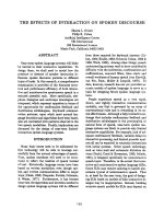
Figure 1. Schematic illustration of putative signals, cytoskeletal
mechanisms, and plasma membrane parameters involved in insulin-
stimulated GLUT4 storage vesicle exocytosis (17).
(A) Activation of GSVs by insulin requires a PI3K signal involving the upstream
IR and IRS activators and the downstream Akt2 target enzyme. TBC1D4 and
TBC1D1, substrates of Akt2, have been suggested to couple the PI3K/Akt2
signal to GSVs via its action on one or more critical Rab proteins. The basal
intracellular pool of GDP-Rab GSVs shown associated with several putative
anchoring systems (e.g., microtubules, Ubc9 (ubiquitin-conjugating enzyme 9),
TUG (tether containing UBX domain for GLUT4)) are activated by the
9
suppression of the Rab-GAP activity of TBC1D4/TBC1D1 by Akt2. Several
putative Rab proteins, the existence of possible calcium regulation, and
mechanisms associating TBC1D4 (and presumably TBC1D1) to the GSV via
IRAP have been suggested (see inset). (B) Cortical F-actin, likely originating at
the neck region of caveolae PM microdomains, plays a critical role in GSV
trafficking. Reorganization of the cortical F-actin meshwork by insulin signaling to
TC10 allows GSV/PM arrival, tethering, and docking. A large number of
proposed insulin-regulated processes occur in this PM vicinity such as TC10-
regulated formation of the exocyst complex and cortical F-actin remodeling,
PI3K/RalA-stimulated transition of trafficking GSVs to tethered GSVs, a role of
ACTN4 and/or the exocyst complex in tethering, and an α-fodrin-mediated
rearrangement of cortical actin filaments in the area of syntaxin 4 to facilitate
GSV/PM SNARE protein interaction and docking (see inset). (C) Insulin
signaling, through two putative PI3K signals that activate PKCδ/λ and PLD1,
prepares GSVs for fusion with the PM. The first PKCδ/λ signal has been
implicated in promoting the dissociation of Munc18c from syntaxin4, contributing
to the fusion-competent SNARE complex. The second PLD1 signal primes the
GSV and PM for fusion by generating PA, which has been suggested to act as a
fusogenic lipid in biophysical modeling studies by lowering
the activation energy
for membrane bending (i.e., negative membrane curvature) during generation
and expansion of fusion pores (see inset).