THE DIRECT REPROGRAMMING OF SOMATIC CELLS: ESTABLISHMENT OF A NOVEL SYSTEM FOR PHOTORECEPTOR DERIVATION
Bạn đang xem bản rút gọn của tài liệu. Xem và tải ngay bản đầy đủ của tài liệu tại đây (16.88 MB, 72 trang )
THE DIRECT REPROGRAMMING OF SOMATIC CELLS: ESTABLISHMENT
OF A NOVEL SYSTEM FOR PHOTORECEPTOR DERIVATION
A Thesis
Submitted to the Faculty
of
Purdue University
by
Melissa Mary Steward
In Partial Fulfillment of the
Requirements for the Degree
of
Master of Science
December 2012
Purdue University
Indianapolis, Indiana
ii
This thesis is dedicated to Anne McSherry Steward and the loving memory of
Eleanor Mary Vahey. The grandmothers of the author provided unflinching support
and inspiring examples of strength while articulating the value of independence,
family and education. Working mothers were my first teachers of the critical
concept ‘necessary and sufficient’. They have my gratitude, respect and love for
everything they shared.
iii
ACKNOWLEDGMENTS
I would like to thank many individuals for their support of this work. Firstly, I must
thank Dr. Jason S. Meyer, my thesis advisor, and friend of over a decade, for always
respecting and soliciting my contributions to science and education. His support and
expertise are deeply appreciated for their influence on me as a developing scientist
and person. I thank the members of my committee, Dr. Stephen Randall and Dr.
Guoli Dai as well as my department chair, Dr. Simon Atkinson, for their support
and expertise. I owe a debt of gratitude for significant support, both technical and
personal, to my friend and collaborator Akshayalakshmi Sridhar. She is wise beyond
her years. I thank Dr. Kathy Marrs, Dr. Mariah Judd and the NSF-funded GK-12
program, for providing professional and financial support, as well as the opportunity
to teach in one of my favorite settings. I thank Meyer lab member Manav Gupta, the
Biology Department and support staff of IUPUI, namely Sue Merrell, Shari Dowell
and Kurt Kulhavy, for their technical contributions and assistance. I must thank my
sister, Jennifer Steward and friend, Matthew Butcher, who have been indefatigable
sources of love, support and motivation. Dr. Mark Kirk deserves special thanks as
the first scientist to provide me a project and kindly, emphatically and ceaselessly
encouraging and supporting my career and graduate studies. Thanks also to my many
inspiring educators and supportive friends over the years, too numerous to name here
and all those who went before me, shining the light as far as they could, allowing me
to press even farther still. It appears to take a village to complete a thesis: I extend
my deep and sincere gratitude to the numerous individuals who contributed to my
success in this endeavor.
iv
TABLE OF CONTENTS
Page
LIST OF TABLES . . . . . . . . . . . . . . . . . . . . . . . . . . . . . . . . vi
LIST OF FIGURES . . . . . . . . . . . . . . . . . . . . . . . . . . . . . . . vii
ABBREVIATIONS . . . . . . . . . . . . . . . . . . . . . . . . . . . . . . . . viii
ABSTRACT . . . . . . . . . . . . . . . . . . . . . . . . . . . . . . . . . . . x
1 INTRODUCTION . . . . . . . . . . . . . . . . . . . . . . . . . . . . . . 1
1.1 Pluripotent stem cells as models and therapeutic agents . . . . . . . 1
1.2 Seminal studies in cellular reprogramming . . . . . . . . . . . . . . 3
1.3 Advantages of direct reprogramming over indirect reprogramming . 4
1.4 Differentiation and direct cellular reprogramming to neural
phenotypes . . . . . . . . . . . . . . . . . . . . . . . . . . . . . . . 5
1.5 Specific neuronal subtypes as phenocopies and replacement cell
sources . . . . . . . . . . . . . . . . . . . . . . . . . . . . . . . . . . 8
1.6 A model that accounts for direct cellular reprogramming . . . . . . 10
1.7 Photoreceptors: A unique opportunity for direct reprogramming . . 13
1.8 The transcriptional dominance model . . . . . . . . . . . . . . . . . 15
2 ESTABLISHMENT OF A NOVEL SYSTEM FOR DERIVATION OF
PHOTORECEPTORS VIA DIRECT REPROGRAMMING . . . . . . . 19
2.1 Selection of candidate genes . . . . . . . . . . . . . . . . . . . . . . 19
2.2 Establishment of a screening system for candidate genes . . . . . . 24
2.3 Lentiviral expression construct modifications . . . . . . . . . . . . . 25
2.4 Cloning strategies for the 23 gene candidate constructs . . . . . . . 31
2.4.1 PCR amplification techniques . . . . . . . . . . . . . . . . . 31
2.4.2 Direct commercial custom gene synthesis . . . . . . . . . . . 35
2.5 Sequence-confirmation of lentiviral expression constructs . . . . . . 36
2.6 Restriction enzyme-excision confirmation of large-scale plasmid
DNA preparations . . . . . . . . . . . . . . . . . . . . . . . . . . . 37
2.7 Lentivirus production: protocol optimization . . . . . . . . . . . . . 40
2.8 Demonstration of experimental feasibility and utility of constructs . 42
2.9 Reprogramming of somatic cells through delivery of transcription
factors . . . . . . . . . . . . . . . . . . . . . . . . . . . . . . . . . . 44
3 DETAILED METHODS . . . . . . . . . . . . . . . . . . . . . . . . . . . 48
3.1 MEF derivation . . . . . . . . . . . . . . . . . . . . . . . . . . . . 48
v
Page
3.2 Cloning strategies . . . . . . . . . . . . . . . . . . . . . . . . . . . . 49
3.2.1 PCR amplification . . . . . . . . . . . . . . . . . . . . . . . 49
3.2.2 Serial bacterial expression vector cloning . . . . . . . . . . . 49
3.2.3 Genes custom ordered from Integrated DNA Technologies . . 50
3.3 Cell culture . . . . . . . . . . . . . . . . . . . . . . . . . . . . . . . 50
3.4 Virus production . . . . . . . . . . . . . . . . . . . . . . . . . . . . 51
3.5 Calcium phosphate transfection . . . . . . . . . . . . . . . . . . . . 51
3.6 Immunocytochemistry . . . . . . . . . . . . . . . . . . . . . . . . . 52
4 CONCLUSIONS, FUTURE EXPERIMENTS AND IMPLICATIONS . . 53
4.1 Conclusions . . . . . . . . . . . . . . . . . . . . . . . . . . . . . . . 53
4.2 Future experiments continuing the project presented herein . . . . . 54
4.3 Implications of work resulting in directly reprogrammed rod
photoreceptors . . . . . . . . . . . . . . . . . . . . . . . . . . . . . 56
LIST OF REFERENCES . . . . . . . . . . . . . . . . . . . . . . . . . . . . 57
vi
LIST OF TABLES
Table Page
2.1 Transcription factors determined from data-mining . . . . . . . . . . . 21
2.2 Candidate genes . . . . . . . . . . . . . . . . . . . . . . . . . . . . . . 23
2.3 Primer Sequences(A-N) . . . . . . . . . . . . . . . . . . . . . . . . . . 32
2.4 Primer Sequences(N-O) . . . . . . . . . . . . . . . . . . . . . . . . . . 33
2.5 Sequencing primers for gene insertion sites . . . . . . . . . . . . . . . . 37
2.6 Custom sequencing primers for BRN2, BLIMP1 and CTCF . . . . . . 38
2.7 Custom sequencing primers for MYBPP1A and MYT1 . . . . . . . . . 39
vii
LIST OF FIGURES
Figure Page
1.1 Simplified schematic of gene circuits and attractor states . . . . . . . . 12
1.2 The transcriptional dominance model . . . . . . . . . . . . . . . . . . . 17
2.1 NrL promoter driven GFP-expression is specific to rods . . . . . . . . . 20
2.2 Rhodopsin-GFP fusion protein: specificity and experimental design . . 25
2.3 The modification and confirmation of the viral expression construct . . 26
2.4 Agarose gel showing the PCR-amplified PGK promoter . . . . . . . . . 28
2.5 Modified pCSC-PGK-IGW viral expression construct . . . . . . . . . . 29
2.6 Agarose gel showing clones 2 and 9 of the pCSC-PGK-IGW . . . . . . 30
2.7 Custom sequencing primers used to sequence confirm PGK promoter . 30
2.8 PCR-amplification and optimization experiments for Olig2 gene . . . . 34
2.9 Proper gene excision from the pCSC-PGK-IGW backbone . . . . . . . 40
2.10 Optimization of the lentivirus production and delivery protocols . . . . 42
2.11 Upregulation of protein expression induced in HEK293 cells . . . . . . 44
2.12 Phenotypic and protein expression changes induced in MEF cells . . . 46
viii
ABBREVIATIONS
ALS amyotrophic lateral sclerosis
BAM A combination of three proneural transcription factors Brn2,
Ascl1 and Myt1l
bHLH basic helix-loop-helix protein
BSC Biological Safety Cabinet
CMV cytomegalovirus
cDNA complementary DNA
DAPI 4
,6-diamidino-2-phenylindole
DMEM Dulbecco’s modified eagle medium
DMSO dimethyl sulfoxide
DNA deoxyribonucleic acid
E16 embryonic day16
EDTA ethylenediaminetetraacetic acid
ESCs embryonic stem cells
FACS fluorescence-activated cell sorting
FAD familial Alzheimer
s disease
FBS fetal bovine serum
GFP green fluorescent protein
HBSS Hank
s balanced salt solution
HEK293 human embryonic kidney cell line 293
iDA induced dopaminergic
iMN induced motor neuron
iN induced neuronal cells
iPSCs induced pluripotent stem cells
ix
LCA Leber
s congential amaurosis
MEF mouse embryonic fibroblast
miRNA micro ribonucleic acid
mRNA messenger ribonucleic acid
MCSs multiple cloning sites
MOIs multiplicities of infection
P2 post-natal day 2
PBS phosphate buffered saline
PGK phosphoglycerate kinase
qRT-PCR quantitative reverse transcriptase polymerase chain reaction
RP retinitis pigmentosa
SCNT somatic cell nuclear transfer
SMA spinal muscular atrophy
x
ABSTRACT
Steward, Melissa Mary. M.S., Purdue University, December 2012. The Direct Re-
programming of Somatic Cells: Establishment of a Novel System for Photoreceptor
Derivation. Major Professor: Jason S. Meyer.
Photoreceptors are a class of sensory neuronal cells that are deleteriously affected in
many disorders and injuries of the visual system. Significant injury or loss of these
cells often results in a partial or complete loss of vision. While previous studies have
determined many necessary components of the gene regulatory network governing
the establishment, development, and maintenance of these cells, the necessary and
sufficient profile and timecourse of gene expression and/or silencing has yet to be
elucidated. Arduous protocols do exist to derive photoreceptors in vitro utilizing
pluripotent stem cells, but only recently have been able to yield cells that are disease-
and/or patient-specific. The discovery that mammalian somatic cells can be directly
reprogrammed to another terminally-differentiated cell phenotype has inspired an ex-
plosion of research demonstrating the successful genetic direct reprogramming of one
cell type to another, a process which is typically both more timely and efficient than
those used to derive the same cells from pluripotent stem cell sources. Therefore,
the emphasis of this study was to establish a novel system to be used to determine
a minimal transcriptional network capable of directly reprogramming mouse embry-
onic fibroblasts (MEFs) to rod photoreceptors. The tools, assays and experimental
design chosen and established herein were designed and characterized to facilitate
this determination and preliminary data demonstrated the utility of this approach
for accomplishing this aim.
1
1 INTRODUCTION
The fields of developmental and regenerative biology have long sought to identify novel
approaches for the repair of damaged and/or diseased tissue, including that of the
nervous system. The mammalian central nervous system has been well documented
as one with limited regenerative capabilities, due at least in part to an inhospitable
environment for regeneration [1, 2]. In cases of injury and neurodegeneration, glial
scarring, the lack of proliferating oligodendrocytes, and the presence of inhibitory
factors can physically block or impair the regrowth of damaged neuronal axons and
pathfinding of growth cones [3, 4]. In both injury-induced and neurodegenerative
disorders, a toxic extracellular environment including widespread cell death and a
general absence of growth-promoting signals has been described [4, 5]. The mulitude
of factors contributing to the lack of regeneration in the mammalian central nervous
system has been a significant limitation for the fields of mammalian developmental
biology and regenerative medicine. A further limitation is a reduced ability to study
the molecular mechanisms and sequelae of disease at the cellular level, in both de-
veloping and adult tissue. A lack of animal models for many disorders, as well as
uncharacterized species differences in the pathways involved in injury, neurodegener-
ation and regeneration have hampered efforts to describe the underlying mechanisms
controlling and contributing to these processes.
1.1 Pluripotent stem cells as models and therapeutic agents
When mouse embryonic stem cells (ESCs) were first derived in 1981 [6], followed by
the derivation of human ESCs in 1998 [7], they provided a new model system for
researchers to study developmental and disease processes at a cellular level. At the
2
same time, they represented a new potential therapeutic cellular agent for clinicians
as a source for replacement cells in cases of neurodegeneration and injury.
ESCs are derived from the inner cell mass of a fertilized oocyte, and have two defin-
ing characteristics. They are pluripotent, which means that they can give rise to
all the cell types of an adult organism, including all of the specific cell types of the
central nervous system. They are also capable of self-renewal, which allows them to
be cultured and expanded in vitro indefinitely, providing an unlimited source of cells
for applications of research or therapeutics. However, one of the two major limit-
ing attributes of ESCs as applied to the field of therapeutics is the fact that they
are not patient-specific. Thus, these cells have an increased risk of rejection when
transplanted into another individual. A second inherent risk involved with the trans-
plantation of cells derived from a pluripotent cell source is the potential for delivering
pluripotent, or undifferentiated and dividing, cells to the body
The derivation of induced pluripotent stem cells (iPSCs) in 2006 [10] represented
a critical advance for regenerative medicine as the first opportunity to derive cells
from a pluripotent source while circumventing the risk of immune rejection due to
the ability to derive patient-specific lines. These iPSCs provided an opportunity to
derive adult cell types via an indirect cellular reprogramming strategy, which could
serve as the basis for pharmacological screening, disease-modeling and therapeutics
such as cellular replacement or cell rescue enabled by transplantation. However, the
second limiting attribute of ESCs as applied to therapeutics was not overcome with
the advent of this new pluripotent cell source. The delivery of mitotically active,
undifferentiated cells to a niche introduces a risk of tumorogenicity, i.e. tumor for-
mation. Unregulated cell division and invasion of undifferentiated or inappropriately
differentiated cells is a hallmark of certain forms of cancers. The advent of iPSCs
did however, open wide the door for further innovative studies in cellular reprogram-
ming. Directed in vitro differentiation of iPS cells prior to transplantation constitutes
3
one mechanism with which to minimize teratogenicity, but it does not exclude the
possibility of even an exceedingly small number of cells avoiding differentiation in
vivo application. An alternate strategy that would eliminate the tetratogenicity of
iPS cell cultures would involve a direct reprogramming strategy. The demonstrated
and replicated ability to genetically reprogram mammalian, adult, somatic cells to a
pluripotent, mitotically-active cellular phenotype stood contrary to the long-standing
tenet of biology that once cells become terminally differentiated, they cannot change
their fate. If adult somatic cells could be genetically reprogrammed to a pluripotent
state and further redifferentiated to specific adult cell phenotypes, the next question
became: could these same adult cells be directly genetically reprogrammed to another
cell fate?
1.2 Seminal studies in cellular reprogramming
Cellular reprogramming experiments conducted over the last 6 decades have laid a
substantial foundation upon which the hypothesis and experimental design of this
study are based. The work of Dr. John Gurdon and Dr. Shinya Yamanaka received
the Noble Prize in Physiology or Medicine in 2012 for their significant and high impact
discoveries in cellular reprogramming. Dr. Gurdon conducted the first experiment
that successfully cloned an organism from a somatic cell source [8]. In this study,
he used the process of somatic cell nuclear transfer (SCNT) established by Briggs
and King [9]. This process involves the transplantation of the nucleus of a somatic
cell to an enucleated, unfertilized oocyte. Cytoplasmic factors in the oocyte were
found to be sufficient to reprogram the somatic nuclei to an effective earlier stage of
development, allowing for the reinitiation of transcription of embryonic genes that
were silenced in the adult cell and initiating cellular division of the oocyte based
upon the genomic DNA of the somatic nucleus. Gurdon exploited this process to
clone a new frog, Xenopus laevis, through the use of a nucleus from a gastrointestinal
cell, removed from an adult frog. Rather than relying on undefined cytoplasmic
4
factors within an oocyte, Yamanaka
s work first demonstrated a genetic approach
to reprogram mouse somatic cells to a pluripotent state via lentiviral delivery of a
cocktail of four genes that govern pluripotency [10]. He dubbed the cells derived via
this process induced pluripotent stem cells (iPSCs). Similar to embryonic stem cells,
they were demonstrated to have the capacity to proliferate indefinitely in culture and
differentiate both in vivo and in vitro to cell types derived from all three germ layers -
ectoderm, mesoderm and endoderm. In between the time of these exciting discoveries,
other groups demonstrated the direct reprogramming of fibroblasts to myoblasts via
delivery of a single master transcriptional regulator MyoD [12], as well as the in vivo
direct reprogramming of exocrine cells from the pancreas to insulin-secreting beta
cells [13]. The implication of studies demonstrating these dramatic cell fate changes
was that direct cellular reprogramming of somatic cells was possible utilizing a genetic
approach.
1.3 Advantages of direct reprogramming over indirect reprogramming
There are several advantages afforded by direct reprogramming strategies when com-
pared to those utilizing a pluripotent stem cell intermediary. While either strategy
could be used to yield patient-specific cell populations, those derived via a direct
reprogramming strategy can remain a mitotically inactive cell population. Indirect
reprogramming strategies utilize pluripotent stem cells, which by definition are pro-
liferative and can give rise to more undifferentiated cells, as well cells that are more
differentiated. While in vitro protocols exist to differentiate these stem cells in sub-
stantial numbers and at high efficiencies and cell sorting using surface markers could
purify these cells for many cell types, there remains an increased risk of transplant-
ing undifferentiated cells, that could lead to tumor formation. Upon transplantation,
directly reprogrammed cells would have a much lower risk of tumorigenicity, as the
likelihood of introducing pluripotent stem cells to a new niche is significantly lower.
Another advantage of using direct genetic reprogramming strategies is that they may
5
uncover novel genes involved in the gene regulatory network of the desired cell type.
Many indirect reprogramming strategies utilizing in vitro differentiation of pluripo-
tent stem cells to the final cell type involve adding soluble mitogens and growth
factors to the cell culture media to differentiate stem cells, potentially activating or
inactivating often innumerable and overlapping pathways in the cell. Direct genetic
reprogramming strategies allow for the definition of elusive gene regulatory networks
that are ‘necessary and sufficient’ for defined cellular phenotypes that are currently
undescribed. Finally, direct reprogramming strategies are faster, more efficient and
less arduous than those involving a pluripotent intermediary. For example, Marius
Wernig
s group saw 20% conversion rates of fibroblasts to neuronal cells in 2 weeks
time utilizing direct genetic reprogramming [18]. This efficiency, similar to that seen
by many others, is orders of magnitude higher than that seen when establishing
pluripotent stem cell lines, and on the order of weeks instead of months. For pho-
toreceptors specifically, after the pluripotent cell lines are established, it takes up to
another three months to derive photoreceptors from them [19]. None of these advan-
tages conferred by direct genetic reprogramming affect their applicability when com-
pared to cells derived via indirect reprogramming strategies. They can still be used
for studies of development such as cell fate specification and for disease-modeling, as
well as therapeutics such as cell replacement and rescue conferred by transplantation
and also used for drug screening. Not only are none of these applications lost, some
- such as transplantation applications - stand to be enhanced when cell populations
are derived via direct reprogramming.
1.4 Differentiation and direct cellular reprogramming to neural phenotypes
Diseases of and injuries to the central and peripheral nervous system devastate the
sensory experience and motor control of a significant portion of the population each
year. Because of the prevalence and ramifications of these injuries and diseases, many
efforts have focused on the replacement or rescue of neural cell populations once they
6
are damaged or lost. In vitro protocols already exist to derive specific neural and
neuronal cell types from pluri- or multi-potent sources such as embryonic stem cells
(ESCs), induced pluripotent stem cells (iPSCs) or neural stem cells [14–17, 19–21].
These protocols are often based upon culturing the stem cells in culture medium with
fetal bovine serum and known proneural soluble growth factors. These factors are
known to be involved in pathways governing neural specification in vivo and induce
expression of neural-specific genes and positive feedback loops leading to the ultimate
differentiation of pluripotent cells to neuronal phenotypes. Until very recently, cells
derived via these protocols provided the best potential source for potential cellular
replacement and rescue strategies, as well as pharmacological screening and disease-
modeling.
The first successful direct, genetic reprogramming of mammalian somatic cells to a
neuronal phenotype was published in 2010 [18] and since that time, many groups
have utilized a similar experimental strategy to derive more specific neuronal cell
types from mammalian somatic sources [22–27]. Vierbuchen et al. first used a strat-
egy similar to the one employed by Yamanaka to derive induced pluripotent stem cells
from fibroblasts [10]. In the landmark studies by Yamanaka group, they sought to
reprogram terminally differentiated, somatic cells to a mitotically active pluripotent
state. Thus, he tested the effects of viral delivery of combinations of transcription
factors known to be active in embryonic stem cells and silenced in quiescent cell popu-
lations. These genes were therefore implicated to be involved in positively regulating
pluripotency. Vierbuchen et al. hypothesized that a similar strategy could be used
to derive neuronal cells directly from fibroblasts [18]. They defined a set of candi-
date transcription factors to test that were known or implicated to be involved in the
processes governing pluripotency or were specific to neural cell populations. They
started with a pool of 19 genes that were virally delivered combinatorially to mouse
embryonic fibroblast (MEF) cells, and screened for neuronal conversion. They ulti-
mately defined a combination of three factors, Brn2, Ascl1 and Myt1l (BAM) that
7
could quickly and efficiently convert fibroblasts to neuronal cells. These neuronal cells
were named induced neuronal (iN) cells and importantly were found to express mul-
tiple neural specific proteins, generate action potentials and form functional synapses
when cultured with cortical neuronal or glial cells. These iN cell cultures contained
inhibitory GABA-ergic neuronal cells, excitatory glutaminergic neuronal cells, as well
as some iN cells expressing markers of cortical interneurons and other neuronal sub-
types. Another important discovery from this study was the marked increase in
efficiency and rapidity of neuronal conversion seen using this direct reprogramming
strategy. They reported an approximate 20% conversion efficiency of infected cells
within 2 weeks, wheras traditional methods for iPSC reprogramming typically report
efficiencies of less than 0.1% and require several weeks for effective reprogramming.
This exciting discovery spurred an explosion of studies in the neurosciences employing
to use a similar approach to derive human iN cells, as well as specific neuronal cell
types utilizing the same strategy. By delivering cell-specific transcription factors in
combination with pro-neural genes such as those in the BAM cocktail to somatic cells,
attempts were made to derive dopaminergic neurons or motor neurons, for example.
When the BAM combination of transcription factors was initially delivered to human
cell cultures, immature neuronal phenotypes were reported, along with significant cell
death [23, 24]. It was quickly determined that the addition of another transcription
factor, NeuroD1 to the BAM cocktail resulted in the same neuronal attributes in
human cells after 5-6 weeks as those seen in the mouse system in 2 weeks with the
BAM combinatorial treatment alone. Neural-specific protein expression, action po-
tentials and post-synaptic currents were observed [23]. The differential time-course
of neuronal maturation seen when comparing the mouse and human system in direct
reprogramming is similar to differences seen using mouse and human derived ESCs
and iPSCs and may be reflective of a longer period of maturation during human ges-
tation and in vivo development.
8
As dopaminergic neurons are affected in many neurodegenerative disorders, such as
Parkinson
s disease and familial Alzheimer
s disease, replacement or rescue of these
specific neurons holds great promise for strategies of regenerative medicine. Dopamin-
ergic neurons were the first neuronal subtypes to be specified through genetic, di-
rect reprogramming strategies [24–27]. Several independent studies reported different
combinations of factors to derive these action potential-firing, tyrosine-hydroxylase
positive, induced dopaminergic (iDA) cells from human and mouse fibroblast cells,
with efficiencies reported approximating 10% of transduced cells, though only the
delivery of Ascl1, Nurr1 and Lmx1a, or the combination of these 3 genes with Pitx3,
Foxa2 and En1 was capable of reprogramning cells that were characterized to release
dopamine [25, 27]. Spinal motor neurons are another specific neuronal cell type that
is known to be affected by disease-states including spinal muscular atrophy (SMA)
and amyotrophic lateral sclerosis (ALS) or Lou Gehrig
s disease. Less than a month
after reports about the direct reprogramming of human and mouse fibroblasts to in-
duced dopaminergic (iDA) neuronal cells were published, the first study characterized
the direct reprogramming of spinal motor neuronal cells as well [22]. Their highest
efficiencies of conversion (around 5-10% in under 2 weeks) from mouse fibroblasts to
induced motor neuron (iMN) cells were reported using the aforementioned BAM com-
bination with the addition of four spinal motor neuron-specific factors, Lhx3, Hb9,
Isl1 and Ngn2. These iMN cells generated action potentials and responded to both
excitatory and inhibitory neurotransmitters in culture, similar to ESC-derived and
embryonic motor neurons. Addition of NeuroD1 to the pool of these seven factors led
to functional iMN cells reprogrammed from human ESC-derived fibroblasts as well,
that were characterized as similar to their mouse counterparts in the study.
1.5 Specific neuronal subtypes as phenocopies and replacement cell sources
Once defined neuronal cell types could be specified using direct genetic reprogram-
ming, the field was poised to ask if these directly reprogrammed iN cells could 1)
9
serve as reliable phenocopies for disease-states, 2) be demonstrated to integrate in
vivo and 3) restore any function that had been lost associated with the particular
disease pathology. Indeed, these questions have been addressed by several studies.
In the first study to derive iMN cells, using both mouse and human cells, iMN cell
sensitivity to growth factor withdrawal was demonstrated similar to embryonic motor
neurons [22]. The significant interest in the factors and pathways that confer neuronal
survival in the context of injury and neurodegenerative disease states makes these cells
a valuable in vitro tool for the study of motor neuron function, survival, disease, in-
jury, and response to exogeneously added or removed defined factors. They further
cocultured their iMN cells with glial cells derived from the SOD1 mutant mouse model
of ALS, as it is known that motor neurons are selectively sensitive to toxic effects of
mutant glia when compared to other neuronal cell types, such as spinal interneu-
rons [28,29]. They indeed demonstrated a reduction in iMN cells to an extent similar
to that seen with ESC-derived motor neurons in this coculture system [28,29]. They
also found that iMNs derived from this mutant mouse model had impaired survival
in culture when compared to wild-type derived iMNs. These findings in combination
suggest that iMNs can serve as phenocopies for “both cell-autonomous and non-cell-
autonomous contributors to motor neuron degeneration in ALS” [22]. Furthermore
this group also used a rigorous test commonly used by the field of neuroscience to
test the in vivo survival, migration ability, and response to in vivo axon guidance
cues of these iMNs, testing their ability to contribute to the developing central ner-
vous system. It was demonstrated that upon injection to the chick embryo neural
tube, iMNs were able to survive in vivo, migrate to appropriate regions to integrate,
and respond appropriately to in vivo axon guidance cues, as demonstrated by their
axonal projections out of the spinal cord via the ventral horn towards the musculature.
Studies of induced dopaminergic (iDA) cells have taken the characterization of the
utility of derived neuronal cells a step farther, demonstrating not only their abil-
10
ity to be derived from human patients with diseases such as familial and sporadic
Alzheimer
s [24] or Parkinson
s [25, 27] and exhibit disease-specific phenotypes in
vitro [24] as well as survive and integrate upon transplantation [25, 27]), but also
that upon transplantation iDA cells were able to alleviate symptoms in a mouse
model of Parkinsons disease [27]. Elevated dopamine levels were detected in the
transplanted striatum of 6OHDA lesioned mice compared to controls and eight weeks
after transplantation the animals with implanted cells showed significant reduction
in amphetamine-induced rotation scores when compared to sham-injected or intact
control-lesioned animals. While further studies need conducted aimed to increase the
efficacy of such treatments, this important proof-of-principle establishes the utility
of transplanted iDA cells to restore function in at least one animal model of human
disease or injury.
All of these studies utilized a genetic approach to induce neuronal cells from fibrob-
lasts. While there has been significant overlap in the particular genes or transcription
factors specifically that were delivered, several groups have demonstrated similar cell
phenotypes using various combinations. Interestingly, the group that reprogrammed
fibroblasts from familial and sporadic Alzheimer
s disease patients used a 5-factor
combination of genes to derive their iN cells that included Brn2, Ascl1, Zic1, Olig2
and Myt1l further demonstrating that there are multiple pathways to a neural - even
specific neuronal subtype - identity [24].
1.6 A model that accounts for direct cellular reprogramming
The paradigm of cellular biology during development once stated that cells undergo an
irreversible process of increasing lineage commitment as they undergo differentiation,
i.e. as cells develop and begin to differentiate, they become increasingly committed
to a particular phenotype and once terminally differentiated, they cannot reinitiate
cellular division or change cellular fate. However, an ever-rapidly growing number
11
of peer-reviewed studies has indicated and even characterized, events and outcomes
completely contrary to this long-standing tenet of biology. If this relatively new, in-
triguing, expansive body of data cannot be reconciled with the previous biological
model of development and cell fate commitment, then what model does exists to
account for these phenomena that are observed and reproduced in such astounding
numbers?
The gene expression network should be conceptualized as, and has indeed been demon-
strated to be, a highly dynamic, multi-dimensional space. As an accepted rule, mi-
croarray data of global gene expression profiles demonstrates the highly dynamic
nature of gene expression over time, as well as the variability within defined cell
populations. For purposes of modeling, one should imagine each individual gene
s
expression level as represented by an axis [31, 32]. The model depicted and defined
by Huang [32] and Zhou and Huang [31] also describe particular positions within
this multi-dimensional space that are states of gene expression that are low-energy
for the cell to maintain. They name these states “attractor states”. (Figure 1.1)
shows a simplified gene network in which genes X1 and X2 cross-inhibit one another
(a) and in (b) also positively feedback upon themselves. The third panel in each of
these schematics graphically depicts the low energy ‘attractor’ states on the Z-axis of
Quasi-potential [energy] (U) occupied by a cell governed by these feedback networks.
Note that high expression of gene X1 along the y-axis coupled with low expression
of gene X2 on the x-axis is depicted as an attractor state, S
1
. A similarly stable but
opposite gene expression profile exists at S
2
, noting a cell
s state when it has a pattern
of gene expression corresponding to low levels of gene X1 and high levels of gene X2.
As noted in the figure legend, the “higher U is, the less stable that state is [31]”.
The cell reaches a low energy state by occupying a gene expression profile of what
the authors named an ‘attractor state’. Other intermediary gene expression profiles
are less energy efficient, as indicated by their higher position on the Z-axis. The cell
is therefore attracted to these basins of stability that are reflected by cellular pheno-
12
types, governed in part by gene expression feedback systems. Direct reprogramming
strategies can therefore be considered two-fold in their approach. They seek to push
the gene expression of a cell far enough out of it
s current attractor state and also
nearest to the attractor state of the cellular phenotype desired.
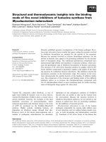
Figure 1.1. Simplified schematic of gene circuits and attractor states.
Reprinted from Trends in Genetics, 27, Zhou JX, Huang S, Understand-
ing gene circuits at cell-fate branch points for rational cell programming,
pages 55-62, Copyright (2011), with permission from Elsevier. The self-
stimulation (positive feedback) of genes X1 and X2 creates attractor state,
S
0
, representing a bipotent progenitor that is less stable that (attractor
states) S
1
or S
2
. “The quasi-potential landscape (right panel) offers a view
on the global dynamics by assigning to each point S in the state space a
‘quasi-potential’ U(S) that is inversely related to the approximate relative
stability of S, hence enabling the comparison of the relative ‘depth’ of
attractors or any other point S. In this two-gene system, the state space
is represented by the XY plane, whereas the Z-axis denotes U(S). The
higher U is, the less stable that state is. Thus, the system is attracted to
the lowest points = stable states = attractor states” [31].
Several useful predictions can be made using this model and many have been demon-
strated to be true by the growing body of evidence put forth by studies of indirect
and direct cellular reprogramming. First, since the state or phenotype of a cell at
13
any point in time is governed to a large extent by it
s gene expression profile, it is not
permanent, even though relatively stable. If acted upon enough by outside factors
that influence gene expression, a cell
s fate could be changed. This change would be
the result of a significant enough change in gene expression, or enough energy added
to the system, to overcome the stability gained by occupying its current phenotype.
This model also predicts that the processes of cellular reprogramming do not need
to be externally regulated throughout the entire process. Rather, it predicts that
enough of a perturbation in the system can remove the cell from it
s current attrac-
tor state and that upon that perturbation, it will seek the nearest attractor state.
This has been demonstrated by several groups that have used forced gene expression
of lineage- and cell-specific genes to induce the cellular phenotypes they sought to
induce from terminally differentiated cell types that typically have little to no ex-
pression of the specific genes delivered. This model also predicts that cells with more
similar gene expression profiles can more easily be transitioned between. Another
prediction of the model would be that the forced expression of specified genes may
not be necessary. Rather, published by many independent groups, various combina-
tions of genes involved in transcriptional regulation of cell-specific genes could provide
enough change, likely due to positive feedback mechanisms and feed-forward systems
that push gene expression towards a particular, desired attractor state.
1.7 Photoreceptors: A unique opportunity for direct reprogramming
Initial studies establishing direct reprogramming as a viable induction method to de-
rive neuronal cell types were enabled by 1) a need for these specific cell types, as dic-
tated by particularly problematic human disease pathologies and 2) a well-established
body of literature identifying and delineating important gene regulatory networks of
14
the final, desired cell types. Photoreceptor cells of the retina constitute an additional
cell type in which both of the requirements also exist, yet direct reprogramming of
somatic cells to a photoreceptor fate has yet to be achieved.
The loss of sight, and the ensuing problems it brings are certainly among our most
basic human fears. Almost 30% of the sensory input to the brain traces back to the
retina, which is commonly referred to as the “window to the brain” [34–36]. The
visual experience begins with photoreceptors, a unique class of neuronal sensory cells
that are responsible for receiving light information that falls on the retina and con-
verting that input to signals that the nervous system can process. The output of pho-
toreceptors is integrated and processed first by interneurons of the retina before the
information is transmitted to visual centers and others in the brain [37]. It should be
no surprise then, that diseases deleteriously affecting photoreceptors are the primary
cause of visual impairment or blindness in most retinal diseases, including macular
degeneration, Lebers congential amaurosis (LCA), and retinal pigmentosa (RP), to
name a few of the more common [36]. Therefore, cellular replacement strategies often
have been aimed at protecting these important sensory cells as well as replacing them
through transplantation, or by stimulating in vivo rescue or replacement by existing
cell populations. Furthermore, studies and models of retinal degeneration could also
provide valuable information about more general features of progressive neurodegen-
eration [38].
Photoreceptors are broadly classified into two main types: cones or rods. Cones
respond to bright light and relay high resolution, color information. Rods on the
other hand, function in low light and are a hundred-fold more light-sensitive than
cones [36, 37, 39]. In mice and humans, 70-80 % of all cells in the neural retina
are photoreceptors, with rods outnumbering cones 30:1 in mice, and 18-20:1 in hu-
mans [36,41,42], indicating that rod photoreceptors are the most abundant cell type
in the retina of both mice and humans. While subtypes of cones exist expressing
15
different and singular visual pigments, the mammalian retina has only one rod opsin,
rhodopsin, with a peak sensitivity around 500 nm [36, 37]. Lastly, and importantly,
transplantation studies have demonstrated that rod precursor cells readily incorpo-
rate in the adult retina, differentiate, and form synaptic connections [43]. This study
contrasted these rod progenitors with other progenitor or stem cells from various alter-
nate stages of development that failed to integrate to the same extent as rod progen-
itors [43–47]. For these reasons- abundance, sensitivity, simplicity, and demonstrated
integration- an abundance of research has focused on the gene regulatory networks
of rod photoreceptors. Furthermore, the aforementioned reasons also make rod pho-
toreceptor cells ideal targets for studies of direct cellular reprogramming, as well as
excellent candidates for the first applications of directly reprogrammed cells to regen-
erative medicine, including transplantation experiments aimed at recovering vision
in genetic or injury models where vision has been lost or impaired due to loss of
photoreceptors.
1.8 Transcriptional dominance model of photoreceptor cell fate determination
Decades of research support the transcriptional dominance model (Figure 1.2) of pho-
toreceptor cell fate determination put forth by Dr. Anand Swaroop [36]. While he
states that “the molecular mechanisms that generate photoreceptor precursors from
retinal progenitor cell remain uncharacterized”, several players, including but not
limited to, CrX, Otx2, NrL, Nr2e3 and RORβ have been implicated as necessary in
rod photoreceptor development [36]. Loss of any one of these genes leads to a com-
plete, or almost complete loss of rod photoreceptors, or lack of expression of many
important rod-specific phototransduction genes [36, 48–52]. It should also be noted
that not one of these single genes has been sufficient to induce the differentiation of
rod photoreceptors. However, the demonstrated overlapping targets of these genes, as
well as the step-wise nature of photoreceptor differentiation from retinal progenitors
and the increasingly likely multifactorial and transient nature of the terminal differ-