ERK1 AND ERK2 IN HEMATOPOIESIS, MAST CELL FUNCTION, AND THE MANAGEMENT OF NF1-ASSOCIATED LEUKEMIA AND TUMORS
Bạn đang xem bản rút gọn của tài liệu. Xem và tải ngay bản đầy đủ của tài liệu tại đây (6.98 MB, 246 trang )
ERK1 AND ERK2 IN HEMATOPOIESIS, MAST CELL FUNCTION, AND THE
MANAGEMENT OF NF1-ASSOCIATED LEUKEMIA AND TUMORS
Karl W. Staser
Submitted to the faculty of the University Graduate School
in partial fulfillment of the requirements
for the degree
Doctor of Philosophy
in the Department of Biochemistry and Molecular Biology,
Indiana University
March, 2012
ii
Accepted by the Faculty of Indiana University, in partial
fulfillment of the requirements for the degree of Doctor of Philosophy.
_________________________
D. Wade Clapp, M.D., Chair
_________________________
Maureen A. Harrington, Ph.D.
_________________________
Mark G. Goebl, Ph.D.
_________________________
Feng Chun Yang, M.D., Ph.D.
July 7, 2011
Doctoral Committee
iii
ACKNOWLEDGEMENTS
I thank my committee members for immeasurable insight, support,
criticisms, and benedictions, which have critically shaped the direction and
discoveries of my graduate research. I also am grateful for the faculty and staff of
the Department of Biochemistry, whose intellectual and financial support
facilitated this project while providing the fundamental didactic and inductive
tutelage that guides meaningful inquiry. Likewise, I thank the students of the
Department of Biochemistry who, through the peculiarities and profundities of
weekly seminar, have expanded the globe of my scientific exploration.
I thank every single member of the Clapp and Yang laboratories, including
several graduate students and technicians who have continued on elsewhere. Of
note, I would like to acknowledge Su-Jung Park, who has challenged and tutored
me, both technically and intellectually. Her mentorship invaluably underpins this
thesis, and I happily anticipate consulting her particular and profound expertise
throughout my career.
I am especially grateful for Dr. Wade Clapp’s guidance and friendship.
Without Wade’s encouragement, this thesis would be absent from the scientific
repertoire. He ardently promoted and ultimately fulfilled my nascent desire to
develop my career goals toward those of a physician-scientist. Thus, from the
depths of a previous obscurity my enduring aim of lifelong scientific discovery
and service has emerged, and I treasure Wade as a mentor and friend.
iv
ABSTRACT
Karl W. Staser
ERK1 AND ERK2 IN HEMATOPOIESIS, MAST CELL FUNCTION, AND THE
MANAGEMENT OF NF1-ASSOCIATED DISEASE
Neurofibromatosis type 1 is a genetic disease that results from either
heritable or spontaneous autosomal dominant mutations in the NF1 gene, which
encodes a protein serving, at least in part, to accelerate the intrinsic hydrolysis of
active Ras-GTP to inactive Ras-GDP. A second-hit NF1 mutation precedes
predominant NF1 neoplasms, including juvenile myelomoncytic leukemia (JMML)
and plexiform neurofibroma formation, potentially fatal conditions with no medical
therapy. While NF1 loss of heterozygosity (LOH) in myeloid progenitor cells
sufficiently engenders leukemogenesis, plexiform neurofibroma formation
depends on LOH in Schwann cells and Nf1 heterozygosity in the hematopoietic
system. Specifically, recruited Nf1
+/-
mast cells accelerate tumorigenesis through
secreted cytokines and growth factors. Nf1
+/-
mast cells depend upon
deregulated signaling in c-kit pathways, a receptor system conserved in
hematopoietic stem cells (HSCs). Accordingly, Nf1
-/-
myeloid progenitor cells,
which can induce a JMML-like disease in mice, also demonstrate deregulated c-
kit receptor signaling. C-kit-activated Nf1
+/-
mast cells and Nf1
-/-
myeloid
progenitors both show increased latency and potency of active Erk1 and Erk2,
the principal cytosolic-to-nuclear effectors of canonical Ras-Raf-Mek signaling.
v
Thus, Erk represents a potential regulator of leukemogenesis and tumor-
associated inflammation. However, single and combined Erk1 and Erk2 roles in
HSC function, myelopoiesis, and mature mast cell physiology remain unknown,
and recent hematopoietic studies relying on chemical Mek-Erk inhibitors have
produced conflicting results. Here, we show that hematopoietic stability,
myelopoiesis, and mast cell generation require Erk1 or Erk2, but individual
isoforms are largely dispensable. Principally, Erk-disrupted hematopoietic stem
cells incorporate BrdU but are incapable of dividing, a novel and cell type-specific
Erk function. Similarly, mast cell proliferation requires Erk but cytokine production
proceeds through other pathways, elucidating molecule-specific functions within
the c-kit cascade. Based on these findings, we have reduced tumor mast cell
infiltration by treating genetically-engineered tumor model mice with PD0325901,
a preclinical Mek-Erk inhibitor. Moreover, we have devised a quadruple
transgenic HSC transplantation model to examine dual Erk disruption in the
context of Nf1 nullizygosity, testing whether diseased hematopoiesis requires
Erk. These insights illuminate cell-specific Erk functions in normal and Nf1-
deficient hematopoiesis, informing the feasibility of targeting Mek-Erk in NF1-
associated disease.
D. Wade Clapp, M.D., Chair
vi
TABLE OF CONTENTS
ABBREVIATIONS x
INTRODUCTION 1
Mast Cells, Tumors, and the NF1 Hematopoietic System 3
NF1 Genetics 8
Nf1 Gene Dosage 10
Mek-Erk Signaling in Mast Cells 12
Mek-Erk Signaling in Hematopoietic Stem and Progenitor Cells 17
Global Observations on the Functions of Erk1 and Erk2 18
THESIS OVERVIEW 22
MATERIALS AND METHODS 23
Mice, Genotyping, and Mx1Cre Induction 23
Marrow Isolation 24
Colony Assays 24
Single Cell Colony Assays 25
Bone Marrow Histology 26
Hematopoietic Stem Cell Transplantation 26
Peripheral Blood Isolation 27
Secondary Transplantation 27
Flow Cytometry 28
Acquisition 28
Analysis 28
Flow Cytometry Antibodies 29
BrdU HSC Analysis 30
PY/Hst HSC Analysis 31
vii
Marrow Enrichment 31
Pcl7CREeGFP Generation 32
Virus Generation 32
Viral Transduction 33
Mast Cell Culture 34
Inhibitors 34
Mast Cell Proliferation Assays 35
Hemcytometer-based 35
MTT-based 35
3H-Thymidine-based 36
Mast Cell Cycle Analysis 37
Mast Cell Survival Assay 38
Deconvolution Microscopy 38
Cytokine Array 39
Multiplex Assay 40
Western Blotting 41
Sample isolation 41
Immunoblotting protocol 42
Quantification of Mast Cells In Vivo 43
PD0325901 Treatment of Plexiform Neurofibroma Model 43
Statistics 44
RESULTS 45
Erk and Hematopoiesis 45
viii
Inducible deletion of Erk1/2 in the bone marrow. 45
Loss of myeloid cellularity and granulocytes in DKO bone marrow. 51
Loss of myeloid colony formation in DKO bone marrow. 64
Stable chimerism requires one isoform of Erk. 71
Erk1/2 disruption rapidly and permanently abolishes myelopoiesis. 88
Erk1/2 disruption abrogates the exponential expansion of hematopoietic
progenitor cells. 98
Erk1/2 disruption prevents stem cell colony formation but not BrdU
incorporation. 109
Erk1/2 control HSC proliferation: additional evidence. 119
Single Erk1 or Erk2 disruption have specific long-term consequences. 127
Erk disruption and Nf1-deficient hematopoiesis. 134
Erk and the mast cell 139
Mast cell cytopoiesis requires Erk. 139
Chemical Mek-Erk inhibition in mast cells. 146
PD0325901 inhibits SCF-mediated Erk1/2 phosphorylation. 149
Single Erk isoforms are dispensable for SCF-mediated mast cell
proliferation. 154
Erk negatively regulates SCF-mediated mast cell cytokine production. 169
Erk-dependent biochemical alterations in the mast cell. 176
Erk1/2 disruption in primary mature mast cells. 189
PD0325901 reduces mast cell infiltration in NF1-associated tumors. 195
DISCUSSION 198
Erk and hematopoiesis 200
ix
Mast cells and future directions 207
Conclusions 213
REFERENCES 216
CURRICULUM VITAE
x
ABBREVIATIONS
7-AAD: 7-Aminoactinomycin D.
APC: Allophycocyanin.
BCA: Bicinchoninic acid.
BSA: Bovine serum albumin.
DAPI: 4',6-diamidino-2-phenylindole.
DMEM: Dulbecco’s Modified Eagle Medium.
EPO: Erythropoietin.
ERK: Extracellular regulated kinase.
FBS: Fetal bovine serum.
FITC: Fluorescein isothiocyanate.
Flt3L: Flt (Fms-like receptor tyrosine kinase 3) ligand.
G-CSF: Granulocyte-colony stimulating factor.
GM-CSF: Granulocyte-macrophage-colony stimulating factor.
GAP: GTPase activating protein.
GDP: Guanosine diphosphate.
GMP: Granulocyte-macrophage progenitor.
GTP: Guanosine triphosphate.
HPPC: High proliferation potential cell.
HSC: Hematopoietic stem cell.
Hst: Hoechst.
IL-3: Interleukin-3.
IL-6: Interleukin-6.
IL-13: Interleukin-13.
IL-17: Interleukin-17.
IMDM: Iscove’s Modified Dulbecco’s Medium.
I.P.: Intraperitoneal.
I.V.: Intravenous (tail vein).
LPPC: Low proliferation potential cell.
MAPK: Mitogen activated protein kinase.
M-CSF: Macrophage-colony stimulating factor.
MCP-1: Monocyte chemotactic protein 1.
MEP: Megakaryocyte-erythroid progenitor.
MIP-1a: Macrophage inflammatory protein 1 alpha.
MIP-1b: Macrophage inflammatory protein 1 beta.
MP: Myeloid progenitor.
MPP: Multipotent progenitor.
MTT: 3-(4,5-dimethylthiazol-2-yl)-2,5-diphenyltetrazolium bromide.
NaN
3
: Sodium azide.
NF1: Neurofibromatosis type 1.
NGF: Nerve growth factor.
PBS: Phosphate buffered saline.
PDGF: Platelet-derived growth factor.
PE: Phycoerythrin.
xi
PEI: Polyethyleneimines.
PerCP: Peridinin chlorophyll protein.
PI: Propidium iodide.
PI-3K: Phosphatidylinositol 3-kinases.
PY: Pyronin Y.
PVDF: Polyvinylidene fluoride.
RANKL: Receptor activator of nuclear factor kappa-B ligand.
RAS: Rat Sarcoma protein.
SCF: Stem cell factor.
SLAM: Signaling lymphocytic activation molecule.
SDS-PAGE: Sodium dodecyl sulfate polyacrylamide gel electrophoresis.
TNF-α: Tumor necrosis factor alpha.
VEGF: Vascular endothelial growth factor.
WT: Wild-type.
1
INTRODUCTION
Neurofibromatosis type 1 (NF1, von Recklinghausen’s disease) is a
genetic disorder caused by autosomal dominant mutations in the NF1 gene,
which encodes Neurofibromin, a protein that accelerates the hydrolysis of Ras
from its GTP- to GDP-bound conformation. The disease afflicts approximately 1
in 3500 persons worldwide in a pandemic fashion, and it is the most common
genetic disorder with a predisposition to cancer (1). NF1 manifests with both non-
tumorigenic and tumorigenic maladies, including learning disabilities, skeletal
dysplasia, non-healing fractures (pseudarthrosis), myeloid leukemia (JMML), and
tumors such as optic glioma and the namesake neurofibroma. The disease’s
hallmark signs include hyper-pigmented areas of the skin (café au lait macules)
and hamartomas on the iris (Lisch nodules), which serve as important diagnostic
criteria and may be observed in infancy or childhood of afflicted individuals (2, 3).
Because prominent NF1 symptoms arise from neural crest-derived tissue (e.g.
glia, Schwann cells, melanocytes), some reports have characterized NF1 as a
disorder of the neural crest. However, NF1 pathologies arise in organs derived
from all embryonic germ layers, and we should consider NF1 not only a tumor
predisposition syndrome but also a systemic developmental disorder (4).
NF1-like cutaneous tumor syndromes appeared in the literature during the
18
th
century (5-7), and in the 1880s Friedrich von Recklinghausen published
seminal observations detailing cutaneous tumors comprised of both neuronal and
fibroblastic tissue, deeming the tumors neurofibromen (8). NF1’s pathognomonic
neurofibromas are slowly progressing, heterogeneous solid tumors comprised of
2
Schwann cells, fibroblasts, vascular cells, and infiltrating hematopoietic cells,
predominantly degranulating mast cells (9-14). Cutaneous and subcutaneous
neurofibromas derive from small peripheral nerve branches during adolescence
or adulthood and are found in nearly all individuals with NF1 (15). By
comparison, plexiform neurofibromas afflict half or fewer individuals with NF1 and
develop from cranial and large-peripheral nerve sheaths, possibly initiating during
gestation or early infancy from abnormally differentiated nonmyelinating
Schwann cells or their less-differentiated precursors (16, 17).
Plexiform neurofibromas are typically a lifelong source of disfigurement,
disability, and mortality. In many cases, plexiform neurofibromas compress
cranial nerves and/or peripheral nerve roots at the vertebral column and create
an array of morbidity, including paresthesia, paralysis, drooling, sleeplessness,
respiratory and gastrointestinal distress, blindness, and loss of bowel and
bladder control (18, 19). A plexiform neurofibroma also has the potential to
transform into a malignant peripheral nerve sheath tumor (MPNST), a highly
morbid, metastatic cancer afflicting up to 10% of NF1 patients in their lifetime (20,
21).
Plexiform neurofibroma treatment consists primarily of symptom
management and/or surgical resection. In many cases, the tumor’s close
involvement with vital nerve tissue, vasculature, or other viscera complicates
surgery (18, 19, 22). Currently, the tumors have no medical therapy or cure,
although several molecularly-targeted compounds are in preclinical or clinical
testing (23-27). Problematically, nerve sheaths and heavily collagenized areas
3
may resist drug bioavailability, complicating direct pharmacological inhibition of
the tumorous mass. Therefore, therapeutic strategies targeting components of
the tumor microenvironment, including vascular cells and infiltrating mast cells,
may prove viable alternatives (28).
Mast Cells, Tumors, and the NF1 Hematopoietic System
Mast cells are granular hematopoietic cells that arise from myeloid
progenitor cells prior to granulocyte/monocyte lineage commitment (29). Mast
cell precursors migrate from the bone marrow into the vasculature and enter
dermal tissue where they mature into immune effector cells. Mast cells fight
pathogens, protect against venoms and toxins, and may perform other
immunomodulatory functions, both pro- and anti-inflammatory (30-33). While
mast cells are predominantly known as the mediators of allergy and allergic
asthma via IgE/FcεR pathways, they additionally depend on stem cell factor
(SCF) signaling at the c-kit receptor tyrosine kinase for their generation and, in
some contexts, pathophysiological activation (34-37). Indeed, mice naturally
mutated at the c-kit receptor tyrosine kinase (W, or “white spotting locus”
mutants, which reduces c-kit kinase activity >85%) exhibit profoundly reduced
numbers of tissue-resident mast cells (35). Some W mice have anemia and
deficient hematopoiesis, as the hematopoietic stem cell (HSC) also depends
upon SCF/c-kit signaling.
The pro-inflammatory activities of recruited mast cells and other immune
effector cells have been shown to sustain tumor microenvironments in various
disease models (reviewed in (38-40)). In this inflammatory microenvironment
4
hypothesis, tumorigenic cells recruit and co-opt the functions of non-tumorigenic
hematopoietic cells via unchecked mitogenic and chemotactic signals. These
recruited cells, in turn, coordinate vascular in-growth, collagen deposition, and
the pathological inflammation promoting extracellular matrix remodeling, tumor
expansion, invasion, and metastasis. Specifically, mast cells can synthesize and
secrete matrix metalloproteinases (MMPs), various cytokines (e.g. IL-6 and TNF-
α), and multiple mitogens (e.g. NGF, VEGF, and PDGF) (32, 33) with putative
roles in tumor initiation, maintenance, and growth.
Mast cells have been associated with NF1 since 1911, when H. Greggio
first noted les cellules granuleuses in neurofibroma tissue (14). Decades later,
several investigators confirmed their presence using traditional histology and
electron microscopy (9-13). By the 1980s, mast cells were widely-recognized
inflammatory effectors and hallmark histological features (albeit of unknown
significance) of the neurofibroma. Vincent Riccardi first hypothesized that mast
cells may critically contribute to neurofibroma formation, proposing that mast cell
degranulation explained his clinical observations of coincident pruritus and
cutaneous neurofibroma formation (41). Indeed, a small human study with a mast
cell granule stabilizer (ketotifen) reduced pruritus and/or slowed neurofibroma
growth (42), but a subsequent multiphase trial confirmed only anti-pruritic and
analgesic effects, not neurofibroma reduction (43). These inquiries provided
important evidence of aberrant mast cell degranulatory activity in neurofibroma
tissue yet suggested that local inhibition of degranulation alone does not change
overall disease course. As discussed in this review, recent biochemical,
5
transplantation, and pharmacological studies have implicated a preponderant
role for SCF-mediated mast cell gain-in-functions in orchestrating the
neurofibroma microenvironment. This SCF-mediated coordination of mast cell
inflammation and tumor growth may inform a novel approach to NF1
therapeutics.
Intriguingly, the mast cell shares functional and phenotypic similarities with
hematopoietic stem and progenitor cells, potentially informing mechanisms of the
coincident occurrence of JMML in NF1 patients. This myelomonocytic neoplasia,
which has no therapy or cure and is uniformly fatal, results from loss of NF1
heterozygosity in hematopoietic stem and progenitor cells, which become
hypersensitive to multiple cytokines, including GM-CSF and SCF (44, 45). Like
mast cells, all hematopoietic stem and progenitor cells express the c-kit receptor
tyrosine kinase and utilize SCF signaling for their proliferation, differentiation, and
survival (46). Thus, we can consider the NF1 hematopoietic system to be one of
myeloid dysfunction at the level of hematopoietic stem and progenitor cells,
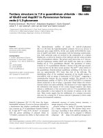
including mast cell precursor cells (Figure 1).
F
F
igure 1
6
7
Figure 1: Myeloid hierarchy with an emphasis on cells known to be
dependent on Neurofibromin signaling. The hematopoietic stem cell (HSC)
gives rise to multipotent progenitors (MPP), which can differentiate to the
common myeloid progenitor (CMP) or the common lymphoid progenitor (not
shown). The CMP gives rise to granulocyte-macrophage progenitors (GMP),
mast cell precursors, and erythroid lineages (not shown). The GMP, in turn, gives
rise to the granulocyte progenitor (GP) and the macrophage/monocyte progenitor
(MP), which can differentiate into multiple cell types, including macrophages,
osteoclasts, and dendritic cells. The shaded box indicates lineages known to be
hyper-responsive to the indicated cytokines, subsequent to mono- or biallelic
inactivation of Nf1/NF1. SCF: stem cell factor, Flt3L: Flt3 (fms-like receptor
tyrosine kinase 3) ligand, Il-3: interleukin-3, GM-CSF: granulocyte-macrophage
colony stimulating factor, M-CSF: macrophage colony stimulating factor, G-CSF:
granulocyte colony stimulating factor.
8
NF1 Genetics
A century after von Recklinghausen’s seminal case reports, genetic
linkage studies in NF1-afflicted families identified the pericentromeric region of
chromosome 17 as the genomic region harboring the gene responsible for the
disease (47, 48). Further studies in patients with translocations of chromosome
17 (49-52) facilitated the identification and full-length sequencing of the NF1
gene (53), which spans 350 kilobases of human chromosome 17 (17q11.2) and
encodes 59 exons producing a 2818 amino acid protein (49, 54-56). Of note,
human Neurofibromin and its mouse homolog share 98% identity at the protein
level (57).
Approximately half of NF1 mutations in humans arise spontaneously (58),
with the majority of mutations leading to premature truncation of the protein
neurofibromin (59, 60). When NF1 mutations occur post-meiotically, individuals
may exhibit segmental NF1 with manifestations confined regionally or to a subset
of normally affected cell types (e.g. only pigmentation defects) (61). Different NF1
frameshift and point mutations do not necessarily correlate with phenotypic
severity, although some studies have shown that microdeletions encompassing
the entire NF1 locus (which account for less than 10% of mutations) associate
with earlier onset and more profound disease manifestations (62, 63). Phenotypic
variation tends to be high even within families, and pedigree analyses indicate
that while NF1 mutations are fully penetrant, variation in genes independent of
the NF1 locus critically modulates time-to-onset and course of the disease (64,
65). Parallel to the human data, different Nf1-mutant mouse strains exhibit both
9
varied expression levels of neurofibromin and variable susceptibility to different
NF1-like disease manifestations (66). Overall, with the exception of the
documented severity associated with NF1 locus-encompassing microdeletions
and a uniquely mild phenotype associated with a 3-base pair deletion in exon 17
(67), particular genetic mutations or genomic variations which may correlate to
specific disease outcomes are largely unknown.
NF1 encodes neurofibromin, a protein which functions, at least in part, as
a p21
ras
(Ras) guanosine tri-phosphatase (GTP) activating protein (GAP) (68-72).
Neurofibromin and other Ras-GAPs logarithmically accelerate the intrinsic
hydrolysis of Ras-GTP to its inactive guanosine di-phosphate- (GDP)-bound
conformation (73). In response to multiple mitogenic stimuli, active Ras-GTP
orchestrates diverse protein signaling networks, including mitogen activated
protein kinase- (MAPK)- and Akt-directed pathways (74-78). Hence, by
accelerating the conversion of Ras-GTP to Ras-GDP, neurofibromin negatively
regulates Ras-dependent signaling cascades and, generally, serves to
downregulate mitogenic events across diverse protein networks. In cases of NF1
heterozygosity or nullizygosity, as observed in somatic cells and in tumor cells of
individuals with NF1, respectively, downstream Ras-mediated phosphorylation
and transcriptional events can increase in duration and total output. This global
upregulation of Ras-dependent activity in NF1/Nf1-disrupted tissue typically leads
to cellular gain-in-functions, including enhanced proliferation, migration, and
survival in multiple cell types (reviewed in (79-83). Of note, the specific Ras
effectors potentiated by loss of NF1 may vary by cell and receptor type, and
10
biochemical consequences in one cell-receptor system may or may not be
observed in another.
Nf1 Gene Dosage
Although NF1 is classified as a classical Knudson tumor suppressor gene,
multiple studies have shown that NF1 heterozygosity critically modulates cell fate
and function by altering Ras-dependent biochemical pathways in distinct cell
types (reviewed in (82)). Moreover, physiological Ras activity regulates
embryogenesis, early development, and normal tissue maintenance. Therefore,
neurofibromin may be viewed not only as a tumor suppressor but also as a
regulator of histiogenesis, cellular maintenance, and repair (4). Accordingly, NF1
is a disorder of both tumor predisposition and of developmental dysplasia.
While somatic cells in an individual with NF1 are heterozygous for NF1,
loss of heterozygosity (LOH) in different cell types typically precedes hallmark
hyperplastic, dysplastic, and neoplastic disease manifestations. LOH has been
shown in human tissue samples and confirmed in NF1 mouse models of certain
NF1 pathologies via multiple molecular techniques, coinciding with NF1’s
designation as a classical tumor suppressor gene. As examples, LOH in
Schwann cells or their precursors permits neurofibroma formation (17, 84-86)
and LOH in myeloid progenitor cells induces myelomonocytic leukemia (44).
Individuals with NF1 also have an increased prevalence of multiple
generalized manifestations which do not appear to require cell-specific biallelic
inactivation of NF1, including skeletal and mesenchymal dysplasia (e.g. short
stature, osteoporosis, and soft tissue malformation), disorders of neurocognitive
11
development (e.g. retardation, spatial/visual coordination, and autism), and
vascular pathologies (e.g. fistulae, infarcts, and aneurysms). Hence, NF1
heterozygosity alone alters Ras-dependent pathways to a degree sufficient for
the pathological alteration of normal developmental and homeostatic processes
in multiple organ systems.
Indeed, Nf1 haploinsufficient mast cells and fibroblasts, major constituents
of the heterogeneous plexiform neurofibroma, demonstrate multiple gain-in-
function phenotypes that include enhanced proliferation, survival, migration, and
cytokine production in response to specific stimuli (87, 88). These data parallel
findings in Nf1 haploinsufficient microglia (89), which critically modulate the
inflammatory microenvironment of NF1-associated optic glioma (90-92).
In some mouse models of plexiform neurofibroma and optic glioma
formation, tumorigenesis requires Nf1 haploinsufficiency in non-tumorigenic cells.
Specifically, hematopoietic stem cell transplantation studies in the Nf1
flox/flox
;
Krox20°Cre and Nf1
flox/flox
;
P0aCre models (which experience biallelic Nf1
inactivation in a subset of Schwann cell/Schwann cell precursors) have shown
that neurofibroma genesis requires Nf1 haploinsufficiency and c-kit-mediated
signaling in the hematopoietic compartment (24). In these experiments, Nf1
flox/flox
;
Krox20°Cre mice required Nf1
+/-
hematopoietic stem cell transplants to engender
tumorigenesis, while WT hematopoietic stem cell transplants protected against
tumorigenesis in Nf1
flox/-
;Krox20°Cre mice. These data, combined with cell culture
studies of Nf1
+/-
mast cells, roundly implicate the Nf1 haploinsufficient
hematopoietic compartment (and, specifically, deregulated myeloid and mast
12
cells), as a principal pathologic component of the plexiform neurofibroma
microenvironment.
By comparison, clonal outgrowth in NF1-associated JMML depends on
Nf1/NF1 LOH in hematopoietic stem and progenitor cells (c-kit
+
cells) with no
requirement for an Nf1
+/-
cellular background. As evidence, transplantation of
Nf1
-/-
hematopoietic stem cells into WT mice engenders a myeloproliferative
disorder (MPD) recapitulating human JMML (44, 45, 93). However, in human
cases of NF1-associated JMML, we expect all surrounding somatic cells in the
individual to be essentially NF1
+/-
. Of note, no report has directly investigated
possible contributions of an Nf1
+/-
stroma to the time-to-onset, progression, and
severity of NF1-associated MPD.
Mek-Erk Signaling in Mast Cells
SCF regulates mast cell and hematopoietic progenitor cell cytopoiesis,
proliferation, survival, and cytokine synthesis, and Nf1 deficiency can potentiate
these functions. In fact, the study of SCF-stimulated Nf1
+/-
mast cells provided
foundational evidence that haploinsufficiency of a “tumor suppressor” could
modulate multi-lineage cell fate and function in tissue culture and in vivo (87).
This study additionally demonstrated that Nf1 haploinsufficiency increases the
latency and potency of GTP-bound Ras in SCF-stimulated cells. Subsequent
studies have detailed the biochemical mechanisms modulating SCF-mediated
gain-in-functions, showing alterations arising from deregulated signaling events
in multiple Ras-dependent networks. In response to ligand binding at diverse cell
surface receptors, Ras activates to its GTP-bound state and promotes
13
phosphorylation in downstream protein networks, including those orchestrated by
MAPKs and phosphoinositide-3-kinase (PI-3K) (75-78). Neurofibromin, which
contains a highly-conserved GAP-related domain (GRD) with homology to the
yeast gene products IRA1 and IRA2, logarithmically accelerates the intrinsic
hydrolysis of active GTP-bound Ras to its GDP-bound state (50, 55, 68, 71, 79,
94). Generally, loss-of-function mutations in genes encoding Ras-GAPs promote
cell growth, proliferation, migration, and survival (40). In myeloid progenitor cells,
microglia, and mast cells, loss of one or both alleles of Nf1 leads to increased
duration of Ras-GTP activity and phosphorylation of specific effectors within Raf-
Mek-Erk, PI-3K-Rac-Pak-P38, and PI-3K-Akt cascades (44, 45, 87, 92, 95-100).
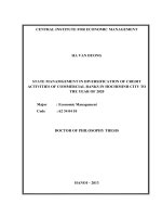
Cell culture and in vivo studies of genetically-disrupted mast cells indicate
that the Raf-Mek-Erk pathway may primarily modulate SCF-mediated
proliferation and protein synthesis while the PI-3K-Rac2-Pak-p38 pathway
controls F-actin dynamics and cellular motility (97, 98, 100-103). Biochemical
investigations have additionally shown that the PI-3K-dependent pathway
reinforces the classical Raf-Mek-Erk cascade through the activity of the p21
activated kinases (Paks) (98, 102). In this schema, PI-3K-activated Rac2 induces
Pak1 to phosphorylate Mek at serine 298 as well as Raf1 at serine 338,
potentiating Raf1’s phosphorylation of Mek at serine 217/222. These activities
potentiate phosphorylation of the extracellular regulated kinases, Erk1 and Erk2.
Erk1 and Erk2 phosphorylate cytoplasmic targets (e.g. p90
rsk
), translocate to the
nucleus, and activate multiple mitogenic transcription factors (e.g. c-Fos, Elk1,
C/EBP).
14
However, direct genetic studies of Mek-Erk signaling in the SCF-
stimulated mast cell are lacking. All prior investigations have relied on chemical
inhibitors of Mek (e.g. PD98059), which are known to have non-selective
inhibitions and cellular toxicities. Moreover, Erk1 and Erk2’s specific modulation
of the mast cell cycle, as well as Erk-dependent transcriptional events, including
the production of inflammatory cytokines, are not documented. Finally, it is
unknown whether Erk1 and Erk2 have isoform specific roles in the modulation of
SCF-mediated mast cell function (Figure 2).