- Trang chủ >>
- Y - Dược >>
- Ngoại khoa
Oxford cases in medicine and surgery new medical books
Bạn đang xem bản rút gọn của tài liệu. Xem và tải ngay bản đầy đủ của tài liệu tại đây (4.5 MB, 589 trang )
Oxford Cases in Medicine and Surgery
This page intentionally left blank
Oxford Cases in
Medicine and Surgery
Hugo Farne
Junior Doctor; St Mary’s Hospital,
Paddington, London, UK
Edward Norris-Cervetto
Junior Doctor; Royal Berkshire Hospital,
Reading, UK
James Warbrick-Smith
Junior Doctor; Gloucestershire Royal Hospital, UK
1
3
Great Clarendon Street, Oxford OX2 6DP
Oxford University Press is a department of the University of Oxford.
It furthers the University’s objective of excellence in research, scholarship,
and education by publishing worldwide in
Oxford New York
Auckland Cape Town Dar es Salaam Hong Kong Karachi
Kuala Lumpur Madrid Melbourne Mexico City Nairobi
New Delhi Shanghai Taipei Toronto
With offi ces in
Argentina Austria Brazil Chile Czech Republic France Greece
Guatemala Hungary Italy Japan Poland Portugal Singapore
South Korea Switzerland ailand Turkey Ukraine Vietnam
Oxford is a registered trade mark of Oxford University Press
in the UK and in certain other countries
Published in the United States
by Oxford University Press Inc., New York
© Oxford University Press, 2010
e moral rights of the author have been asserted
Crown copyright material is reproduced under Class Licence
Number C01P0000148 with the permission of OPSI
and the Queen’s Printer for Scotland
Database right Oxford University Press (maker)
First published 2010
All rights reserved. No part of this publication may be reproduced,
stored in a retrieval system, or transmitted, in any form or by any means,
without the prior permission in writing of Oxford University Press,
or as expressly permitted by law, or under terms agreed with the appropriate
reprographics rights organization. Enquiries concerning reproduction
outside the scope of the above should be sent to the Rights Department,
Oxford University Press, at the address above
You must not circulate this book in any other binding or cover
and you must impose the same condition on any acquirer
British Library Cataloguing in Publication Data
Data available
Library of Congress Cataloging in Publication Data
Data available
Typeset by MPS Limited, A Macmillan Company
Printed in Italy
on acid-free paper by LEGO SpA-Lavis, TN
ISBN 978–0–19–956052–3
10 9 8 7 6 5 4 3 2 1
Oxford University Press makes no representation, express or implied, that the drug dosages in this book
are correct. Readers must therefore always check the product information and clinical procedures with
the most up to date published product information and data sheets provided by the manufacturers
and the most recent codes of conduct and safety regulations. e authors and publishers do not accept
responsibility or legal liability for any errors in the text or for the misuse or misapplication of material
in this work.
Foreword
ere is an abundance of excellent medical and surgical textbooks, written in both
traditional and more novel formats. However, in a climate in which the content
and mode of delivery of medical education remain in constant fl ux there remains
a need for new resources that stimulate interest in the reader as well as providing
the important and relevant facts. Oxford Cases in Medicine and Surgery fulfi ls this
need. is book’s uniqueness – and its educational value – stems from the way that
the authors have approached the learning aspect from direct clinical symptoms,
highlighting the most important diff erential diagnoses but also explaining how to
diff erentiate them. is approach represents the book’s real strength, mirroring
as it does the integrated systems-based approach that is commonly used by many
medical schools.
In my experience as a clinical teacher, course organiser, and examiner over the
past decade, this is the fi rst book that has attempted to bring together, and explain
from a basic science concept, the reasons for the clinical picture or condition. is
will help readers enormously, whether they are under-graduate or post-graduate
medical, dental, or nursing students. It is an important book for those who wish to
understand the reasons for clinical presentations and their diff ering management.
Mr Christopher LH Chan
Senior Lecturer/Honorary Consultant Surgeon
Barts and e London School of Medicine and Dentistry
This page intentionally left blank
Introduction
Why we wrote this book
e inspiration for this book comes from our time as medical students. e
problem we found with existing textbooks was twofold.
Firstly, most books are organized by pathology. For example, they may have chap-
ters on ‘cardiology’ that then discuss specifi c conditions, like ‘myocardial infarction’,
in detail. But patients do not present with ready-made diagnoses like ‘myocardial
infarction’. ey present with symptoms, such as ‘chest pain’, which could be a myo-
cardial infarction – but could also be anything from refl ux oesophagitis to aortic
dissection.
Secondly, there are also textbooks based around cases rather than pathologies.
Our experience is that these tend to skip over the diagnostic approach too quickly,
in order to move on to a discussion of the underlying disease. Many give the reader
so much information in the initial case presentation that the diagnosis is virtually
made for you. For example, a ‘62-year-old diabetic male with sudden onset, crush-
ing chest pain; tachycardia on examination; ST elevation on his ECG, plus raised
troponins’ has a myocardial infarction. But by giving so much information upfront,
these books neglect to address what many students fi nd most challenging – how
do you decide what information to collect in order to make a diagnosis? Patients
present with symptoms such as ‘chest pain’ and it is your job to elicit the key clues
on history and examination, and to arrange the key investigations that will confi rm
that this is a myocardial infarction and rule out other diagnoses.
Knowing what to do when faced simply with ‘confusion’ or ‘abdominal pain’ can be
daunting and tricky – we know, and that is what motivated us to write this book.
We hope this book will help you start thinking like a diagnostician from your fi rst
day on the wards. us, we hope you will be able to work out why your patient is
short of breath or has abdominal pain in a way that is safe and effi cient, and avoids
you missing important diagnoses. Even with detailed knowledge of anatomy, physi-
ology, biochemistry, pathology, history-taking, examination skills, and data inter-
pretation, it can be diffi cult to integrate everything when faced with acutely ill
patients on the wards. We benefi ted greatly from case-based seminars that taught
us a hypothesis-driven, logical, step-by-step approach to diagnosis. Our hope is that
this book emulates the teaching that we found so benefi cial.
Finally, we wanted to write a workbook that students will enjoy using and where
even the simplest concepts are clearly explained.
The need for a logical diagnostic approach
Looks like an elephant. Sounds like an elephant. Smells like an elephant. Prob-
ably an elephant. Experienced clinicians often use pattern recognition to guide
diagnosis. As a student, you will begin to do this rapidly for conditions that you
will encounter frequently – chances are that, by now, you easily recognized that
the 62-year-old diabetic male mentioned above was having a myocardial infarction.
viii Introduction
Pattern recognition is useful and effi cient, and we have tried to illustrate stereotypical
presentations of some diseases in our short cases.
Looks like a crocodile. Sounds like a crocodile. Smells like a crocodile . . .
but is actually an alligator. Pattern recognition is a problem when a disease presents
in a way that mimics another disease. For example, patients with oesophageal spasm
may describe the same pain as those with an acute coronary syndrome. Such diag-
nostic puzzles are the stuff that hospital grand rounds and television shows are made
of. But misdiagnosis due to (incorrect) pattern recognition can have disastrous con-
sequences – you could inadvertently thrombolyse a patient you thought was having
a myocardial infarction but actually had an aortic dissection. is is one reason why
it is important to always follow a logical diagnostic approach.
Looks like an elephant. Sounds like a lion. Not sure what it smells like. Must be
a . . . ? You cannot recognize a pattern you have never seen before, an especially big
problem for the inexperienced medical student starting their clinical placements.
On other occasions, the symptoms may not fi t any known pattern, and even experi-
enced clinicians may struggle initially with the diagnosis. is is another reason for
having a logical diagnostic approach.
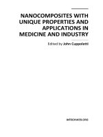
A logical approach to diagnosis
Below is an outline of the diagnostic strategy we have used throughout this book. We
recognize that, over time, everyone develops their own diagnostic strategy and that
tutors may teach you diff ering approaches. is is simply one that has worked for us.
‘50-year-old male with chest pain’. It is tempting to assume that he is having a myo-
cardial infarction, like the 62-year-old diabetic male mentioned above. However, . . .
Step 1
•
: ink of all the things that could cause this presentation. Use anatomy,
a surgical sieve (e.g. INVITED MD), etc. to come up with as long a list as
possible.
Step 2
•
: Highlight from your list the most common causes. For example, acute
coronary syndrome is a common cause of chest pain, viral costochondritis is
not. Mark the ones that you must exclude because they are lethal. In the case
of chest pain, Boerhaave’s perforation of the oesophagus is important as, if
untreated, it carries a 100% mortality.
Step 3
•
: ink of key clues in the patient history for each of the diagnoses. For
example, patients with Boerhaave’s perforation of the oesophagus invariably
give a history of vomiting immediately before onset of the pain. Now take a
history that deliberately tries to pick up these clues, rather than just going
through a set of ‘standard’ questions which may miss things. Also consider the
patient themselves (e.g. their age, occupation, etc.) and how this aff ects the
relative likelihoods of your diff erential diagnoses. Has the patient’s history or
epidemiological factors made any diagnoses more/less likely?
Step 4
•
: ink of key clues on examination for your diagnoses. For example,
patients with a pneumothorax will have an area of the chest that is hyperex-
panded, hyper-resonant to percussion, with absent breath sounds. Perform a
thorough examination looking for these clues. Have your examination fi nd-
ings made any diagnoses more/less likely?
Introduction ix
Step 1: What could it
be?
Step 2: What is most likely?
What must I exclude (*)?
Step 3: Key clues on
history?
Step 4: Key clues on
examination?
Step 5: Key clues on basic
investigations?
Step 6: Patient improving
with management?
Acute coronary
syndrome
Pneumothorax
Aortic dissection
Boerhaave’s perforation
Peptic ulcer disease
Stable angina
Musculoskeletal
Oesophagitis (e.g. due
to refl ux)
Oesophageal spasm
Pulmonary embolism
Pleurisy (secondary to
infection)
Anxiety
Myopericarditis
Aortic aneurysm
Coronary spasm
Cholecystitis
Pancreatitis
Acute coronary
syndrome*
Pneumothorax*
Aortic dissection*
Boerhaave’s perforation*
Peptic ulcer disease
Stable angina*
Musculoskeletal
Oesophagitis
Oesophageal spasm
Pulmonary embolism*
Pleurisy
Anxiety
Myopericarditis
Aortic aneurysm
Coronary spasm
Cholecystitis
Pancreatitis
Acute coronary
syndrome*
Pneumothorax*
Aortic dissection*
Boerhaave’s perforation*
Peptic ulcer disease
Stable angina*
Musculoskeletal
Oesophagitis
Oesophageal spasm
Pulmonary
embolism*
Pleurisy
Anxiety
Myopericarditis
Aortic aneurysm
Coronary spasm
Cholecystitis
Pancreatitis
Acute coronary
syndrome*
Pneumothorax*
Aortic dissection*
Boerhaave’s
perforation*
Peptic ulcer disease
Stable angina*
Musculoskeletal
Oesophagitis
Oesophageal spasm
Pulmonary embolism*
Pleurisy
Anxiety
Myopericarditis
Aortic aneurysm
Coronary spasm
Cholecystitis
Pancreatitis
Acute coronary
syndrome*
Pneumothorax*
Aortic dissection*
Boerhaave’s perforation*
Peptic ulcer disease
Stable angina*
Musculoskeletal
Oesophagitis
Oesophageal spasm
Pulmonary embolism*
Pleurisy
Anxiety
Myopericarditis
Aortic aneurysm
Coronary spasm
Cholecystitis
Pancreatitis
Acute coronary
syndrome*
Pneumothorax*
Aortic dissection*
Boerhaave’s perforation*
Peptic ulcer disease
Stable angina*
Musculoskeletal
Oesophagitis
Oesophageal spasm
Pulmonary embolism*
Pleurisy
Anxiety
Myopericarditis
Aortic aneurysm
Coronary spasm
Cholecystitis
Pancreatitis
50-year-old male with
chest pain
50-year-old male with
chest pain
50-year-old male with
sharp, left-sided chest
pain, came on suddenly
whilst watching TV.
Smoker with known
COPD. No risk factors
for venous thrombosis.
Upper left zone of
chest is hyper- resonant
with reduced air entry
and reduced vocal
resonance. Chest not
tender to palpation. No
signs of DVT in calves.
Chest radiograph shows
air in pleural space on left,
with lung collapsing away
from the upper left apex.
Patient improves after
insertion of a chest drain
for pneumothorax. Chest
pain resolves completely.
x Introduction
Step 5
•
: Don’t order a set of ‘standard’ investigations. ink about those inves-
tigations that will help confi rm or dismiss each diagnosis. Also include those
that are relevant for management. us urea and electrolytes are necessary if a
patient is put nil by mouth and on intravenous fl uids, or started on drugs that
are renally excreted or potentially nephrotoxic. Try to prioritize investigations
into those that are more readily available (e.g. an MRI head scan is not a viable
option for everyone who presents with a fall). Also think about which investi-
gations are safe for this patient – is radiation exposure necessary, is the woman
pregnant, do they have contraindications to MRI? en ask yourself, have your
investigation results made any diagnoses more/less likely?
Step 6
•
: Always try to confi rm your diagnosis. Is the patient getting better with
your management for your proposed diagnosis? If not, why not?
What this book is about
✓Common acute presentations: We cover 29 of the most common patient
presentations in acute general medicine (internal medicine for our American
readers) and general surgery. ese refl ect both the general medical and surgi-
cal syllabus at UK medical schools and those presentations that you are most
likely to encounter during clinical attachments.
✓Diagnostic strategy: is book is primarily a diagnostic manual. It should
equip the student with a framework for thinking about the most common
general medical and surgical presentations.
✓Pattern recognition: e cases are loosely based on real clinical scenarios,
although any likeness to a particular patient or individual is unintended. Some
cases represent stereotypical presentations of diseases, from which the stu-
dent may begin to pick up pattern recognition skills. Others illustrate more
unusual presentations, and are designed to keep readers on their toes and
remind them to keep an open mind at all times.
✓Basic management: For completeness, we include a discussion of the basic
management for many of the diseases featured in the cases. Points on man-
agement cover the core knowledge expected of a medical student but are nec-
essarily brief. We have tried to highlight areas of contention, and to refer to
landmark trials and guidelines where relevant. Some of this is covered under
our ‘viva questions’.
What this book is not about
✗Every possible diagnosis: It is not logistically possible to condense the
entirety of the medical and surgical syllabus into a book of this style – indeed
such an attempt would run counter to the aims of this book. Our aim is to
cover the most common presentations, and in so doing we also cover the most
common diagnoses. We are fully aware that many diagnoses are not covered.
But our hope is that we have provided a framework that will enable the reader
to exclude the more common conditions, and be able to deal intelligently with
clinical conundrums. e reader should be equipped to recognize the salient
features of the case in question, and know when to ask for specialist help.
Introduction xi
A case-based book which attempted to cover all possible diagnoses which may
be encountered would not only be so long as to be unwieldy, but would also
run the risk of suggesting that pattern recognition is a surrogate for a rational
diagnostic strategy.
✗Basic sciences and clinical skills: is book does not aim to teach disease
pathology or how to take a history, examine a patient, and how to interpret
basic investigations (biochemistry, haematology, radiology). However, we
believe that this book can be fruitfully read alongside books and teaching
about basic science and clinical skills.
✗Specialities: It should also be noted that the cases covered refl ect only a
selection, albeit a broad one, of the diseases and presentations that a medical
student needs to cover. e bulk of the omissions relate to the specialities (e.g.
obstetrics and gynaecology, paediatrics, ear, nose, and throat, ophthalmology,
etc.) and general practice (family medicine).
✗Epidemiology: is book does not contain detailed epidemiological data
on the exact likelihood of diagnoses, because such data are rarely available
and hardly memorable. Instead, we consider diagnoses to be either ‘common’,
‘occasional’, or ‘very rare’. is is based on data, where available, or the cumula-
tive experience of our senior reviewers (all of them consultants of many years’
standing).
✗Detailed management: is book does not focus on drug doses, surgical
techniques, or other details of management, because these can already be
found in other textbooks.
How to use this book
A workbook, not a reference text: is is intended to be an exercise text
where you ‘learn by doing’. Try to cover up the answers and work through the
text (without cheating!) to get the most out of it.
Find a presentation: We have structured this book by presenting com-
plaint, rather than pathology, because patients present with ‘chest pain’
rather than ‘aortic dissection’. For ease of reference there is also an index
by disease. Each chapter can be read individually, so the student can read
those that relate to the presentation they last encountered or that was most
recently discussed. Every chapter contains a core case, short cases, and viva
questions, in that order.
Core case: Each core case is a clinical problem that walks you through the
diagnostic approach. e information the clerking doctor might receive is
provided in an initial box, followed by a question. e answer follows, with
another section with clinical information and another question, and so on.
Short cases: e short case ‘vignettes’ are designed to highlight some of the
other conditions that can present in a similar manner (indeed, with the same
symptom). ey will help develop your ‘pattern recognition’ of some diseases,
but also remind you that pathologies can masquerade as one another, hence
the need for a logical approach.
Viva questions: ese questions are designed to test aspects of anatomy,
pharmacology, physiology, etc. related to the cases. We hope they will prepare
xii Introduction
the reader for the inevitable quizzing that occurs on teaching ward rounds or
in theatres/operating rooms.
Graphical features: Questions are on a red background. Font sizes in a diff er-
ential diagnosis illustrate how likely a diagnosis is (or isn’t). Important points
are in red or bold text.
Acknowledgements
Miss P. J. Clarke, Dr J. Dwight, Mr A. Handa, Dr T. Lancaster, and Dr T. Littlewood:
thank you for sharing your invaluable clinical and educational experience with us,
and for tirelessly reviewing all of the chapters over the past 2 years.
Dr P. Dennis and Dr T. Lancaster: thank you for your case-based seminars, which
inspired this book. We hope to have captured the essence of what you taught us as
medical students.
Dr C. Conlon, Professor T. Hope, Dr N. Meston, Mr R. Mihai, Dr A. Slater, Profes-
sor C. Tapper, Dr W. evathasan, Dr C. M. Norris, and Dr T. Walker: thank you for
specialist advice when we were out of our depth.
Dr R. Graham and Dr J. Teh: thank you for helping us obtain elusive images.
Miss C. Connelly, Miss H. Edmundson, and the staff from OUP: thank you for
believing in our project, encouraging us, and mentoring us as fi rst time authors.
Emily: thank you for being so patient and discreet.
Rachel: thank you for cooking your boys endless amounts of brain food.
This page intentionally left blank
Dr J Dwight MD FRCP
Consultant Cardiologist, Department
of Cardiology, John Radcliff e Hospital,
Oxford
Dr T Lancaster MRCP MRCGP
General Practitioner and Director
of Clinical Studies, John Radcliff e
Hospital, Oxford
Dr T Littlewood MD FRCP FRCPath
Consultant Haematologist,
Department of Haematology, John
Radcliff e Hospital, Oxford
Mr A Handa MBBS FRCS
Clinicial Tutor & Consultant Vascular
Surgeon, Nuffi eld Dept of Surgery,
John Radcliff e Hospital, Oxford
Miss P J Clarke MD FRCS
Consultant Breast & General Surgeon,
John Radcliff e Hospital, Oxford
Editorial advisors
This page intentionally left blank
Contents
Abbreviations
1 Headache 1
2 Confusion 19
3 Blackout 41
4 Neck lump 61
5 Haematemesis 87
6 Dysphagia 107
7 Cough 123
8 Haemoptysis 139
9 Chest pain 159
10 Shortness of breath 181
11 Breast lump 211
12 Epigastric pain 229
13 Nausea and vomiting 251
14 Jaundice 269
15 Right upper quadrant (RUQ) pain 291
16 Right iliac fossa (RIF) pain 307
17 Left iliac fossa (LIF) pain 323
18 Flank pain 337
19 Constipation 353
20 Diarrhoea 373
21 Rectal bleeding 395
22 Poor urinary output 417
23 Polyuria 439
24 Groin lump 451
25 Scrotal mass 469
26 Limb weakness 481
27 Acute joint pain 505
28 Swollen calf 521
29 Leg ulcer 535
Index
5-6
4
26-29
22-25
12-14
15-18
19-21
7-11
1-3
This page intentionally left blank
List of abbreviations
AAA abdominal aortic aneurysm
ABC airways, breathing, and circulation
ABG arterial blood gas
ABPI ankle–brachial pressure index
ACA anterior cerebral artery
ACE angiotensin-converting enzyme
ACEi angiotensin-converting enzyme inhibitor
ACTH adrenocorticotropic hormone
ADH antidiuretic hormone
ADP adenosine diphosphate
A&E Accident and Emergency [Department]
AFP alpha-fetoprotein
ALP alkaline phosphatase
ALT alanine aminotransferase
AMA antimitochondrial antibodies
AMTS Abbreviated Mental Test Score
ANA antinuclear antibodies
APTT activated partial thromboplastin time
ARBs angiotensin II receptor blockers
ARDS acute respiratory distress syndrome
ASIS anterior superior iliac spine
ASMA antismooth muscle antibodies
AST aspartate aminotransferase
ATLS advanced trauma life support
AV [node] atrioventricular [node]
BCG bacille Calmette–Guérin [vaccine against tuberculosis]
b.d. twice a day [drug dosing]
BMI body mass index
BNF British National Formulary
BP blood pressure
BPH benign prostatic hyperplasia
bpm beats per minute
BPPV benign paroxysmal positional vertigo
BTS British oracic Society
CABG coronary artery bypass graft
cANCA cytoplasmic-staining antineutrophil cytoplasmic antibodies
CBT cognitive behavioural therapy
CCB calcium-channel blocker
CCK cholecystokinin
CCP cyclic citrullinated peptide
CEA carcinoembryonic antigen
CLO columnar lined oesophagus
CMV cytomegalovirus
xx List of abbreviations
CNS central nervous system
COCP combined oral contraceptive pill
COPD chronic obstructive pulmonary disease
COX cyclooxygenase
CRP C-reactive protein
CSF cerebrospinal fl uid
CT computed tomography
CT-KUB CT of kidneys, ureters, and bladder
CTPA CT pulmonary angiogram
CVP central venous pressure
DCBE double-contrast barium enema
DIC disseminated intravascular coagulation
DSM-IV Diagnostic and Statistical Manual of Mental Disorders, 4th
edition
DVLA Driver and Vehicle Licensing Agency
DVT deep vein thrombosis
EBV Epstein–Barr virus
ECG electrocardiogram
EEG electroencephalogram
eGFR estimated glomerular fi ltration rate
ELISA enzyme linked immunosorbent assay
ENT ear, nose, and throat
ERCP endoscopic retrograde cholangiopancreatography
ESR erythrocyte sedimentation rate
ESWL extracorporeal shock wave lithotripsy
EUS endoscopic ultrasound
FAP familial adenomatous polyposis
FATP1 fatty acid transporter protein 1
FBC full blood count
FER forced expiratory ratio
FEV
1
forced expiratory volume in 1 second
FiO
2
fraction of inspired oxygen
FMTC familial medullary thyroid carcinoma
FNA fi ne needle aspiration
FOBT faecal occult blood test
FVC forced vital capacity
G6PDH glucose-6-phosphate dehydrogenase
GCA giant cell arteritis
GCS Glasgow Coma Scale
GGT gamma-glutamyl transferase
GI gastrointestinal
GORD gastro-oesophageal refl ux disease
GP general practitioner
GTN glyceryl trinitrate
GUM genitourinary medicine
Hb haemoglobin
HDL high-density lipoprotein
β-HCG β-human chorionic gonadotropin
HDU high-dependency unit
List of abbreviations xxi
HiB Haemophilus infl uenzae B
HIT heparin-induced thrombocytopenia
HNPCC hereditary non-polyposis colorectal cancer
HOCM hypertrophic obstructive cardiomyopathy
HONK hyperosmotic non-ketotic [coma/acidosis]
HPOA hypertrophic pulmonary osteoarthropathy
HR heart rate
HRT hormone replacement therapy
IBD infl ammatory bowel disease
IBS irritable bowel syndrome
ICD-10 WHO International Statistical Classifi cation of Diseases and
Related Health Problems, 10th revision
ICDs implantable cardioversion devices
Ig immunoglobulin
IM intramuscular
INO internuclear ophthalmoplegia
INR international normalized ratio
IPAA ileal pouch–anal anastomosis
ITP immune thrombocytopenic purpura
ITU intensive therapy unit
IV intravenous
IVC inferior vena cava
IVF in vitro fertilization
IVP intravenous pyelogram/pyelography
IVU intravenous urogram/urography
JVP jugular venous pressure
LABA long-acting β
2
-agonist
LACA long-acting anticholinergic
LAD left anterior descending coronary artery
LDH lactate dehydrogenase
LDL low-density lipoprotein
LFTs liver function tests
LHRH luteinizing hormone-releasing hormone
LIF left iliac fossa
LMN lower motor neuron
LMWH low-molecular weight heparin
LNG-IUD levonorgestrel-releasing intrauterine device
LP lumbar puncture
LPL lipoprotein lipase
LUQ left upper quadrant
LUTS lower urinary tract symptoms
MCA middle cerebral artery
MC&S microscopy, culture, and sensitivities
MCPJs metacarpophalangeal joints
MCV mean corpuscular volume
MDT multidisciplinary team
MEN multiple endocrine neoplasia
MI myocardial infarction
MMSE Mini Mental State Exam
xxii List of abbreviations
MRCP magnetic resonance cholangiopancreatography
MRI magnetic resonance imaging
MRU magnetic resonance urogram
MSU mid-stream urine
MTC medullary thyroid carcinoma
NBM nil by mouth
NG nasogastric [tube]
NICE National Institute for Health and Clinical Excellence
NIV non-invasive ventilation
NSAID non-steroidal anti-infl ammatory drug
NSCLC non-small cell lung cancer
NSTEMI non-ST elevation myocardial infarction
NUD non-ulcer dyspepsia
NYHA New York Heart Association
o.d. once a day [drug dosing]
OGD oesophagogastroduodenoscopy
OSCE Objective Structured Clinical Examination
P
a
CO
2
arterial partial pressure of carbon dioxide
P
a
O
2
arterial partial pressure of oxygen
pANCA perinuclear-staining antineutrophil cytoplasmic autoantibodies
PBC primary biliary cirrhosis
PCA posterior cerebral artery
PCD primary ciliary dyskinesia
PCNL percutaneous nephrolithotomy
PCOM posterior communicating artery
PE pulmonary embolism
PEF peak expiratory fl ow
PEFR peak expiratory fl ow rate
PET positron emission tomography
P
i
inorganic phosphate
PK pyruvate kinase
PMN polymorphonuclear leucocyte
PPAR peroxisome proliferator-activated receptor
PPI proton-pump inhibitor
PSA prostate-specifi c antigen
PSC primary sclerosing cholangitis
PT prothrombin time
PTH parathyroid hormone
PTHrP parathyroid hormone-related peptide
PUJ pelvi-ureteric junction
q.d.s. four times a day [drug dosing]
RAPD relative aff erent papillary defect
RBC red blood cell [count]
RCA right coronary artery
RIF right iliac fossa
RLN recurrent laryngeal nerve
RUQ right upper quadrant
SACD subacute combined degeneration of the cord
SAH subarachnoid haemorrhage
List of abbreviations xxiii
SALT speech and language therapist
SC subcutaneous
SCLC small cell lung cancer
SIADH syndrome of inappropriate ADH secretion
SLE systemic lupus erythematosus
SOL space-occupying lesion
SSRV small structured round virus
STEMI ST elevation myocardial infarct
SVC superior vena cava
T
3
tri-iodothyronine
T
4
thyroxine
TB tuberculosis
t.d.s. three times a day [drug dosing]
TFTs thyroid function tests
TG thyroglobulin
TIA transient ischaemic attack
TIBC total iron-binding capacity
TIPS/TIPSS transjugular intrahepatic portosystemic shunt
TMJ temporomandibular joint
TNF tumour necrosis factor
tPA tissue plasminogen activator
TPN total parenteral nutrition
TRAM tranverse rectus abdominis myocutaneous [fl ap]
TSH thyroid-stimulating hormone
TTG tissue transglutaminase
TURP transurethral resection of the prostate
TWOC trial without catheter
UC ulcerative colitis
U&Es urea and electrolytes
UMN upper motor neuron
UTI urinary tract infection
VTE venous thromboembolism
WCC white cell count
WLE wide local excision
This page intentionally left blank