dynamic echocardiography expert consult premium edition enhanced online features and print
Bạn đang xem bản rút gọn của tài liệu. Xem và tải ngay bản đầy đủ của tài liệu tại đây (38.86 MB, 455 trang )
Dynamic Echocardiography
ii
Section XIV—Congenital Heart Disease
Dynamic Echocardiography
Roberto M. Lang,
MD, FASE, FACC, FAHA, FESC, FRCP
Professor of Medicine
President, American Society of Echocardiography
Director, Noninvasive Cardiac Imaging Laboratories
University of Chicago Medical Center
Chicago, Illinois
Steven A. Goldstein,
MD, FACC
Director, Noninvasive Cardiology Lab
Washington Hospital Center
Washington, District of Columbia
Itzhak Kronzon,
MD, FASE, FACC, FAHA, FESC, FACP
Professor of Medicine
Director, Non Invasive Cardiology
New York University Medical Center
New York, New York
Bijoy K. Khandheria,
MD, FASE, FACC, FESC, FACP
Director, Echocardiography Services
Aurora Health Care, Aurora Medical Group
Aurora/St. Luke Medical Center, Aurora/Sinai Medical Center
Milwaukee, Wisconsin
3251 Riverport Lane
St. Louis, Missouri 63043
DYNAMIC ECHOCARDIOGRAPHY
ISBN: 978-1-4377-2262-8
Copyright © 2011 by Saunders, an imprint of Elsevier Inc.
No part of this publication may be reproduced or transmitted in any form or by any means, electronic or
mechanical, including photocopying, recording, or any information storage and retrieval system, without
permission in writing from the publisher. Details on how to seek permission, further information about the
Publisher’s permissions policies and our arrangements with organizations such as the Copyright Clearance
Center and the Copyright Licensing Agency, can be found at our website: www.elsevier.com/permissions.
This book and the individual contributions contained in it are protected under copyright by the Publisher
(other than as may be noted herein).
Notices
Knowledge and best practice in this field are constantly changing. As new research and experience broaden
our understanding, changes in research methods, professional practices, or medical treatment may become
necessary.
Practitioners and researchers must always rely on their own experience and knowledge in evaluating
and using any information, methods, compounds, or experiments described herein. In using such
information or methods they should be mindful of their own safety and the safety of others, including
parties for whom they have a professional responsibility.
With respect to any drug or pharmaceutical products identified, readers are advised to check the most
current information provided (i) on procedures featured or (ii) by the manufacturer of each product to be
administered, to verify the recommended dose or formula, the method and duration of administration,
and contraindications. It is the responsibility of practitioners, relying on their own experience and
knowledge of their patients, to make diagnoses, to determine dosages and the best treatment for each
individual patient, and to take all appropriate safety precautions.
To the fullest extent of the law, neither the Publisher nor the author assumes any liability for any injury
and/or damage to persons or property as a matter of products liability, negligence or otherwise, or from
any use or operation of any methods, products, instructions, or ideas contained in the material herein.
Library of Congress Cataloging-in-Publication Data
Dynamic echocardiography / American Society of Echocardiography ; [edited by] Roberto M. Lang.—1st ed.
p. ; cm.
Includes bibliographical references and index.
ISBN 978-1-4377-2262-8 (hardcover : alk. paper)
1. Echocardiography. I. Lang, Roberto M. II. American Society of Echocardiography.
[DNLM: 1. Cardiovascular Diseases—ultrasonography. 2. Echocardiography—methods. WG 141.5.E2
D997 2010]
RC683.5.U5D96 2010
616.1′207543—dc22
2010017586
Vice President and Publisher: Linda Belfus
Executive Editor: Natasha Andjelkovic
Editorial Assistant: Bradley McIlwain
Publishing Services Manager: Patricia Tannian
Project Manager: Carrie Stetz
Design Direction: Steven Stave
Working together to grow
libraries in developing countries
Printed in the United States of America
Last digit is the print number: 9 8 7 6 5 4 3 2
www.elsevier.com | www.bookaid.org | www.sabre.org
Preface
For more than a quarter of a century, echocardiography has
made unparalleled contributions to clinical cardiology as a
major tool for real-time imaging of cardiac dynamics. Echocardiography is currently widely used every day in hospitals
and clinics around the world for assessing cardiac function
while simultaneously providing invaluable noninvasive information for the diagnosis of multiple disease states.
The American Society of Echocardiography (ASE) is an
organization of professionals committed to excellence in
cardiovascular ultrasound and its application to patient
care through education, advocacy, research, innovation,
and service to our members and public. ASE’s goal is to
be its members’ primary resource for education, knowledge
exchange, and professional development. This comprehensive
textbook on echocardiography constitutes a major step toward
the achievement of this goal.
Dynamic Echocardiography is a comprehensive project
several years in the making. This text and the companion
online library of cases together comprise a state-of-the-art
publication on all aspects of clinical echocardiography written
by more than 100 medical experts affiliated with ASE. The
book consists of 111 chapters divided into 14 sections: Native
Valvular Heart Disease: Aortic Stenosis/Aortic Regurgitation;
Native Valvular Heart Disease: Mitral Stenosis/Mitral Regurgitation; Prosthetic Heart Valve Disease; Interventional/
Intraoperative Echocardiography; Transesophageal Echocardiography; Coronary Artery Disease; Mechanical Complications of Myocardial Infarction; Pericardial Disease and
Intracardiac Masses; Myocardial Diseases; Heart Failure
Filling Pressures/Diastology; Cardiac Resynchronization
Therapy; New Technology; Cases From Around the World;
and Congenital Heart Disease. Most sections include a commentary chapter written by a leading authority summarizing
the current knowledge on each topic as well as a chapter
written by a sonographer describing the technical aspects
required for optimal data acquisition and display.
Each of the 111 chapters has a companion online library of
didactic slides that include multiple cases. Once readers have
completed review of the written chapter, we encourage
them to review the accompanying slides and case presentations. This exercise will allow the reader to visualize dynamic
echocardiographic clips of multiple cardiac pathologies.
We believe that this combined approach is the most
effective way of learning clinical echocardiography. Our
hope is that physicians and cardiac sonographers will use this
text and the companion online materials as a reference and
self-assessment tool.
The editors and the authors wish to thank the sonographers
with whom we have had the privilege of working throughout
the years. Without their daily pursuit of quality, hard work,
and desire to continuously learn, this project would never
have been completed.
We also especially want to thank each of the section editors:
Randolph Martin, Patricia Pellikka, Fausto Pinto, Mani
Vannan, Neil Weissman, Malissa Wood, and William Zoghbi
for their time and expertise in bringing this product to fruition. We owe a great debt to our ASE staff, who has collaborated with us closely in every aspect of this project, including
Chelsea Flowers, who helped obtain the required permissions;
Hilary Lamb, who assisted us with all aspects of the artwork;
and Anita Huffman and Debra Fincham, who assembled the
list of contributors. In particular, we would like to acknowledge the tireless and invaluable help of Andrea Van Hoever
and Robin Wiegerink, who helped us complete this project in
a timely and effective manner. We would like to also thank
Dr. Harry Rakowski, who has provided us with practical, positive encouragement and advice.
We also wish to thank our families for their continuous
support while we worked on this project—our wives Lili,
Simoy, Ziva, and Priti; our children Daniella, Gabriel, Lauren,
Derek, Iris, Rafi, Shira, Vishal, and Trishala; and our grandchildren Ella, Adam, Lucy, and Eli.
Roberto M. Lang, MD, FASE
Steven Goldstein, MD
Itzhak Kronzon, MD, FASE
Bijoy Khandheria, MD, FASE
v
Foreword
It is difficult for contemporary cardiology fellows to imagine
a day when echocardiography was not the focal point for
patient diagnosis and management, but cardiovascular ultrasound is still a relatively young discipline. It has been less than
60 years since Inge Edler and Helmuth Hertz first directed a
shipyard reflectoscope toward their own hearts and noted
moving echoes on an oscilloscope screen, a development that
the normally clairvoyant Paul Dudley White termed “ingenious” but of little clinical value. Even by the 1980s, when
two-dimensional echocardiography and continuous wave
Doppler were well established, one of the factors influencing
me to choose an echo fellowship over electrophysiology was
that echocardiography was so little regarded clinically that
echo fellows were never called in at night or on the weekends!
I recall the day in 1988 when this all changed for me. I was
attending the weekly catheterization laboratory conference at
Massachusetts General Hospital, traditionally a setting for
pointing out the perceived failings of the echo lab. On that
fateful day, however, Peter Block, director of the cath lab,
announced that in his mind echo was the gold standard for
quantification of aortic stenosis, leading to Ned Weyman
nearly falling out of his chair! Flash forward to 2010. At the
Cleveland Clinic, we now do approximately 100,000 cardiovascular ultrasound studies, more than five times the combined total of nuclear, magnetic resonance, and computed
tomographic studies. The echo lab is the hub of decision
making in valvular heart disease, adult and pediatric congenital abnormalities, congestive heart failure, arrhythmia management, aortic and vascular disease, and cardiac ischemia.
And, in a cruel irony, it is now the echo lab that is far more
likely to be called in after hours than electrophysiology!
As the utility of echocardiography has expanded, the
technical and clinical knowledge base required to apply this
technique to its fullest potential has grown exponentially.
Learning the many nuances of echocardiography must be a
lifelong commitment. With this goal in mind, the American
Society of Echocardiography has published Dynamic Echocar
diography, a comprehensive text and atlas of echocardiography. Conceived and executed by editor in chief Roberto Lang,
2009/2010 President of ASE, and senior editors Steven
Goldstein, Itzhak Kronzon, and Bijoy Khandheria, this book
provides a comprehensive and practical approach to the basic
principles and clinical application of echocardiography. This
really is two educational products in one. First is an expansive
book with more than 100 chapters that detail the myriad ways
that echocardiography can be used to solve clinical problems.
Complementing this book is the accompanying online library
that provides a wealth of classic examples of the various
pathologies likely to be encountered clinically. Combined, the
book and online library provide the perfect study guide for
fellows initially learning echocardiography, those studying for
the echocardiography boards, and practicing cardiologists
looking for a refresher and update to improve their clinical
echo skills.
In this era of multimodality imaging, many have predicted
the decline of echocardiography. Those of us who have spent
our careers in the field, however, have long marveled at the
capacity of echo for reinvention, most obviously in its technical capabilities but even more impressively in its expanded
clinical applications. By publishing Dynamic Echocardio
graphy, the American Society of Echocardiography continues
its commitment to educational excellence. I commend this
resource to you with great enthusiasm.
James D. Thomas, MD, FACC, FAHA, FESC
Cleveland, Ohio
vii
Contributors
Theodore P. Abraham, MD, FASE, FACC
Sripal Bangalore, MD, MHA
Hari P. Chaliki, MD, FASE, FACC
Director, Hypertrophic Cardiomyopathy Clinic
Division of Cardiology
Johns Hopkins University
Baltimore, Maryland
Division of Cardiology
Brigham and Women’s Hospital
Harvard Medical School
Boston, Massachusetts
Assistant Professor of Medicine
Division of Cardiovascular Diseases
Mayo Clinic
Scottsdale, Arizona
Harry Acquatella, MD, FASE, FACC, FAHA
Manish Bansal, MD, DNB
Kwan-Leung Chan, MD, FACC, FRCPC
Professor of Medicine
Universidad Central de Venezuela, Caracas
Department of Echocardiography
Centro Medico de Caracas
Caracas, Venezuela
Consultant Cardiologist
Indraprastha Apollo Hospital
New Delhi, India
Professor of Medicine
Division of Cardiology
University of Ottawa Heart Institute
Ottawa, Ontario, Canada
David Adams, RCS, RDCS, FASE
Cardiac Sonographer
Duke Echocardiography Laboratory
Duke University Hospital
Durham, North Carolina
Deborah A. Agler, RCT, RDCS, FASE
Coordinator of Education and Training
Cardiovascular Imaging
Cleveland Clinic
Cleveland, Ohio
Josef Aichinger, MD
Senior Cardiologist
Cardiology, Angiology, Intensive Care
Elisabethinen Hospital Linz
Linz, Austria
Bilal Shaukat Ali, MD
Fellow in Advanced Cardiac Imaging
Division of Cardiology
Brigham and Women’s Hospital
Boston, Massachusetts
Samuel J. Asirvatham, MD, FACC, FHRS
Consultant, Division of Cardiovascular Diseases
and Internal Medicine
Division of Pediatric Cardiology
Professor of Medicine
Mayo Clinic College of Medicine
Vice Chair, Cardiovascular
Division—Innovations
Program Director, EP Fellowship Program
Mayo Clinic
Rochester, Minnesota
David S. Bach, MD, FASE
Professor
Department of Internal Medicine, Division of
Cardiovascular Medicine
University of Michigan
Ann Arbor, Michigan
viii
Helmut Baumgartner, MD, FACC, FESC
Professor of Cardiology
Adult Congenital and Valvular Heart
Disease Center
University Hospital Muenster
Muenster, Germany
Sonal Chandra, MD
Advanced Imaging Fellow
Section of Cardiology
University of Chicago
Chicago, Illinois
Jeroen J. Bax, MD, PhD
Krishnaswamy Chandrasekaran,
Professor of Cardiology
Department of Cardiology
Leiden University Medical Center
Leiden, The Netherlands
MD, FASE
S. Michelle Bierig, MPH, RDCS, RDMS, FASE
Manager, Core Echocardiography Laboratory
St. John’s Mercy Heart and Vascular Hospital
St. Louis, Missouri
Gabe B. Bleeker, MD, PhD
Department of Cardiology
Leiden University Medical Center
Leiden, The Netherlands
Professor of Medicine
Mayo Clinic College of Medicine
Consultant, Division of Cardiovascular Diseases
Mayo Clinic
Scottsdale, Arizona
Nithima Chaowalit, MD
Assistant Professor
Division of Cardiology, Department of Medicine
Siriraj Hospital
Mahidol University
Bangkok, Thailand
Farooq A. Chaudhry, MD, FASE, FACC,
William B. Borden, MD
FAHA, FACP
Assistant Professor of Medicine
Cardiovascular Disease
Weill Cornell Medical College
New York, New York
Associate Professor of Medicine
Columbia University College of Physicians
and Surgeons
Associate Chief of Cardiology
Director of Echocardiography
St. Luke’s Roosevelt Hospital Center
New York, New York
Darryl J. Burstow, MBBS, FRACP
Senior Staff Cardiologist
Associate Professor of Medicine
Department of Cardiology
The Prince Charles Hospital
Brisbane, Queensland, Australia
Scipione Carerj, MD
Professor of Cardiology
Department of Medicine and Pharmacology
University of Messina
Messina, Italy
Namsik Chung, MD, PhD, FASE, FAHA
Dean
Yonsei University College of Medicine
Professor of Cardiology
Yonsei University College of Medicine
Seoul, Korea
Patrick D. Coon, RDCS, FASE
Program Director, Echocardiography
Division of Cardiology, Department of
Pediatrics
The Cardiac Center at The Children’s Hospital
of Philadelphia
Philadelphia, Pennsylvania
Contributors
ix
Ronan J. Curtin, MD, MSc
Mario J. Garcia, MD, FACC, FACP
Judy W. Hung, MD, FASE
Consultant Cardiologist
Department of Cardiology
Cork University Hospital
Cork, Ireland
Chief, Division of Cardiology
Montefiore Medical Center
Albert Einstein College of Medicine
Bronx, New York
Jeanne M. DeCara, MD, FASE
Eulogio García-Fernández, MD
Associate Director, Echocardiography
Assistant Professor of Medicine
Harvard Medical School
Cardiology Division, Department of Medicine
Massachusetts General Hospital
Boston, Massachusetts
Associate Professor of Medicine
University of Chicago Medical Center
Chicago, Illinois
Cardiology Department
Hospital General Universitario “Gregorio
Marón”
Madrid, Spain
Geneviève Derumeaux, MD, PhD, FESC
Professor of Physiology
Explorations Fonctionnelles Cardiovasculaires
Lyon University
Lyon, France
Veronica Lea J. Dimaano, MD
Senior Research Fellow
Division of Cardiology
Johns Hopkins University, School of Medicine
Baltimore, Maryland
Jean G. Dumesnil, MD, FACC, FRCPC
Professor of Medicine
Laval University
Cardiologist, Quebec Heart and Lung Institute,
Laval University
Quebec City, Quebec, Canada
Miguel Angel García-Fernandez,
James G. Jollis, MD, FACC
Cardiology Department
Hospital General Universitario “Gregorio
Marón”
Madrid, Spain
Professor of Medicine and Radiology
Duke University
Durham, North Carolina
José Antonio García-Robles, MD
Cardiology Department
Hospital General Universitario “Gregorio
Marón”
Madrid, Spain
Steven A. Goldstein, MD, FACC
Director, Noninvasive Cardiology Lab
Washington Hospital Center
Washington, District of Columbia
José Luis Zamorano Gomez,
Senior Cardiologist
Cardiology, Angiology, Intensive Care
Elisabethinen Hospital Linz
Linz, Austria
MD, PhD, FESC
West-German Heart Center
University Duisburg-Essen
Essen, Germany
Raimund Erbel, MD, FACC, FAHA, FESC
Professor of Medicine/Cardiology
European Cardiologist
Department of Cardiology
West-German Heart Center
University Duisburg-Essen
Essen, Germany
Rebecca B. Fountain, RN, BSN
Section of Internal Medicine and Cardiovascular
Diseases
Mayo Clinic
Rochester, Minnesota
Andreas Franke, MD, FESC
Department of Cardiovascular Medicine
Okayama University
Okayama, Japan
MD, PhD
Christian Ebner, MD
Holger Eggebrecht, MD
Hiroshi Ito, MD, PhD
Professor of Medicine
Universidad Complutense de Madrid
Director, Cardiovascular Institute
University Clinic San Carlos
Madrid, Spain
José Juan Gómez de Diego, MD
Cardiology Staff
Laboratorio de Imagen Cardíaca
Hospital Universitario La Paz
Madrid, Spain
Jose Luis Gutierrez-Bernal, MD
Hospital Español
Mexico City, Mexico
Jong-Won Ha, MD, PhD, FESC
Cardiology Division
Professor of Medicine
Yonsei University College of Medicine
Seoul, South Korea
David R. Holmes, Jr., MD, FACC
Medical Clinic I
RWTH University Hospital
Aachen, Germany
Consultant, Cardiovascular Diseases
Professor of Medicine
Mayo Clinic
Rochester, Minnesota
William K. Freeman, MD, FACC
Kenneth Horton, RCS, RCIS, FASE
Associate Professor of Medicine
Division of Cardiovascular Diseases
Mayo Clinic
Rochester, Minnesota
Echo/Vascular Research Coordinator
Intermountain Healthcare
Salt Lake City, Utah
Christine Attenhofer Jost, MD, FESC
Professor of Cardiology
Cardiovascular Center Zurich
Zurich, Switzerland
Gudrun Kabicher, MD
Senior Cardiologist
Cardiology, Angiology, Intensive Care
Elisabethinen Hospital Linz
Linz, Austria
Sanjiv Kaul, MD, FASE, FACC
Professor and Division Head, Cardiovascular
Medicine
Oregon Health & Sciences University
Portland, Oregon
Bijoy K. Khandheria, MD, FASE, FACC,
FACP, FESC
Director, Echocardiography Services
Aurora Health Care, Aurora Medical Group
Aurora/St. Luke Medical Center, Aurora/Sinai
Medical Center
Milwaukee, Wisconsin
James N. Kirkpatrick, MD, FASE, FACC
Assistant Professor of Medicine
Division of Cardiovascular Medicine
University of Pennsylvania
Philadelphia, Pennsylvania
Allan L. Klein, MD, FASE, FACC,
FAHA, FRCP(C)
Director of Cardiovascular Imaging Research
Director of the Center for the Diagnosis and
Treatment of Pericardial Diseases
Cardiovascular Medicine
Cleveland Clinic
Cleveland, Ohio
Smadar Kort, MD, FASE, FACC
Associate Professor of Medicine
Director, Cardiovascular Imaging
Division of Cardiology
Stony Brook University Medical Center
Stony Brook, New York
x
Contributors
Itzhak Kronzon, MD, FASE, FACC, FAHA,
Boris S. Lowe, BHB, MB ChB, FRACP
Hector I. Michelena, MD, FACC
FESC, FACP
Consultant Cardiologist
Green Lane Cardiovascular Service
Auckland City Hospital
Auckland, New Zealand
Assistant Professor of Medicine
Mayo Clinic College of Medicine
Consultant, Division of Cardiovascular Diseases
Mayo Clinic
Rochester, Minnesota
Professor of Medicine
Director, Non Invasive Cardiology
New York University Medical Center
New York, New York
Karla M. Kurrelmeyer, MD, FASE
Assistant Professor of Medicine
Weill Cornell Medical College
Department of Cardiology
Methodist DeBakey Heart & Vascular Center
Houston, Texas
Roberto M. Lang, MD, FASE, FACC, FAHA,
Joan L. Lusk, RN, RDCS, ACS, FASE
Registered Adult and Pediatric Cardiac
Sonographer
Adult Congenital Heart Disease Clinic Advanced
Clinical/Research Sonographer
Mayo Clinic Cardiac Ultrasound Imaging and
Hemodynamic Laboratory
Mayo Clinic
Scottsdale, Arizona
FESC, FRCP
Professor of Medicine
President, American Society of
Echocardiography
Director, Noninvasive Cardiac Imaging
Laboratories
University of Chicago Medical Center
Chicago, Illinois
Pui Lee, MBChB
Advanced Fellow in Echocardiography
Echocardiography Laboratory
Mayo Clinic
Rochester, Minnesota
Vera Lennie, MD, FESC
Cardiologist
Department of Cardiac Imaging
Hospital Carlos III
Madrid, Spain
Steven J. Lester, MD, FASE, FACC, FRCPC
Consultant, Department of Medicine, Division
of Cardiology
Associate Professor of Medicine, College of
Medicine
Director of Echocardiography
Mayo Clinic
Scottsdale, Arizona
Dominic Y. Leung, MBBS, PhD, FACC,
FRCP(Edin), FRACP, FHKCP, FCSANZ
Professor of Cardiology, Department of
Cardiology
Liverpool Hospital, University of New South
Wales
Sydney, New South Wales, Australia
Jonathan R. Lindner, MD, FASE
Professor and Associate Chief for Education
Cardiovascular Division
Oregon Health & Science University
Portland, Oregon
Joseph A. Lodato, MD
Section of Cardiology
Department of Medicine
University of Chicago Medical Center
Chicago, Illinois
Joseph F. Malouf, MD
Professor of Medicine
Mayo Clinic College of Medicine
Department of Internal Medicine
Mayo Clinic
Rochester, Minnesota
Randolph P. Martin, MD, FASE,
FACC, FESC
Medical Director, Cardiovascular Imaging
Piedmont Hospital
Chief, Structural & Valvular Heart Disease
Piedmont Heart Institute
Professor of Medicine, Emeritus
Emory University School of Medicine
Atlanta, Georgia
Thomas H. Marwick, MBBS, PhD
Victor Mor-Avi, PhD, FASE
Professor
Section of Cardiology, Department of Medicine
Director of Cardiac Imaging Research
University of Chicago
Chicago, Illinois
Sherif F. Nagueh, MD, FASE
Professor of Medicine
Weill Cornell Medical College
Associate Director of Echocardiography
Laboratory
Methodist DeBakey Heart and Vascular Center
Houston, Texas
Hans Joachim Nesser, MD, FASE,
FACC, FESC
Professor of Medicine
Head, Department of Cardiology, Angiology,
Intensive Care
Hospital Vice Director
Cardiology, Angiology, Intensive Care
Elisabethinen Hospital Linz
Linz, Austria
Johannes Niel, MD
Professor of Medicine
Institution University of Queensland
Brisbane, Queensland, Australia
Senior Cardiologist
Cardiology, Angiology, Intensive Care
Elisabethinen Hospital Linz
Linz, Austria
Gerald Maurer, MD, FACC, FESC
Steve R. Ommen, MD
Professor of Medicine
Director, Division of Cardiology
Chair, Department of Medicine II
Medical University of Vienna
Vienna, Austria
Consultant
Vice-Chair for Education
Director, Hypertrophic Cardiomyopathy Clinic
Division of Cardiovascular Diseases
Professor of Medicine
Mayo Clinic
Rochester, Minnesota
Patrick M. McCarthy, MD, FACC
Chief of Cardiac Surgery Division, Director of
the Bluhm Cardiovascular Institute, and
Heller-Sacks Professor of Surgery
Division of Cardiac Surgery
Northwestern University/Northwestern
Memorial Hospital
Chicago, Illinois
Alan S. Pearlman, MD, FASE, FACC, FAHA
Professor of Medicine
Division of Cardiology
University of Washington School of Medicine
Seattle, Washington
Patricia A. Pellikka, MD, FASE, FACC,
Ivàn Melgarejo, MD
FAHA, FACP
Cardiologist, Echocardiographer
Department of Noninvasive Cardiology
Fundaciòn A. Shaio
Professor of Cardiology
Universidad del Rosario
Bogotà, Colombia
Professor of Medicine
Mayo Clinic College of Medicine
Co-Director, Echocardiography Laboratory
Division of Cardiovascular Diseases and
Internal Medicine
Mayo Clinic
Rochester, Minnesota
Contributors
xi
Esther Pérez-David, MD, PhD
Ricardo E. Ronderos, MD, PhD
Masaaki Takeuchi, MD, PhD, FASE
Cardiology Department
Hospital General Universitario “Gregorio
Marón”
Madrid, Spain
Associate Professor of Cardiology
Director, Instituto de Cardiologia La Plata
Chief, Cardiovascular Imaging Department
Instituto Cardiovascular de Buenos Aires
Universidad Nacional de La Plata
La Plata, Buenos Aires, Argentina
Associate Professor
Second Department of Internal Medicine
University of Occupational and Environmental
Health
School of Medicine
Kitakyushu, Japan
Muhamed Saric, MD, PhD, FASE, FACC
Hélène Thibault, MD, PhD
Associate Professor of Medicine
Noninvasive Cardiology
New York University
New York, New York
Docteur of Cardiology
Echocardiography Laboratory
Hôpital Louis Pradel
Lyon, France
Partho P. Sengupta, MD, DM
Wolfgang Tkalec, MD
Assistant Professor of Medicine
Mayo Clinic College of Medicine
Cardiovascular Division
Mayo Clinic
Scottsdale, Arizona
Senior Cardiologist
Cardiology, Angiology, Intensive Care
Elisabethinen Hospital Linz
Linz, Austria
Philippe Pibarot, DVM, PhD, FACC, FAHA
Professor of Medicine
Department of Medicine
Laval University
Québec City, Quebec, Canada
Michael H. Picard, MD, FASE, FACC, FAHA
Director, Echocardiography
Massachusetts General Hospital
Associate Professor
Harvard Medical School
Boston, Massachusetts
Fausto J. Pinto, MD, PhD, FASE, FACC,
FESC, FSCAI
Professor of Cardiology/Medicine
Department of Cardiology
Lisbon University Medical School
Lisbon, Portugal
Heidi Pollard, RDCS
Cardiac Sonographer
Department of Cardiology
University of Chicago Medical Centers
Chicago, Illinois
Tamar S. Polonsky, MD
Post Doctoral Fellow
Cardiovascular Epidemiology and Prevention
Northwestern University
Chicago, Illinois
Thomas R. Porter, MD, FASE
Professor of Cardiology
Courtesy Professor of Radiology and Pediatric
Cardiology
Department of Internal Medicine–Division of
Cardiology
University of Nebraska Medical Center
Omaha, Nebraska
Brian D. Powell, MD
Assistant Professor of Medicine
Cardiovascular Division
Mayo Clinic
Rochester, Minnesota
Jose E. Riarte, MD
Staff, Cardiovascular Ultrasound Service
Cardiac Imaging Department
Instituto Cardiovascular de Buenos Aires
Ciudad de Buenos Aires, Argentina
Vera H. Rigolin, MD, FASE, FACC, FAHA
Associate Professor of Medicine
Northwestern University Feinberg School of
Medicine
Medical Director, Echocardiography Laboratory
Northwestern Memorial Hospital
Chicago, Illinois
Dipak P. Shah, MD
Cardiology Fellow
Section of Cardiology
University of Chicago
Chicago, Illinois
Paul A. Tunick, MD
Professor, Department of Medicine
Noninvasive Cardiology Laboratory
New York University Medical Center
New York, New York
Matt M. Umland, RDCS, FASE, RT(R),
Stanton K. Shernan, MD, FASE, FAHA
(CT), (QM)
Associate Professor of Anesthesia
Director of Cardiac Anesthesia Department
of Anesthesiology, Perioperative and
Pain Medicine
Brigham and Women’s Hospital
Harvard Medical School
Boston, Massachusetts
Echocardiography Quality Coordinator
Advanced Hemodynamic and Cardiovascular
Laboratory
Aurora Medical Group
Advanced Cardiovascular Services
Milwaukee, Wisconsin
Kirk T. Spencer, MD, FASE
Associate Professor of Medicine
University of Chicago
Chicago, Illinois
Monvadi B. Srichai, MD
Assistant Professor
Department of Radiology and Medicine,
Cardiology Division
New York University School of Medicine
New York, New York
Kathleen Stergiopoulos, MD, PhD,
Mani A. Vannan, MBBS, FACC
Professor of Clinical Internal Medicine
Joseph M. Ryan Chair in Cardiovascular
Medicine
Director, Cardiovascular Imaging
The Ohio State University
Columbus, Ohio
Philippe Vignon, MD, PhD
Professor of Critical Care Medicine
Medical-Surgical ICU and Clinical Investigation
Center
Teaching Hospital of Limoges
Limoges, France
FASE, FACC
Assistant Professor of Medicine
Director, Inpatient Cardiology Consultation
Stony Brook University School of Medicine
SUNY Health Sciences Center
Stony Brook, New York
G. Monet Strachan, RDCS, FASE
Supervisor, Echocardiography Lab
University of California San Diego Medical
Center
San Diego, California
Lissa Sugeng, MD, MPH
Assistant Professor of Clinical Medicine
Non-Invasive Cardiovascular Imaging Lab
University of Chicago Medical Center
Chicago, Illinois
Hector R. Villarraga, MD, FASE, FACC
Assistant Professor of Medicine
Mayo Clinic College of Medicine
Division of Cardiovascular Diseases and Internal
Medicine
Mayo Clinic
Rochester, Minnesota
R. Parker Ward, MD, FASE, FACC
Associate Professor of Medicine
Non-Invasive Imaging Laboratories
Section of Cardiology
University of Chicago Medical Center
Chicago, Illinois
xii
Contributors
Nozomi Watanabe, MD, PhD, FACC
Malissa J. Wood, MD, FASE, FACC
Qiong Zhao, MD, PhD, FASE
Department of Cardiology
Kawasaki Medical School
Kurashiki, Japan
Co-director MGH Heart Center Corrigan
Women’s Heart Health Program
Assistant Professor of Medicine
Harvard Medical School
Departments of Medicine/Cardiology
Massachusetts General Hospital
Boston, Massachusetts
Assistant Professor of Medicine
Cardiology Division, Department of Medicine
Northwestern University, Feinberg School of
Medicine
Chicago, Illinois
Kevin Wei, MD
Associate Professor of Medicine
Cardiovascular Division
Oregon Health & Science University
Portland, Oregon
Neil J. Weissman, MD, FASE
Professor of Medicine, Georgetown University
President, MedStar Health Research Institute at
Washington Hospital Center
Washington, District of Columbia
Siegmund Winter, MD
Senior Cardiologist
Cardiology, Angiology, Intensive Care
Elisabethinen Hospital Linz
Linz, Austria
Feng Xie, MD
Associate Professor of Medicine
Division of Cardiology
University of Nebraska Medical Center
Omaha, Nebraska
Hyun Suk Yang, MD, PhD
Division of Cardiovascular Diseases
Mayo Clinic
Scottsdale, Arizona
Danita M. Yoerger Sanborn,
MD, FASE, MMSc
Assistant Physician, Instructor in Medicine
Cardiology Division
Massachusetts General Hospital
Harvard Medical School
Boston, Massachusetts
Concetta Zito, MD
Cardiology Assistant
Unit of Intensive and Invasive Heart Care
Department of Medicine and Pharmacology
University of Messina
Messina, Italy
William A. Zoghbi, MD, FASE, FACC, FAHA
William L. Winters Endowed Chair in CV
Imaging
Professor of Medicine
Weill-Cornell Medical College
Director, Cardiovascular Imaging Institute
The Methodist DeBakey Heart & Vascular
Center
Houston, Texas
I
Chapter
1
Morphologic Variants
of the Aortic Valve
Steven A. Goldstein, MD
Valvular aortic stenosis (AS), a chronic progressive disease,
usually develops over decades. The majority of cases of AS are
acquired and result from degenerative (calcific) changes in an
anatomically normal trileaflet aortic valve that becomes gradually dysfunctional. Congenitally abnormal valves may be
stenotic at birth but usually become dysfunctional during
adolescence or early adulthood. A congenitally bicuspid
aortic valve is now the most common cause of valvular AS in
patients younger than 65 years. Rheumatic AS is now much
less common than in prior decades and is almost always
accompanied by mitral valve disease. Table 1.1 lists the
most common causes of valvular AS. These are illustrated in
Figs. 1.1 to 1.4.
Bicuspid Aortic Valve
Congenital aortic malformation reflects a phenotypic continuum of unicuspid valve (severe form), bicuspid valve
(moderate form), tricuspid valve (normal, but may be abnormal), and the rare quadricuspid forms. Bicuspid aortic valves
(BAVs) are the result of abnormal cusp formation during the
complex developmental process. In most cases, adjacent cusps
fail to separate, resulting in one larger conjoined cusp and a
smaller one. Therefore BAV (or bicommissural aortic valve)
has partial or complete fusion of two of the aortic valve leaflets, with or without a central raphe, resulting in partial or
complete absence of a functional commissure between the
fused leaflets.1
The accepted prevalence of BAV in the general population
is 1% to 2%, which makes it the most common congenital
heart defect. Information on the prevalence of BAV comes
primarily from pathology centers.1-7 The most reliable estimate of BAV prevalence is often considered the 1.37%
reported by Larson and Edwards.3 These authors have special
expertise in aortic valve disease and found BAVS in 21,417
consecutive autopsies. An echocardiographic survey of
primary schoolchildren demonstrated a BAV in 0.5% of boys
and 0.2% of girls.8 A more recent study detected 0.8% BAVs
in nearly 21,000 men in Italy who underwent echocardiographic screening for the military.9 Table 1.2 summarizes data
on the prevalence of bicuspid valves. BAV is seen predominantly in males with a 2-4 : 1 male/female ratio.10-12 Although
a BAV may occur in isolation, it may be associated with many
forms of congenital heart disease.
2
Other less common congenital abnormalities of the aortic
valve include the unicuspid valve and the quadricuspid
valve. The unicuspid valve is dome shaped and has a central
stenotic orifice. These valves generally become stenotic during
adolescence or early adulthood and are seldom seen in
older adults. Quadricuspid valves are rare and may be either
d
regurgitant or stenotic.13-17 With advances in echocar io
graphy, more cases of quadricuspid aortic valves (QAVs) are
being diagnosed antemortem. The preoperative diagnosis
of QAV is important because it can be associated with
abnormally located coronary ostia.14 Echocardiographic
diagnosis can be established by either transthoracic or transesophageal echocardiography (Fig. 1.5). On the short-axis
view of the aortic valve in diastole, the commissural lines
formed by the adjacent cusps result in an X confi
guration
rather than the Y of the normal tricuspid aortic valve (Tables
1.3 to 1.5).
Natural History of Bicuspid Valves
Although BAVs in some patients may go undetected or present
no clinical consequences over a lifetime, complications that
usually require treatment, including surgery, develop in most
patients. The most important clinical consequences of BAV
are valve stenosis, valve regurgitation, infective endocarditis,
and aortic complications such as dilation, dissection, and
rupture (Table 1.6).
Isolated AS is the most frequent complication of BAV,
occurring in approximately 85% of all BAV cases.10 BAV
accounts for the majority of patients aged 15 to 65 years with
significant AS. The progression of the congenitally deformed
valve to AS presumably reflects its propensity for premature
fibrosis, stiffening, and calcium deposition in these structurally abnormal valves. The specific anatomy may influence the
propensity for obstruction. Stenosis may be more rapid if the
aortic cusps are asymmetric or in the anteroposterior position.2 Novaro and colleagues18 suggest that stenosis was more
frequent in females and in patients with fusion of the right
and noncoronary cusps. In addition, patients with abnormal
lipid profiles and those who smoke may be at increased risk
of development of significant stenosis.12 In fact, some recent
evidence indicates that statins may slow the progression of
AS.18,19 However, more evidence is needed before evidencebased therapy can be recommended.
Section I—Native Valvular Heart Disease: Aortic Stenosis/Aortic Regurgitation
COMMON CAUSES OF VALVULAR AORTIC STENOSIS
Degenerative
Bicuspid
Rheumatic
Fig. 1.1 Diagram showing the three major causes of valvular aortic
stenosis. Degenerative: commissures not fused; calcium deposits in
cusps. Bicuspid: two cusps and a raphe in the fused cusps. Rheumatic:
fused commissures with central round or oval opening.
3
Aortic regurgitation, present in approximately 15% of
patients with BAV,10 is usually due to dilation of the sinotubular junction of the aortic root, preventing cusp coaptation.
It may also be caused by cusp prolapse, fibrotic retraction of
leaflet(s), or by damage to the valve from infective endocarditis. Aortic regurgitation tends to occur in younger patients
than in those with AS.
Why stenosis develops in some patients with a BAV and
regurgitation develops in others is unknown. As mentioned,
in rare cases no hemodynamic consequences develop. Roberts
et al.21 reported three congenital BAVs in nonagenarians
undergoing surgery for AS. Why some patients with a
congenital BAV do not experience symptoms until they are in
their 90s and others have symptoms in early life is also unclear.
Table 1.1 Etiology of Aortic Stenosis
Congenital (unicuspid, bicuspid)
Degenerative (sclerosis of previously normal valve)
Rheumatic
A
B
Fig. 1.3 Stenotic and calcified bicuspid aortic valve. Note the median
raphe (arrow) in the larger, conjoined cusp.
C
Fig. 1.2 A to C, Degenerative aortic stenosis in the elderly. A, Transesophageal echocardiographic cross-sectional view of an elderly patient
with degenerative aortic stenosis illustrating relative absence of commissured fusion. The resulting orifice is composed of three “slits” between
each pair of cusps. B, Same view illustrates planimetry of the aortic valve
area. C, Pathologic specimen from a different patient illustrates similar
rigid leaflets caused by fibrosis and calcium deposition (seen from aortic
side of the valve).
4
Section I—Native Valvular Heart Disease: Aortic Stenosis/Aortic Regurgitation
Area ϭ 0.95 cm2
Fig. 1.4 A and B, Typical rheumatic
aortic stenosis with commissural
fusion resulting in a central triangular (as shown here) or oval or circular orifice. Typical rheumatic aortic
stenosis with commissured fusion
resulting in a central triangular
orifice as shown in the transesophageal echocardiogram (A) and a
pathologic specimen (B).
A
B
Table 1.2 Prevalence of Bicuspid Aortic
Valves
BAV
Prevalence
(%)
Author
Year
No.
Pts.
Wauchope4
1928
9,996
0.5
Necropsy
Gross5
1937
5,000
0.56
Necropsy
Larsen and
Edwards3
1984
21,417
1.37
Necropsy
1988
8,800
0.59
Necropsy
Pauperio et al.
1999
2,000
0.65
Necropsy
Basso et al.8
2004
817
0.5
2D echo
9
2005
20,946
0.8
2D echo
Datta et al.6
7
Nistri et al.
Method
2D echo: Two-dimensional echocardiography.
Fig. 1.5 Quadricuspid aortic valve. Transesophageal echocardiographic
short-axis view (37 degrees) illustrates failure of leaflet coaptation in
diastole (arrow) with a square-shaped central opening and typical
X-shaped configuration of the four commissures.
Table 1.3 Prevalence of Quadricuspid Aortic Valves
Author
Year
Method
Cases/Total No.
%
17
Simonds
1923
Necropsy
0/2000
0.013
Simonds17
1923
Necropsy
2/25,666
0.000
13
Feldman et al.
1990*
Literature review
8/60,446
0.043
Feldman et al.13
1990†
2D-echo
6/13,805
0.008
Olson et al.19
1984
2D-echo
Surgery for pure AR
2/225
1.000
* Study period 1982-1988.
† Study period 1987-1988.
Section I—Native Valvular Heart Disease: Aortic Stenosis/Aortic Regurgitation
Table 1.7 Frequency of Aortic Dissection in
Persons With a Bicuspid Aortic Valve
Table 1.4 Function of Quadricuspid Aortic
Valves
Author
Year
Fenoglio et al.36
1977
8/152 (5)
Larson and
Edwards3
1984
18/293 (6)
Necropsy all ages
1977
14/328 (4)
Necropsy >15
years old
No. (%)
Aortic regurgitation
Frequency
of Aortic
Dissection
in BAV (%)
Roberts and
Roberts37
Valve Function
115 (75)
Aortic stenosis + aortic regurgitation
13 (8)
Aortic stenosis
1 (1)
Normal
25 (16)
From Tutarel O: The quadricuspid aortic valve: a comprehensive review.
J Heart Valve Dis 2004;13:534-537.
Table 1.5 Quadricuspid Aortic Valves:
Morphologic Types
Anatomic Variation: Cusps
5
No.
Population
Necropsy ≥20
years old
Table 1.8 Frequency of Bicuspid Aortic Valve
in Aortic Dissection
4 equal
51
Author
Year
3 equal, 1 smaller
43
Gore and Seiwert38
1952
11/85 (13)
2 equal larger, 2 equal smaller
10
Edwards et al.
1978
11/119 (9)
1 large, 2 intermediate, 1 small
7
Larson and Edwards3
1984
18/161 (11)
3 equal, 1 larger
4
Roberts and Roberts
2 equal, 2 unequal smaller
4
Totals
4 unequal
5
39
37
1991
No. BAV/ Dissection (%)
14/186 (7.5)
54/551 (9.8)
From Hurwitz LE, Roberts WC: Quadricuspid semilunar valve. Am J Cardiol
1973;31:623-626.
is highly recommended by the American Heart Association/
American College of Cardiology (AHA/ACC) Guidelines.25
Table 1.6 Complications of Bicuspid Aortic
Valves
Valve Complications
Aortic Complications
Stenosis
Dilation
Regurgitation
Aneurysm
Infection
Dissection, rupture
Infective Endocarditis
Patients with BAVs are particularly susceptible to infective
endocarditis. Although the exact incidence of endocarditis
remains controversial, the population risk, even in the presence of a functionally normal valve, may be as high as 3% over
time.22 In a series of 50 patients with native valve endocarditis,
12% had a BAV.23 In a similar study, BAV accounted for 70%
of all native valve endocarditis cases and was the single most
important predisposing factor.24
In many cases of BAV, endocarditis is the first indication
of structural valve disease, which emphasizes the importance
of either clinical or echocardiographic screening for the diagnosis of BAV. Unexplained systolic ejection murmurs, diastolic decrescendo murmurs, and/or aortic ejection sounds
(clicks) should prompt echocardiographic evaluation. Bacterial endocarditis prevention is vital for patients with BAV and
Aortic Complications
BAV is associated with several additional abnormalities,
including displaced coronary ostia, left coronary artery dominance, and a shortened left main coronary artery; coarctation
of the aorta; aortic interruption; Williams syndrome; and
most important, aortic dilation, aneurysm, and dissection.
Given these collective findings, BAV may the result of a developmental disorder involving the entire aortic root and arch.
Although the pathogenesis is not well understood, these associated aortic malformations suggest a genetic defect.26
Although they are less well-understood, these aortic complications of BAV disease can cause significant morbidity and
mortality. BAV may also be associated with progressive dilation, aneurysm formation, and dissection (Tables 1.7 and
1.8). These vascular complications may occur independent of
valvular dysfunction9,11 and can manifest in patients without
significant stenosis or regurgitation. According to Nistri and
colleagues,9 50% or more of young patients with normally
functioning BAV have echocardiographic evidence of aortic
dilation.
The diameter of the ascending aorta measured at the level
of the sinuses of Valsalva appears to be the best predictor of
the occurrence of aortic complications.1-3 However, no consensus exists regarding the threshold value of the diameter of
the ascending aorta that should not be exceeded. Nevertheless,
there is a general trend toward aggressive treatment of ascending aortic dilation in patients with BAV using criteria similar
6
Section I—Native Valvular Heart Disease: Aortic Stenosis/Aortic Regurgitation
Fig. 1.6 A to C, Bicuspid aortic valve.
A, Short-axis view shows “fish
mouth” or football-shaped opening.
B, Long-axis view shows systolic
doming. C, Color Doppler shows
eccentric aortic regurgitant jet.
A
B
C
to those for patients with Marfan syndrome.26-34 However,
evidence supporting this approach does not exist and the
optimal diameter at which replacement of the ascending aorta
should be performed in patients with BAV is not known. The
recent ACC/AHA guidelines for the management of patients
with valvular heart disease recommend surgery to prevent
dissection or rupture when the diameter of the ascending
aorta exceeds 50 mm (a lower threshold value should be considered for patients of small stature) or if the rate of increase
in diameter is ≥5 mm per year.27 These indications are based
largely on criteria from echocardiographic studies.
Coarctation
BAV may occur in isolation or with other forms of congenital
heart disease. The association of BAV with coarctation is well
documented.3,35-45 An autopsy study found coexisting coarctation of the aorta in 6% of cases of BAV,1 and an echocardiographic study found coarctation in 10% of patients with
BAV.43 On the other hand, as many as 30% to 55% of patients
with coarctation have a BAV.42,45 Therefore, when a BAV is
detected on an echocardiogram, coarctation of the aorta
should always be sought.
Echocardiographic Findings
The importance of diagnosing BAV should be evident from
the previous discussions; BAV is common, requires endocarditis prophylaxis, can develop into stenosis or regurgitation,
and is associated with aortic complications. Echocardiography
remains the most practical and widely available method for
detecting BAV. An outline of the role of echocardiography for
detecting and evaluating BAVs is listed in Table 1.9.
M-mode echocardiography of a BAV may demonstrate an
eccentric diastolic closure line. However, an eccentric closure
line also may be seen in patients with a normal tricuspid aortic
valve; and a normal, central closure line is often present in
patients with a BAV. Therefore two-dimensional echocardiography is required for reliable detection of a BAV. The
most reliable and useful views are the parasternal long-axis
and short-axis views.
The long-axis view typically shows systolic doming (Figs.
1.6 to 1.8) resulting from the limited valve opening; normally
Fig. 1.7 Bicuspid aortic valve. Systolic doming with small, stenotic
opening at the apex of the dome (arrow).
Table 1.9 Bicuspid Aortic Valve: Role of
Echocardiography
Evaluation for aortic stenosis/regurgitation
Careful measurements of aortic root
Search for coarctation
Consider screening first-degree family members
the leaflets are parallel to the aortic walls (Fig. 1.9). In diastole,
one of the leaflets (the larger, conjoined cusp) may prolapse.
The parasternal long-axis (PLAX) view with color Doppler is
also useful to evaluate for aortic regurgitation (diastolic aortic
regurgitant jet) and AS (turbulence in the aortic root and
ascending aorta in systole). Lastly, the PLAX view is also
important for sizing the sinus of Valsalva, sinotubular junction, and ascending aorta.
The parasternal short-axis (SAX) view is useful in examining the number and position of the commissures, the opening
pattern, the presence of a raphe, and the leaflet mobility. The
Section I—Native Valvular Heart Disease: Aortic Stenosis/Aortic Regurgitation
AO
LVOT
Fig. 1.8 A and B, Bicuspid aortic
valve. Transesophageal echocardio
graphy demonstrates several features of BAV: “fish mouth” opening
in systole (white arrow) and median
raphe (yellow arrow) (A) and systolic
doming of the leaflets (red arrow)
and dilated ascending aorta (doubleheaded arrow) (B). AO, Aorta;
LVOT, left ventricular outflow tract.
A
Fig. 1.9 Normal tricuspid valve opens normally. Note that the aortic
leaflets are parallel to the aortic walls.
normal (trileaflet) aortic valve appears like a Y in diastole with
the commissures at the 10 o’clock, 2 o’clock, and 6 o’clock
positions. When the commissures deviate from these clockface positions, BAV should be suspected with subsequent
careful evaluation. In systole, the BAV opens with a “fish
mouth” or football shape appearance (Figs. 1.10 and 1.11).
There is typically a raphe (region where the cusps failed
to separate), which is usually distinct and extends from the
free margin to the base. Calcification generally occurs first
along this raphe, ultimately hindering the motion of the conjoined cusp.46
False-positive diagnosis of BAV may occur if all three leaflets are not imaged in systole or if their closure lines are not
imaged in diastole. If images are suboptimal or heavily fibrotic/
sclerotic, then transesophageal echocardiography may be
helpful for accurate evaluation of the aortic valve anatomy
and confirmation of a BAV. Diastolic images in the parasternal SAX view can also be misleading if the raphe is mistaken
for a third commissural closure line.
7
B
Fig. 1.10 Transesophageal echocardiographic short-axis view illustrates
typical football-shaped opening and median raphe at the 5 o’clock
position.
n ϭ 315
P
R
P
L
270 (86%)
R
P
L
37 (12%)
R
L
8 (3%)
Fig. 1.11 Variations in bicuspid valves. Relative positions of raphe and
conjoined cusp. (Adapted from Sabet HY, Edwards WD, Tazelaar HD, et al.
Congenitally bicuspid aortic valves: a surgical pathology study of 542 cases (1991
through 1996) and a literature review of 2,715 additional cases. Mayo Clin Proc
1999;74:14-26.)
8
Section I—Native Valvular Heart Disease: Aortic Stenosis/Aortic Regurgitation
Aortic root measurements should be made in the PLAX
view at four levels: the annulus, sinuses of Valsalva, sinotu
bular junction, and proximal ascending aorta (Fig. 1.12).
The aortic arch and descending thoracic aorta should be
imaged from the suprasternal notch view, looking for
coarctation.
4
3
Because of the risk of progressive aortic valve disease (stenosis
and/or regurgitation) and ascending aortic disease, serial
echocardiographic monitoring is warranted in patients with
BAV even when no symptomatic are reported. The 2006 ACC/
AHA guidelines recommend monitoring of adolescents and
young adults, older patients with AS, and patients with a BAV
and dilation of the aortic root or ascending aorta.27 If the
aortic root is poorly visualized on echocardiography, cardiac
computed tomography or magnetic resonance imaging are
excellent substitutes.
Indications for Echocardiography
for Incidental Murmurs
2
1
Surveillance
Ao
The 2006 ACC/AHA guidelines on the management of
patients with valvular heart disease recommend the use of
echocardiography in patients with symptomatic and asymptomatic murmurs and ejection sounds (Table 1.10 and
Fig. 1.13).27 A diagram (Fig. 1.14) and actual phonocardiogram (Fig. 1.15) illustrate typical physical findings in patients
with BAV.
LV
LA
Fig. 1.12 Aortic dimensions: measurement locations. 1, Annulus; 2,
midpoint of sinuses of Valsalva; 3, sinotubular junction; 4, ascending
aorta at level of its largest diameter. LV, Left ventricle; Ao, aorta; LA, left
atrium.
STRATEGY FOR EVALUATING HEART MURMURS
Cardiac Murmur
Systolic Murmur
Midsystolic,
grade 2 or
less
Asymptomatic &
no associated
findings
Diastolic Murmur
• Early systolic
• Midsystolic,
grade 3 or more
• Late systolic
• Holosystolic
Symptomatic or
other signs of
cardiac disease*
Continuous Murmur
Echocardiography
Catheterization
and angiography
if appropriate
• Venous hum
• Mammary souffle
of pregnancy
No further
workup
* If an ECG or CXR has been obtained and is abnormal, echo is indicated
Fig. 1.13 Strategy for evaluating heart murmurs. ECG, Electrocardiogram; CXR, chest x-ray. (Adapted from Bonow RO, Carabello BA, Chatterjee K, et al: ACC/
AHA 2006 Guidelines for the management of patients with valvular heart disease: a report of the American College of Cardiology/American Heart Association Task Force
on Practice Guidelines. J Am Coll Cardiol 2006;48:e1-e148.)
Section I—Native Valvular Heart Disease: Aortic Stenosis/Aortic Regurgitation
S1
S2
9
Table 1.10 Evaluation of Heart Murmurs:
Role of Echocardiography
Differentiate pathologic vs. physiologic cause
Define the etiology
Determine severity of the lesion
Determine the hemodynamics
Detect secondary or coexisting lesions
Evaluate chamber sizes and function
Establish reference point for future
a
Adapted from Bonow RO, Carabello BA, Chatterjee K, et al: ACC/AHA 2006
guidelines for the management of patients with valvular heart disease: a
report of the American College of Cardiology/American Heart Association
Task Force on Practice Guidelines. J Am Coll Cardiol 2006;48:e1-e148.
EC
A
P
Fig. 1.14 Valvular aortic stenosis auscultatory features.
P2
A2
PAMF
ES
S1
SBMF
CAR
Fig. 1.15 Aortic ejection sound in a patient with a bicuspid aortic valve. S1, first heart sound; ES, ejection sound (red arrows); A2, aortic closure; P2,
pulmonic closure; CAR, carotid pulse; PAMF, pulmonic area, medium frequency; SBMF, left sternal border, medium frequency.
10
Section I—Native Valvular Heart Disease: Aortic Stenosis/Aortic Regurgitation
References
1. Roberts WC: The congenitally bicuspid aortic valve. A study of 85 autopsy
cases. Am J Cardiol 26:72-83, 1970.
2. Ward C: Clinical significance of the bicuspid aortic valve. Heart 83:81-85,
2000.
3. Larson EW, Edwards WD: Risk factors for aortic dissection: a necropsy study
of 161 cases. Am J Cardiol 53:849-855, 1984.
4. Wauchope GM: The clinical importance of variations in the number of
cusps forming the aortic and pulmonary valves. Quart J Med 21:383-399,
1928.
5. Gross L: So-called congenital bicuspid aortic valve. Arch Pathol 23:350-362,
1937.
6. Datta BN, Bhusnurmah B, Khatri HN, et al: Anatomically isolated aortic
valve disease. Morphologic study of 100 cases at autopsy. Jpn Heart J
29:661-670, 1988.
7. Pauperio HM, Azevedo AC, Ferreira C: The aortic valve with two leaflets.
A study in 2000 autopsies. Cardiol Young 9:488-498, 1999.
8. Basso C, Boschello M, Perrone C, et al: An echocardiographic survey of
primary school children for bicuspid aortic valve. Am J Cardiol 93:661-663,
2004.
9. Nistri S, Basso C, Marzari C, et al: Frequency of bicuspid aortic valve in
young male conscripts by echocardiogram. Am J Cardiol 96:718-721, 2005.
10. Sabet HY, Edwards WD, Tazelaar HD, et al: Congenitally bicuspid aortic
valves: a surgical pathology study of 542 cases (1991 through 1996) and a
literature review of 2,715 additional cases. Mayo Clin Proc 74:14-26, 1999.
11. Keane MG, Wiegers SE, Plappert T, et al: Bicuspid aortic valves are associated
with aortic dilatation out of proportion to coexistent valvular lesions.
Circulation 102:1135-1139, 2000.
12. Chan KL, Ghani M, Woodend K, et al: Case-controlled study to assess risk
factors for aortic stenosis in congenitally bicuspid aortic valve. Am J Cardiol
88:690-693, 2001.
13. Feldman BJ, Khandheria BK, Warnes CA, et al: Incidence, description and
functional assessment of isolated quadricuspid valves. Am J Cardiol
65:937-938, 1990.
14. Timperly J, Milner R, Marshall AJ, Gilbert TJ: Quadricuspid aortic valves.
Clin Cardiol 25:548-552, 2002.
15. Hurwitz LE, Roberts WC: Quadricuspid semilunar valve. Am J Cardiol
31:623-626, 1973.
16. Moore GW, Hutchins GM, Brito JC, et al: Congenital malformations of the
semilunar valves. Hum Pathol 11:367-372, 1980.
17. Simonds JP: Congenital malformations of the aortic and pulmonary valves.
Am J Med Sci 166:584-595, 1923.
18. Novaro GM, Tiong IY, Pearce GL, et al: Features and predictors of ascending
aortic dilatation in association with a congenital bicuspid aortic valve. Am J
Cardiol 92:99-101, 2003.
19. Olson LJ, Subramanian MB, Edwards WD: Surgical pathology of pure aortic
insufficiency: a study of 725 cases. Mayo Clin Proc 59:835-841, 1984.
20. Tutarel O: The quadricuspid aortic valve: a comprehensive review. J Heart
Valve Dis 13:534-537, 2004.
21. Roberts WC, Ko JM, Matter GJ: Isolated aortic valve replacement without
coronary bypass for aortic valve stenosis involving a congenitally bicuspid
aortic valve in a nonagenarian. Am J Geriatr Cardiol 15:389-391, 2006.
22. Rosenhek R, Rader F, Loho N, et al: Statins but not angiotensin-converting
enzyme inhibitors delay progression of aortic stenosis. Circulation
110:1291-1295, 2004.
23. Bellamy MF, Pellikka PA, Klarich KW, et al: Association of cholesterol levels,
hydroxymethylglutaryl coenzyme A reductase inhibitor treatment, and
progression of aortic stenosis in the community. J Am Coll Cardiol
40:1723-1730, 2002.
24. Mills P, Leech G, Davies M, et al: The natural history of a non-stenotic
bicuspid valve. Br Heart J 40:951-957, 1978.
25. Bonow RO, Carabello BA, Chatterjee K, et al: ACC/AHA 2006 Guidelines for
the management of patients with valvular heart disease: a report of the
American College of Cardiology/American Heart Association Task Force on
Practice Guidelines. J Am Coll Cardiol 48:e1-e148, 2006.
26. Lamas CC, Eykyn SJ: Bicuspid aortic valve—a silent danger: analysis of 50
cases of infective endocarditis. Clin Infect Dis 30:336-341, 2000.
27. Dyson C, Barnes RA, Harrison GA: Infective endocarditis: an
epidemiological review of 128 episodes. J Infect 38:87-93, 1999.
28. Fedak PW, Verma S, David TE, et al: Clinical and pathophysiological
implications of a bicuspid aortic valve. Circulation 106:900-904, 2002.
29. Gott.VL, Greene PS, Alejo DE, et al: Replacement of the aortic root in
patients with Marfan’s syndrome. N Engl J Med 341:1473-1474, 1999.
30. Leggett ME, Unger TA, O’Sullivan CK, et al: Aortic root complications in
Marfan’s syndrome: identification of a lower risk group. Heart 75:389-395,
1996.
31. Devereux RB, Roman MJ: Aortic disease in Marfan’s syndrome. N Engl J
Med 340:1307-1313, 1999.
32. Ergin MA, Spielvoge D, Apaydin A, et al: Surgical treatment of the dilated
ascending aorta: when and how? Ann Thorac Surg 67:1834-1839, 1999.
33. Svensson LG, Kim KH, Lytle BW, et al: Relationship of aortic cross-sectional
area to height ratio and the risk of aortic dissection in patients with bicuspid
aortic valves. J Thorac Cardiovasc Surg 26:892-893, 2003.
34. Borger MA, Preston M, Ivanov J, et al: Should the ascending aorta be
replaced more frequently in patients with bicuspid aortic valve disease?
J Thorac Cardiovasc Surg 128:677-683, 2004.
35. Isselbacher EM: Thoracic and abdominal aortic aneurysms. Circulation
111:816-828, 2005.
36. Fenoglio JJ, McAllister HA, DeCastro CM, et al: Congenital bicuspid aortic
valve after age 20. Am J Cardiol 39:164-169, 1977.
37. Roberts CS, Roberts WC: Dissection of the aorta associated with congenital
malformation of the aortic valve. J Am Coll Cardiol 17:712-716, 1991.
38. Gore I, Seiwert VJ: Dissecting aneurysm of the aorta, pathologic aspects: an
analysis of eighty-five fatal cases. Arch Pathol 53:121-141, 1952.
39. Edwards WD, Leaf DS, Edwards JE: Dissecting aortic aneurysm associated
with congenital bicuspid aortic valve. Circulation 57:1022-1025, 1978.
40. Liberthson RR, Pennington DG, Jacobs ML, et al: Coarctation of the aorta:
review of 234 patients and clarification of management problem. Am J
Cardiol 43:835-840, 1979.
41. Folger GM Jr, Stein PD: Bicuspid aortic valve morphology when associated
with coarctation of the aorta. Cathet Cardiovasc Diagn 10:17-25, 1984.
42. Nihoyannopoulos P, Karas S, Sapsford RN, et al: Accuracy of twodimensional echocardiography in the diagnosis of aortic arch obstruction.
J Am Coll Cardiol 10:1072-1077, 1987.
43. Huntington K, Hunter AG, Chan KL: A prospective study to assess the
frequency of familial clustering of congenital bicuspid aortic valve. J Am Coll
Cardiol 30:1809-1812, 1997.
44. Warnes CA: Bicuspid aortic valve and coarctation: two villains part of a
diffuse problem. Heart 89:965-966, 2003.
45. Fernandes SM, Sanders SP, Khairy P, et al: Morphology of bicuspid aortic
valve in children and adolescents. J Am Coll Cardiol 44:1648-1651, 2004.
46. Waller BF, Carter JB, Williams HJ Jr, et al: Bicuspid aortic valve. Comparison
of congenital and acquired types. Circulation 48:1140-1150, 1973.
Chapter
2
I
Aortic Stenosis Quantitation
Steven A. Goldstein, MD
Aortic stenosis is the most common valvular heart disease
requiring valve replacement. The indications for valve replacement depend on symptoms and hemodynamic variables, such
as aortic valve area (AVA) and transaortic gradients. Therefore accurate hemodynamic evaluation of the aortic valve is
important for clinical decision-making. Transthoracic echocardiography (TTE) is used most frequently in clinical practice to quantify the severity of aortic valve stenosis because it
is noninvasive, widely available, and generally reliable. With
careful attention to technique, an experienced echocardiography laboratory can accurately measure transaortic pressure
gradients and AVA in nearly all patients. The accuracy of both
Doppler-determined pressure gradients (using the Bernoulli
equation) and the calculation of AVA (using the continuity
equation) is well established and provides sufficient information in most instances.
However, these methods are limited in some patients with
poor acoustic windows and by several technical issues.1,2 Some
of the technical limitations and pitfalls are listed in Table 2.1.
A major source of error can result from imprecision in the
measurement of the cross-sectional area of the left ventricular
outflow tract (LVOT). Generally measured in the parasternal
long-axis view, the LVOT diameter can be difficult to measure
in patients for whom limitations are imposed by poor acoustic
window(s) or those with heavy calcium deposits in the aortic
annulus, especially when the calcium extends onto the anterior mitral leaflet. In the latter case, reverberations can obscure
the true dimension.1 Moreover, the LVOT is assumed to be
circular, although this is not always the case. Furthermore, the
LVOT diameter can be difficult to measure in patients with
subaortic obstruction. Another important source of error is
failure to display and measure the highest velocity signals in
either the LVOT or the transvalvular velocity. If the echo
beam is not parallel to the velocity jet, peak transvalve velocity
is underestimated, and thus the calculated peak and mean
gradients also are underestimated. On occasion, when the
nonimaging transducer (Pedoff transducer) is used, a mitral
regurgitant jet or a tricuspid regurgitant jet can be mistaken
for the transvalvular aortic jet. This can be recognized since
generally both the mitral regurgitant and tricuspid regurgitant
jets are longer in duration and begin during isovolumic
relaxation.
Indications for Transesophageal
Echocardiography Planimetry
When the reliability of TTE in estimating the degree of aortic
stenosis is questioned, a second noninvasive modality may be
necessary. Transesophageal echocardiography (TEE), by providing superior image resolution, enables direct aortic valve
planimetry in the majority of patients.3-11 The anatomic area
measured with multiplane TEE correlates well with calculation of AVA using the Gorlin formula at catheterization and
with the continuity equation.4-8,11 In another study, good
correlation was also obtained when the planimetered valve
area was compared with direct intraoperative measurement
of the anatomic area by the surgeon.12 The indications for use
of TEE to assess the severity of aortic stenosis are listed in
Table 2.2.
Method for Optimal Transesophageal
Echocardiography Planimetry
Optimal positioning for planimetry of the aortic valve first
requires visualization of the aortic valve and ascending aorta
in the long-axis view (usually between 100 and 150 degrees).
Then the leaflet tips should be positioned in the center of the
two-dimensional sector (to take advantage of axial resolution). With the TEE probe held stable, the ultrasound beam
should be electronically steered to obtain the short-axis view
of the aortic valve (generally 90 degrees less than the long-axis
view). The true short-axis of the aortic valve is between 30 and
60 degrees in most cases; but in individual patients may be
found anywhere between 0 and 90 degrees. Minimal probe
manipulations are then made to ensure that the smallest
orifice of the aortic valve (at its tips) is identified. In the
optimal view for planimetry, the aortic wall has a circular
shape and all aortic cusps are visualized simultaneously.
Special care should be taken to optimize gain settings. The
gain should be reduced to the lowest value that permits complete delineation of the cusps. Maximal opening of the aortic
valve generally occurs in early systole. The smallest orifice
during maximum opening of the aortic valve in systole should
be measured using a magnified image in the zoom mode. The
11
12
Section I—Native Valvular Heart Disease: Aortic Stenosis/Aortic Regurgitation
Table 2.1 Technical Limitations and Pitfalls of
Quantitating Valvular Aortic Stenosis by
Transthoracic Echo-Doppler
Table 2.4 Feasibility of Determining AVA by
TEE Using Continuity Equation
Author
Year
Feasibility (%)
1. Intercept angle between AS jet and Doppler beam
2. Outflow tract diameter
• Heavily calcified aortic annulus
• Upper septal bulge (“sigmoid septum”)
3. Coexisting subaortic obstruction
4. Flow signal origin (AS vs. MR or TR)
5. Beat-to-beat variability (atrial fibrillation, PVCs)
Stoddard et al.14
First 43 patients
Last 43 patients
1996
62/86 (72)
24/43 (56)
38/43 (88)
Blumberg et al.14
1998
25/28 (89)
AS, Aortic stenosis; MR, mitral regurgitation; PVCs, premature ventricular
contractions; TR, tricuspid regurgitation.
Table 2.2 Indications for Using TEE to Assess
Severity of Aortic Stenosis
1. Suboptimal transthoracic echo-Doppler study
a. Heavy aortic valve calcification, especially when
extending onto base of anterior mitral leaflet
b. Poor-quality LVOT velocity (V1) or transvalvular velocity
(V2)
c. Coexisting subaortic obstruction
d. “Sigmoid” septum
2. Conflict between invasive and noninvasive date
3. Patients undergoing CABG with coexisting aortic stenosis
CABG, Coronary artery bypass grafting; LVOT, left ventricular outflow tract.
Table 2.3 Feasibility of Planimetry of AVA by
TEE
Author
Year
Feasibility (%)
Hofmann et al.3
1987
20/24 (83)
4
Stoddard et al.
1991
64/67 (95)
Hoffmann et al.5
1993
38/41 (93)
Tribouilly et al.6
1994
51/55 (94)
Stoddard et al.9
1996
81/86 (94)
Cormier et al.8
1996
41/45 (91)
Bernard et al.11
1997
48/52 (92)
Chandrasekaran et al.
1991
85/95 (89)
Tardif et al.7
1995
32/32 (100)
12
area can then be measured by tracing the contours of the inner
cusps with a digitizing caliper. It is advisable to measure and
average several consecutive beats. On occasion, color Doppler
can be useful in helping to identify the stenotic opening. It is
important that the minimal orifice size be measured. This
detail is particularly important in congenital bicuspid aortic
valves, in which case the smallest orifice is at the apex of a
domed valve. Planimetry at a more basal level appears larger
and can be misleading. The feasibility of planimetry of the
AVA by TEE is listed in Table 2.3.
Although the results obtained with TEE two-dimensional
echocardiography planimetry are encouraging, potential
sources of error exist with this method. Measuring the
smallest AVA accurately requires the imaging plane to be
located at the tips of the valve leaflets. The longitudinal motion
of the aortic root during the cardiac cycle can make this difficult. Confirmation that the image plane is positioned at the
smallest anatomic area is subjective and requires careful
manipulation of the TEE probe. Heavy calcification of the
aortic valve also presents problems. Acoustic shadowing
behind the calcification often projects into the AVA, resulting
in gaps in the outline of the orifice. In addition, prominent
reverberations may lead to underestimation of the AVA.
Gradients by Transesophageal
Echocardiography
Measurement of gradients and valve area by the continuity
equation by means of TEE requires proper alignment of the
continuous wave Doppler beam to obtain peak aortic valve
velocity. Although this measurement is difficult and technically demanding by TEE, it can be performed in many
patients.13,14 Stoddard et al.13 have demonstrated that a significant learning curve exists, but this technique appears feasible
in the majority of patients.14
Continuous wave Doppler of the transaortic valve flow and
pulsed wave Doppler of the LVOT flow can be performed
from at least two views:
1. Deep transgastric apical four-chamber view
2. Transgastric long-axis view
Ideally, the continuous wave cursor should be parallel to the
aortic stenotic jet; color flow Doppler can be used to assist this
alignment. The diameter of the LVOT can be measured from
an esophageal long-axis view. The zoom mode can be used to
maximize the LVOT. The diameter of the LVOT should be
measured immediately beneath the insertion of the aortic
valve leaflets in the LVOT during early systole using the “inner
edge–to–inner edge” technique.
The feasibility of this method is listed in Table 2.4. The
feasibility, accuracy, and reproducibility of measurements of
the AVA are being evaluated by three-dimensional echocardiography15-19 and magnetic resonance imaging.20-22 Comparative studies among these different diagnostic methods are
needed.
Reporting and Classification
of Severity
The ACC/AHA Practice Guidelines, revised in 2006, re
commend grading the severity of aortic stenosis based on a
variety of hemodynamic and natural history data, using definitions of aortic jet velocity, mean pressure gradient, and AVA
Section I—Native Valvular Heart Disease: Aortic Stenosis/Aortic Regurgitation
Table 2.5 Classification of the Severity of
Aortic Stenosis in Adults
Severity
Aortic Jet
Velocity
(m/sec)
Mean
Gradient
(mm Hg)
Valve Area
(cm2)
Normal
≤1.5
<5
3.0-4.0
Mild
<3.0
<25
>1.5
3.0-4.0
25-40
1.0-1.5
>4.0
>40
<1.0
Moderate
Severe
Adapted from Bonow RO, Carabello BA, Chatterjee K, et al. ACC/AHA 2006
guidelines for the management of patients with valvular heart disease: a
report of the American College of Cardiology/American Heart Association
Task Force on Practice Guidelines. J Am Coll Cardiol 2006;48:e1-148.
(Table 2.5).23 In applying these definitions, the examiner
should recognize the potential for imprecision in the measurements for both catheterization and echo-Doppler techniques. Therefore particular attention should be paid to the
technical quality of these studies in individual patients. In
addition, transvalvular pressure gradients depend on and vary
with stroke volume. Decisions about intervention are based
predominantly on symptom status, and because symptom
onset does not correspond to a single hemodynamic value in
all patients, no absolute breakpoints define severity.
References
1. Zoghbi WA, Farmer KL, Soto JG, et al: Accurate noninvasive quantification
of stenotic valve area by Doppler echocardiography. Circulation 73:452-459,
1986.
2. Zhou YQ, Faerestrand S, Matre K: Velocity distributions in the left
ventricular outflow tract in patients with valvular aortic stenosis. Effect on
the measurement of aortic valve area by using the continuity equation. Eur
Heart J 16:383-393, 1995.
3. Hofmann T, Kasper W, Meinertz T, et al: Determination of aortic valve
orifice area in aortic valve stenosis by two-dimensional transesophageal
echocardiography. Am J Cardiol 59:330-335, 1987.
4. Stoddad MF, Arce J, Liddell NE, et al: Two-dimensional echocardiographic
determination of aortic valve area in adults with aortic stenosis. Am Heart J
122:1415-1422, 1991.
5. Hoffmann R, Flachskampf FA, Hanrath P: Planimetry of orifice area in
aortic stenosis using multiplane transesophageal echocardiography. J Am
Coll Cardiol 22:529-534, 1993.
6. Tribouilloy C, Shen WF, Peltier M, et al: Quantitation of aortic valve area in
aortic stenosis with multiplane transesophageal echocardiography:
comparison with monoplane transesophageal approach. Am Heart J
128:526-532, 1994.
13
7. Tardif JC, Miller DS, Pandian NG, et al: Effects of variations in flow on
aortic valve area in aortic stenosis bases on in vivo planimetry of aortic
valve area by transesophageal echocardiography. Am J Cardiol 76:193-198,
1995.
8. Cormier B, Iung B, Porte JM, et al: Value of multiplane transesophageal
echocardiography in determining aortic valve area in aortic stenosis. Am J
Cardiol 77:882-885, 1996.
9. Stoddard MF, Hammons RT, Longaker RA: Doppler transesophageal
echocardiographic determination of aortic valve area in adults with aortic
stenosis. Am Heart J 132:337-342, 1996.
10. Kim KS, Maxted W, Nanda NC, et al: Comparison of multiplane and
biplane transesophageal echocardiography in the assessment of aortic
stenosis. Am J Cardiol 79:436-441, 1997.
11. Bernard Y, Meneveau N, Vuillemenot A, et al: Is planimetry of aortic valve
area using multiplane transesophageal echocardiography a reliable method
for assessing severity of aortic stenosis? Heart 78:68-73, 1997.
12. Chandrasekaran K, Foley R, Weintraub A, et al: Evidence that
transesophageal echocardiography can reliably and directly measure the
aortic valve area in patients with aortic stenosis—a new application that is
independent of LV function and does not require Doppler data. J Am Coll
Cardiol 17:Suppl A:20A, 1991.
13. Stoddard MF, Prince CR, Ammash N, et al: Pulsed Doppler transesophageal
echocardiographic determination of cardiac output in human beings:
comparison with thermodilution technique. Am Heart J 126:956-962,
1993.
14. Blumberg FC, Pfeifer M, Holmer SR, et al: Quantification of aortic stenosis
in mechanically ventilated patients using multiplane transesophageal
Doppler echocardiography. Chest 114:94-97, 1998.
15. Nanda NC, Roychoudhry D, Chung S, et al: Quantitative assessment of
normal and stenotic aortic valve using transesophageal three-dimensional
echocardiography. Echocardiography 11:617-625, 1994.
16. Menzel T, Mohr-Kahaly S, Kolsch B, et al: Quantitative assessment of aortic
stenosis by three-dimensional echocardiography. J Am Soc Echocardiogr
10:215-223, 1997.
17. Kasprzak JD, Nosir YFM, dall’Agata A, et al: Quantification of the aortic
valve area in three-dimensional echocardiographic datasets: analysis of
orifice overestimation resulting from suboptimal cut plane selection.
Am Heart J 135:995-1003, 1998.
18. Ge S, Warner JG Jr, Abraham TP, et al: Three-dimensional surface area of
the aortic valve orifice by three-dimensional echocardiography: clinical
validation of a novel index for assessment of aortic stenosis. Am Heart J
136:1042-1050, 1998.
19. Handke M, Shafer DM, Heinrichs G, et al: Quantitative assessment of aortic
stenosis by three-dimensional anyplane and three-dimensional volumerendered echocardiography. Echocardiography 19:45-53, 2002.
20. Friedrich MG, Schulz-Menger J, Poetsch T, et al: Quantification of valvular
aortic stenosis by magnetic resonance imaging. Am Heart J 144:329-334,
2002.
21. John AS, Dill T, Brandt RR, et al: Magnetic resonance to assess the aortic
valve area in aortic stenosis: how does it compare to current diagnostic
standards? J Am Coll Cardiol 42:519-526, 2003.
22. Kupfahl C, Honold M, Meinhardt G, et al: Evaluation of aortic stenosis by
cardiovascular magnetic resonance imaging: comparison with established
routine clinical techniques. Heart 90:893-901, 2004.
23. Bonow RO, Carabello BA, Chatterjee K, et al: ACC/AHA 2006 Guidelines for
the management of patients with valvular heart disease: a report of the
American College of Cardiology/American Heart Association Task Force on
Practice Guidelines. J Am Coll Cardiol 48:e1-e148, 2006.
I
Chapter
3
Aortic Stenosis:
Subaortic Membrane
Vera Lennie, MD, and José Luis Zamorano Gomez, MD
Subvalvular (subaortic) stenosis (SAS) is the second most
common form of aortic stenosis. It is considered an acquired
lesion with genetic predisposition because it is rarely found in
the embryologic or neonatal period (Table 3.1). Up to 50%
of all cases are associated with other congenital abnormalities
(ventricular septal defect, aortic coarctation, atrioventricular
septal defect, patent ductus arteriosus, bicuspid aortic valve).1
SAS can develop after acquired heart diseases in rare instances.
Morphologic Variants of Subaortic
Membrane
An extensive range of lesions has been described to cause SAS.
The classification has always been controversial. Kelly’s morphologic classification in type I (thin membrane) and type II
(fibromuscular stenosis) lesions is currently underused.2 Choi
and Sullivan3 presented a classification based on echocardiographic features:
1. Short-segment subaortic obstruction (length less
than one third of the aortic valve diameter) includes
previous membranous, diaphragmatic, discrete, fixed,
fibrous, or fibromuscular stenosis. Short-segment
obstruction can be complete (annular) or incomplete
(semilunar) (Fig. 3.1), as well as fibrous or muscular
(Fig. 3.2).
2. Long-segment subaortic obstruction (length greater
than one third of the aortic valve diameter) is usually
tunnel-like and diffuse. It usually coexists with hypoplasia of the aortic valve annulus (Fig. 3.3).
3. SAS can result from a malalignment of septal structures in the presence of a ventricular septal defect
(VSD) (Fig. 3.4). It can include a posterior malalignment with obstruction above the VSD, usually associated with aortic arch interruption, and anterior
malalignment with obstruction below the VSD.
4. SAS resulting from atrioventricular valve tissue in
the left ventricular outflow tract (LVOT) (Fig. 3.5)
includes accessory mitral valve tissue, anomalous
attachment of mitral valve chordae, tricuspid valve
tissue prolapsing through a VSD, and abnormal left
atrioventricular valve in the atrioventricular septal
defect (AVSD).
14
The clinical features of SAS are determined by the severity
of the LVOT obstruction. Patients with mild gradients normally have no symptoms; the defect is often diagnosed when
proceeding for surgery of another congenital defect. In patients
with symptoms, the most common presentation is limited
exercise tolerance, but syncope and angina pectoris have also
been described.4
Diagnosis
On physical examination, a “harsh” systolic ejection murmur—best heard at the left sternal border—is characteristically found. A thrill can be palpable in the same position.
An early diastolic murmur is also present in cases of aortic
regurgitation. The diagnosis through physical examination
remains a challenge because this feature can also be found in
other causes of LVOT obstruction. The electrocardiographic
findings are usually abnormal, with nonspecific findings
including left ventricular hypertrophy (LVH), strain patterns,
and left atrial enlargement. Chest radiographic findings are
often normal.
The echocardiogram is the cornerstone of diagnosis of SAS.
It defines the anatomy and type of defect as well as functioning
of the LVOT. Associated cardiac defects can also be diagnosed
with this imaging technique. The objective measurements of
systolic Doppler pressure gradient, aortic regurgitation, or
mitral regurgitation are vital to establishing patient treatment
and follow-up. If surgery has already been performed, the
echocardiogram should help the physician to determine the
type of intervention (simple resection, myotomy, myectomy,
Konno’s intervention, valve prosthesis, and so on) and rule
out the existence of iatrogenic VSD.
Three-Dimensional
Echocardiography of the
Subaortic Membrane
Two-dimensional (2-D) transthoracic echocardiography
(TTE) and transesophageal echocardiography (TEE) are the
standard techniques for diagnosing SAS. However, these
methods are often limited in their ability to visualize the
Section I—Native Valvular Heart Disease: Aortic Stenosis/Aortic Regurgitation
Table 3.1 Subaortic Stenosis: Summary
Acquired lesion With strong genetic predisposition
ECHOCARDIOGRAPHIC FINDINGS
Morphology (classification)
Congenital defects (50%)
LVOT obstruction: severity
Aortic regurgitation
DIFFERENT NATURAL HISTORY DEPENDING ON AGE
Children: rapid hemodynamic deterioration
Adults: slow course
INDICATIONS FOR SURGERY: CONTROVERSIAL
Children: LVOT obstruction >30 mm Hg
Adults: LVOT obstruction >40 mm Hg
High incidence of recurrences
Surgery does not prevent the appearance of AR
Fig. 3.2 Muscular membrane.
Fig. 3.1 Subaortic membrane (short segment).
details of SAS and the LVOT.5 Three-dimensional TEE can
accurately diagnose and measure SAS and in the future could
be a useful tool for guiding transcatheter interventions.6 The
“aortotomy view” just below the plane of the aortic valve
provides an excellent perspective for assessing the entire SAS
and quantifying the LVOT obstruction by planimetry.7
Cardiac catheterization was the classic technique for diagnosis of SAS before the development of 2-D echocardiography. Catheterization provides anatomic and hemodynamic
data but lacks good definition of small anatomic structures
Fig. 3.3 Tunnel-like subaortic membrane (long segment).
15
16
Section I—Native Valvular Heart Disease: Aortic Stenosis/Aortic Regurgitation
and assessment of mitral apparatus. The measurement of the
peak-to-peak gradient at catheterization has no good correlation with the maximum instantaneous gradient of the echocardiogram, and therefore these should not be compared.8
The Doppler mean pressure gradient correlated well with
mean pressure gradient measured at catheterization, as Bengur
et al.9 studied. The presence of low cardiac output or arrhythmias could mask the presence of significant gradient across
the LVOT. Leichter, Sullivan, and Gersony10 described 35
patients with no significant LVOT obstruction at initial
cardiac catheterization but who later were shown to have significant SAS. Today catheterization is performed only when
multiple levels of obstruction are suspected.
LA
RA
Pathophysiology and Natural
History
VSD
LV
RV
Fig. 3.4 Subaortic stenosis caused by malalignment of septal structures
in the presence of a ventricular septal defect. RA, Right atrium;
LA, left atrium; RV, right ventricle; VSD, ventricular septal defect;
LV, left ventricle.
LA
RA
LV
RV
Fig. 3.5 Subaortic stenosis resulting from atrioventricular valve tissue in
the left ventricular outflow tract. RA, Right atrium; LA, left atrium;
RV, right ventricle; LV, left ventricle.
The development of a subaortic lesion is genetically influenced. Nevertheless, various abnormal flow patterns are
believed to take part in the process: septal ridge, malalignment
of the septum, elongated or hypoplastic LVOT, apical muscular band, an abnormality between the LVOT axis and aortic
axis, and so on.4 All such phenomena have the potential to
harm the endothelium of the LVOT, where fibrosis would
take place as a result of the chronic contact with the flow. As
a result, a fibrous or muscular structure would display in the
LVOT, causing the clinical and hemodynamic compromise.
Progression of SAS occurs, but the rate is variable and the
factors influencing it are unknown. SAS usually causes LVOT
obstruction of various degrees. If it manifests during early
childhood, it is normally accompanied with a rapid hemodynamic worsening of symptoms and more severe gradient of
LVOT obstruction. In adults, it can have a slow course (over
several decades). Therefore SAS in patients with initially mild
stenosis is likely to progress less rapidly than in those who
initially have a higher gradient. Patients with an increasing
gradient need early surgery, but surgery in mild cases may be
delayed if close follow-up can be ensured.11
Aortic regurgitation (AR) is present in half of patients with
SAS but is usually mild. The mechanism is thought to be
damage to the aortic valve resulting from the repetitive trauma
of the subvalvular jet or direct extension of subvalvular tissue
into the aortic valve.4 AR is also associated with bicuspid
aortic valve.12 The existing literature shows correlation
between the severity of stenosis and the severity of AR in both
children and in adults. However, Oliver et al.12 found no relationship between AR and age. Surgical repair in children does
not prevent the development of AR in adults. It appears that
significant AR is more likely to be found in patients who have
undergone surgical intervention than those who have not had
surgery. In some annular forms, extension to the anterior
mitral leaflet may exist, causing various degrees of fibrosis and
deficits in coaptation. This extension is believed to be related
to longer distances from the membrane to the valves.
A high rate of restenosis after surgery also has been reported.
The simple resection of the ridge renders it more likely to
develop restenosis (the ventricular geometry has not been
modified and the hemodynamics of the forces continue to
act the same way). In some cases, SAS appears after VSD
repair; the risk of endocarditis is especially high among these
patients.


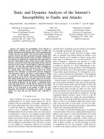


![conn's current therapy 2011 [expert consult] - e. bope, et. al., (saunders, 2011)](https://media.store123doc.com/images/document/14/y/tc/medium_sn0OGS08eq.jpg)