The action of snake venom phospholipase a and trypsin on purified myelin in vitro
Bạn đang xem bản rút gọn của tài liệu. Xem và tải ngay bản đầy đủ của tài liệu tại đây (2.06 MB, 7 trang )
Biochem.
J.
(1976)
159,
273-277
Printed
In
Great
Bitain
The
Action
of
Snake
Venom,
Phospholipase
A
and
Trypsin
on
Purified
Myelln
in
vitro
By
NAREN
L.
BANIK,*
KISHOR
GOHIL
and
A.
N.
DAVISON
Miriam
Marks
Department
of
Neurochemistry,
Institute
ofNeurology,
The
National
Hospital,
Queen
Square,
London
WC1N
3BG,
U.K.
(Received
3
May
1976)
1.
Purified
myelin
was
incubated
with
snake
venom
or
phospholipase
A
in
the
presence
of
or
absence
of
trypsin
at
37°C,
pH7.4,
for
different
times.
2.
Analysis
of
the
myelin
pellet
obtained
after
centrifugation
of
the
myelin
sample
incubated
with
snake
venom
or
phospholipase
A
alone
showed
conversion
of
phosphatidylcholine,
phosphatidylethanol-
amine
and
phosphatidylserine
into
their
corresponding
lyso
compounds.
No
significant
loss
of
myelin
protein
was
observed
in
these
samples.
3.
A
marked
digestion
of
basic
protein
and
proteolipid
protein
was
observed
from
the
myelin
pellet
when
trypsin
was
present
in
the
incubation
mixture.
4.
The
digestion
of
basic
protein
and
particularly
of
proteolipid
from
myelin
suggests
that
phospholipases
may
make
protein
more
exposed
to
proteolytic
enzyme
for
its
digestion.
5.
The
relevance
of
the
co-operative
effect
of
phospholipases
and
proteinases
as
a
model
system
of
the
mechanism
of
myelin
break-
down
in
degenerative
brain
diseases
is
discussed.
Radioisotopic
studies
of
myelin
constituents
indicate
that
at
least
part
of
the
structure
is
meta-
bolically
rather
stable
(Davison,
1961;
Smith,
1972;
Sabri
et
al.,
1974;
Agrawal
et
al.,
1976).
However,
in
multiple
sclerosis
and
other
demyelin-
ating
conditions
there
is
primary
dissolution
of
the
myelin
lamellae,
with
early
loss
of
basic
protein.
As
this
protein
is
susceptible
to
proteolysis,
proteinases
have
been
implicated
in
the
demyelinating
process
(Einstein
et
al.,
1972;
Adams
et
al.,
1971).
Previous
studies
on
isolated
myelin
showed
that
the
basic
protein
was
partially
lost
on
treatment
with
trypsin,
but
unexpectedly
the
myelin-sheath
ultrastructure
appears
to
be
unaltered
(Raghavan
et
al.,
1973;
Banik
&
Davison,
1974;
Wood
et
al.,
1974).
Since
phospholipase
A
incubated
with
isolated
myelin
causes
changes
in
its
lipid
composition
(Coles
et
al.,
1974),
we
have
investigated
the
possibility
that
phospholipases,
together
with
proteo-
lytic
enzymes,
may
cause
the
more
complete
destruction
of
the
myelin
sheath.
Thus
the
purpose
of
the
present
work
was
to
study
the
co-operative
effect
of
phospholipases
and
proteinases
on
the
dissolution
of
the
myelin
membrane
in
the
hope
that
it
will
provide
an
experimental
model
for
the
degenerative
process.
A
preliminary
report
of
this
work
has
appeared
elsewhere
(Banik
&
Davison,
1975).
*
Present
address:
Neurological
Unit,
Veterans
Ad-
ministration
Hospital,
Stanford
University
School
of
Medicine,
3801
Miranda
Avenue,
Palo
Alto,
CA
94304,
U.S.A.
Vol.
159
Experimental
Materials
Acetylated
trypsin,
lysophosphatidylcholine,
crude
snake
(Naja
naja)
venom
and
purified
phospho-
lipase
A
were
obtained
from
Sigma
(London)
Chemical
Co.
(Kingston-upon-Thames,
Surrey,
U.K.).
All
other
chemicals
were
AnalaR
grade
(BDH
Chemicals
Ltd.,
Poole,
Dorset,
U.K.).
Methods
Preparation
of
myelin.
Adult
Wistar
rats
of
either
sex
were
used
throughout
these
experiments.
Rats
were
anaesthetized
with
chloroform
before
exsan-
guination.
Brains
were
quickly
removed,
weighed
and
transferred
into
ice.
The
tissue
was
homo-
genized
in
0.32M-sucrose
and
purified
myelin
was
prepared
as
described
by
Norton
(1971).
Incubation
ofmyelin.
Purified
myelin
was
suspended
in
water.
The
suspended
myelin
was
incubated
with
crude
snake
venom
(10-150,ug/mg
of
myelin
protein),
lysophosphatidylcholine
(20,cg-1.0mg/mg
of
myelin
protein)
and
phospholipase
A
(80,cg/mg
of
myelin
protein)
in
the
presence
or
absence
of
acetylated
trypsin
(10-25pg/g
of
myelin
protein)
in
50mM-
Tris/HCl
buffer,
pH7.4
(Coles
et
al.,
1974),
at
37°C
with
constant
shaking.
Myelin
with
or
without
tryp-
sin,
lysophosphatidylcholine
or
snake
venom
or
phospholipase
A
at
zero
time
served
as
controls.
After
the
incubation
the
experimental
and
control
tubes
were
quickly
chilled
in
ice
and
centrifuged
at
273
N.
L.
BANIK,
K.
GOHIL
AND
A.
N.
DAVISON
12000g
for
10min.
A
firm
myelin
pellet
and
super-
natant
were
obtained
on
centrifugation
and
were
analysed.
Determination
ofprotein
and
adenosine
2':
3'-cyclic
monophosphate
3'-phosphodiesterase
(EC
3.1.4.16)
activity.
Protein
was
determined
by
the
method
of
Lowry
et
al.
(1951),
with
albumin
as
standard,
and
adenosine
2':
3'-cycic
monophosphate
3'-
phosphohydrolase
activity
was
measured
by
the
method
of
Banik
&
Davison
(1969).
Lipid
extraction
and
separation.
Lipid
was
extracted
by
the
method
of
Folch
et
al.
(1957)
and
was
separated
by
t.l.c.
as
described
previously
(Banik
&
Davison,
1971).
Lipids
were
separated
by
t.l.c.
in
the
solvent
system
chloroform/methanol/aq.
12%
(w/v)
NH3
(17:7:1,
by
vol.).
In
this
system
the
lysoethanolamine
phosphoglyceride
was
found
to
co-migrate
with
sphingomyelin,
lysophosphatidylcholine
and
phos-
phatidylinositol;
lysophosphatidylserine
moved
as
a
separate
band.
When
plates
were
stained
with
iodine
vapour
the
loss
of
phosphoglyceride
and
con-
comitant
appearance
of
darkly
stained
bands
for-
corresponding
lyso
compounds
were
observed
(see
Plate
2).
Lysophosphatidylcholine
was
also
separated
by
t.l.c.
by
the
method
of
Coles
et
al.
(1974).
Gel
electrophoresis.
Electrophoresis
of
the
de.
lipidized
samples
in
a
sodium
dodecyl
sulphate
medium
was
carried
out
by
the
method
of
Banik
et
al.
(1974).
Gels
were
stained
with
Coomassie
Brilliant
Blue
overnight
and
de-stained
as
descibed
by
Agrawal
et
al.
(1972).
After
de-staining
gels
were
scanned
in
a
u.v.
spectrophotometer
at
595nm
fitted
with
a
scanner.
Electron
microscopy.
The
pelleted
fractions
were
fixed
overnight
in
4.0%
(whv)
glutaraldehyde
in
0.1
M-potassium
phosphate
buffer,
pH7.4,
then
washed
three
times
in
the
same
buffer
and
fixed
in
1.0%
(w/v)
0S04
for
2h.
Results
Effect
of
lysophosphatidylcholine,
snake
venom
and
phospholipase
A
in
the
presence
or
absence
of
trypsin
on
incubated
myelin
In
our
experiments,
when
myelin
preparations
were
incubated
for
60min
in
Tris/HCI
buffer
at
37°C,
some
digestion
of
both
basic
proteins
occurred,
suggesting
the
presence
of
an
endogenous
proteinase.
All
our
experiments
were
therefore
repeated
in
dupli-
cate
and
data
were
corrected
for
changes
in
control
preparations.
No
apparent
loss
of
membrane
protein
occurred
when
myelin
was
incubated
for
different
time-intervals
separately
with
either
lyso-
phosphatidylcholine
A.
A
9%/
loss
of
protein
from
myelin
was
observed
when
it
was
incubated
with
snake
venom
alone.
However,
there
was
a
marked
loss
of
protein
(17%)
compared
with
controls
when
myelin
was
incubated
with
crude
snake
venom
in
the
presence
of
acetylated
trypsin
(Table
1).
Digestion,
particularly
of
basic
protein,
was
observed
in
these
samples
in
the
presence
of
trypsin,
and
the
appearance
offaster-moving
protein
bands
was
noted.
This
loss
of
protein
was
greater
(25%)
when
the
concentration
of
snake
venom
and
trypsin
was
increased
or
the
time
of
incubation
extended
(Table
1).
An
extensive
digestion
of
high-molecular-
weight
Wolfgram
protein
was
evident
from
the
electrophoretic
pattern
of
incubated
samples
treated
with
either
phospholipase
A
or
snake
venom.
When
trypsin
was
incubated
for
30min
with
myelin
previously
exposed
to
snake
venom,
the
loss
of
protein
was
25%.
In
experiments
in
which
both
phospholipase
A
and
trypsin
were
present,
extensive
loss
of
proteolipid
protein
and
basic
protein
from
myelin
preparations
resulted.
The
loss
of
basic
and
especially
proteolipid
protein
appeared
to
be
greater
when
myelin
preincubated
with
snake
venom
or
phospholipase
A
was
further
incubated
with
trypsin.
The
digestion
of
proteolipid
protein
compared
with
controls
was
60%,
and
bothhigh-
and
low-molecular-
weight
basic
proteins
were
extensively
degraded
when
myelin
was
incubated
with
either
phospholipase
A
or
snake
venom
in
the
presence
of
trypsin.
The
extent
of
digestion
of
high-molecular-weight
basic
protein
was
higher
in
the
presence
of
phospholipase
A
than
with
snake
venom
(Table
2).
A
similar
amount
of
low-molecular-weight
basic
protein
was
digested
in
the
presence
of
either
snake
venom
or
phospholipase
A.
Morphology
Electron-microscope
observations
of
the
washed
myelin
pellet
after
treatment
with
snake
venom
or
phospholipase
A
did
not
reveal
any
structural
difference
compared
with
controls,
and
the
myelin
lamellae
remained
tightly
packed.
However,
the
washed
myelin
residues
after
treatment
with
trypsin
together
with
phospholipase
A
or
snake
venom
revealed
less
densely
packed
myelin.
There
was
extensive
splitting
of
myelin
lamellae
at
the
intra-
period
line
and
numerous
dissociated
single
lamellae
or
free
strands
were
also
present
(Plate
1).
The
periodicity
of
the
myein
lamellae,
trypsin-
and
phospholipase
A-treated
and
control
samples
re-
mained
unaltered.
Effect
on
myelin
2':
3'-cyclicphosphohydrolase
activity
The
total
phosphohydrolase
activity
remained
unchanged
when
myelin
was
incubated
with
lysophos-
phatidylcholine,
snake
venom
or
phospholipase
A.
However,
a
15-20%
loss
of
enzyme
activity
was
observed
when
trypsi'
was
incubated
with
these
reagents
(Table
1).
1976
274
The
Biochemical
Journal,
Vol.
159,
No.
2
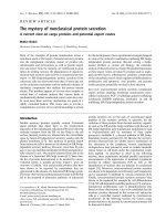
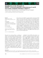
Plate
1
EXPLANATION
OF
PLATE
I
Electron
micrograph
of
the
myelin
pellet
obtained
after
incubation
of
myelin
with
phospholipase
A
in
the
presence
of
acetylated
trypsin
Extensive
splitting
and
dissociation
of
the
myelin
lamellae
can
be
seen
after
incubation
with
trypsin
and
phospholipase
A.
In
normal
rat
myelin
fractions
after
incubation
in
buffer
alone,
splitting
of
the
lamellae
is
minimal
and
few
single
membrane
vesicles
are
present.
Sections
were
70-80nm
thick.
The
horizontal
bar
represents
0.5,m.
N.
L.
BANIK,
K.
GOHIL
AND
A.
N.
DAVISGN
(facing
p.
274)
The
Biochemical
Journial,
Vol.
1
59,
No.
2
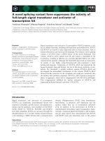
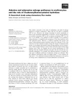
Plate
2
-ch
ol
**.*i
C'
(
r-
cr
b
'.
.:i:;:.Y.r,::?.s::
8!::&::i:
ra
'.n.':'^#
.
-t'
t
j:
w
|
|
_.'.
rs
|
s
i3
111
X
.:x,
#
<S.:.
.:
s
:.'
?:
'
PC
.:.,£,
?lLi
Ig
-
?,.:.
'gL
X
|
|
|
W'aS
l'
|
|
i
::
'.': j
L
y
s
o
P
L
Lyso-pc
1
2
3
4
S
6
7~~~~~~~
A
EXPLANATION
OF
PLATE
2
T.l.c.
of
lipids
extracted
from
mryelin
residue
obtained
after
incubation
of
myelin
with
either
snake
venom
or
phospholipase
A
in
the
presence
or
absence
of
trypsin
The
method
of
separation
of
lipids
is
described
in
the
text.
I,
Myelin
with
snake
venom
(lOO,ug/mg
of
myelin
protein)
at
0min;
2,
as
1,
plus
trypsin
(lSpg/mg
of
myelin
protein);
3,
as
2
at
60min;
4,
myelin
with
trypsin
(15,ug/mg
of
myelin
protein)
at
60min;
5,
myelin
with
phospholipase
A
(80,ug/mg
of
myelin
protein)
at
0min;
6,
same
as
5
at
60mi;
7,
lipids
from
whole
brain.
Abbreviations:
Chol,
cholesterol;
Cereb,
cerebrosides;
PE,
ethanolamine
phospholipid;*
PC,
phosphatidyl-
choline;
Sph,
sphingomyelin;
PS,
phosphatidylserine;
P1,
phosphatidylinositol.
In
this
solvent
system
lyso-PE,
lyso-PC
and
Iyso-PS
in
samples
1,
2,
3,
5
and
6
moved
with
sphingomyelin,
PS
and
PI
respectively.
The
mobility
of
PE,
PC
sphingomyelin,
PS
and
PI
can
be
seen
in
samples
4
and
7.
N.
L.
BANIK,
K.
GOHIL
AND
A.
N.
DAVISON
DISSOLUTION
OF
MYELIN
Table
1.
Loss
ofprotein
and
2':3'-cyclic
AMP
phosphohydrolase
activity
on
incubation
ofpurified
myelin
with
snake
venom,
phospholipase
A
and
lysophosphatidyicholine
in
the
presence
or
absence
of
acetylated
trypsin
Purified
myelin
alone
incubated
in
buffer
for
60min,
and
also
myelin
under
various
conditions
at
zero
time,
served
as
controls.
*,
Myelin
preincubated
with
snake
venom
for
60min
was
further
incubated
with
trypsin
for
30min;
t,
myelin
preincubated
with
snake
venom
for
60min
was
pelleted
and
the
pellet
was
incubated
with
trypsin
for
30min.
Conditions
Total
protein
in
myelin
residue
(mg/sample)
.~~~~~
Incubation
time
(min)
0
Incubation
of
purified
myelin
with:
1.
Lysophosphatidylcholine
(20,ug/mg
of
myelin
2.56
protein)
Lysophosphatidylcholine
(20,ug/mg)+trypsin
2.63
(lOpg/mg
of
myelin
protein)
2.
Snake
venom
(lOO1ug/mg
of
myelin
protein)
2.26
Snake
venom
(lOO,ug/mg)+trypsin
(15#ug/mg
2.36
of
myelin
protein)
3.
Phospholipase
A
(80,cg/mg
of
myelin
protein)
2.44
Phospholipase
A
(80jug/mg)+trypsin
(I5,pg/
2.38
of
myelin
protein)
4.
Snake
venom
(1504ug/mg
of
myelin
protein)
1.10
Snake
venom
(lSO,g/mg)+trypsin
(25pug/mg
1.17
of
myelin
protein)
*Snake
venom
(1
50ug/mg)
+
trypsin
(154ug/mg
0.92
of
myelin
protein)
tSnake
venom+trypsin
(l50.ug/mg)+trypsin
0.92
(lSgg/mg
of
myclin
protein)
5.
Trypsin
(15gg/mg
of
myelin
protein)
0.92
60
2.52
2.32
2.06
1.96
2.36
2.00
1.00
0.87
0.70
0.71
0.81
2':
3'-CyclicAMP
Loss
of
phosphohydrolase
protein
(pmol
of
product/
(Y.
of
h
per
sample)
control
value)
0
60
1.6
3738
3532
12.0
9.0
17.0
3.3
16.0
9.0
25.6
24.0
4576
4068
4219
3872
4366
3360
3986
3740
4135
3167
1907
1812
2138
1590
24.0
2079
1542
12.0
1987
1620
Loss
of
enzyme
activity
(Y.
of
control
value)
5
12
8
23
6
14
5
25
26
18
Table
2.
Loss
of
myelin
proteins
on
incubation
with
snake
venom,
phospholipase
and
lysophosphatidylcholine
in
the
presence
or
absence
of
acetylated
trypsin
Results
are
expressed
as
percentage
loss
of
different
protein
compared
with
control.
The
symbols
*
and
t
are
as
in
Table
1.
Loss
of
myelin
proteins
(Y.
of
control
value)
Conditions
Myelin
incubated
with:
1.
Snake
venom
(l00pg/mg
of
myelin
protein)
Snake
venom
(l00g,g/mg)+trypsin
(15,g/mg
of
myelin
protein)
2.
Phospholipase
A
(80,ug/mg
of
myelin
protein)
Phospholipase
A
(80pg/mg)+trypsin
(15,g/
mg
of
myclin
protein)
3.
Snake
venom
(l50g/mg
of
myelin
protein)
Snake
venom
(150,ug/mg)+trypsin
(lSpg/mg
of
myelin
protein)
4.
Lysophosphatidylcholine
(20,ug/mg
of
myelin
protein)
Lysophosphatidylcholine
(20pg/mg)+trypsin
(lOpg/mg
of
myelin
protein)
Vol.
159
Incubation
I
A
time
Wolfgram
Proteolipid
Basic
protein
Basic
protein
(min)
protein
protein
(large)
(small)
60
15
60
85
60
60
<5
<5
<5
60
42
76
10
<5
<5
54
46
66
<5
69
60
20
<5
7
11
*
71
66
68
74
t
58
61
63
70
60
7
<5
<5
<5
60
15
8
25
28
275
N.
L.
BANIK,
K.
GOHIL
AND
A.
N.
DAVISON
Effect
of
phosphatidylkholine
on
myelin
lipids
Lysophosphatidylcholine
had
a
less
marked
effect
than
crude
venom
enzyme
or
pure
phospholipase
A
on
the
composition
of
myelin
lipids.
On
incubation
with
lysophosphatidylcholine
no
significant
change
in
the
turbidity
of
myelin
was
found
compared
with
controls.
Although
the
higher
amount
of
lysophos-
phatidylcholine
(1mg/mg
of
myelin
protein,
incu-
bated
for
14h)
had
an
effect
on
myelin
proteins,
the
effect
was
less
than
that
obtained
with
crude
snake
venom
or
pure
phospholipase
A.
Action
of
crude
snake
venom
and
phospholipase
A
on
myelin
lipids
Although
there
was
bome
loss
of
lipid
found
in
the
samples
treated
with
trypsin
alone,
no
formation
of
lyso
compounds
was
detected.
When
myelin
preparations
were
incubated
with
either
crude
snake
venom
or
phospholipase
A
in
the
presence
or
absence
of
trypsin,
the
myelin
phospho-
lipids,
ethanolamine-containing
phospholipids,
phos-
phatidylcholine
and
phosphatidylserine
were
found
to
have
been
converted
into
the
corresponding
lyso
compounds
(Plate
2).
The
relative
rates
of
hydrolysis
of
phosphoglycerides
to
lysophosphoglycerides
in
the
membrane
were
phosphatidylserine>
phosphatidyl-
choline
>
ethanolamine
phospholipid.
The
treatment
of
myelin
(1
mg
of
myelin
protein)
with
snake
venom
(100,cg)
showed
that
74%
of
phosphatidyl-
choline,
58
%
of
ethanolamine
phosphoglyceride
and
83%
of
phosphatidylserine
were
cleaved,
and
with
phospholipase
A
(80,ug),
57
%
of
phosphatidylcholine,
40%
of
ethanolamine
phosphoglyceride
and
63%Yo
of
phosphatidylserine
were
hydrolysed
compared
with
the
control.
Most
of
the
lysophosphoglycerides
were
present
in
the
pellet
obtained
after
centri-
fugation
of
the
incubated
myelin
sample,
and
only
a
negligible
amount
of
lysophosphatidylcholine
could
be
demonstrated
in
the
supernatant
fraction
on
t.l.c.
Thus
phospholipase
A
present
in
the
crude
snake
venom
was
active
for
the
conversion
of
myelin
phos-
phoglycerides
into
their
lyso
derivatives,
whereas
galactolipid
and
cholesterol
contents
remained
un-
changed.
The
change
observed
in
cholesterol
and
cerebroside
concentration
after
incubation
with
either
snake
venom
(100,ug)
or
phospholipase
A
(80,cg)
was
less
than
5
%
compared
with
the
control.
No
complete
hydrolysis
of
myelin
phospho-
glycerides
was
obtained
even
when
the
amount
of
crude
snake
venom
was
increased
to
100-150,ccg/mg
of
myelin
protein.
Under
these
experimental
conditions
the
extent
of
hydrolysis
was
greater
than
that
found
with
lesser
amount
of
crude
venom
(20,ug/mg
of
myelin
protein).
The
rate
of
hydrolysis
of
phosphoglycerides
was
obtained
by
incubating
myelin
at
different
times
either
with
snake
venom
(20pg)
or
phospholipase
A.
Phosphatidylserine
was
hydrolysed
more
rapidly
than
phosphatidylcholine
and
ethanolamine
phospholipid,
and
phosphatidyl-
choline
was
hydrolysed
faster
than
ethanolamine
phosphoglyceride.
Discussion
Since
it
has
been
proposed
that
proteolytic
enzymes
are
involved
in
the
breakdown
of
the
myelin
sheath
in
demyelinating
diseases
(Einstein
et
al.,
1969;
Hallpike
et
al.,
1970;
Ramsey
et
al.,
1974;
Smith
&
Rauch,
1974),
we
have
previously
taken
the
effect
of
a
proteolytic
enzyme,
trypsin,
on
myelin
in
vitro
as
a
possible
model
system
(Banik
&
Davison,
1974;
Wood
et
al.,
1974).
Although
our
studies
with
trypsin
showed
the
loss
of
lipids,
including
neutral
lipid,
and
basic
encephalitogenic
protein
from
myelin,
there
was
unexpectedly
no
alteration
in
the
ultrastructure
of
the
myelin
sheath.
Wood
et
al.
(1974)
had
noted
the
same
in
their
experiments.
We
therefore
extended
this
study
by
adding
phospho-
lipase
A
or
crude
snake
venom
to
our
incubation
medium
in
the
presence
of
trypsin,
to
evaluate
the
combined
effect
of
these
enzymes
on
the
dissolution
of
myelin.
When
isolated
myelin
is
incubated
with
either
lysophosphatidylcholine
or
phospholipase
A,
there
is
no
apparent
loss
of
protein
(small
corrections
are
made
for
endogenous
myelin
proteinase
activity).
In
the
presence
of
trypsin
there
is
a
15-30%
loss
of
protein
from
the
membrane.
After
incubation
of
myelin
with
phospholipase
A
or
snake
venom
in
the
presence
of
trypsin,
this
loss
of
myelin
protein
is
shown
to
be
due
to
digestion
not
only
of
basic
protein
but
also
of
proteolipid
protein.
The
lipid
profile
of
the
pelleted
myelin
fractions
showed
a
loss
of
all
classes
of
lipids
and
also
showed
the
conversion
of
myelin
phosphoglycerides
into
their
corresponding
lyso
compounds.
Lysophospholipids
were
found
to
have
remained
with
the
pelleted
myelin
membrane,
and
only
small
amounts
were
detectable
in
the
supernatant.
These
results
are
in
agreement
with
Coles
et
al.
(1974),
where
they
incubated
myelin
preparations
with
phospholipase
A.
There
is
evidence
from
the
findings
of
Poduslo
&
Braun
(1973)
that
basic
protein
is
localized
on
the
cytoplasmic
side
(dense
period
line)
of
the
myelin
and
is
therefore
available
in
myelin
preparations
to
tryptic
digestion,
whereas
proteolipid
protein
may
be
protected
by
its
hydrophobic
lipid
environment
(Folch,
1971).
Once
these
lipids
are
removed
the
proteolipid
protein
becomes
exposed
to
proteolytic
attack,
leading,
it
is
postulated,
to
the
disintegration
of
the
membrane.
The
disintegration
of
the
myelin
sheath
was
observed
in
the
electron
micrograph
of
the
incubated
myelin
sample,
where
splitting
of
the
myelin
lamellae
was
evident
(Plate
1).
After
a
split
1976
276
DISSOLUTION
OF
MYELIN
277
of
the
intraperiod
line
or
dense
line,
the
peeled-off
myelin
lamellae
was
found
to
have
formed
vesicular
structures.
This
type
of
dissolution
of
the
myelin
sheath
has
been
demonstrated
in
experimental
allergic
encephalomyelitis
(Lampert
&
Carpenter,
1965;
Lampert
&
Kies,
1967).
The
vesicular
myelin
debris
as
well
as
the
part
of
the
intact
sheath
are
probably
later
removed
by
activated
macrophages
in
the
diseased
condition.
Elevated
activities
of
phospholipase
have
since
been
demonstrated
in
tissues
from
patients
with
experimental
allergic
encephalomyelitis
and
also
in
tissues
from
patients
with
multiple
sclerosis
(Woelk
&
Kanig,
1974;
Woelk
&
Peiler-Ichikawa,
1974).
Increased
proteinase
has
also
been
found
in
experimental
allergic
encephalomyelitis
and
de-
myelinating
tissues,
both
histochemically
and
bio-
chemically,
by
various
investigators
(Einstein
et
a!.,
1969;
Hallpike
et
a!.,
1970;
Cuzner
&
Davison,
1973;
Ramsey
et
al.,
1974).
In
view
of
these
findings,
phospholipase
and
proteinases
may
be
jointly
involved
in
the
degradation
of
the
myelin
sheath
in
demyelinating
diseases.
These
hydrolases
present
in
activated
macrophages
(David,
1975)
may
well
be
responsible
for
the
primary
attack
on
the
myelin
sheath
in
the
demyelinating
process.
We
thank
the
Multiple
Sclerosis
Society
of
Great
Britain
and
Northern
Ireland
for
financial
support,
and
Dr.
D.
London
for
performing
the
electron
microscopy.
References
Adams,
C.
W.
M.,
Hallpike,
J.
F.
&
Bayliss,
0.
B.
(1971)
J.
Neurochem.
18,
1479-1483
Agrawal,
H.
C.,
Burton,
R.
M.,
Fishman,
M.
A.,
Mitchell,
R.
F.
&
Prensky,
A.
L.
(1972)
J.
Neurochem.
19,
2083-2089
Agrawal,
H.
C.,
Fujimoto,
K.
&
Burton,
R.
M.
(1976)
Biochem.
J.
154,
265-269
Banik,
N.
L.
&
Davison,
A.
N.
(1969)
Biochem.
J.
115,
1051-1062
Banik,
N.
L.
&
Davison,
A.
N.
(1971)
Biochem.
J.
122,
751-758
Banik,
N.
L.
&
Davison,
A.
N.
(1974)
Biochem.
J.
143,
39-45
Banik,
N.
L.
&Davison,
A.
N.
(1975)
Proc.
Meet.
Int.
Soc.
Neurochem.
5th,
Barcelona,
p.
397
Banik,
N.
L.,
Davison,
A.
N.,
Ramsey,
R.
B.
&
Scott,
T.
(1974)
Dev.
Psychobiol.
7,
539-549
Coles,
E.,
Mcllwain,
D.
L.
&
Rapport,
M.
M.
(1974)
Biochim.
Biophys.
Acta
337,
68-78
Cuzner,
M.
L.
&
Davison,
A.
N.
(1973)
J.
Neurol.
Sci.
19,
29-36
David,
J.
(1975)
Fed.
Proc.
Fed.
Am.
Soc.
Exp.
Biol.
34,
1730-1736
Davison,
A.
N.
(1961)
Biochem.
J.
78,
272-282
Einstein,
E.
R.,
Csejtey,
J.,
Davis,
W.
J.,
Lajtha,
A.
&
Mark,
N.
(1969)
Int.
Arch.
Allergy
Suppl.
36,363-375
Einstein,
E.
R.,
Csejtey,
J.,
Dalal,
K.
B.,
Adams,
C.
W.
M.,
Bayliss,
0.
B.
&
Hallpike,
J.
F.
(1972)
J.
Neurochem.
19,
653-662
Folch,
J.
(1971)
Proc.
Meet.
Int.
Soc.
Neurochem.
3rd,
Budapest,
p.
413
Folch,
J.,
Lees,
M.
B.
&
Sloane-Stanley,
G.
H.
(1957)
J.
Biol.
Chem.
226,497-509
Hallpike,
J.,
Adams,
C.
W.
M.
&
Bayliss,
0.
(1970)
Histochem.
J.
2,
199-208
Lampert,
P.
&
Carpenter,
S.
(1965)
J.
Neuropathol.
Exp.
Neurol.
24,
11-24
Lampert,
P.
&
Kies,
M.
W.
(1967)
Exp.
Neurol.
18,
210-223
Lowry,
0.
H.,
Rosebrough,
N.
J.,
Farr,
A.
L.
&
Randall,
R.
J.
(1951)
J.
Biol.
Chem.
193,
265-275
Norton,
W.
T.
(1971)
in
Chemistry
and
Brain
Development
(Paoletti,
R.
&
Davison,
A.
N.,
eds.),
pp.
327-337,
Plenum
Press,
New
York
Poduslo,
J.
F.
&Braun,
P.
E.
(1973)
Trans.
Meet.
Am.
Soc.
Neurochem.
4th,
p.
124
Raghavan,
S.
S.,
Rhoads,
D.
B.
&
Kanfer,
J.
N.
(1973)
Biochim.
Biophys.
Acta
328,
205-212
Ramsey,
R.
B.,
Banik,
N.
L.,
Bowen,
D.
M.,
Scott,
T.
&
Davison,
A.
N.
(1974)J.
Neurol.
Sci.
21,
213-225
Sabri,
M.
I.,
Bone,
A.
H.
&
Davison,
A.
N.
(1974)
Biochem.
J.
142,
499-507
Smith,
M.
E.
(1972)
Neurobiology
2,
35-40
Smith,
M.
E.
&
Rauch,
H.
C.
(1974)
J.
Neurochem.
23,
775-783
Woelk,
H.
&
Kanig,
K.
(1974)
J.
Neurochem.
23,
739-743
Woelk,
H.
&
Peiler-Ichikawa,
K.
(1974)
J.
Neurol.
207,
319-326
Wood,
J.
G.,
Dawson,
R.
M.
C.
&
Hauser,
H.
(1974)
J.
Neurochem.
22,
637-643
Vol.
159