optimization of protein and rna detection methodologies and a new approach for manipulating protein activity in living cells
Bạn đang xem bản rút gọn của tài liệu. Xem và tải ngay bản đầy đủ của tài liệu tại đây (3.77 MB, 144 trang )
UNIVERSITY OF CALIFORNIA, SAN DIEGO
Optimization of protein and RNA detection methodologies and a new
approach for manipulating protein activity in living cells
A dissertation submitted in partial satisfaction of the requirements
for the degree Doctor of Philosophy
in
Biomedical Sciences
by
Brent R. Martin
Committee in charge:
Professor Roger Tsien, Chair
Professor Mark Ellisman
Professor Xiang-Dong Fu
Professor Gerald Joyce
Professor Susan Taylor
Professor Inder Verma
2006
UMI Number: 3208094
3208094
2006
UMI Microform
Copyright
All rights reserved. This microform edition is protected against
unauthorized copying under Title 17, United States Code.
ProQuest Information and Learning Company
300 North Zeeb Road
P.O. Box 1346
Ann Arbor, MI 48106-1346
by ProQuest Information and Learning Company.
Copyright
Brent R. Martin, 2006
All rights Reserved
iii
This dissertation of Brent R. Martin is approved,
and is acceptable in quality and form for publication on microfilm:
Chair
University of California, San Diego
2006
iv
I dedicate this work to my father, Albert Martin, my first mentor and most
successful collaborator.
v
Table of Contents
Signature Page iii
Dedication
iv
Table of Contents
v
List of Figures and Tables vii
Acknowledgements x
Vita and Publications xi
Abstract of the Dissertation xiii
Chapter 1: Mammalian Cell-Based Optimization of the Biarsenical-binding
Tetracysteine Motif for Improved Fluorescence and Affinity
Abstract 1
Introduction 1
Results and Discussion 3
Materials and Methods 22
References
33
Chapter 2: Inducible aggregation of tetracysteine-GFP fusion proteins for
reversible protein inactivation
Abstract
36
Introduction
37
Results
40
Discussion 61
vi
Materials and Methods 65
References 69
Chapter 3: New strategies and progress towards enhancing the specificity of
trans-splicing RNAs in mammalian cells
Abstract 72
Introduction 73
Results 84
Discussion 111
Materials and Methods 116
References 125
vii
List of Figures and Tables
Chapter 1
Figure 1.1 RRL1 selection for improved tetracysteine sequences 4
Figure 1.2 Multiple tetracysteines do not enhance contrast in cells
7
Figure 1.3 RRL2 selection and analysis of optimized flanking residues
9
Figure 1.4 Analysis of unique sequences isolated in sort 16
10
Figure 1.5 Inhibition of tetracysteine-specific membrane localization
12
Table 1.1 Quantum yields of FlAsH and ReAsH
13
Figure 1.6 Contrast improvement quantified by flow cytometry
14
Figure 1.7 Tetracysteine-GFP based fluorescence pulse chase
16
Figure 1.8 FRET-mediated Photoconversion of Cx43-GFP-tetracysteine
17
Figure 1.9 Fusion of optimized tetracysteines to β-actin 18
Figure 1.10 Correlated fluorescence and EM of tetracysteine-tagged β-actin
19
Figure 1.11 Dithiol resistance of alanine mutants point to key residues
21
Table 1.2 Primer sequences
31
Chapter 2
Figure 2.1 YRE#MWR-GFP aggregates following ReAsH labeling 41
Figure 2.2 FACS analysis of YRE#MWR-GFP expressing cells
43
Figure 2.3 Chemical structures of three biarsenical dyes
44
Figure 2.4 Detergents and salts alter properties of YRE#MWR-GFP
45
Figure 2.5 Aggregation is blocked in some fluorescent protein mutants
46
Figure 2.6 Location of Y66W and N146I on GFP
47
Figure 2.7 YRE#MWR-GFP aggregates are released by photobleaching
48
Figure 2.8 YRE#MWR-GFP re-aggregation blocked after photobleaching
49
viii
Figure 2.9 Timecourse of labeling and bleaching of ReAsH 50
Figure 2.10 ReAsH labeling of YRE#MWR-GFP tagged β-actin and α-tubulin
51
Figure 2.11 CHoXAsH labeled tetracysteine-mGFP-β-lactamase
52
Figure 2.12 YRE#MWR-GFP fusions to PKA regulatory subunits
54
Figure 2.13 Timecourse of YRE#MWR-mGFP-RI aggregation
55
Figure 2.14 Co-localization of RIα and Cα in aggregates
56
Figure 2.15 Cytosolic PKA is partially inhibited by RIα aggregation
57
Figure 2.16 Nuclear PKA activity further inactivated by RIα aggregation
58
Figure 2.17 Inactivation of PKA by Cα aggregation
59
Figure 2.18 RIα fusions restore cAMP regulation in RIα null cells
60
Figure 2.19 Cytoskeletal morphology is rescued by tagged RIα expression
61
Table 2.1 Microscope filter sets
67
Table 2.2 Primer sequences
68
Chapter 3
Figure 3.1 Mammalian cell-based libraries for optimizing trans-splicing 83
Figure 3.2 Designed dsRed targeting trans-splicing ribozymes
85
Figure 3.3 DsRed targeted IGS library for in vitro IGS mapping
87
Figure 3.4 In vitro trans-splicing targeting dsRed using the IGS library
89
Figure 3.5 Trans-splicing in the context of total cellular RNA
90
Figure 3.6 In vitro transcription and reaction using newer protocols
91
Figure 3.7 Construction and testing the Dimer2-intron
92
Figure 3.8 Virus-transduced Dimer2-intron-PEST cells have no intron
94
Figure 3.9 Spliceosome-mediated trans-splicing targeting Dimer2-intron
95
Figure 3.10 Analysis of a Dimer2-intron targeted PTM in HeLa cells
96
ix
Figure 3.11 Trans-splicing by tethering β-lactamase gene fragments 98
Figure 3.12 Testing β-lactamase intron insertions at three positions
99
Figure 3.13 Detection of β-lactamase fragment expression in cells
100
Figure 3.14 Schematic of the 3’ER split β-lactamase reporter
101
Figure 3.15 Spontaneous β-lactamase activity in 3’ER HeLa cells
102
Figure 3.16 Activity of 3’ER in HeLa cells
103
Figure 3.17 RT-PCR analysis of 3’ER from transfected 293T cells
104
Figure 3.18 No specific trans-splicing is detectable by western blotting
105
Figure 3.19 Design and testing of split β-lactamase reporters for 5’ER
107
Figure 3.20 Double trans-splicing generates background activity
109
Figure 3.21 Segmental trans-splicing is only partially sequence dependent
111
Table 3.1 Primer sequences
121
x
Acknowledgements
I would like to thank several former members of the Tsien lab, including Grant
Walkup, David Zacharias, Alice Ting, Robert Campbell, Jin Zhang, Amy Palmer, and
Coyt Jackson for passing down their knowledge and helping me achieve my research
goals. Also, many thanks to Oded Tour, Christina Hauser, Paul Steinbach, Qing Xiong,
Tom Deerinck, and other members of the FlAsHers group for assistance and advice.
Most importantly, I would like to acknowledge my closest collaborators, Stephen Adams
and Ben Giepmans whose guidance and encouragement has been invaluable. I would
also like to thank James Lim for assistance with several experiments discussed in
Chapter 3, specifically those involving RIα knockout fibroblasts. Finally I would like to
thank Roger Tsien for supporting me and placing me in such an excellent research
environment and the members of my thesis committee for making time and giving
invaluable advice. On a personal note, I would like to thank my parents and wife Monica
for their constant support.
The text of Chapter 1, in part, is a reprint of the material as it appears in Nature
Biotechnology (Citation: Martin, B.R., Giepmans, B.N.G., Adams, S.R., Tsien, R.Y.
Nature Biotechnology 23, 1308-1314 (2005), I was the
primary researcher and author and the co-authors listed in this publication contributed or
supervised the research which forms the basis for this chapter.
xi
Vita
Education
9/95 – 6/99 B.S. in Molecular Biology, Cum Laude, Provost Honors, University
of California, San Diego
9/99 – present Graduate Student, Biomedical Sciences Graduate Program,
University of California, San Diego
5/06 Ph.D., Biomedical Sciences, University of California, San Diego
Publications
Adams SR, Campbell RE, Gross LA, Martin BR, Walkup GK, Yao Y, Llopis J, and Tsien
RY. 2002. New biarsenical ligands and tetracysteine motifs for protein labeling in vitro
and in vivo: synthesis and biological applications. J. Am. Chem. Soc., 124: 6063-6076.
Martin BR, Giepmans BN, Adams SR, Tsien RY. 2005 Mammalian cell-based
optimization of the biarsenical-binding tetracysteine motif for improved fluorescence and
affinity. Nature Biotechnol. 10:1308-1314.
Martin BR and Tsien RY. Inducible aggregation of tetracysteine-GFP fusion proteins for
reversible protein inactivation. In Preparation.
Poster Presentations
Martin BR, Giepmans BN, Adams SR, Tsien RY. Optimization of the Biarsenical Binding
Tetracysteine Motif for Fluorescence and Affinity and Discovery of a Reversible Tag for
Protein Aggregation. Imaging Technology, The American Society for Cell Biology Annual
xii
Meeting (2005), San Francisco. (Highlighted in Nature Chemical Biology 2, 119-122
(2006)).
Martin BR, Jackson WC, Tsien RY. Mammalian cell-based directed evolution of the
biarsenical binding tetracysteine peptide for improved fluorescence and dithiol
resistance. Program No. 124.11, Society for Neuroscience, Annual Meeting (2004), San
Diego.
Giepmans BN, Martin BR, Gaietta GM,
Deerinck TJ, Adams SR, Tsien RY, Ellisman MH
Visualizing Cytoskeletal Dynamics and Ultrastructure at Cell-cell Junctions Using
Genetically Encoded Tags. Cytoskeleton-Membrane Interactions I, The American
Society for Cell Biology Annual Meeting (2004), Washington D.C. (Presented by BN
Giepmans).
Martin BR, Jackson WC, Tsien RY. Mammalian cell-based directed evolution of the
biarsenical-binding tetracysteine motif for improved fluorescence and affinity. Imaging
Technology, The American Society for Cell Biology Annual Meeting (2003), San
Francisco.
xiii
ABSTRACT OF THE DISSERATION
Optimization of protein and RNA detection methodologies and a new approach
for manipulating protein activity in living cells
by
Brent R. Martin
Doctor of Philosophy in Biomedical Sciences
University of California, San Diego, 2006
Professor Roger Tsien, Chair
The orchestrations that underlie the existence of even the simplest organisms
are quite complex and extremely dynamic. In order to gain a greater understanding of
the biochemistry underlying many unsolved biological problems, new tools are required
to first visualize a phenomenon, and then perturb it to study its significance. Visualizing
the dynamics of intracellular biochemistry has been enhanced greatly with widespread
adoption of genetically encoded fluorescent proteins.
Due to the large size of fluorescent proteins and their lack of chemical flexibility,
the tetracysteine-biarsenical system was developed. This technology uses the
combination of a short genetically encoded tag and a specific class of exogenous,
membrane-permeant dyes. Since its introduction, the biarsenical-tetracysteine system
has suffered from spontaneous background staining, preventing the detection of dilute
proteins. To remedy this problem, a library of tetracysteine sequences was screened for
improved dithiol resistance and brightness. Several new sequences were discovered
xiv
and background staining was reduced significantly, resulting in a 20-fold increase in
labeling contrast.
One unique sequence isolated from the library found an unexpected mechanism
to ensure its selection. Upon ReAsH labeling, YRECCPGCCMWR-GFP rapidly
aggregates into tiny, highly fluorescent speckles. Upon bleaching ReAsH, the
aggregates dissociate, dispersing the tagged protein throughout the cell. Fusions of this
tag on several cellular proteins led to ReAsH dependent aggregation of the tagged
protein as wells as endogenous binding partners. By sequestering protein complexes in
the aggregates, activity is inhibited.
Finally, the detection of specific RNAs in living cells remains a major challenge in
biology with numerous potential applications. Trans-splicing repair of clinically relevant
transcripts has been reported as an efficient and specific method for delivering
exogenous message for translation. Therefore, a crippled reporter gene lacking
translation initiation sites gene was targeted using existing trans-splicing techniques to
an expressed RNAs. Trans-splicing then leads to the conversion of the targeted mRNA
into a chimeric mRNA capable of translating an active protein. After significant effort and
several novel approaches to enhance specificity, it became clear that new methods of
RNA detection will be required to prevent non-specific splicing of cargo RNAs in cells.
1
Chapter 1
Mammalian Cell-Based Optimization of the Biarsenical-binding Tetracysteine
Motif for Improved Fluorescence and Affinity
Abstract
Membrane-permeant biarsenical dyes such as FlAsH and ReAsH fluoresce upon
binding to genetically encoded tetracysteine motifs expressed in living cells
1,2
, yet
spontaneous non-specific background staining conceals weakly expressed or dilute
proteins from detection
1,3
. If the affinity of the tetracysteine peptide could be increased,
then stringent dithiol washes should increase the contrast between specific and
nonspecific staining. Residues surrounding the tetracysteine motif were randomized and
fused to GFP, then retrovirally transduced into mammalian cells and iteratively sorted by
fluorescence-activated cell sorting for high FRET from GFP to ReAsH despite increasing
concentrations of dithiol competitors. Selected sequences demonstrate higher
fluorescence quantum yields and drastically improved dithiol resistance, culminating in a
>20-fold increase in contrast. The best tetracysteine sequences, FLNCCPGCCMEP and
HRWCCPGCCKTF, maintain their enhanced properties as fusions to either termini of
GFP or directly to β-actin. The new biarsenical-tetracysteine motif and should enable
detection of a much broader spectrum of cellular proteins.
Introduction
Biarsenical-tetracysteine labels are analogous to fluorescent protein fusions
2
, yet
offer several unique capabilities such as correlative fluorescence and electron
microscopy (EM)
4
, determination of protein age by multi-color fluorescence pulse
chase
4,5
, chromophore-assisted light inactivation (CALI) for spatio-temporal inactivation
2
of proteins
6-8
, and numerous other in vitro applications
1
. Additionally, the tetracysteine
sequence consists of only a few amino acids, far smaller and potentially less
perturbative than incorporation of a fluorescent protein
9-13
. Furthermore, biarsenical-
tetracysteines are detectable immediately following tetracysteine translation
14,15
, allowing
visualization of early events in protein synthesis, in contrast to the intrinsic delays
required for fluorescent protein maturation
16
. Several other protein fusion partners can
also trap distinctive tags in or on living cells
17
, however, these proteins are either an
order of magnitude larger than tetracysteines, require secondary processing enzymes,
lack a general ability to label intracellular targets, or have no demonstrated expanded
functionality.
The earliest designs of tetracysteine sequences were intended to encourage α-
helicity under the assumption that the biarsenical would ideally fit into the i, i+1, i+4, and
i+5 positions of an α-helix
18
. With these sequences, non-specific biarsenical background
staining was estimated to equal the fluorescence of several micromolar of labeled
protein
1,3
, and was partially reduced by increasing the concentration of the dithiols 1,2-
ethanedithiol (EDT) or 2,3-dimercaptopropanol (BAL) in washes to remove thiol
dependent background or by including non-fluorescent dyes to block hydrophobic
binding sites
2
. When the helix-breaking amino acids PG were inserted between the CC
pairs, the resulting tetracysteine significantly enhanced the affinity and contrast of FlAsH
labeled tetracysteine fusion proteins in cells, increasing the tolerable concentration of
dithiol competitors without detrimental loss of specific fluorescence
1
. However, only a
few pairs of amino acids were tested between the cysteines, and the surrounding
residues were left unaltered, maintaining the α-helical bias.
To optimize the tetracysteine sequence for improved ReAsH affinity and
fluorescence, we developed a retrovirally transduced mammalian cell-based library
3
approach for fluorescent selection of optimal residues surrounding the tetracysteine
motif by fluorescence-activated cell sorting (FACS). Other complementary approaches,
such as surface display on phage or bacteria
19
, pan libraries for high affinity binders in
vitro, disregarding maintenance of desirable fluorescence properties. By performing
these selections in the reducing environment of mammalian cytosol, we intended to
avoid disulfide formation and evolve peptides with improved specificity, activity, toxicity,
and expression in the environment most important to us.
Results and Discussion
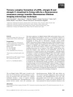
We created our first library, ReAsH Retroviral Library 1 (RRL1), by ligating a
semi-randomized oligonucleotide cassette to the C terminus of green fluorescent protein
(GFP) in a retroviral cloning vector (Fig. 1.1a). NIH3T3 cells were infected with the
recombinant viral library at a low multiplicity of infection (MOI), stained with ReAsH and
analyzed by flow cytometry. Measurement of the GFP quench and GFP-sensitized
FRET (fluorescence resonance energy transfer) to ReAsH emission allows for
determination of the kinetics and extent of ReAsH labeling in a single cell
1
. ReAsH
binding was detectable in cells expressing GFP fused to AEAAARECCRECCARA
18
(αRE), our first generation tetracysteine sequence, and RRL1 cells, as compared to cells
expressing GFP alone (Fig. 1.1b). Interestingly, the RRL1 cells showed varying levels of
FRET after dithiol washing, indicating different amino acid combinations near the
tetracysteine are capable of modulating dithiol resistance and/or fluorescent properties
of the complex. FRET positive RRL1 cells were collected and expanded (Fig. 1.1c, left).
To discriminate higher affinity peptides, three additional rounds of sorting were
performed, each time increasing the selection pressure by escalating the dithiol
4
concentration during washing. Finally, single cells were sorted (Fig. 1.1c, right) on a 96-
well plate.
Figure 1.1. RRL1 selection for improved tetracysteine sequences. (a) Schematic of RRL1 cloning
strategy. Unique restriction sites (italic), randomized codons (blue), and cysteine codons (red) are
indicated. (b) FACS analysis of ReAsH binding by FRET. NIH3T3 cells virally transduced with
either GFP-RRL1 (red), GFP-αRE (blue), or GFP alone (green) following ReAsH staining and a
30 min 0.1 mM BAL wash. ReAsH binding is characterized by an increase in FRET (630/22 nm)
and a decrease in GFP (530/30 nm) fluorescence, which appear on a log scale as a shifts
upwards and leftwards. (c) RRL1 FACS selections. Cells collected (red) in sort 1 (left) and sort 4
(right). (d) Sequence results and analysis for dithiol resistance. Unique sequences isolated in the
RRL1 selection are listed, with the number of identical clones isolated in parenthesis. (-) indicates
an additional peptide deliberately generated rather than isolated from the selection. The dithiol
resistance of ReAsH fluorescence is shown for each sequence determined from live cell imaging
experiments. Measurements represent the average of more than five cells following acute
treatment with 0.4 mM and 1.0 mM EDT to ReAsH labeled cells. Background subtracted, total
ReAsH fluorescence before washing is normalized to 1, representing saturated labeling.
5
Sequence analysis of the isolated clones established ten novel tetracysteine
sequences (Fig. 1.1d). MPCCPGCCGC was highly resistant to EDT, maintaining 50% of
its total ReAsH fluorescence in the face of 1.0 mM EDT, while αRE and
AEAAARECCPGCCARA
1
(αPG), our second generation tetracysteine, both retained
less than 20% (Fig. 1.1d). The next best four peptides all contained either internal
prolines or glycines, corroborating the superiority of hairpin turns over helical
conformations. Replacement of the cysteine in the final randomized position of
MPCCPGCCGC to a serine showed no effect on dithiol resistance, proving the last
cysteine was a fortuitous non-participant in arsenical binding.
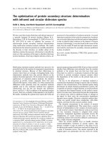
Instead of optimizing a single tetracysteine sequence for improved contrast, an
overlooked approach for increasing the biarsenical-tetracysteine contrast is to attach
multiple tetracysteines to a single protein. By fusing several tetracysteines locally to a
single protein, it should be possible to increase the local concentration of the fluorescent
complex, enhancing the relative brightness as compared to non-specific background
fluorescence. To test this idea, a series of tandem tetracysteines was constructed as C-
terminal fusions to ECFP, diagramed as ECFP-ESSGS(MPCCPGCCGS)
n
. Expression
levels in both bacteria and mammalian cells were inhibited by increasing multiples of
tetracysteines. N-terminally 6-his-tagged recombinant protein was expressed in bacteria,
labeled with FlAsH, and then purified by a Ni-NTA column. The resulting protein was
nearly completely composed of non-oxidized, monomeric, FlAsH-labeled protein (Fig.
1.2a). After measuring the quantum yield of each multiple FlAsH-tetracysteine complex,
it was observed that by increasing the number of fluorophores, the quantum yield was
quenched (Fig. 1.2b). Two tetracysteines on ECFP doubled the overall brightness of
FlAsH, yet further addition of tetracysteines gave diminishing results. When expressed in
cells, no additional brightness was observed with two tetracysteines versus one
6
tetracysteine following FlAsH labeling (Fig. 1.2c). To explore this effect further, GFP with
5 tandem tetracysteines was attached to the C-terminus of the gap junction protein Cx43
(Fig. 1.2d-e). Following addition of ReAsH, the fluorescence increased quickly, then
slowly decayed as labeling became saturated. Following incremental dithiol washing, the
fluorescence increased again. These results imply that the level of tetracysteine
occupancy correlates with the output fluorescence. Early in staining as ReAsH-
tetracysteine complexes first form, no fluorescence quenching occurs. Later in staining,
as each vacant tetracysteine site is filled, dye-dye interactions lead to strong
fluorescence quenching and diminished fluorescence signal. This quenching can be
relieved by the incremental disruption of ReAsH-tetracysteine complexes using dithiol
washes. Overall, as the number of tetracysteines linked in tandem increases, the
fluorescence diminishes. Furthermore, no increases in contrast were observed following
ReAsH photo-oxidation for EM (data not shown). Because of the lower expression and
decreased fluorescence, this avenue towards increased contrast was set aside, and
attention was refocused at increasing the brightness and affinity of a single tetracysteine
tag.
7
Figure 1.2. Multiple tetracysteines do not enhance contrast in cells. (a) Gel analysis of bacterially
expressed, FlAsH-labeled and purified 6-His-ECFP-(TC)
n
protein. Coomassie stained SDS-PAGE
gel of purified protein (left), with contrast enhanced. The protein runs predominantly as a
monomer, yet some oxidized dimer is visible in samples containing tetracysteine sequences,
implying incomplete or partial FlAsH labeling. This incomplete binding is due to the Ni-NTA
purification scheme, which does not exclude oxidized or modified tetracysteines, as does the
FlAsH bead purification protocol. Also, FlAsH fluorescence quantum yields resulting from Ni-NTA
purifications are generally lower, due to the mixture of oxidized tetracysteines in the protein
sample. FlAsH fluorescence of the labeled protein is also observed (right) and shows a trend of
increased fluorescence, relative to the amount of protein seen in the Coomassie stained gel, as
the number of tetracysteines increases. (b) Quantum yields of multiple tandem tetracysteines. Φ
= fluorescence quantum yield, n = number of tetracysteines. The improvement in the overall
fluorescence output of a single protein is written as Φ•n / Φ
TC1
. (c) FlAsH brightness is decreased
in cells expressing multiply tetracysteine tagged CFP. Background subtracted FlAsH fluorescence
per measured amount of pre-stained CFP fluorescence is lower with multiple tetracysteine tags.
(d) HeLa cells expressing Cx43-GFP-TC5 are linked by a large gap junction, as seen by GFP
sensitized ReAsH FRET emission approximately 1000 sec after labeling. (e) Timecourse of
ReAsH staining of Cx43-GFP-TC5 in HeLa cells shows quenching of fluorophores after initial
increases in florescence. Regions were drawn around each cell and the shared gap junction.
8
The results from the RRL1 selection confirmed the consensus sequence
CCPGCC and verified the usefulness of the mammalian cell-based library approach for
optimization of the tetracysteine motif. In an effort to further optimize the ReAsH binding
tetracysteine peptide, a new library, RRL2, was devised, fixing PG as the internal
residues, while randomizing the three external residues on either side of the
tetracysteine, XXXCCPGCCXXX, and abbreviated XXX#XXX (# = CCPGCC) (Fig.
1.3a).
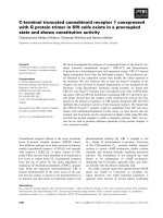
Three hundred million HEK293 cells were infected with RRL2 virus and pre-
sorted for GFP expression, isolating thirty million cells. A 568 nm laser was added to
directly excite ReAsH, allowing cells to be sorted based on two criteria: FRET ratio
(GFP-sensitized ReAsH emission divided by GFP emission) and directly-excited ReAsH
emission. The GFP+ cells were stained with ReAsH and sorted for multiple rounds, each
round selecting the best 10-15% of the total population (Fig. 1.3b) with the goal of
eliminating unfavorable cells over the course of several selections, each time amplifying
the pool of selected cells in culture for better sampling of each genotype. After ten
rounds, the population fell into two categories: one exhibiting high ReAsH fluorescence
and a low FRET ratio, and the other displaying a high FRET ratio but lower ReAsH
fluorescence. Each phenotype was simultaneously separated by sorting with two
streams, one pool for each phenotype. After four more rounds of sorting, cells were
washed with a low concentration of dithiol and sorted into 96-well plates to determine the
composition of each population (Fig. 1.3b, middle).
9
Figure 1.3. RRL2 selection and analysis of optimized flanking residues. (a) Schematic of RRL2
cloning strategy. Notation as in Fig. 1.1a. (b) RRL2 sort history. The FRET ratio is plotted versus
ReAsH intensity in individual cells following a given BAL wash. In sort 1 (left), cells with high
FRET ratios and ReAsH intensities were collected (red). Sort 14 (middle) shows the separation of
high FRET ratio cells (↑Ratio, red/black) from high ReAsH intensity cells (↑ReAsH, blue/green)
and the corresponding sorted fraction (***). In sort 16 (right), the final clones were selected from
the top few percent in the high FRET ratio population. (c) Dithiol resistance of final optimized
tetracysteines. BAL titration of ReAsH (left) and FlAsH (right) labeled N and C terminal optimized
tetracysteine fusions to GFP and Cerulean respectively. Tetracysteine color notation is the same
in both ReAsH and FlAsH titrations. Dithiol resistance is shown as the average fraction of the
FRET ratio remaining following incremental washes with high concentrations of BAL, shown with
corresponding standard deviations derived from three or more duplicate wells on a 96-well plate.
Following sequencing and analysis, the high ReAsH intensity, low FRET ratio
clones displayed massive over-expression of the tetracysteine-GFP fusion, but weak
dithiol resistance and poor labeling efficiency (data not shown). By excluding the GFP
excitation data during the selections, protein expression levels were left uncorrected,
leading to overexpression rather than high affinity. On the contrary, the high FRET ratio,
lower ReAsH fluorescence population was dominated by sequences with better or equal
10
dithiol resistance as MP#GS (Fig. 1.4), while still expressing high levels of the fusion
protein.
Figure 1.4. Analysis of unique sequences isolated in Sort 14. Unique sequences are listed next
to their frequency of occurrence, in parenthesis. Dithiol resistance is shown as the fraction of the
FRET ratio remaining following washes with high concentrations of EDT, and corresponding
standard deviations calculated from three or more duplicate wells on a 96-well plate.
After two more rounds of selection with higher stringency, single cells were
sorted onto 96-well plates for clonal expansion (Fig. 1.3b, right). All twenty-two isolated
clones converged on three sequences, HRW#KTF, FLN#MEP, and YRE#MWR. Each
peptide was tested as both N and C terminal fusions to GFP (Emerald) and CFP
(Cerulean
20
), and exhibited significantly improved dithiol resistance for both ReAsH and
FlAsH (Fig. 1.3c), while demonstrating little preference for either biarsenical and
confirming the weaker dithiol resistance of ReAsH as compared to FlAsH
1
. Upon ReAsH
labeling, cells expressing YRE#MWR, but neither of the other two sequences, quickly
formed tiny subcellular, highly red fluorescent aggregates capable of evading even the
highest dithiol washes. This sequence was therefore excluded from further analysis,
though the ability to precipitate a protein in living cells merely by addition of a permeant
fluorogenic small molecule may be useful in other contexts.