the regulation of genes involved in trichome development
Bạn đang xem bản rút gọn của tài liệu. Xem và tải ngay bản đầy đủ của tài liệu tại đây (17.02 MB, 186 trang )
THE REGULATION OF GENES INVOLVED IN TRICHOME DEVELOPMENT
A Dissertation
Submitted to the Graduate Faculty of the
Louisiana State University and
Agricultural and Mechanical College
in partial fulfillment of the
requirements for the degree of
Doctor of Philosophy
in
The Department of Biological Sciences
by
Matthew Lloyd Brown
B.S., Louisiana State University, 1996
May, 2006
UMI Number: 3208148
3208148
2006
UMI Microform
Copyright
All rights reserved. This microform edition is protected against
unauthorized copying under Title 17, United States Code.
ProQuest Information and Learning Company
300 North Zeeb Road
P.O. Box 1346
Ann Arbor, MI 48106-1346
by ProQuest Information and Learning Company.
ii
This work is dedicated to my mother and father,
Brenda and Jerry Brown,
for the support they have given me in all my endeavors.
iii
ACKNOWLEDGEMENTS
There are so many people that contributed to my professional and
personal development over the last seven years that it would be difficult to
mention them all. My parents have been, and continue to be, a constant source
of encouragement and support. My friend Jared Patterson provided an important
role model for my pursuit of this degree. My fiancé, Emily McMains, has provided
an enormous amount of emotional support to me for the past three years. My
labmates, Ginger Brininstool, Michelle Speckhart, and Remmy Kasili, have all
provided me with counsel and assistance in the laboratory for my entire graduate
career. I feel especially lucky that I chose to study at Louisiana State University. I
feel that all of the professors and graduate students with whom I interacted were
always eager to make available their knowledge and resources to me. This spirit
of cooperation makes LSU a special place. Lastly, I would like to thank my major
professor, John Larkin. Without his direction my graduate career would have
been much less fulfilling.
iv
TABLE OF CONTENTS
ACKNOWLEDGEMENTS iii
LIST OF FIGURES
viii
ABSTRACT
xi
CHAPTER 1. INTRODUCTION 1
1.1 Arabidopsis as a Model Organism 2
1.2 Trichomes as a Model System. 5
1.3 Generation of the Trichome Spacing Pattern 6
1.4 Process of Trichome Morphogenesis 15
1.5 Modulation of the Cell Cycle During Trichome Development 20
1.6 Function of Trichomes 23
CHAPTER 2. MATERIALS AND METHODS 25
2.1 Recombinant DNA Construction Techniques 25
2.1.1 Restriction Enzyme Digests 25
2.1.2 Purifying Vector and Insert DNA 25
2.1.3 Ligation of DNA Fragments 26
2.1.4 Transformation of Escherichia coli
and Agrobacterium tumefaciens 26
2.1.5 Molecular Analysis of Bacterial Transformants 27
2.2 Plant Growth Methods 29
2.2.1 Plant Growth Conditions 29
2.2.2 Agrobacterium-Mediated Transformation
of Arabidposis. 29
2.3 Polymerase Chain Reaction (PCR) Techniques 30
2.3.1 Standard PCR Reactions 30
2.3.2 Quantitative PCR Techniques
31
2.4 DNA, RNA, and cDNA Techniques 35
2.4.1 RNA Purification
35
2.4.2 Reverse Transcription
35
2.4.3 Genomic DNA Extraction 35
2.4.4 Nucleic Acid Quantification 36
2.5 Microscopy Techniques
36
2.5.1 Light Microscopy and Leaf Measurement 36
2.5.2 GUS Staining 37
2.5.3 GFP and Fluorescent Imaging
37
2.5.4 Scanning Electron Microscopy (SEM) 37
2.6 Specific Methods for Each Chapter
37
2.6.1 Chapter 3 Methods 37
2.6.1.1 Construction of GL3 Genomic
Rescue Fragment 37
2.6.1.2 Construction of pGL3::GL3::GR.
38
v
2.6.1.3 Construction of 35S::GL3::GR 40
2.6.1.4 Creation of the gl3 egl3 Double Mutant
Containing pGL3::GL3::GR. 41
2.6.1.5 Examination of T-DNA Levels
Using Quantitative PCR
42
2.6.1.6 Analysis of Gene Expression Between
gl3 egl3, col, GL3
OE
, and pMB02 Transformants
and in the Dex-Inducible Experiment 42
2.6.2 Chapter 4 Methods 42
2.6.2.1 Comparison of Expression of Genes Flanking the
E938 Insert 42
2.6.2.2 Analysis of At2g28210 Message Levels
in Different Plant Tissues and in the Dex-Inducible
System. 43
2.6.2.3 Effect of Ethoxyzolamide on Trichome Growth 44
2.6.3 Chapter 5 Methods 44
2.6.3.1 Construction of a SIM Rescue Plasmid 44
2.6.3.2 Absolute Quantitation of SIM and Its
Arabidopsis Homologues in Various Tissues 45
2.6.3.3 Absolute Quantitation of SIM in gl3 egl3,
sim-1, wild-type, SIM
OE
, and GL3
OE
Plants 46
CHAPTER 3. THE ROLE OF GLABRA3 IN TRICHOME
DEVELOPMENT AND THE RELATION OF GLABRA3
FUNCTION TO THE EXPRESSION OF GENES INVOLVED
IN TRICHOME DEVELOPMENT 47
3.1 Introduction 47
3.2 Results 50
3.2.1 Complementation of GLABRA3 50
3.2.2 Plants with Both 35S::GL1 and Extra Copies of GL3
Produce Ectopic Trichomes
56
3.2.3 Identification of Genes Exhibiting Trichome-Specific
Expression.
58
3.2.4 Construction of a Dex-Inducible GL3 for Use in
Identifying Downstream Targets.
63
3.2.5 Expression of GL3::GR Using the 35S Promoter 72
3.3 Discussion
75
3.3.1 Determination of the Targets of the Trichome
Initiation Complex 75
3.3.2 Use of a Steroid-Inducible Trichome Initiation
System to Determine Direct Targets of GL3
81
3.3.3 A Case for Auto-Regulation?
83
3.3.4 The Effect of Increasing Amounts of pGL3:GL3. 84
3.3.5 The Effect of GL3 Amount on Trichome Morphology.
86
3.3.6 GL1/GL3 Association May Occur in the Cytoplasm.
87
3.3.7 GL1 Dictates which Tissues in the Early Shoot
vi
Produce Trichomes. 88
3.3.8 Plants Containing Both 35S::GL1 and
35S:GL3:GR Produce Prodigious Numbers of Trichomes
in Response to Dex Exposure 89
CHAPTER 4. IDENTIFICATION OF A CARBONIC
ANHYDRASE IN TRCIHOME DEVELOPMENT 90
4.1 Introduction 90
4.2 Results 92
4.2.1 Identification of an Enhancer Trap Line with
a Developing Trichome Phenotype 92
4.2.2 Determination of Which Gene Shares the E938
Expression Pattern 94
4.2.3 Expression of At2g28210 in the Plant. 97
4.2.4 Response of At2g28210 Expression to Trichome
Induction 97
4.2.5 Identification of the At2g28210 Gene 100
4.2.6 Function of At2g28210 104
4.2.7 Identity of At2g28210 Homologs in Arabidopsis 109
4.3 Discussion 110
4.3.1 An Alpha Carbonic Anhydrase Is Upregulated
During Trichome Development. 110
4.3.2 Carbonic Anhydrase Activity Is Required for
Trichome Expansion. 115
4.3.3 Possible Roles for an α-Carbonic Anhydrase
in Trichome Development. 116
4.3.4 Conclusion 118
CHAPTER 5. IDENTIFICATION OF SIAMESE, A GENE
CONTROLLING ENDOREPLICATION IN TRICHOMES 120
5.1 Introduction
120
5.2 Results 122
5.2.1 Identification of the SIAMESE Gene
122
5.2.2 SIAMESE Is Predicted to Encode a
Novel Class of Protein
125
5.2.3 Tissue Level Distribution of the SIAMESE
Transcript
127
5.2.4 Tissue Level Distribution of the Transcripts of
the SIAMESE Homologues 134
5.2.5 Sub-Cellular Localization of SIM
137
5.2.6 The Effect of Ectopic Over-Expression of SIM 140
5.3 Discussion
141
CHAPTER 6: SUMMARY AND FUTURE DIRECTIONS
152
WORKS CITED 155
vii
APPENDIX: LIST OF PCR PRIMERS USED IN THIS WORK 170
VITA 174
viii
LIST OF FIGURES
1-1: Number of citations found on
PubMed for Arabidopsis and Drosophila every
5 Years for the past 15 years
4
1-2: Model of the Lateral inhibition
mechanism of trichome development
8
1-3: Hypothetical Model of Epidermal Cell Fate
Determination By Interactions between proteins
involved in trichome patterning 13
2-1 Sample standard curve for absolute quantitation 34
3-1: Schematics of the pMB02, pMB014,
and the pMB016 constructs. 51
3-2: Phenotypes of plants transformed with pGL3::GL3. 52
3-3: Comparison of the number of copies of
GL3 DNA within different transformant lines. 55
3-4: Change in GL3 expression between GL3
transformants and gl3-1 mutant plants. 57
3-5: Plants harboring 35S::GL1 and multiple
copies of pMB02 have ectopic trichomes. 59
3-6: Fold change in expression of genes
involved in trichome initiation and development.
62
3-7: Proposed GR mediated GL3 action.
64
3-8: Behavior of the pGL3::GL3::GR construct in gl3-1.
66
3-9: Behavior of the pGL3::GL3::GR construct
in the gl3 egl3 background. 69
3-10: Cycloheximide exposure inhibits trichome formation.
71
3-11: Preliminary experiments indicate that MYB23
and GL2 levels increase significantly after exposure
to dex regardless of the presence of chx. 73
3-12: Behavior of the pMB016 construct. 76
ix
4-1: GFP phenotype of E938. 93
4-2: Comparison of expression of genes flanking
the E938 insertion site in glabrous and pubescent plants.
95
4-3: At2g28210 expression as determined by
in situ hybridization.
96
4-4: Expression of At2g28210 in various organs of the plant. 98
4-5: Analysis of the expression of At2g28210 using
the dex-inducible system. 99
4-6: Correct annotation of At2g28210. 101
4-7: ClustalW alignment of genes related to At2g28210. 103
4-8: The carbonic anhydrase inhibitor ethoxyzolamide
inhibits trichome development. 106
4-9: Close ups of GL3::GR leaves exposed to EZ
and dex for two, three, and four days. 107
4-10: Effect of the carbonic anhydrase inhibitor
EZ upon leaf growth. 108
4-11: ClustalW alignment of all alpha carbonic
anhydrases in Arabidopsis. 111
4-12: Analysis of expression of the other
alpha CAs in Arabidopsis.
112
5-1: Location of the siamese mutation. 124
5-2: The sequence of the At5g04470 transcript
and the location of single-base-pair changes found in
the sim-1 and sim-3 alleles.
126
5-3: A 3500 base pair genomic fragment containing
At504470 rescues the sim phenotype.
128
5-4: SIAMESE encodes a protein of a previously
undescribed class. 129
5-5: GUS expression in the sim-2 plant. 130
x
5-6: Distribution of SIM messge as reported by
in situ hybridization. 132
5-7: Absolute quantitation reveals tissue-specific
differences in expression in SIM and its homologs. 133
5-8: Quantiative RT-PCR determination of
trichome-dependent expression of SIM and its homologs. 136
5-9: An N-terminal GFP-SIM fusion localizes to the nucleus. 138
5-10: Phenotypes of the SIM over-expresser. 142
5-11: Expression of SIM in sim-1, wild-type and SIM
OE
lines. 144
xi
ABSTRACT
Arabidopsis thaliana is an organism that can be used as a model for most
of the processes that occur in flowering plants. The leaf hairs, or trichomes, of
Arabidopsis thaliana are macroscopic single cells that have been used as a
model system for cell fate determination, cell expansion, cell cycle regulation, cell
wall deposition, as well as other processes. Initiation of the trichome cell fate is
controlled by a complex of genes including GLABRA1 (GL1), TRANSPARENT
TESTA GLABRA (TTG), and GLABRA3 (GL3). This work examines the role of
GL3 in trichome initiation and uses plants expressing varying levels of GL3 to
determine if genes involved in trichome development are regulated by GL3.
Though several genes are given a cursory examination, the regulation of two
genes, an α–carbonic anhydrase and a novel cell-cycle regulator called
SIAMESE, are given a thorough examination. The α–carbonic anhydrase
At2g28210 was previously not known to be involved in trichome development. Its
involvement in trichome development was discovered with the aid of an enhancer
trap line with robust reporter gene expression in developing trichomes.
Pharmacological studies indicate that this α–carbonic anhydrase may play a role
in trichome expansion. SIAMESE (SIM) was first identified in a mutant screen in
the Larkin lab. This dissertation demonstrates that this gene encodes a novel
type of cell-cycle regulator with several homologs in Arabidopsis and other plant
species. SIM and one of its homologs in Arabidopsis were shown to be
expressed in a trichome-dependent manner. These investigations shed new light
into the molecular process of trichome development.
CHAPTER 1. INTRODUCTION
A central question of biology is that of development. Even the most
complex organism begins life as a single cell, but in most multicellular organisms,
this cell’s descendents differentiate into a myriad of cell types required to
produce a mature organism. How these different fates are realized when all of
the daughter cells of that original zygote have the same genes is the result of two
general processes: cell fate selection and activation of a discrete subset of genes
once this cells fate is determined. These processes have been investigated in
many different systems from heterocyst formation in cyanobacteria (Meeks and
Elhai, 2002) to neural development in the mouse brain (Hirabayashi et al., 2004).
These investigations have revealed many different strategies of differentiation
ranging from those which are entirely lineage-dependent to those which depend
solely upon positional cues provided by neighboring cells. Trichome development
in Arabidopsis thaliana has been studied for over fifteen years (Haughn and
Somerville, 1988; Herman and Marks, 1989). This system is an excellent model
for developmental questions because trichome development is confined to the
two-dimensional plane of the developing epidermis and trichomes are not
essential to the survival of the plant. In this work, I investigate both the machinery
that governs trichome differentiation and the genetic consequences of adopting
the trichome cell fate. To address these issues, I have examined downstream
events regulated by the transcription factors controlling trichome initiation.
2
1.1 Arabidopsis as a Model Organism
Arabidopsis thaliana is one of the most studied organisms in the world.
This plant has not received this attention because of its agricultural importance,
but rather because of its use as a model organism. A model organism is one that
is used as the focus of intense study by investigators with the assumption that
discoveries in the model will help to elucidate similar properties in related
organisms. A. thaliana is a small winter annual crucifer, a member of the mustard
family Brassicaceae. It is related to several crop plants such as broccoli,
cabbage, and radish.
A. thaliana was first described by Johannes Thal in 1577 in a book
describing the plant life of the Harz Mountains (Koncz et al., 1992). It was not
until 1907, however, that Friedrich Laibach published the first paper describing
experimental research using Arabidopsis thaliana. In 1935, Koncz and
colleagues published a paper suggesting Arabidopsis as a model organism for
the study of plants analogous to the use of Drosophila for the study of animals
(Koncz et al., 1992). However, widespread adoption of Arabidopsis thaliana as a
model organism would not be realized for several decades. In the early 1970’s,
two particularly influential reviews by G.P. Rédei proclaimed the benefits of using
Arabidopsis thaliana as a plant model system (Rédei, 1970; Rédei, 1975). In
addition to the lobbying by Rédei and subsequent investigators, the production of
a linkage map of A. thaliana by Koornneef et al (1982) helped to establish it as a
widely used research tool. Application of molecular biology methods during the
1980’s and 1990’s (Chang and Meyerowitz, 1986) and the subsequent
3
sequencing of the genome (The Arabidopsis Genome Initiative, 2000) also
served as powerful catalysts to attract interest in Arabidopsis. Changes in the
scientific culture of the 1980s which paralleled these technological advances also
increased interest in the plant (Fink, 1998) and set the stage for a boom in
Arabidopsis research in the proceeding decade (Figure 1-1).
Arabidopsis thaliana has many qualities that make it an organism well-
suited for scientific research. Unlike crop plants, A. thaliana is very small and it
requires only a minimal amount of care. Furthermore, A. thaliana can produce a
prodigious amount of seed (over 50,000 per plant, Redei, 1975); it can be grown
year-round under laboratory conditions, and it has a relatively short life cycle of
6-8 weeks. It has a small, relatively compact genome (Pruitt and Meyerowitz,
1986; Sparrow and Miksche, 1961) consisting of 5 chromosomes (Steinitz-Sears,
1963). In 1999-2000, the Arabidopsis genome sequence was completed (Lin et
al., 1999; Mayer et al., 1999; Odell et al., 1985; Salanoubat et al., 2000; Tabata
et al., 2000; Theologis et al., 2000), and the total number of base pairs was found
to be 125 Mbp, only about 25 times greater that of E. coli (Blattner et al., 1997),
and twenty times smaller than maize (Palmer et al., 2003). As of July 2004 there
were 31,270 genes annotated in Arabidopsis
( illustrating the compact nature of the
genome. Arabidopsis thaliana is also easy to transform with foreign DNA by
utilizing Agrobacterium tumefaciens-mediated floral dip transformation
4
The increasing research interest in Arabidopsis thaliana in
the last 15 years.
0
200
400
600
800
1000
1200
1400
1600
1989 1994 1999 2004
Number of publicati
Arabidopsis
Drosophila
Figure 1-1: Number of citations found on PubMed for Arabidopsis and
Drosophila every 5 years for the past 15 years. This is a graph of the number
of publications resulting from a PubMed search containing the word Arabidopsis
or Drosophila in the title within the calendar year listed.
5
(Clough and Bent, 1998; Desfeux et al., 2000; Feldmann and Marks, 1987), thus
eliminating the need for more labor-intensive tissue culture transformation to
generate transgenic organisms.
The greatest resource to an Arabidopsis researcher today is a website
called The Arabidopsis Information Resource, or TAIR (www.arabidopsis.org).
TAIR contains the complete annotated genome sequence indexed by several
different criteria, including name, accession number, or keyword. An investigator
can also use an Arabidopsis-specific version of the Basic Local Alignment
Search Tool (BLAST) to explore the genome. TAIR also includes collections of
tools available for a nominal fee to any researcher, such as library clones and
seed stocks. This website also acts as a repository of Arabidopsis knowledge
and a focal point so that investigators can find others working on the plant. For all
of the reasons stated above, Arabidopsis thaliana was the obvious choice as the
organism upon which to base this body of work.
1.2 Trichomes as a Model System
The epidermal surfaces of the leaves and stem of Arabidopsis thaliana are
covered by hair-like projections called trichomes. In Arabidopsis thaliana,
trichomes are large single cells that project perpendicularly from the epidermis;
these structures are so large that they can be easily seen with the naked eye and
a dissecting microscope allows for easy description. Trichomes located on
different tissues have different shapes. Leaf trichomes have two to four branches
while trichomes found on the stem are typically unbranched (Marks et al., 1991).
Trichomes are found on the adaxial surface of early leaves, and on both surfaces
6
of leaf pairs that arise later in the development of the rosette. Importantly,
trichomes are not essential for the survival of the plant. This allows for easy
genetic manipulation of processes involving trichome development (Haughn and
Somerville, 1988). Because trichomes are easy to observe and can be
manipulated with minimal effect on the physiology of the plant, these cells make
an excellent model system for studying a variety of processes including cell fate
determination, cytoskeletal function, and cell cycle regulation.
1.3 Generation of the Trichome Spacing Pattern
The mechanism responsible for generating the spacing pattern on the
surface of first true leaves has been intensely investigated during the past
decade (Larkin et al., 2003; Marks, 1994; Marks, 1997). Trichomes arising on the
leaf surface are found adjacent to one another less than one percent of the time
(Larkin et al., 1994). Trichome initiation occurs in a non-random, non-cell lineage
dependent manner (Larkin et al., 1996). Currently, a lateral-inhibition model is
used to explain cell fate determination in Arabidopsis trichomes (Larkin et al.,
1997). A spacing pattern governed by lateral inhibition would consist of an
initiation factor and inhibitory factor acting within a field of equipotent precursor
cells. The initiation factor would be produced by all cells in an auto-regulatory
manner and would be cell-autonomous. The activity or synthesis of the inhibitory
factor would be under the control of the initiation factor and this inhibitory factor
would be able to diffuse to other cells to counteract the action of the initiatior.
This would set up a “dead-locked” situation by which all cells are producing the
initiation factor, but are being inhibited from adopting the specified fate by their
7
neighbors until the stalemate is broken by one cell producing more of the
initiation factor than its neighbors though stochastic changes in gene expression.
This would cause some cells among the equipotent precursors to adopt a
particular identity while maintaining a minimum distance between cells that
adopted this fate (see Figure 1-2). This basic mechanism has also been shown
to explain the distribution of Drosophila neural bristles (Portin, 2002) and the
spacing of cyanobacteria heterocysts (Wilcox et al., 1973).
Several genetic components that could initiate or inhibit Arabidopsis
trichome production have been discovered. There is a great deal of redundancy
in both types of components. The initiation decision requires products from at
least five different genes: GLABRA1 (GL1), AtMYB23 (MYB23), GLABRA3
(GL3), ENHANCER OF GLABRA3 (EGL3), and TRANSPARENT TESTA
GLABRA (TTG). GL1 encodes a R2R3 MYB transcription factor (Oppenheimer et
al., 1991). Mutants of GL1 have no trichomes at all on early leaves, but do have
trichomes on the edges of later rosette leaves. This lack of trichomes (making the
plants bald, or glabrous) is the only reported phenotype of gl1 (Koornneef et al.,
1982). MYB23 also encodes a R2R3 MYB transcription factor (Kirik et al., 2001),
and the protein is functionally equivalent to GL1 with respect to trichome initiation
(Kirik et al., 2005). MYB23 mutants display reduced trichome branching, while
gl1 myb23 double mutants lack all trichomes, including those few trichomes
found on the edges of later leaves of gl1 mutant plants (Kirik et al., 2005). GL3 is
a basic helix-loop-helix (bHLH) transcription factor (Payne et al., 2000). GL3 loss-
8
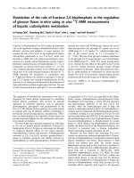
Figure 1-2: Model of the lateral inhibition mechanism of trichome
development. Early in leaf development all cells produce about the same
amount of initiation complex, but the cell is prevented from adopting the trichome
fate by inhibitor elements produced by its neighbors. Slight increases in the
amount of initiation complex or decreases in inhibitor activity could easily disrupt
this equilibrium owing to the auto-regulatory nature of the initiation complex and
its subsequent control over the inhibitor. This would quickly give rise to a single
cell which adopts the trichome fate while strongly inhibiting its neighbors from
adopting the same fate. Black arrowheads denote activation and red blunt arrows
denote inhibition.
9
of-function mutants have a reduced number of trichomes, and the trichomes that
do form have fewer branches than do wild-type trichomes (Bowman, 1994).
EGL3 is also a bHLH transcription factor that is 75% similar to GL3 at the amino
acid level (Zhang et al., 2003). EGL3 mutants have slightly reduced trichome
initiation, and they exhibit a subtle branching defect as well as reduced
anthocyanin pigmentation, reduced seed coat mucilage and altered root hair
positioning. Double mutants of gl3 egl3 are completely glabrous (Zhang et al.,
2003). TTG encodes a WD40 repeat protein (Walker et al., 1999). TTG loss-of-
function mutants are not only glabrous, but they also lack anthocyanin pigment
and seed coat mucilage (Koornneef, 1981), and they have defects in root-hair
patterning (Galway et al., 1994). GL1, MYB23, GL3/EGL3, and TTG are thought
to act together to promote transcription of downstream genes required for
trichome initiation (Ramsay and Glover, 2005).
The inhibitory module of trichome initiation appears to be comprised of a
highly redundant group of proteins that contain a R3 MYB-DNA activation
domain, but lack a transcription activation domain. So far, four genes have been
found that play a role in trichome inhibition: TRIPTYCHON (TRY), CAPRICE
(CPC), ENHANCER OF TRY AND CPC (ETC), and ENHANCER OF TRY AND
CPC2 (ETC2). TRY loss-of-function mutants have larger trichomes with more
branches than wild-type and these trichomes often are found in clusters of
adjacent trichomes (Hulskamp et al., 1994). CPC was first identified because it
has a reduction in the amount of root hairs that it produces (Wada et al., 1997),
but a more careful examination of its phenotype revealed that it produces more
10
trichomes than wild type (Schellmann et al., 2002). The trichomes of try cpc
double mutants exist primarily in clusters and these plants have no root hairs,
indicating the redundant nature of these two proteins (Schellmann et al., 2002).
In both try and try cpc trichome clusters, the extra trichomes at a single initiation
site seem to arise in the location normally occupied by one of the accessory cells
found at the base of the trichome. ETC1 has no apparent single mutant
phenotype, but enhances either the cpc phenotype (with respect only to root
hairs) or the try cpc double mutant phenotype (Kirik et al., 2004a). The try cpc
etc1 triple mutant phenotype is dramatic: giant clusters consisting of hundreds of
trichomes cover the entire leaf surface leaving only the midrib and most basal
portion of the leaf bare (Kirik et al., 2004a). The only ETC2 mutant phenotype is
a small increase in trichome production which is enhanced when combined with
a cpc mutant (Kirik et al., 2004b). Combining the etc2 mutation with try and cpc
does not produce the great trichome clusters seen in try;cpc;etc1 triple mutants;
rather the phenotype of the try;cpc;etc2 mutant resembles the try;cpc double but
with trichome clusters also appearing on the petiole (Kirik et al., 2004b). Thus,
these four genes seem to function as partially redundant inhibitors of trichome
initiation.
The key to understanding the process by which a cell adopts the trichome
fate lies not only in finding the components of the initiation and inhibitory
elements, but also in uncovering how these elements interact. Based upon data
found in Arabidopsis and other species, a model of the interaction of these
elements is shown in Figure 1-3. A functional trichome initiation element is
11
thought to be created by a complex of two bHLH proteins (GL3 or EGL3) with a
single GL1 and TTG protein attached to this dimer. MYB-bHLH-WD40 protein
complexes are involved in controlling developmental and biochemical processes
in several species (Ramsay and Glover, 2005). In Petunia, maize and
snapdragon a network of proteins that includes a WD repeat protein, an R2R3
MYB transcription factor, and two bHLH transcription factors is involved in
regulating anthocyanin pigmentation (Mol et al., 1998). The fact that these
proteins have been found in some cases to be interchangeable between these
three species emphasizes the universality of this motif (Mol et al., 1998, Ramsay
and Glover, 2005). Using the preceding information and data presented below, I
have made a model of the process of the molecular basis of trichome cell fate
selection, which is shown in Figure 1-3.
The function of TTG in this process is rather mysterious. It has been
suggested that TTG-like WD repeat proteins mediate protein-protein interactions
(Mol et al., 1998), or possibly function in a signal transduction pathway (Walker et
al., 1999). The ttg loss-of-function phenotype is phenocopied by a gl3 egl3 tt8
triple mutant (TT8 is TRANSPARENT TESTA 8; a gene putatively encoding a
bHLH protein similar to GL3 and EGL3) (Zhang et al., 2003) and ttg mutants can
be rescued by the maize R gene under the control of the cauliflower mosaic virus
35S promoter (Lloyd et al., 1992; Zhang et al., 2003), which is a promoter that
directs constitutive, ectopic expression of genes under its control (Benfey and
Chua, 1990). Given that GL1 over-expression has no effect on the ttg phenotype
(Larkin et al., 1994) while GL3 or EGL3 over-expression can suppress the ttg
12
phenotype (Lloyd et al., 1992; Zhang et al., 2003), it is likely that TTG somehow
affects the bHLH proteins in such a way as to lower the amount of these proteins
needed to generate the initiation signal. This idea is supported by yeast two-
hybrid studies that show that GL3, but not GL1 interacts with TTG (Payne et al.,
2000). The sub-cellular localization of TTG has not been determined directly.
Preliminary evidence, such as lack of an identifiable nuclear localization signal,
an unpublished result indicating cytoplasmic localization, and the localization of
the TTG homolog AN11 to the cytoplasm in Petunia, all imply that TTG may be
restricted to the cytoplasm and thus its exact role in regulating bHLH/MYB
transcription factor complexes remains to be determined (Mol et al., 1998;
Walker et al., 1999).
The exact assortment of MYB and bHLH proteins that make up the
trichome initiation complex is unknown, but it seems as though both bHLH
proteins are interchangeable with one another. Expression of EGL3 under the
control of the 35S promoter appears to be functionally redundant with expression
of GL3 under control of the same promoter (Zhang et al., 2003). A similar
situation exists among the MYB proteins, though expression of MYB23 using the
35S promoter only rescues the trichome initiation phenotype; the resulting
trichomes from these plants still have fewer branches (Kirik et al., 2005). These
collected facts imply a molecular mechanism of trichome initiation in which any
combination of the bHLH proteins GL3 or EGL3 form a dimer in the cytoplasm,
which is facilitated by TTG. Simultaneously, a MYB protein, either GL1 or
MYB23, associates with the bHLH dimer, and if this MYB/bHLH/bHLH complex
13
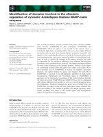
Figure 1-3: Hypothetical model of epidermal cell fate determination by
interactions between proteins involved in trichome patterning. A
combination of a MYB protein (either GL1 or MYB23) with a bHLH dimer (any
combination of GL3 or EGL3) produces the trichome initiation complex. This
complex activates genes involved in trichome development as well as both
initiation genes and an inhibitory signal. This inhibitory signal would be exported
to neighboring cells, where it would interact preferentially with the bHLH dimer,
preventing expression of genes involved in trichome development. TTG would
act in the cytoplasm most probably aiding in the association of the bHLH dimer.