a role for the endosomal snare complex and tethers in autophagy
Bạn đang xem bản rút gọn của tài liệu. Xem và tải ngay bản đầy đủ của tài liệu tại đây (5.84 MB, 220 trang )
Glasgow Theses Service
Cowan, Marianne (2014) A role for the endosomal SNARE complex and
tethers in autophagy. PhD thesis.
Copyright and moral rights for this thesis are retained by the author
A copy can be downloaded for personal non-commercial research or
study, without prior permission or charge
This thesis cannot be reproduced or quoted extensively from without first
obtaining permission in writing from the Author
The content must not be changed in any way or sold commercially in any
format or medium without the formal permission of the Author
When referring to this work, full bibliographic details including the
author, title, awarding institution and date of the thesis must be given
A ROLE FOR THE ENDOSOMAL
SNARE COMPLEX AND
TETHERS IN AUTOPHAGY
A thesis submitted to the
INSTITUTE OF MOLECULAR, CELL AND SYSTEMS BIOLOGY
For the degree of
DOCTOR OF PHILOSOPHY
by
Marianne Cowan
College of Medical, Veterinary and Life Sciences
Institute of Molecular, Cell and Systems Biology
University of Glasgow
October 2013
2
Autophagy is a major route for lysosomal and vacuolar degradation in mammals
and yeast respectively. It is involved in diverse physiological processes and
implicated in numerous pathologies. The process of autophagy is initiated at the
pre-autophagosomal structure and is characterised by the formation of a double
membrane vesicle termed the autophagosome which sequesters cytosolic
components and targets them for lysosomal/vacuolar degradation. The molecular
mechanisms that regulate autophagosome formation are not fully understood. The
conserved oligomeric Golgi (COG) complex is a hetero-octameric tethering factor
implicated in autophagosome formation which interacts directly with the target
membrane SNARE proteins Syntaxin 6 and Syntaxin 16 via the Cog6 and Cog4
subunits respectively. The work presented in this thesis demonstrates direct
interaction of the yeast orthologue of Syntaxin 16, Tlg2, with Cog2 and Cog4. In
addition, I investigated binding of the COG complex subunits to Tlg1, Vti1 and
Snc2, the partner SNARE proteins of Tlg2. Direct interaction of Tlg1, the yeast
orthologue of Syntaxin 6, with Cog1, Cog2 and Cog4 were observed. Given that
Tlg2 has previously been shown to regulate autophagy in yeast, these data
support a conserved role for the COG complex in mediating autophagosome
formation through regulation of SNARE complex formation.
In addition to investigating binding of COG complex subunits to the endosomal
SNARE complex, I have also investigated a role for autophagy in regulating Tlg2
levels. The SM protein Vps45 has previously been shown to stabilise Tlg2 cellular
levels. Our laboratory has demonstrated a role for both the proteasome and
vacuole in the degradation of Tlg2. Here I demonstrated a role for autophagy in
the regulation of Tlg2 levels and show that Swf1-mediated palmitoylation may
serve to protect Tlg2 from being selectively targeted for autophagy. I also
investigated the effects of the yeast T238N mutation on Vps45 function. The
analogous mutation in human Vps45 has recently been associated with congenital
neutropenia. Vps45 function is best characterised in yeast where it associates with
membranes via Tlg2 and is required for membrane traffic from the trans-Golgi
network into the endosomal system. Cellular levels of Vps45 T238N were
destabilised and a concomitant reduction in Tlg2 levels was also observed.
Vacuolar protein sorting remained unaffected in yeast cells harboring Vps45
Abstract
3
T238N but was subjected to increased apoptosis under hydrogen peroxide-
mediated stress. This identifies a novel role for Vps45 in maintaining cell viability.
Finally, I also investigated a role for endosomal trafficking and autophagy in
C.elegans post-embryonic development and identified a role for these pathways in
the clearance of the pre-moult increase in intracellular membranes and cuticular
formation.
4
Table of Contents
Abstract 2
List of Tables 8
List of Figures 9
Acknowledgements 13
Author’s Declaration 14
Definitions/Abbreviations 15
Chapter 1 – Introduction 18
1.1 Autophagy 19
1.1.1 Identification of autophagy 19
1.1.2 Functional significance of autophagy 20
1.1.3 Autophagy versus the cytosol-to-vacuole targeting pathway 21
1.1.4 The process of autophagy 22
1.1.5 Ubiquitination and selective autophagy 28
1.1.6 Regulation of autophagy by signalling pathways 30
1.1.7 Autophagy in disease and development 31
1.2 SNARE proteins 32
1.2.1 Structure and function of SNARE proteins 32
1.2.2 Expression and localisation of SNARE proteins 34
1.2.3 The endosomal SNARE complex 34
1.2.4 Syntaxin 16 is the mammalian orthologue of Tlg2 35
1.2.5 Regulation of Tlg2 cellular levels 36
1.2.5.1 Protein palmitoylation 37
1.3 The SM family of proteins 38
1.3.1 SM protein structure 38
1.3.2 Regulation of membrane fusion by SM proteins 39
1.3.3 Other SM protein interactions 40
1.3.4 Identification of the SM protein Vps45 41
1.4 Tethering proteins 42
1.4.1 Function of the COG tethering complex 43
1.4.2 Molecular structure of the COG complex 44
1.5 C.elegans: An introduction 45
1.5.1 C.elegans post-embryonic development 46
1.5.2 C.elegans cuticle 47
1.5.3 Temporal expression of cuticle collagen genes 48
5
1.5.4 Collagen protein structure 48
1.5.5 UNC-51 is the C.elegans ortholog of yeast Atg1 49
1.5.6 VPS-45 function in C.elegans 50
1.6 Project aims 51
Chapter 2 – Materials and Methods 53
2.1 Materials 53
2.1.1 Antibodies 54
2.1.2 Bacterial, yeast and nematode strains 55
2.1.3 Growth media 57
2.2 Molecular Biology 58
2.2.1 Purification of plasmid DNA from E.coli 58
2.2.2 Agarose gel electrophoresis 61
2.2.3 Gel extraction and purification of DNA 62
2.2.4 Polymerase Chain Reaction 62
2.2.5 Site-directed mutagenesis 65
2.2.6 Restriction endonuclease digestion of DNA 66
2.2.7 Ligation of DNA 67
2.3 Protein analysis 68
2.3.1 SDS-polyacrylamide gel electrophoresis 68
2.3.2 Coomassie™ blue staining 68
2.3.3 Western blot transfer 69
2.3.4 Immunological detection of proteins 69
2.4 IgG affinity purification 70
2.5 General yeast methods 71
2.5.1 Cryopreservation and maintenance of yeast cell stock 71
2.5.2 Preparation of competent yeast cells 71
2.5.3 Transformation of competent yeast cells 72
2.5.4 Preparation of yeast whole cell lysates 72
2.5.4.1 Rapid Twirl buffer lysis procedure 73
2.5.4.2 Glass bead lysis procedure 73
2.5.5 Isolation of yeast genomic DNA 74
2.6 Production of mutant yeast strains by homologous recombination 75
2.7 Carboxypeptidase Y overlay assay 76
2.8 Palmitoylation assays 77
2.8.1 Hydroxylamine treatment 77
2.8.2 Acyl resin-assisted capture 78
2.9 Bradford protein assay 80
2.10 Hydrogen peroxide halo assay 81
2.11 Purification of recombinant fusion proteins from E.coli 81
6
2.11.1 Preparation of competent bacterial cells 81
2.11.2 Transformation of competent bacterial cells 82
2.11.3 Cryopreservation and maintenance of plasmid DNA 82
2.11.4 Expression of recombinant fusion proteins 82
2.11.5 Purification of GST fusion proteins 84
2.11.6 Purification of Protein A fusion proteins 85
2.12 Protein interaction assays 86
2.12.1 GST and Protein A pull-down assays 86
2.12.2 Yeast two-hybrid assay 87
2.13 C.elegans methods 89
2.13.1 Maintenance of C.elegans in culture 89
2.13.2 Preparation of E.coli OP50-1 liquid culture 89
2.13.3 Cryopreservation and recovery of C.elegans 90
2.13.4 Isolation of C.elegans genomic DNA 90
2.13.5 Preparation of C.elegans whole animal lysates 91
2.13.6 C.elegans genetic crosses 91
2.13.7 Nomarski microscopy 91
2.13.8 Immunofluorescence of C.elegans 92
Chapter 3 – Endosomal SNAREs and autophagy 93
3.1 Overview and aims 93
3.2 Results 94
3.2.1 Yeast two-hybrid assays 94
3.2.1.1 Summary of yeast two-hybrid interactions 109
3.2.2 Pull-down assays 110
3.2.2.1 Expression and purification of recombinant fusion proteins 110
3.2.2.2 Detection of chromosomally expressed HA-tagged Cog proteins
116
3.2.2.3 Tlg2 directly associates with COG complex subunits 117
3.2.2.4 Tlg1 directly associates with Cog1 122
3.2.2.5 Functional significance of the Tlg1 and Cog1 interaction 123
3.2.2.6 Tlg1 directly associates with Cog2 and Cog4 125
3.2.2.7 Summary of pull-down interactions 128
3.3 Chapter summary 129
Chapter 4 – Regulation of Tlg2 steady-state levels 131
4.1 Overview and aims 131
4.2 Results 132
4.2.1 Vps45 regulates Tlg2 steady-state protein levels 132
4.2.2 Tlg2 steady-state protein levels are regulated by the vacuole 133
4.2.3 Tlg2 is regulated in an autophagy-dependent manner 134
7
4.2.4 A role for palmitoylation in the regulation of Tlg2 141
4.3 Chapter summary 148
Chapter 5 – The T238N mutation in yeast Vps45 150
5.1 Overview and aims 150
5.2 Results 151
5.2.1 Generation of the Vps45 T238N mutation in yeast 151
5.2.2 The yeast Vps45 T238N position localises to domain 3a 153
5.2.3 Tlg2 is destabilised by the Vps45 T238N mutation in yeast 154
5.2.4 CPY is correctly sorted in yeast harboring the Vps45T238N mutation
156
5.2.5 The T238N mutation in yeast VPS45 leads to increased apoptosis
158
5.2.6 Chapter summary 162
Chapter 6 – Autophagy and endosomal trafficking in C.elegans development
164
6.1 Overview and aims 164
6.2 Results 165
6.2.1 Disruption of autophagy in dpy-10 mutant backgrounds 167
6.2.2 Disruption of endosomal trafficking in dpy-10 mutant backgrounds
170
6.2.3 Characterisation of C.elegans strains 175
6.2.4 C.elegans development and a role for autophagy 178
6.2.4.1 Morphological characterisation of autophagy deficient C.elegans
179
6.2.4.2 Cuticular localisation of DPY-7 in autophagy deficient C.elegans
182
6.2.5 C.elegans development and a role for endosomal trafficking 184
6.2.5.1 Cuticular localisation of DPY-7 in endosomal trafficking deficient
C.elegans 184
6.2.5.2 Monitoring soluble DPY-7 in endosomal trafficking deficient
C.elegans 185
6.3 Chapter summary 189
Chapter 7 – Discussion 190
7.1 Endosomal SNAREs and autophagy 190
7.2 Regulation of Tlg2 steady-state levels 194
7.3 The T238N mutation in yeast Vps45 195
7.4 Autophagy and endosomal trafficking in C.elegans development 197
References 200
Publications 219
8
List of Tables
Table 2-1 Antibiotics used in this study 53
Table 2-2 Antibodies used in this study 54
Table 2-3 E.coli strains used in this study 55
Table 2-4 S.cerevisiae strains used in this study 56
Table 2-5 C.elegans strains used in this study 57
Table 2-6 List of plasmids used in this study 59
Table 2-7 Oligonucleotides used in this study 63
Table 2-8 Standard PCR reaction mix 64
Table 2-9 Standard PCR conditions 64
Table 2-10 SDM PCR conditions 65
Table 2-11 Standard restriction enzyme digest 66
Table 2-12 DNA ligation reaction 67
Table 3-1 Summary of yeast two-hybrid interactions 109
Table 3-2 Summary of pull-down interactions 128
9
Figure 1-1. The process of autophagy 18
Figure 1-2. Schematic representation of the endosomal system, autophagy and
the Cvt pathway in yeast. 26
Figure 1-3. Schematic overview of ubiquitination 29
Figure 1-4. Regulation of autophagy by TORC1 30
Figure 1-5. Domain structure of the syntaxin proteins 32
Figure 1-6. Closed and open conformations of the SNARE proteins 33
Figure 1-7. Transmembrane domain protein sequence alignment of yeast SNARE
proteins 38
Figure 1-8. Modes of SM protein binding to SNARE proteins 40
Figure 1-9. Schematic diagram of membrane fusion 44
Figure 1-10. Architecture of the COG complex 45
Figure 1-11. C.elegans development 46
Figure 1-12. Structural organisation of the C.elegans cuticle 47
Figure 2-1. One-step gene replacement primers 75
Figure 2-2. One-step gene replacement by homologous recombination 76
Figure 2-3. Summary flow chart of hydroxylamine treatment protocol 78
Figure 2-4. Recombinant fusion protein expression summarised 83
Figure 2-5. Summary flow chart of yeast two-hybrid protocol 88
Figure 3-1. Yeast two-hybrid schematic 95
Figure 3-2. Yeast two-hybrid plasmids 96
Figure 3-3. Yeast two-hybrid interactions between AD-Tlg2
cyto
and BD Cog
constructs 99
Figure 3-4. Yeast two-hybrid interactions between AD Tlg2
cyto
∆N36 and BD Cog
constructs 100
Figure 3-5. Yeast two-hybrid interactions between AD-Tlg2
cyto
∆Habc and BD Cog
constructs 101
Figure 3-6. Yeast two-hybrid positive and negative interaction controls for BD Cog
constructs 102
Figure 3-7. Expression of the yeast two-hybrid AD-Tlg2
cyto
, AD-Tlg2
cyto
∆N36 and
AD-Tlg2
cyto
∆Habc fusion proteins 103
Figure 3-8. Yeast two-hybrid interactions between BD-Tlg2
cyto
and AD Cog
constructs 105
List of Figures
10
Figure 3-9. Yeast two-hybrid interactions between BD-Tlg2
cyto
∆N36 and AD Cog
constructs 106
Figure 3-10. Yeast two-hybrid interactions between BD-Tlg2
cyto
∆Habc and AD Cog
constructs 107
Figure 3-11. Yeast two-hybrid negative and positive interaction controls for AD
Cog constructs 108
Figure 3-12. Expression of the yeast two-hybrid BD-Tlg2
cyto
, BD-Tlg2
cyto
∆N36, BD-
Tlg2
cyto
∆Habc and BD-p53 fusion proteins 109
Figure 3-13. Expression and purification of PrA and PrA-tagged Tlg2 constructs
112
Figure 3-14. Expression and purification of PrA-tagged Snc2
cyto
and Vti1
cyto
113
Figure 3-15. Expression and purification of GST-tagged proteins 115
Figure 3-16. Detection of HA-tagged Cog1 to Cog4 117
Figure 3-17. Tlg2
cyto
-PrA associates with HA-tagged Cog2 and Cog4 118
Figure 3-18. Normalised protein concentration for PrA-tagged Tlg2 fusion proteins
120
Figure 3-19. The Tlg2 SNARE domain mediates binding to HA-tagged Cog2 and
Cog4 121
Figure 3-20. Normalised recombinant protein concentration for Tlg2 partner
SNARE proteins 122
Figure 3-21. HA-Cog1 associates with GST-Tlg1
cyto
123
Figure 3-22. Tlg1 whole cell protein levels are selectively reduced in cog1 deficient
yeast 124
Figure 3-23. HA-Cog2 associates with GST-Tlg1
cyto
125
Figure 3-24. HA-Cog3 does not associate with GST-Tlg1
cyto
, Snc2
cyto
-PrA or
Vti1
cyto
-PrA 126
Figure 3-25. HA-Cog4 interacts with GST-Tlg1
cyto
but not with Snc2
cyto
-PrA or
Vti1
cyto
-PrA 127
Figure 3-26. HA-Cog6 does not associate with GST-Tlg1
cyto
, Snc2
cyto
-PrA or
Vti1
cyto
-PrA 128
Figure 4-1. Vps45 deficient cells exhibit reduced cellular levels of Tlg2 132
Figure 4-2. Endogenous levels of Tlg2 is elevated in cells deficient in vacuolar
activity 133
Figure 4-3. Regulation of Tlg2 steady-state levels by the vacuole is dependent on
Vps45 134
Figure 4-4. COG1 and ATG1 KanR modules produced by PCR 135
11
Figure 4-5. Integration of the COG1 KanR module into the COG1 locus 136
Figure 4-6. Integration of the ATG1 KanR module into the ATG1 locus 138
Figure 4-7. Tlg2 steady-state levels are increased in autophagy deficient cells 140
Figure 4-8. Cellular levels of HA-Tlg2 are reduced following treatment with
hydroxylamine in wild type cells 143
Figure 4-9. Endogenous levels of Tlg2 and Tlg1 are reduced in Swf1 deficient cells
144
Figure 4-10. Schematic overview of resin-assisted capture of S-acylated proteins
145
Figure 4-11. Endogenous Tlg2 is palmitoylated in wild type but not Swf1 deficient
cells 146
Figure 4-12. Levels of Tlg2 palmitoylation is comparable in wild type and atg1∆
cells 148
Figure 5-1. Products of site-directed mutagenesis for the production of yeast
Vps45 T238N 151
Figure 5-2. Partial DNA sequence alignment for pMC007 and yeast wild type
VPS45 152
Figure 5-3. Sequence alignment of yeast Vps33 domain 3a with yeast and human
Vps45 153
Figure 5-4. Yeast cells harboring the Vps45T238N mutation exhibit reduced
cellular levels of Vps45 and Tlg2 155
Figure 5-5. Cellular levels of Vps45 and Tlg2 are reduced in cells harboring low
copy yeast expression plasmids encoding Vps45T238N 156
Figure 5-6. CPY is correctly sorted in yeast harboring the Vps45T238N mutation
157
Figure 5-7. H
2
O
2
halo assay template 159
Figure 5-8. vps45∆ and Vps45T238N lead to increased apoptosis 160
Figure 5-9. Vps45, but not Vps21 or Vps27 deficient cells, lead to increased H
2
O
2
-
induced apoptosis 161
Figure 6-1. Summary of C.elegans genetic crosses 168
Figure 6-2. Phenotypic identification of C.elegans strain IA835 169
Figure 6-3. Phenotypic identification of C.elegans strain IA836 170
Figure 6-4. Schematic diagram of vps-45 and vps-45(tm246) PCR analysis 172
Figure 6-5. PCR analysis confirming homozygosity of vps-45(tm246) in strains
IA779 and IA823 173
Figure 6-6. Phenotypic identification of C.elegans strain IA779 174
12
Figure 6-7. Phenotypic identification of C.elegans strain IA823 175
Figure 6-8. Mutant C.elegans body size 176
Figure 6-9. Larval development for endosomal trafficking deficient C.elegans 177
Figure 6-10. C.elegans embryonic viability measured at 15°C 178
Figure 6-11. The IA835 and I836 dumpy phenotypes at 15°C, 20°C and 25°C 180
Figure 6-12. IA835 phenotypic characteristics 181
Figure 6-13. IA836 phenotypic characteristics 182
Figure 6-14. DPY-7 cuticular localisation in the IA835 and IA836 double mutant
strains 183
Figure 6-15. DPY-7 cuticular localisation in the IA779 and IA823 double mutant
strains 185
Figure 6-16. Soluble DPY-7 accumulates in strain IA779 187
Figure 6-17. Soluble DPY-7 is undetectable in strain IA823 188
13
First and foremost I would like to thank my supervisor Dr Nia Bryant for allowing
me to undertake my PhD under her exceptional supervision. Your continuous
guidance, support and constructive feedback during this time have greatly
contributed to my development as a scientist and for this I am most grateful.
I would also like to thank Dr Iain Johnstone for overseeing my C.elegans project
and members of my academic panel, Dr Mike Blatt and Dr Joanna Wilson, for your
suggestions. I owe my thanks to Martin Werno in the Chamberlain lab (University
of Strathclyde) for showing me how to perform acyl-Rac experiments and to
Stephanie Evans for your patience and advice with yeast dissections. Other
contributions in the form of yeast strains have also been greatly appreciated and I
would like to thank Dr Joe Gray (University of Glasgow) and Dr Daniel Klionsky
(University of Michigan) for these.
Thanks to all the members of lab 241 for your kind help and advice when needed.
In particular, thanks to Dr Scott Shanks for teaching me everything yeast related
during my early days in the lab. Also, thanks to my bench buddy Laura Stirrat for
your fine company – you have provided me with the necessary laughs to see me
through my more challenging days in the lab.
It is fair to say that all of this would not have been possible without the financial
assistance received from the University of Glasgow and as such, I would like to
say a very big thank you!
Last but not least, a special thanks to my wonderful family for your support and
continued interest in my studies. My dear husband, Douglas – I owe you an
especially BIG thank you for your never-ending patience, encouragement and love
throughout my PhD and beyond.
Acknowledgements
14
I declare that the work presented in this thesis has been carried out by me, unless
otherwise cited or acknowledged. It is entirely of my own composition and has not,
in whole or in part, been submitted for any other degree.
Marianne Cowan
October 2013
Author’s Declaration
15
°C degree Celsius
3AT 3-aminotriazole
Acyl-Rac acyl resin-assisted capture
AD activation domain
APS ammonium persulphate
ATG autophagy related gene
ATP adenosine triphosphate
BD binding domain
bp base pairs
BSA bovine serum albumin
CaCl
2
calcium chloride
C.elegans Caenorhabditis elegans
CEN centromeric
CGC C.elegans Genetics Centre
COG conserved oligomeric Golgi
COP coat protein complex
CPY carboxypeptidase Y
C-terminal carboxy-terminal
CuCl
2
copper chloride
Cvt cytoplasm-to-vacuole targeting
dATP deoxyadenosine triphosphate
dCTP deoxycytidine triphosphate
DFCP1 double FYVE domain containing protein 1
dGTP deoxyguanosine triphosphate
dH
2
O distilled water
DMSO dimethyl sulfoxide
DNA deoxyribonucleic acid
dNTP deoxyribonucleotide triphosphate
DslI dependence on SLY1-20
DTT ditiothreitol
dTTP deoxythymidine triphosphate
Dpy dumpy
E1 ubiquitin activating enzyme
E2 ubiquitin conjugating enzyme
E3 ubiquitin ligase
ECL enhanced chemiluminescence
E.coli Escherichia coli
EDTA ethylenediaminetetraacetic acid
ER endoplasmic reticulum
Fc fragment crystallisable
g gravity
GARP Golgi-associated retrograde protein
GFP green fluorescent protein
GST glutathione S-transferase
GTPase guanosine triphosphatase
H
2
O water
H
2
O
2
hydrogen peroxide
HA hemagglutinin
Definitions/Abbreviations
16
Habc helices a, b and c
HCl hydrogen chloride
HEPES 4-(2-hydroxyethyl)-1-piperazineethanesulfonic acid
HRP horseradish peroxidise
IgG immunoglobulin G
IPTG isopropyl β-D-1-thiogalactopyranoside
KanR kanamycin resistant
kb kilobase pair
KCl potassium chloride
kDa kilodalton
K
2
HPO
4
dipotassium hydrogen orthophosphate
KH
2
PO
4
potassium dihydrogen orthophosphate
KOAc potassium acetate
KPO
4
potassium phosphate buffer
L stage larval stage
LC3 microtubule-associated protein 1 light chain 3
Lon long
LSB Laemmli sample buffer
M moles
mA milliAmperes
mM millimoles
MMTS methyl methanethiosulfonate
mg milligrams
MgCl
2
magnesium chloride
MgSO
4
magnesium sulphate
ml millilitres
mm millimetres
mRNA messenger ribonucleic acid
MVB multivesicular body
N-terminal amino-terminal
NaCl sodium chloride
Na
2
HPO
4
disodium hydrogen orthophosphate
NaOH sodium hydroxide
NEM N-ethylmalemide
ng nanograms
NGM nematode growth media
NH
2
OH hydroxylamine
nM nanomoles
nm nanometres
NP-40 nonyl phenoxypolyethoxylethanol
NSF N-ethylmalemide sensitive factor
OD
600
optical density at 600 nanometres
ORF open reading frame
PAS pre-autophagosomal structure
PBS phosphate buffered saline
PBS-T phosphate buffered saline containing 0.1% Tween-20
PCR polymerase chain reaction
Pep12 carboxypeptidase Y-deficient protein 12
PIPES 1,4-piperazinediethanesulfonic acid
pmol picomoles
PrA protein A
PtdIns(3)K phosphatidylinositol 3-kinase
PtdIns(3)P phosphatidylinositol 3-phosphate
17
Raff raffinose
Rol roller
S.cerevisiae Saccharomyces cerevisiae
SD synthetic defined
SDM site-directed mutagenesis
SDS sodium dodecyl sulphate
SDS-PAGE sodium dodecyl sulphate polyacrylamide gel electrophoresis
Sec secretory
SH sulfhydryl
Sly1 suppressor of loss of Ypt1
SM Sec1/Munc18
Sma small
SNAP synaptosomal-associated protein
SNARE soluble NEM sensitive factor attachment protein receptor
Snc suppressor of the null allele of CAP
Sorb sorbitol
Swf1 spore wall formation protein 1
SWLB single worm lysis buffer
TAE Tris-acetic acid EDTA
TB Terrific broth
TBS-T Tris-buffered saline Tween-20
TE Tris-EDTA
TEMED tetramethylethylenediamine
TGN trans-Golgi network
Tlg target-SNARE of the late Golgi compartment protein
TMD transmembrane domain
TORC1 target of rapamycin complex 1
t-SNARE target-SNARE
TST Tris-saline-Tween-20
Tul1 transmembrane ubiquitin ligase protein 1
Unc uncoordinated
µg micrograms
µm micrometres
UV ultraviolet
V volts
VAMP vesicle-associated membrane protein
Vps vacuolar protein sorting
v-SNARE vesicle-SNARE
Vti1 Vps10 (ten) interacting protein 1
v/v volume per volume
w/v weight per volume
YPD yeast extract peptone dextrose
YPG yeast extract peptone galactose
Marianne Cowan, 2013 Chapter 1 - Introduction
18
Cellular housekeeping and energy homeostasis plays an important role in
maintaining eukaryotic cell viability. Macroautophagy, henceforth referred to as
autophagy, assists in this function by sequestering cytosolic components into
double-membrane vesicles called autophagosomes and targeting them for
lysosomal/vacuolar degradation (Mizushima et al., 2008).
Autophagy (Figure 1-1) is initiated by the formation of an isolation membrane
which expands sufficiently to accommodate its content. The defining feature of this
pathway is the formation of the autophagosome which results from fusion of the
two leading edges of the expanding isolation membrane. Delivery of the internal
vesicle of the autophagosome, or autophagic body, to the lysosome and vacuole
in mammals and yeast, respectively, defines the terminal step of autophagy (Baba
et al., 1994; Baba et al., 1995). Mutations in autophagy related genes (ATG) have
highlighted the importance of this pathway in a number of physiological processes
and pathologies (section 1.1.7) (Mizushima et al., 2008).
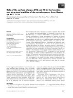
Figure 1-1. The process of autophagy
Autophagy is initiated at a perivacuolar site termed the pre-autophagosomal structure (PAS) by the
formation of an isolation membrane which expands and non-selectively engulfs cytosolic
components in the process. Fusion of the two leading edges of the isolation membrane results in
the formation of a double-membrane vesicle termed the autophagosome. Fusion between the
external membrane of the autophagosome and the lysosome results in the formation of the
autolysosome. The internal vesicle of the autophagosome, or autophagic body, and its contents are
subsequently degraded by the autolysosome and recycled by the cell. Adapted from (Mizushima,
2005).
Chapter 1 – Introduction
Marianne Cowan, 2013 Chapter 1 - Introduction
19
I am particularly interested in the mechanisms that underlie membrane fusion and
during the course of this project I became interested in the generation of the
isolation membrane and subsequent formation of the autophagosome. Evidence
suggests that expansion of the isolation membrane is followed by fusion of the
leading edges to form an autophagosome (Geng & Klionsky, 2010; Geng et al.,
2010; van der Vaart & Reggiori, 2010). The molecular fusion machinery involved in
the generation and subsequent formation of autophagosomes remain unknown
however a number of key players are thought to be involved during these early
stages including soluble N-ethylmalemide (NEM) sensitive factor (NSF)
attachment protein receptor (SNARE) proteins (section 1.2) and tethering
complexes (section 1.4).
1.1 Autophagy
1.1.1 Identification of autophagy
Autophagosomes were initially described in the newborn mouse kidney as being
“large bodies that represent vacuoles which have accumulated a high
concentration of amorphous material” and “that sometimes contain… altered
mitochondria”(Clark, 1957). Cytoplasmic granules were observed to decrease in
abundance (within a week postnatally) as cells differentiated. This observation
corresponds to recent data describing a homeostatic role for autophagy during the
early stages of development (Kuma et al., 2004; Saitoh et al., 2009; Sato & Sato,
2013). In 1962, electron microscopy data obtained by Ashford and Porter
demonstrated a glucagon-mediated increase in the lysosomal content of cells
examined from perfused rat livers (Ashford & Porter, 1962). It was reported that
these so called ‘lysosomes’ preferentially engulfed mitochondria. Other identifiable
content within these lysosomes included small vesicles and endoplasmic reticulum
(ER). The term ‘autophagy’ was subsequently coined in 1963 by de Duve to
describe novel double-membrane vesicles related to lysosomes that contain parts
of the cytosolic content including organelles in varying degrees of structural decay
(Clark, 1957; Ashford & Porter, 1962; De Duve, 1963; De Duve & Wattiaux, 1966).
The sequestering vesicles involved were termed autophagosomes; the biogenesis
of these structures remain controversial.
Marianne Cowan, 2013 Chapter 1 - Introduction
20
Since the term ‘autophagy’ was introduced, the process of autophagy has been
shown to be up-regulated in hepatic cells of starved animals (Novikoff et al., 1964)
and that the size of hepatic lysosomes increase as a result of glucagon
administration (Deter & De Duve, 1967). Using a quantitative morphological
approach Deter and colleagues confirmed this observed increase in autophagy to
be glucagon-mediated.
1.1.2 Functional significance of autophagy
Autophagy is an evolutionary conserved and adaptive catabolic process that plays
a central role in maintaining intracellular homeostasis and thereby cellular health.
The term ‘autophagy’ directly translates to ‘self-eating’ and it is a major route for
lysosomal/vacuolar degradation in eukaryotes (Reggiori & Klionsky, 2002;
Yorimitsu & Klionsky, 2005b; Yang & Klionsky, 2010).
Autophagy is a ubiquitous degradative process that occurs at a basal level and
can be rapidly up-regulated in response to cellular stress. For instance, nutrient
deprivation is the most common trigger of autophagy induction (section 1.1.6) and
in yeast nitrogen starvation represents the most potent stimulus of this pathway
(Takeshige et al., 1992). Basal levels of autophagy play an important role in
constitutive turnover of cytosolic components. Up-regulation of this process is
important in providing amino acids derived from degraded proteins and/or
organelles which in turn are utilised to provide cells with the necessary chemical
energy that is required for cellular maintenance and growth (Mizushima, 2005).
Although recent evidence suggest a link between autophagy and ubiquitin-
mediated degradation via the proteasome (Zhao et al., 2007), these two processes
are functionally distinct. Autophagy shares some functional overlap with the yeast
biosynthetic pathway known as the cytoplasm-to-vacuole targeting (Cvt) pathway
(Klionsky et al., 1992; Scott et al., 1996; Hutchins & Klionsky, 2001). The Cvt
pathway is unique to yeast and both autophagy and the Cvt pathway coexist is
yeast (section 1.1.3) (Klionsky, 2005).
Marianne Cowan, 2013 Chapter 1 - Introduction
21
1.1.3 Autophagy versus the cytosol-to-vacuole targeting pathway
Significant breakthrough in our understanding of autophagy came from genetic
screens in yeast, such as Saccharomyces cerevisiae (S.cerevisiae) (Thumm et al.,
1994; Harding et al., 1995). Autophagy and the yeast Cvt pathway are
morphologically similar thus the latter is considered to be an autophagy-related
pathway (Baba et al., 1997). It was not until the identification of the ATG genes in
yeast (Matsuura et al., 1997) and subsequent molecular analysis of autophagy in
higher eukaryotes (Mizushima et al., 1998) that these two pathways were shown
to share some common molecular machinery that is involved in the formation of
the autophagosome (Harding et al., 1996; Scott et al., 1996; Baba et al., 1997).
This subset of ‘core’ Atg proteins all function during the early phases of
autophagosome formation and include the Atg1-Atg13-Atg17 kinase complex
(Scott et al., 2000), the class III phosphatidylinositol 3-kinase (PtdIns3K) complex I
(Petiot et al., 2000; Kihara et al., 2001), the Atg8 (Kirisako et al., 1999) and Atg12
(Mizushima et al., 1998) ubiquitin-like conjugation systems and the integral
membrane protein Atg9 (Noda et al., 2000). In addition to these core Atg proteins,
autophagy- and Cvt-specific proteins have also been identified (Kawamata et al.,
2008).
Despite sharing similar morphological features, important differences exist
between autophagy and the Cvt pathway. The Cvt pathway is a constitutively
active biosynthetic pathway that serves to selectively sequester and deliver
specific enzymes, such as aminopeptidase I (Klionsky et al., 1992) and α-
mannosidase (Yoshihisa & Anraku, 1990), from the cytosol to the vacuole; in
contrast, autophagy is an inducible degradative pathway that terminates in the
lysosomal/vacuolar compartment (Yang & Klionsky, 2010). Transport vesicle
formation is a key regulatory step of the Cvt and autophagic pathways and the pre-
autophagosomal structure (PAS) represents the site for vesicle formation (Suzuki
et al., 2001; Kim et al., 2002). However, the diameter of the sequestering vesicles
involved differs; in the Cvt pathway, the diameter of the vesicle measures
approximately 140-160 nanometers (nm) (Kim et al., 2002) compared to 400-900
nm for the autophagosome (Takeshige et al., 1992). This difference in size reflects
the ability of the autophagosome to adjust its size appropriately in order to
accommodate its cargo.
Marianne Cowan, 2013 Chapter 1 - Introduction
22
1.1.4 The process of autophagy
In yeast, autophagy is initiated by nucleation of the isolation membrane at a
perivacuolar site termed the PAS (Figure 1-1) (Noda et al., 2000; Suzuki et al.,
2001; Kim et al., 2002). The PAS was originally identified based on observations
using fluorescence microscopy that core Atg components, including Atg1, Atg8
and Atg9, exhibit perivacuolar punctate structures that co-localise with
aminopeptidase I (section 1.1.3) under autophagy inducing conditions. The PAS
therefore defines the focal point for the assembly of Atg proteins which are
recruited in a hierarchical fashion during the early stages of autophagy.
The hierarchical relationship between the core Atg proteins has been determined
by systematic synthetic disruption of each ATG gene followed by morphometric
analysis (Suzuki et al., 2007). This analysis revealed that Atg17, which forms a
complex with Atg29 and Atg31 (Kabeya et al., 2007; Kawamata et al., 2008;
Kabeya et al., 2009), is required for the recruitment of all downstream Atg proteins.
Specifically, the PAS localisation of Atg17 is unaffected in core atg mutant strains;
in contrast, the PAS localisation of the remaining core Atg proteins is impaired in
atg17 (Suzuki et al., 2007). The PAS localisation of the Atg17-Atg29-Atg31
complex and its subsequent binding to Atg11 via Atg17 (Yorimitsu & Klionsky,
2005a) is regulated by phosphorylation of Atg29 (Mao et al., 2013). Binding
between Atg11 and the Atg17-Atg29-Atg31 complex is required for recruiting Atg1-
Atg13 (refer to section 1.1.6) to the PAS. Yeast two-hybrid analyses and co-
immunoprecipitation experiments have demonstrated that the recruitment of Atg1-
Atg13 to the PAS is mediated by a direct interaction between Atg17 and Atg13
(Kabeya et al., 2005). Furthermore, complex formation between Atg17-Atg29-
Atg31 and Atg1-Atg13 is required for Atg1 kinase activity and thereby autophagy
(Kamada et al., 2000; Kabeya et al., 2005). Downstream Atg proteins are
subsequently recruited in the following order: the integral membrane protein Atg9
is recruited to the PAS via direct association with Atg11 (He et al., 2006), which
plays a role in linking cargo to the vesicle-forming machinery at the PAS, possibly
via its coiled-coil tethering actions (Yorimitsu & Klionsky, 2005a; Lipatova et al.,
2012). In turn, recruitment of the autophagy-specific PtdIns(3)K complex 1,
composed of Vps34, Vps15, Atg6 and At14, to the PAS is mediated by direct
association between Atg13 and Atg14 (Jao et al., 2013). The ubiquitin ligase-like
system composed of Atg12-Atg5-Atg16 localises to the developing
Marianne Cowan, 2013 Chapter 1 - Introduction
23
autophagosome where it facilitates lipidation and correct subcellular localisation of
Atg8 (Mizushima et al., 1998; Mizushima et al., 1999; Hanada et al., 2007). Atg8
functions downstream from Atg12-Atg5-Atg16 and the PtdIns(3)K complex 1 and
is recruited to the PAS via an Atg9-dependent mechanism (Suzuki et al., 2001;
Suzuki et al., 2007). Expression of Atg8 is upregulated in response to autophagy
induction and levels of Atg8 directly correlate with autophagosome size (Xie et al.,
2008).
To date, 33 ATG genes have been identified in the yeast model system
S.cerevisiae, which is extensively used for studying autophagy (Kanki et al., 2009;
Okamoto et al., 2009). Homologs of the yeast ATG genes exist in other
eukaryotes, including mammals (Reggiori & Klionsky, 2002). The corresponding
gene products are often orthologs that perform similar functions and their
hierarchical relationship is consistent with that of yeast [reviewed in (Suzuki &
Ohsumi, 2010)]. Emerging evidence suggests that the previously unidentified
mammalian PAS equivalent may also exist in mammals. The double FYVE
domain-containing protein 1 (DFCP1) is a novel phospholipid binding protein that
translocates to a sub-domain of the ER, termed the omegaosome, under
autophagy-inducing conditions. Omegasomes partially co-localise with the
autophagosomal marker green fluorescent protein microtubule-associated protein
1 light chain 3 (GFP-LC3) as well as Vps34-containing vesicles under these same
conditions (Axe et al., 2008; Itakura & Mizushima, 2010). Three-dimensional
electron tomography has confirmed a physical connection between omegasomes
and the isolation membrane complex. This is suggestive of a role for the ER in
autophagosome formation in mammalian cells (Yla-Anttila et al., 2009).
Following the organisation of the vesicle-formation complex at the PAS, the
isolation membrane sequesters various cytosolic components within its boundaries
and expands sufficiently prior to vesicle completion to accommodate its cargo. The
source from which the membranes are acquired and which are required for the
expansion of the isolation membrane remain controversial. Evidence to date have
supported a role for the Golgi (Geng & Klionsky, 2010; van der Vaart & Reggiori,
2010), ER (Young et al., 2006), mitochondria (Hailey et al., 2010) and plasma
membrane (Ravikumar et al., 2010) in the expansion of the isolation membrane.
Recent progress in this field lean towards a role for post-ER Golgi compartments
in the formation of the isolation membrane in yeast. Atg9, which is an integral
Marianne Cowan, 2013 Chapter 1 - Introduction
24
membrane protein (Noda et al., 2000), localises to the Golgi apparatus and late
endosome (Young et al., 2006). Under nutrient replete conditions, Atg9 cycles
between the Golgi apparatus and late endosomes however under nutrient
starvation conditions, and when autophagy is induced, Atg9 relocalise to a
peripheral punctate compartment that is within close proximity of the vacuole and
which is consistent with the PAS (Young et al., 2006; Mari et al., 2010). Based on
these observations it has been proposed that Atg9 sources pre-existing
membranes from the Golgi apparatus and late endosomes and subsequently
transports these membranes to the PAS under autophagy inducing conditions.
Acquisition of these Golgi and late endosome derived membranes results in
expansion of the isolation membrane. This is a necessary step in the elongation of
the isolation membrane and therefore the formation of autophagosomes.
Furthermore, autophagosomes exhibit many of the properties which are likely
derived from an endocytic compartment including enrichment in
phosphatidylinositol 3-phosphate [PtdIns(3)P] (Obara et al., 2008).
The target-SNARE Tlg2 (t-SNARE of the late Golgi compartment protein 2), its SM
protein Vps45 and the COG complex regulate membrane traffic within the Golgi
and endosomal systems (Abeliovich et al., 1998; Holthuis et al., 1998a;
VanRheenen et al., 1998; Whyte & Munro, 2001). Consistent with a role for post-
ER Golgi compartments in autophagosome formation, the PAS localisation of Atg9
is reduced and redistributed throughout the cytosol in both cog (Yen et al., 2010)
and tlg2 (Ohashi & Munro, 2010; Nair et al., 2011) deficient yeast. Atg9 cycles
between peripheral structures and the PAS and its retrieval from the PAS is
dependent on Atg1 (Reggiori et al., 2004). An epistasis assay that relies on the
atg1∆ phenotype has been employed in recent years to investigate anterograde
transport of Atg9 to the PAS. Yen and colleagues demonstrated that Atg9-GFP
localises to multiple puncta in an atg1∆cog1∆ strain under autophagy inducing
conditions (Yen et al., 2010). This observation is indicative of impaired
anterograde movement of Atg9 to the PAS thereby implicating a role for the COG
complex in Atg9 trafficking. Similarly, the tlg2∆atg24∆ mutant combination exhibits
a strong autophagy deficient phenotype as defined by the GFP-Atg8 processing
assay and in combination with atg1∆ results in inhibition of Atg9 accumulation at
the PAS (Ohashi & Munro, 2010). In a separate study Nair and colleagues
demonstrated that the frequency of colocalisation between Atg9-GFP and red
fluorescent protein (RFP)-aminopeptidase 1, a marker for the PAS, was reduced