Phương pháp phổ khối lượng
Bạn đang xem bản rút gọn của tài liệu. Xem và tải ngay bản đầy đủ của tài liệu tại đây (1.38 MB, 34 trang )
Phương pháp phổ khối lượng
Bách khoa toàn thư mở Wikipedia
Bước tới: menu, tìm kiếm
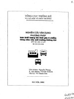
Mô hình cơ bản của một khối phổ kế.
Phương pháp phổ khối là một kĩ thuật dùng để đo đạc tỉ lệ khối lượng-trên-điện tích của
ion; dùng thiết bị chuyên dụng là khối phổ kế. Kĩ thuật này có nhiều ứng dụng, bao gồm:
• Xác định các hợp chất chưa biết bằng cách dựa vào khối lượng của phân tử hợp chất hay
từng phần tách riêng của nó
• Xác định kết cấu chất đồng vị của các thành phần trong hợp chất
• Xác định cấu trúc của một hợp chất bằng cách quan sát từng phần tách riêng của nó
• Định lượng lượng hợp chất trong một mẫu dùng các phương pháp khác (phương pháp phổ
khối vốn không phải là định lượng)
• Nghiên cứu cơ sở của hóa học ion thể khí (ngành hóa học về ion và chất trung tính trong
chân không)
• Xác định các thuộc tính vật lí, hóa học hay ngay cả sinh học của hợp chất với nhiều hướng
tiếp cận khác nhau.
Một khối phổ kế là một thiết bị dùng cho phương pháp phổ khối, cho ra phổ khối lượng của
một mẫu để tìm ra thành phần của nó. Có thể ion hóa mẫu và tách các ion của nó với các khối lượng
khác nhau và lưu lại thông tin dựa vào việc đo đạc cường độ dòng ion. Một khối phổ kế thông
thường gồm 3 phần: phần nguồn ion, phần phân tích khối lượng, và phần đo đạc.
Mục lục
[ẩn]
• 1 Ví dụ về cách hoạt động
• 2 Ứng dụng sinh học
o 2.1 Khối phổ của protein
o 2.2 Protein và các phân mảnh peptit
o 2.3 Xác định Protein
• 3 Xem thêm
• 4 Liên kết ngoài
• 5 Tham khảo
[sửa] Ví dụ về cách hoạt động
Các hóa chất khác nhau thì có khối lượng phân tử khác nhau. Dựa vào đó, khối phổ kế sẽ xác định
chất hóa học nào có nằm trong mẫu. Ví dụ, muối NaCl hấp thụ năng lượng (năng lượng hấp thụ tùy
theo nguồn ion, ví dụ MALDI năng lượng là tia laser) tách ra thành các phân tử tích điện, gọi là
ion), trong giai đoạn đầu của phương pháp phổ khối. Các ion Na
+
, Cl
-
có trọng lượng nguyên tử
khác biệt. Do chúng tích điện, nghĩa là đường đi của chúng có thể được điều khiển bằng điện trường
hoặc từ trường. Các ion được đưa vào buồng gia tốc và đi qua một khe vào miếng kim loại. Một từ
trường được đưa vào buồng đó. Từ trường sẽ tác động vào mỗi ion với cùng một lực và làm trệch
hướng chúng về phía đầu đo. Ion nhẹ hơn sẽ bị lệnh nhiều hơn ion nặng vì theo định luật chuyển
động của Newton gia tốc tỉ lệ nghịch với khối lượng của phân tử. Đầu đo sẽ xác định xem ion bị
lệnh bao nhiêu, và từ giá trị đo này, tỉ lệ khối lượng-trên-điện tích của ion có thể được tính toán. Từ
đó, có thể xác đinh được thành phần hóa học của một mẫu gốc. Trên thực tế thì hai ion Na
+
và Cl
-
sẽ
không được đo trong cùng một lần, vì các máy đo chỉ có thể nhận ra ion điện tích dương hoặc điện
tích âm nên nếu máy khối phổ kế được điều chỉnh để đo các ion điện tích dương thì chỉ có ion Na
+
là được nhận ra bởi máy. .Một trong những tính năng lớn của khối phổ lượng là có thể tìm thấy cấu
tạo không gian của phân tử ví dụ phân tử C
7
H
14
O
2
có thể là acid hoặc ester Và khả năng phát hiện
ra hợp chất với độ nhậy cực cao từ 10
-6
dến 10
-12
gram. Dưới đây là một khối phổ (electrospray)của
phân tử Kaempferol-rhamnose-rhamnose-glucose(m/z 741) trong loại cỏ thaliana, phân tích với
5.10
-6
L (nếu dùng máy MALDI thì chỉ cần 0,5.10
-6
L).
[sửa] Ứng dụng sinh học
[sửa] Khối phổ của protein
[sửa] Protein và các phân mảnh peptit
Protein mà các nhà nghiên cứu sinh học quan tâm thường là sự kết hợp phức tạp của nhiều protein
và phân tử khác nhau, cùng tồn tại trong một môi trường sinh học. Điều này đặt ra hai vấn đề chính.
Thứ nhất, hai kĩ thuật ion hóa dùng cho các phân tử lớn chỉ làm việc tốt khi mà hỗn hợp từ các
thành phần có cấu tạo gần giống, trong khi trong các mẫu sinh học, các protein khác nhau thường là
có lượng khác biệt nhau lớn. Nếu hỗn hợp được ion hóa dùng phương pháp phun ion hay MALDI,
thì những protein dạng mà dư thừa nhiều có xu hướng giảm tín hiệu so với những cái ít dư thừa
hơn. Vấn đề thứ hai, quang phổ khối từ hỗn hợp phức tạp là rất khó để nghiên cứu do có quá nhiều
thành phần phức hợp. Đó là vì với tác động của enzym, một protein tạo ra hàng loạt sản phẩm
peptit.
Để giải quyết vấn đề này, hai phương pháp được sử dụng rộng rãi để phân mảnh protein, hay các
sản phẩm peptit từ sự tác động của enzym. Phương pháp đầu tiên sẽ phân mảnh toàn bộ protein và
được gọi là điện chuyển gel hai chiều (2-DE: two-dimensional gel electrophoresis). Phương pháp
thứ hai, ghi sắc lỏng hiệu suất cao (HPLC) được dùng với các phân mảnh peptit sau khi protein
phân tách bởi tác động của enzym. Trong một số tình huống, có thể cần phải kết hợp cả hai phương
pháp.
Các vết gel được xác định trên 2D Gel thường là thuộc về một protein. Nếu cần biết định danh của
protein đó, thì có thể xem xét vết gel đó. Khối peptit kết quả từ tác động của enzym lên protein có
thể được xác định bằng khối phổ dùng lấy dấu khối peptit. Nếu thông tin này không cho phép xác
định danh tính của protein một cách chính xác, các peptit của nó có thể xem là thuộc về đo phổ khối
tandem.
Việc xác định đặc tính của hỗn hợp protein dùng HPLC/MS còn được gọi là shotgun proteomics và
mudpit. Một hỗn hợp là kết quả của sự tác động của enzym lên hỗn hợp protein sẽ được phân mảnh
theo một hay hai bước bằng ghi sắc lỏng. Chất tách rửa từ giai đoạn ghi sắc có thể hoặc là trực tiếp
đưa vào máy đo phổ khối thông qua ion hóa phun điện tử (ESI), hay tách ra thành một loạt các vết
nhỏ để sử dụng sau này trong phân tích khối bằng MALDI.
[sửa] Xác định Protein
Có 2 cách chính trong khối phổ để xác định protein.
• Lấy dấu khối peptit (PMF) dùng khối của các peptit đã phân giải làm đầu vào để tìm kiếm
trong CSDL của các khối đã biết trước từ danh sách các protein đã biết. Nếu một chuỗi
protein trong danh sách tham khảo trùng khớp với giá trị thử nghiệm thì có lí do để tin rằng
protein đó có tồn tại trong mẫu gốc.
• Tandem MS đang trở thành một phương pháp thử nghiệm phổ biến để xác định protein.
Phân ly do va chạm (CID) được dùng trong các ứng dụng chính để khởi tạo một tập các
phân mảnh từ một ion peptit cụ thể. Quá trình phân tách chủ yếu dựa vào các chế phẩm phân
tách để bẻ gãy liên kết peptit. Vì sự đơn giản của việc phân tách này, nó có thể dùng khối
của các phân mảnh quan sát được để so trùng CSDL của các khối đã biết với một hay nhiều
chuỗi peptit.
An Introduction to Mass Spectrometry
(giới thiệu về Khối phổ)
Dr Alison E. Ashcroft,
Mass Spectrometry Facility Manager,
Astbury Centre for Structural Molecular Biology,
Astbury Building ,
The University of Leeds.
CONTENTS Nội dung
1. What is mass spectrometry (MS)? What Information does mass spectrometry provide?
Khối phổ (MS) là gì? Thông tin không gì khối phổ cung cấp
2. Where are mass spectrometers used? Trường hợp được quang phổ kế khối lượng được sử
dụng
3. How can mass spectrometry help biochemists? Làm thế nào khối phổ có thể giúp các nhà
sinh hóa học
4. How does a mass spectrometer work? Làm thế nào một công việc phổ khối lượng
5. Introduction Giới thiệu
1. Sample introduction Mẫu giới thiệu
2. Methods of sample ionisation Phương pháp ion hoá mẫu
3. Analysis and separation of sample ions Phân tích và tách các ion mẫu
4. Detection and recording of sample ions Phát hiện và thu âm của các ion mẫu
6. Electrospray ionisation Electrospray ion hóa
1. Electrospray ionisation Electrospray ion hóa
2. Nanospray ionisation Nanospray ion hóa
3. Data processing Xử lý dữ liệu
7. Matrix assisted laser desorption ionisation Ma trận giúp giải hấp laser ion hóa
8. Positive or negative ionisation? Tích cực hay tiêu cực ion hoá
9. Tandem mass spectrometry (MS-MS): Structural and sequence information from
mass spectrometry Tandem khối phổ (MS-MS): Kết cấu và trình tự thông tin từ các phép đo
phổ khối lượng
1. Tandem mass spectrometry Tandem khối phổ
2. Tandem mass spectrometry analyses Tandem khối phổ phân tích
3. Peptide sequencing by tandem mass spectrometry Peptide trình tự do đo phổ khối
tandem
4. Oligonucleotide sequencing by tandem mass spectrometry Oligonucleotide trình tự do đo
phổ khối tandem
10. Background reading Nền đọc
1. What is mass spectrometry (MS)? What information does mass spectrometry
provide?
Mass spectrometry is an analytical tool used for measuring the molecular mass of a sample.
Khối phổ là một công cụ phân tích được sử dụng để đo khối lượng phân tử của một mẫu
For large samples such as biomolecules, molecular masses can be measured to within an
accuracy of 0.01% of the total molecular mass of the sample i.e. within a 4 Daltons (Da) or atomic
mass units (amu) error for a sample of 40,000 Da. This is sufficient to allow minor mass changes to
be detected, e.g. the substitution of one amino acid for another, or a post-translational modification.
Đối với những mẫu lớn như phân tử sinh học, khối lượng phân tử có thể đo được để trong độ chính
xác 0,01% tổng khối lượng phân tử của các ví dụ mẫu trong vòng có 4 Dalton (Da: đơn vị cacbon), đơn vị
khối lượng nguyên tử (amu) lỗi cho một mẫu của 40.000 Da. Điều này là đủ để cho phép thay đổi khối lượng
nhỏ được phát hiện, ví dụ: sự thay thế của axit amin một chổ khác, hoặc sửa đổi một bài viết-tịnh.
For small organic molecules the molecular mass can be measured to within an accuracy of
5 ppm or less, which is often sufficient to confirm the molecular formula of a compound, and is
also a standard requirement for publication in a chemical journal.
Đối với các phân tử hữu cơ nhỏ khối lượng phân tử có thể đo được để trong một độ chính xác của 5
ppm hoặc ít hơn, mà thường là đủ để xác nhận công thức phân tử của hợp chất là một, và cũng là một yêu
cầu tiêu chuẩn để công bố trên một tạp chí hóa học
Structural information can be generated using certain types of mass spectrometers, usually
those with multiple analysers which are known as tandem mass spectrometers. This is achieved
by fragmenting the sample inside the instrument and analysing the products generated. This
procedure is useful for the structural elucidation of organic compounds and for peptide or
oligonucleotide sequencing.
Kết cấu thông tin có thể được tạo ra bằng cách sử dụng một số loại quang phổ kế khối lượng,
thường là những người có nhiều phân tích được biết đến như quang phổ kế khối song song. Điều này đạt
được bằng cách phân mảnh các mẫu trong các nhạc cụ và phân tích các sản phẩm tạo ra. Thủ tục này rất
hữu ích cho sự giải thích cấu trúc của các hợp chất hữu cơ và cho peptide hoặc trình tự oligonucleotide
2. Where are mass spectrometers used?
Mass spectrometers are used in industry and academia for both routine and research purposes. The
following list is just a brief summary of the major mass spectrometric applications: Phổ MS được sử
dụng quang phổ kế trong ngành công nghiệp và hàn lâm cho cả hai mục đích thông thường và nghiên cứu.
Danh sách dưới đây chỉ là một bản tóm tắt ngắn gọn về các ứng dụng phổ khối lượng lớn
• Biotechnology: the analysis of proteins, peptides, oligonucleotides.
Công nghệ sinh học: các phân tích của các protein, peptide, oligonucleotides
• Pharmaceutical: drug discovery, combinatorial chemistry, pharmacokinetics, drug
metabolism
Dược phẩm: thuốc phát hiện, tổ hợp hóa học, dược động học, sự trao đổi chất ma túy
• Clinical: neonatal screening, haemoglobin analysis, drug testing
Lâm sàng: trẻ sơ sinh sàng lọc, phân tích huyết cầu tố, kiểm nghiệm thuốc
• Environmental: PAHs, PCBs, water quality, food contamination
Môi trường: PAHs, PCBs, chất lượng nước, thức ăn ô nhiễm
• Geological: oil composition
Địa chất: thành phần dầu
3. How can mass spectrometry help biochemists?
• Accurate molecular weight measurements: Trọng lượng phân tử chính xác các phép đo
sample confirmation, to determine the purity of a sample, to verify amino acid substitutions,
to detect post-translational modifications, to calculate the number of disulphide bridges
mẫu xác nhận, để xác định độ tinh khiết của một mẫu, để xác minh sự thay thế acid amin, để phát hiện
thay đổi sau tịnh, để tính số cầu disulphide
• Reaction monitoring: Phản ứng theo dõi
to monitor enzyme reactions, chemical modification, protein digestion
theo dõi các phản ứng enzyme, biến đổi hóa học, tiêu hóa protein
• Amino acid sequencing: Amino acid trình tự
sequence confirmation, de novo characterisation of peptides, identification of proteins by
database searching with a sequence "tag" from a proteolytic fragment
trình tự xác nhận, de novo mô tả đặc điểm của peptide, xác định các protein của cơ sở dữ liệu tìm kiếm
với một "tag" trình tự từ một đoạn phân giải protein
• Oligonucleotide sequencing: Oligonucleotide trình tự
the characterisation or quality control of oligonucleotides
kiểm soát đặc tính hoặc chất lượng của oligonucleotides
• Protein structure: Protein cấu trúc
protein folding monitored by H/D exchange, protein-ligand complex formation under
physiological conditions, macromolecular structure determination
protein gấp theo dõi bởi H trao đổi D /, protein-phối tử hình phức tạp trong điều kiện sinh lý, xác định
cấu trúc phân tử
4. How does a mass spectrometer work? Làm thế nào một công việc phổ khối lượng
4.1 Introduction Giới thiệu
Mass spectrometers can be divided into three fundamental parts, namely the ionisation
source , the analyser , and the detector. Lễ quang phổ kế có thể được chia thành ba phần cơ bản, cụ
thể là nguồn ion hóa, phân tích, và phát hiện này
The sample has to be introduced into the ionisation source of the instrument. Once inside the
ionisation source, the sample molecules are ionised, because ions are easier to manipulate than
neutral molecules. These ions are extracted into the analyser region of the mass spectrometer where
they are separated according to their mass (m) -to-charge (z) ratios (m/z) . The separated ions are
detected and this signal sent to a data system where the m/z ratios are stored together with their
relative abundance for presentation in the format of a m/z spectrum. Mẫu đã được giới thiệu vào
nguồn ion hóa của nhạc cụ. Một khi bên trong các nguồn ion hóa, các phân tử mẫu được ion hóa, bởi vì các
ion dễ thao tác hơn so với các phân tử trung tính. Những ion này được tách ra thành các khu vực phân tích
của pháp phổ khối lượng, nơi họ được phân theo khối lượng của chúng (m)-phí-to (z) tỷ lệ (m / z). Các ion
được phát hiện và tách tín hiệu này được gửi đến một hệ thống dữ liệu mà m / z tỷ số được lưu trữ cùng với
sự phong phú tương đối của họ trình bày trong các định dạng của sáng / z phổ
The analyser and detector of the mass spectrometer, and often the ionisation source too, are
maintained under high vacuum to give the ions a reasonable chance of travelling from one end of
the instrument to the other without any hindrance from air molecules. The entire operation of the
mass spectrometer, and often the sample introduction process also, is under complete data system
control on modern mass spectrometers. Các phân tích và phát hiện của pháp phổ khối lượng, và thường
là nguồn ion hóa cũng vậy, được duy trì dưới chân không cao để cung cấp cho các ion có cơ hội đi du lịch
hợp lý từ một đầu của thiết bị để các khác mà không có bất kỳ trở ngại từ các phân tử không khí. Toàn bộ
hoạt động của máy quang phổ khối lượng, và thường giới thiệu mẫu quá trình cũng có, đang được điều
khiển hoàn toàn hệ thống dữ liệu về quang phổ kế khối hiện đại
Simplified schematic of a mass spectrometer
4.2 Sample introduction mẫu giới thiệu
The method of sample introduction to the ionisation source often depends on the ionisation
method being used, as well as the type and complexity of the sample. Phương thức giới thiệu mẫu cho
các nguồn ion hóa thường phụ thuộc vào phương pháp ion hóa được sử dụng, cũng như loại và sự phức
tạp của mẫu
The sample can be inserted directly into the ionisation source, or can undergo some type of
chromatography en route to the ionisation source. This latter method of sample introduction usually
involves the mass spectrometer being coupled directly to a high pressure liquid chromatography
(HPLC), gas chromatography (GC) or capillary electrophoresis (CE) separation column, and hence
the sample is separated into a series of components which then enter the mass spectrometer
sequentially for individual analysis. Mẫu này có thể được chèn trực tiếp vào nguồn ion hóa, hoặc có thể
trải qua một số loại sắc ký trên đường đến các nguồn ion hóa. Phương pháp thứ hai giới thiệu mẫu thường
liên quan đến pháp phổ khối lượng được kết trực tiếp đến một sắc ký lỏng cao áp (HPLC), sắc ký khí (GC)
hoặc điện mao dẫn (CE) tách cột, và do đó mẫu được tách thành một loạt các thành phần đó sau đó nhập
vào các máy phổ khối tuần tự để phân tích cá nhân
4.3 Methods of sample ionisation Các phương pháp ion hoá mẫu
Many ionisation methods are available and each has its own advantages and disadvantages
("Ionization Methods in Organic Mass Spectrometry", Alison E. Ashcroft, The Royal Society
of Chemistry, UK, 1997; and references cited therein). Nhiều phương pháp ion hoá có sẵn và từng có
những lợi thế riêng của mình và bất lợi ("Đầu báo phương pháp hữu khối phổ", Alison E. Ashcroft, Hội Hóa
học Hoàng gia, Vương quốc Anh năm 1997; và tài liệu tham khảo trích dẫn trong đó).
The ionisation method to be used should depend on the type of sample under investigation
and the mass spectrometer available. Các phương pháp ion hóa được sử dụng nên phụ thuộc vào loại
mẫu điều tra và phổ kế khối lượng sẵn có
Ionisation methods include the following: Ion hoá các phương pháp bao gồm
Atmospheric Pressure Chemical Ionisation (APCI) Áp suất khí quyển Hóa chất ion hoá (APCI)
Chemical Ionisation (CI) Hóa chất ion hoá (CI)
Electron Impact (EI) Tác động điện tử (EI)
Electrospray Ionisation (ESI) Electrospray ion hoá (ESI)
Fast Atom Bombardment (FAB) Atom nhanh Ném bom (FAB)
Field Desorption / Field Ionisation (FD/FI) Lĩnh vực giải hấp / trường ion hoá (FD / FI)
Matrix Assisted Laser Desorption Ionisation (MALDI) Ma trận Laser hỗ trợ giải hấp ion hoá
(MALDI)
Thermospray Ionisation (TSP) Thermospray ion hoá (TSP)
The ionisation methods used for the majority of biochemical analyses are Electrospray
Ionisation (ESI) and Matrix Assisted Laser Desorption Ionisation (MALDI) , and these are
described in more detail in Sections 5 and 6 respectively. Các phương pháp ion hóa được sử dụng cho
phần lớn các phân tích sinh hóa được Electrospray ion hoá (ESI) và Matrix Laser hỗ trợ giải hấp ion hoá
(MALDI), và đây là những mô tả chi tiết tại mục 5 và 6 tương ứng
With most ionisation methods there is the possibility of creating both positively and
negatively charged sample ions, depending on the proton affinity of the sample. Before embarking
on an analysis, the user must decide whether to detect the positively or negatively charged ions (see
section 7). Với phương pháp ion hóa nhất có khả năng tạo ra cả tích cực và tiêu cực ion mẫu tính phí, tùy
thuộc vào mối quan hệ proton của mẫu. Trước khi bắt tay vào phân tích, người sử dụng phải quyết định để
phát hiện các ion tích cực hay tiêu cực phí (xem phần 7).
4.4 Analysis and Separation of Sample Ions Phân tích và tách ion mẫu
The main function of the mass analyser is to separate , or resolve , the ions formed in the
ionisation source of the mass spectrometer according to their mass-to-charge (m/z) ratios. There
are a number of mass analysers currently available, the better known of which include quadrupoles
, time-of-flight (TOF) analysers, magnetic sectors , and both Fourier transform and quadrupole
ion traps. Chức năng chính của máy phân tích khối lượng là riêng biệt, hoặc giải quyết, các ion được hình
thành trong nguồn ion hóa của pháp phổ khối lượng theo khối lượng của mình phụ trách để (m / z) tỷ lệ. Có
một số phân tích khối lượng hiện có, thì tốt hơn được biết đến trong đó bao gồm quadrupoles, thời gian của
chuyến bay (tvà) phân tích, thành phần từ tính, và cả hai biến đổi Fourier và tứ cực bẫy ion
These mass analysers have different features, including the m/z range that can be covered,
the mass accuracy, and the achievable resolution. The compatibility of different analysers with
different ionisation methods varies. For example, all of the analysers listed above can be used in
conjunction with electrospray ionisation, whereas MALDI is not usually coupled to a quadrupole
analyser. Những phân tích khối lượng có tính năng khác nhau, bao gồm các m / z có thể phạm vi được bảo
hiểm, tính chính xác khối lượng, và độ phân giải đạt được. Sự phù hợp của phân tích khác nhau với các
phương pháp ion hoá khác nhau có khác nhau. Ví dụ, tất cả các phân tích nêu trên có thể được sử dụng kết
hợp với ion hoá electrospray, trong khi MALDI thường không đi đôi với một phân tích tứ cực
Tandem (MS-MS) mass spectrometers are instruments that have more than one analyser
and so can be used for structural and sequencing studies. Two, three and four analysers have all
been incorporated into commercially available tandem instruments, and the analysers do not
necessarily have to be of the same type, in which case the instrument is a hybrid one. More popular
tandem mass spectrometers include those of the quadrupole-quadrupole, magnetic sector-
quadrupole , and more recently, the quadrupole-time-of-flight geometries. Tandem (MS-MS)
quang phổ kế khối lượng là những công cụ mà có nhiều hơn một phân tích và do đó có thể được sử dụng
cho nghiên cứu cấu trúc và trình tự. Hai, ba và bốn phân tích có tất cả được kết hợp vào công cụ thương
mại song song, và các phân tích không nhất thiết phải cùng loại, trong trường hợp cụ này là một lai một.
Phổ biến hơn dù cùng quang phổ kế khối lượng bao gồm những người của tứ cực-tứ cực, từ khu vực kinh
tế-tứ cực, và gần đây, các tứ cực-thời gian-hình học-bay
4.5 Detection and recording of sample ions. phát hiện và ghi của các ion mẫu.
The detector monitors the ion current, amplifies it and the signal is then transmitted to the
data system where it is recorded in the form of mass spectra . The m/z values of the ions are
plotted against their intensities to show the number of components in the sample, the molecular
mass of each component, and the relative abundance of the various components in the sample.
Phát hiện này theo dõi các ion hiện nay, khuyếch đại và sau đó tín hiệu được truyền đến hệ thống dữ liệu
mà nó được ghi lại trong các hình thức phổ khối lượng. Các m / z các giá trị của các ion là các âm mưu
chống lại cường độ của họ để chỉ số lượng các thành phần trong mẫu, khối lượng phân tử của mỗi thành
phần, và sự phong phú tương đối của các thành phần khác nhau trong mẫu
The type of detector is supplied to suit the type of analyser; the more common ones are the
photomultiplier , the electron multiplier and the micro-channel plate detectors. Các loại máy phát
hiện được cung cấp cho phù hợp với các loại phân tích, những người phổ biến hơn là những, quang
electron nhân và các thiết bị dò tấm vi kênh
5. Electrospray ionisation Electrospray ion hóa
5.1 Electrospray ionisation Electrospray ion hóa
Electrospray Ionisation (ESI) is one of the Atmospheric Pressure Ionisation (API)
techniques and is well-suited to the analysis of polar molecules ranging from less than 100 Da to
more than 1,000,000 Da in molecular mass. Electrospray ion hoá (ESI) là một trong những áp lực không
khí ion hóa (API) kỹ thuật và rất phù hợp cho việc phân tích các phân tử phân cực khác nhau, từ nhỏ hơn
100 Da cho hơn 1.000.000 Da khối lượng phân tử
Standard electrospray ionisation source (Platform II)
Standard electrospray ion hoá nguồn (Platform II)
During standard electrospray ionisation (J. Fenn, J. Phys. Chem., 1984, 88, 4451), the
sample is dissolved in a polar, volatile solvent and pumped through a narrow, stainless steel
capillary (75 - 150 micrometers i.d.) at a flow rate of between 1 L/min and 1 mL/min. A high �
voltage of 3 or 4 kV is applied to the tip of the capillary, which is situated within the ionisation
source of the mass spectrometer, and as a consequence of this strong electric field, the sample
emerging from the tip is dispersed into an aerosol of highly charged droplets, a process that is aided
by a co-axially introduced nebulising gas flowing around the outside of the capillary. This gas,
usually nitrogen, helps to direct the spray emerging from the capillary tip towards the mass
spectrometer. The charged droplets diminish in size by solvent evaporation, assisted by a warm
flow of nitrogen known as the drying gas which passes across the front of the ionisation source.
Eventually charged sample ions, free from solvent, are released from the droplets, some of which
pass through a sampling cone or orifice into an intermediate vacuum region, and from there through
a small aperture into the analyser of the mass spectrometer, which is held under high vacuum. The
lens voltages are optimised individually for each sample.
Trong quá trình
ion hóa
electrospray
tiêu chuẩn
(
J.
Fenn
,
J.
Phys
Chem
,
năm 1984,
88,
4451
),
mẫu
được
hòa tan
trong
một
vùng cực
,
dễ bay hơi
dung môi
và
bơm
qua
một
thép
không gỉ
hẹp
,
mao dẫn
(
75-150
micromet
id
)
tại
tốc độ
dòng chảy
của
từ 1
L
/
phút
và
1
ml
/ phút
.
Một
điện áp
cao
của
3 hoặc
4
kV
được áp dụng cho
các
tip
của
mao mạch
,
mà
là
nằm
trong
nguồn
ion hóa
của
pháp phổ khối lượng
,
và
như
một
hệ quả
của
điện trường
mạnh
,
các
mẫu
đang nổi lên
từ đầu
được phân tán
vào
một
bình phun
các
giọt
rất
phí
,
một quá trình được
hỗ trợ bởi
một
đồng
trục
giới thiệu
nebulising
khí
chảy
xung quanh
bên
ngoài
của
các
mao mạch
.
Khí này
,
thường là
nitơ
,
giúp
chỉ đạo
phun
mới nổi
từ
đầu
mao mạch
đối với
các
máy phổ
khối
.
Những
giọt nước
làm giảm
tính
kích thước
bằng cách
bốc hơi
dung môi
,
sự hỗ trợ
của
một
dòng chảy
ấm áp
của
nitơ
được gọi
là
khí
khô
mà
đi
qua
mặt trước
của
các
nguồn
ion hóa
.
Cuối cùng
mẫu
ion
sạc
, không
dung môi
,
được
thả ra từ
các giọt nước
,
một số trong đó
đi
qua
một
hình nón
lấy mẫu
hoặc
lỗ
vào
một
vùng
chân không
trung
,
và
từ đó
thông qua
một
lỗ
nhỏ
vào
các
phân tích
của
pháp phổ khối
lượng
, được
tổ chức
theo
chân không
cao
.
Các
điện áp
ống kính
được tối ưu hóa
riêng biệt cho
mỗi mẫu
The electrospray ionisation process
Các quá trình ion hóa electrospray
5.2 Nanospray ionisationNanospray ion hóa
Nanospray ionisation (M. Wilm, M. Mann, Anal. Chem., 1996, 68, 1) is a low flow rate
version of electrospray ionisation. A small volume (1-4 microL) of the sample dissolved in a
suitable volatile solvent, at a concentration of ca. 1 - 10 pmol/microL, is transferred into a
miniature sample vial. A reasonably high voltage (ca. 700 - 2000 V) is applied to the specially
manufactured gold-plated vial resulting in sample ionisation and spraying. The flow rate of solute
and solvent using this procedure is very low, 30 - 1000 nL/min, and so not only is far less sample
consumed than with the standard electrospray ionisation technique, but also a small volume of
sample lasts for several minutes, thus enabling multiple experiments to be performed. A common
application of this technique is for a protein digest mixture to be analysed to generate a list of
molecular masses for the components present, and then each component to be analysed further by
tandem mass spectrometric (MS-MS) amino acid sequencing techniques (see Section 8).
Nanospray ion hoá (M. Wilm, M. Mann, vây hậu môn Chem , Năm 1996, 68, 1) là một dòng chảy
của phiên bản tốc độ thấp của ion hoá electrospray. Một khối lượng nhỏ (1-4 microL) của mẫu hoà tan trong
dung môi bay hơi phù hợp, ở nồng độ ca. 1-10 pmol / microL, được chuyển vào một lọ mẫu thu nhỏ. Một
điện áp cao, hợp lý (khoảng 700-2000 V) được áp dụng cho các lọ mạ vàng đặc biệt sản xuất dẫn đến ion
hóa mẫu và phun. Tốc độ dòng chảy của chất tan và dung môi bằng cách sử dụng thủ tục này là rất thấp, 3-
10 phút / nL, và do đó không chỉ là mẫu tiêu thụ ít hơn so với tiêu chuẩn kỹ thuật ion hoá electrospray, mà
còn một lượng nhỏ mẫu kéo dài trong vài phút tạo điều kiện cho nhiều thí nghiệm được thực hiện. Một ứng
dụng phổ biến của kỹ thuật này là dành cho tiêu hóa protein hỗn hợp được phân tích để tạo ra một danh
sách các khối lượng phân tử cho các thành phần hiện nay, và sau đó mỗi thành phần được phân tích thêm
theo khối lượng song song phổ (MS-MS) amino acid kỹ thuật lập trình tự (xem Phần 8)
ESI and nanospray ionisation are very sensitive analytical techniques but the sensitivity
deteriorates with the presence of non-volatile buffers and other additives, which should be avoided
as far as possible. ESI và ion hoá nanospray rất nhạy cảm với kỹ thuật phân tích nhưng độ nhạy càng thấp
sự hiện diện của các bộ đệm không dễ bay hơi và các phụ gia khác, mà nên tránh càng xa càng tốt
In positive ionisation mode, a trace of formic acid is often added to aid protonation of the
sample molecules; in negative ionisation mode a trace of ammonia solution or a volatile amine is
added to aid deprotonation of the sample molecules. Proteins and peptides are usually analysed
under positive ionisation conditions and saccharides and oligonucleotides under negative
ionisation conditions. In all cases, the m/z scale must be calibrated by analysing a standard sample
of a similar type to the sample being analysed (e.g. a protein calibrant for a protein sample), and
then applying a mass correction.Trong chế độ ion hóa tích cực, một dấu vết của acid formic thường được
thêm vào để hỗ trợ proton hóa của các phân tử mẫu, trong chế độ ion hóa tiêu cực một chút dung dịch
amoniac hoặc một amin dễ bay hơi được thêm vào để hỗ trợ khử proton của các phân tử mẫu. Protein và
peptide này thường được phân tích theo các điều kiện ion hóa tích cực và saccharides và oligonucleotides
ion hoá trong điều kiện tiêu cực. Trong mọi trường hợp, m / quy mô z phải được hiệu chuẩn bằng cách phân
tích một mẫu chuẩn của một loại tương tự như các mẫu được phân tích (ví dụ như một protein calibrant cho
một mẫu protein), và sau đó áp dụng một điều chỉnh hàng loạt
5.3 Data processing
ESI and nanospray ionisation generate the same type of spectral data for samples, and so
the data processing procedures are identical. ESI và nanospray ion hóa tạo ra cùng một loại dữ liệu
quang phổ của các mẫu, và do đó các thủ tục xử lý dữ liệu giống hệt nhau
In ESI, samples (M) with molecular masses up to ca. 1200 Da give rise to singly charged
molecular-related ions, usually protonated molecular ions of the formula (M+H)+ in positive
ionisation mode, and deprotonated molecular ions of the formula (M-H)- in negative ionisation
mode. Trong ESI, mẫu (M) với khối lượng phân tử lên đến ca. 1200 Đà làm phát sinh đơn lẻ ion phân tử
liên quan, thường là proton phân tử ion của công thức (M + H) + trong chế độ ion hóa tích cực, và các ion
phân tử deprotonated của công thức (MH) - trong chế độ ion hóa tiêu cực
An example of this type of sample analysis is shown in the m/z spectrum of the pentapeptide
leucine enkephalin, YGGFL. The molecular formula for this compound is C28H37N5O7 and the
calculated monoisotopic molecular weight is 555.2692 Da. Một ví dụ của loại phân tích mẫu được thể
hiện trong m / z phổ của leucine enkephalin pentapeptide, YGGFL. Công thức phân tử của hợp chất này là
C28H37N5O7 và tính toán trọng lượng phân tử monoisotopic là 555,2692 Da
The m/z spectrum shows dominant ions at m/z 556.1, which are consistent with the expected
protonated molecular ions, (M+H+). Protonated molecular ions are expected because the sample
was analysed under positive ionisation conditions. These m/z ions are singly charged, and so the
m/z value is consistent with the molecular mass, as the value of z (number of charges) equals 1.
Hence the measured molecular weight is deduced to be 555.1 Da, in good agreement with the
theoretical value. M Các / z quang phổ cho thấy ưu thế tại các ion m / z 556,1, đó là phù hợp với dự kiến
phân tử ion proton, (M + H +). Proton ion phân tử dự kiến sẽ vì mẫu được phân tích theo các điều kiện ion
hóa tích cực. Những m / z ion là tính đơn lẻ, và vì vậy m / z giá trị là phù hợp với khối lượng phân tử, là giá
trị của z (số loại phí) bằng 1. Do đó trọng lượng phân tử đo được suy ra là 555,1 Đa, trong hợp đồng tốt với
các giá trị lý thuyết
Positive ESI-MS m/z spectrum of leucine enkaphalin, YGGFL.
The m/z spectrum also shows other ions of lower intensity (ca. 25 % of the m/z 556.1 ions)
at m/z 557.2. These represent the molecule in which one 12C atom has been replaced by a 13C
atom, because carbon has a naturally occurring isotope one atomic mass unit (Da) higher. The
intensity of these isotopic ions relates to the relative abundance of the naturally occurring isotope
multiplied by the total number of carbon atoms in the molecule. Additionally the fact that the 13C
ions are one Da higher on the m/z scale than the 12C ions is an indication that z = 1, and hence the
sample ions are singly charged. If the sample ions had been doubly charged, then the m/z values
would only differ by 0.5 Da as z, the number of charges, would then be equal to 2. m Các / z quang
phổ cũng cho thấy các ion khác có cường độ thấp hơn (khoảng 25% các m / z 556,1 ion) tại m / z 557,2.
Những đại diện cho các phân tử trong đó một nguyên tử 12C đã được thay thế bởi một nguyên tử 13C, bởi
vì carbon có một đơn vị đồng vị tự nhiên xảy ra một khối lượng nguyên tử (Đà) cao hơn. Cường độ của các
ion đồng vị liên quan đến sự phong phú tương đối của các đồng vị tự nhiên nhân với tổng số nguyên tử
cacbon trong phân tử. Ngoài ra một thực tế là các ion 13C là một Đà cao hơn trên m / z quy mô hơn so với
các ion 12C là một dấu hiệu cho thấy z = 1, và do đó các ion mẫu được đơn phương tính phí. Nếu các ion
mẫu đã được gấp đôi tính, sau đó là m / z các giá trị chỉ có thể khác nhau từ 0,5 Đà là z, số phí, sau đó sẽ là
bằng 2
The m/z spectrum also contains ions at m/z 578.1, some 23 Da higher than the expected
molecular mass. These can be identified as the sodium adduct ions, (M+Na)+, and are quite
common in electrospray ionisation. Instead of the sample molecules being ionised by the addition
of a proton H+, some molecules have been ionised by the addition of a sodium cation Na+. Other
common adduct ions include K+ (+39) and NH4+ (+18) in positive ionisation mode and Cl- (+35)
in negative ionisation mode. Các m / z quang phổ cũng chứa các ion tại m / z 578,1, một số 23 Đà cao
hơn so với khối lượng phân tử dự kiến. Đây có thể được xác định là các ion natri adduct, (M + Na) +, và là
khá phổ biến trong ion hoá electrospray. Thay vì các phân tử bị ion hóa mẫu bằng cách cho thêm một proton
H +, một số phân tử đã được ion hóa bằng cách cho thêm một cation Na Na +. Ion khác adduct phổ biến
bao gồm K + (39) và NH4 + (18) trong chế độ ion hóa tích cực và-ion hoá Cl (35) trong chế độ tiêu cực
Electrospray ionisation is known as a "soft" ionisation method as the sample is ionised by
the addition or removal of a proton, with very little extra energy remaining to cause fragmentation
of the sample ions. ion hoá Electrospray được biết đến như một phương pháp "mềm" ion hóa như mẫu
được ion hóa bằng cách thêm hoặc loại bỏ các proton, với rất ít thêm năng lượng còn lại để gây ra sự phân
mảnh của các ion mẫu
Samples (M) with molecular weights greater than ca. 1200 Da give rise to multiply charged
molecular-related ions such as (M+nH)n+ in positive ionisation mode and (M-nH)n- in negative
ionisation mode. Proteins have many suitable sites for protonation as all of the backbone amide
nitrogen atoms could be protonated theoretically, as well as certain amino acid side chains such as
lysine and arginine which contain primary amine functionalities. Mẫu (M) với trọng lượng phân tử lớn
hơn ca. 1200 Đà làm tăng nhân ion phân tử liên quan như (M + nH) n + trong chế độ ion hóa tích cực và (M-
nH) n, trong chế độ ion hóa tiêu cực. Protein có nhiều trang web phù hợp với proton như tất cả các nguyên
tử nitơ amide xương sống có thể được proton về mặt lý thuyết, cũng như một số acid amin chuỗi phụ như
lysine và arginine có chứa chức năng amin đầu
An example of multiple charging, which is practically unique to electrospray ionisation, is
presented in the positive ionisation m/z spectrum of the protein hen egg white lysozyme. Một ví dụ
về tính phí nhiều, đó là thực tế duy nhất để ion hóa electrospray, được trình bày trong các m ion hóa tích
cực phổ z / của protein trứng gà trắng lysozyme
Positive ESI-MS m/z spectrum of the protien hen egg white lysozyme.
The sample was analysed in a solution of 1:1 (v/v) acetonitrile : 0.1% aqueous formic acid
and the m/z spectrum shows a Gaussian-type distribution of multiply charged ions ranging from
m/z 1101.5 to 2044.6. Each peak represents the intact protein molecule carrying a different number
of charges (protons). The peak width is greater than that of the singly charged ions seen in the
leucine enkephalin spectrum, as the isotopes associated with these multiply charged ions are not
clearly resolved as they were in the case of the singly charged ions. The individual peaks in the
multiply charged series become closer together at lower m/z values and, because the molecular
weight is the same for all of the peaks, those with more charges appear at lower m/z values than do
those with fewer charges (M. Mann, C. K. Meng, J. B. Fenn, Anal. Chem., 1989, 61, 1702). mẫu
được phân tích trong một giải pháp của (v / v) 01:01 acetonitrile: 0,1% dung dịch acid formic và m / z quang
phổ cho thấy một phân phối Gaussian-ion nhân loại phí khác nhau, từ m / z 1101,5-2044,6. Mỗi đỉnh cao đại
diện cho các phân tử protein nguyên vẹn mang theo một số khác nhau về chi phí (proton). Chiều rộng đỉnh
cao lớn hơn so với các ion đơn lẻ tính nhìn thấy trong quang phổ leucine enkephalin, như các đồng vị kết
hợp với các ion nhân tính không rõ ràng họ đã được giải quyết như trong trường hợp của các ion đơn lẻ tính
phí. Các cá nhân đỉnh trong loạt tính nhân trở nên gần gũi hơn với nhau tại m thấp hơn / z các giá trị và, vì
trọng lượng phân tử là như nhau cho tất cả các đỉnh núi, những người có chi phí nhiều hơn xuất hiện tại
thấp hơn m / z các giá trị hơn so với những người có chi phí ít hơn ( M. Mann, CK Meng, JB Fenn, vây hậu
môn Chem , năm 1989, 61, 1702).
The m/z values can be expressed as follows: m Các / z các giá trị có thể được thể hiện như sau:
m/z = (MW + nH+)/n
where m/z = the mass-to-charge ratio marked on the abscissa of the spectrum; nơi m / z = khối
lượng đến tỷ lệ phí được đánh dấu trên đường ngang của quang phổ
MW = the molecular mass of the sample khối lượng phân tử của mẫu
n = the integer number of charges on the ions là số nguyên của các khoản phí trên các ion
H = the mass of a proton = 1.008 Da. khối lượng của proton
If the number of charges on an ion is known, then it is simply a matter of reading the m/z
value from the spectrum and solving the above equation to determine the molecular weight of the
sample. Usually the number of charges is not known, but can be calculated if the assumption is
made that any two adjacent members in the series of multiply charged ions differ by one charge.
Nếu số lượng các khoản phí trên một ion được biết đến, sau đó nó chỉ đơn giản là vấn đề đọc m / z giá trị từ
quang phổ và giải quyết các phương trình trên để xác định trọng lượng phân tử của mẫu. Thông thường số
tiền là không biết đến, nhưng có thể được tính toán, nếu giả thiết được thực hiện rằng bất kỳ hai thành viên
liền kề trong chuỗi các ion nhân tính khác nhau của một phí
For example, if the ions appearing at m/z 1431.6 in the lysozyme spectrum have "n"
charges, then the ions at m/z 1301.4 will have "n+1" charges, and the above equation can be written
again for these two ions: Ví dụ, nếu các ion xuất hiện tại m / z 1431,6 trong quang phổ lysozyme có "n"
phí, sau đó các ion tại m / z 1301,4 sẽ có "n +1" phí, và các phương trình trên có thể được viết lại cho hai
các ion
1431.6 = (MW + nH
+
)/n and 1301.4 = [MW + (n+1)H
+
] /(n+1)
These simultaneous equations can be rearranged to exclude the MW term: Những phương
trình này đồng thời có thể được sắp xếp lại để loại trừ các hạn MW
n(1431.6) - nH
+
= (n+1)1301.4 - (n+1)H
+
and so:
n(1431.6) = n(1301.4) +1301.4 - H
+
therefore:
n(1431.6 - 1301.4) = 1301.4 - H
+
and so:
n = (1301.4 - H
+
) / (1431.6 - 1301.4)
hence the number of charges on the ions at m/z 1431.6 = 1300.4/130.2 = 10. do đó số lượng
các khoản phí trên các ion tại
Putting the value of n back into the equation: Đưa giá trị của n trở lại vào phương trình
1431.6 = (MW + nH
+
) n
gives 1431.6 x 10 = MW + (10 x 1.008)
and so MW = 14,316 - 10.08
therefore MW = 14,305.9 Da
The observed molecular mass is in good agreement with the theoretical molecular mass of
hen egg lysozyme (based on average atomic masses) of 14305.14 Da. The individual isotopes
cannot be resolved when the ions have a large number of charges, and so for proteins the average
mass is measured. Khối lượng phân tử quan sát được trong thỏa thuận tốt với phân tử lượng lý thuyết của
lysozyme trứng gà (dựa trên khối lượng nguyên tử trung bình) của Đà 14.305,14. Các đồng vị cá nhân
không thể được giải quyết khi các ion có một số lượng lớn các khoản phí, và để cho các protein khối lượng
trung bình được đo.
This may seem long-winded but fortunately the molecular mass of the sample can be
calculated automatically, or at least semi-automatically, by the processing software associated with
the mass spectrometer. This is of great help for multi-component mixture analysis where the m/z
spectrum may well contain several overlapping series of multiply charged ions, with each
component exhibiting completely different charge states. Điều này có vẻ dài dòng nhưng may mắn thay
khối lượng phân tử của mẫu có thể được tính toán tự động, hoặc ít nhất là bán tự động, bởi các phần mềm
xử lý liên quan đến việc phổ khối lượng. Điều này giúp ích rất nhiều cho việc phân tích hỗn hợp nhiều thành
phần, nơi m / z quang phổ cũng có thể chứa một số chuỗi chồng chéo của các ion nhân tính, với mỗi thành
phần tham gia triển lãm quốc gia chịu trách nhiệm hoàn toàn khác nhau
Using electrospray or nanospray ionisation, a mass accuracy of within 0.01% of the
molecular mass should be achievable, which in this case represents +/- 1.4 Da. Sử dụng electrospray
hoặc nanospray ion hoá, độ chính xác khối lượng của trong vòng 0,01% khối lượng phân tử nên được đạt
được, mà trong trường hợp này đại diện cho + / - 1,4 Da
In order to clarify electrospray/nanospray data, molecular mass profiles can be generated
from the m/z spectra of high molecular mass, multiply charged samples. To achieve this, all the
components are transposed onto a true molecular mass (or zero charge state) profile from which
molecular masses can be read directly without any amendments or calculations. Để làm rõ dữ liệu
electrospray nanospray /, hồ sơ khối lượng phân tử có thể được tạo ra từ m phổ z / khối lượng phân tử cao,
nhân mẫu tính phí. Để đạt được điều này, tất cả các thành phần được hoán lên một khối lượng phân tử
đúng (hoặc không tính phí nhà nước) thông tin mà từ đó chúng phân tử có thể được đọc trực tiếp mà không
có bất kỳ sửa đổi hoặc tính toán
The m/z spectrum of lysozyme has been converted to a molecular mass profile using
Maximum Entropy processing and the data are shown. The mass profile is dominated by a
component of molecular mass 14,305.7 Da, with a series of minor peaks at higher mass, which is
usually indicative of salt adducting e.g. Na (M+23), K (M+39), H2SO4 or H3PO4 (M+98). The
molecular masses can be read easily and unambiguously, and a good idea of the purity of the
protein is obtained on inspection of the molecular mass profile. M Các / z quang phổ của lysozyme đã
được chuyển đổi thành một hồ sơ khối lượng phân tử bằng cách sử dụng tối đa Entropy chế biến và các dữ
liệu được hiển thị. Các thông tin đại chúng được thống trị bởi một thành phần của phân tử lượng 14,305.7
Đa, với một loạt các đỉnh núi nhỏ ở khối lượng cao hơn, mà thường chỉ mang tính ví dụ như muối adducting
Na (M 23), K (M 39), H2SO4 hoặc H3PO4 (M 98). Các khối lượng phân tử có thể được đọc dễ dàng và rõ
ràng, và một ý tưởng tốt đẹp của sự tinh khiết của protein thu được về kiểm tra hồ sơ khối lượng phân tử
Molecular mass profile of lysozyme obtained by maximum entropy processing of the m/z
spectrum Khối lượng phân tử thông tin về lysozyme thu được bằng cách xử lý entropy tối đa m / z phổ
Proteins in their native state, or at least containing a significant amount of folding, tend to
produce multiply charged ions covering a smaller range of charge states (say two or three). These
charge states tend to have fewer charges than an unfolded protein would have, due to the
inaccessibility of many of the protonation sites. In such cases, increasing the sampling cone
voltage may provide sufficient energy for the protein to begin to unfold and create a wider charge
state distribution centering on more highly charged ions in the lower m/z region of the spectrum.
The differences in m/z spectra due to the folded state of the protein are illustrated with the
m/z spectra of the protein apo-pseudoazurin acquired under different solvent conditions.
Analysis of the protein in 1:1 acetonitrile : 0.1% aqueous formic acid at pH2 gave a
Gaussian-type distribution with multiply charged states ranging from n = 9 at m/z 1487.8 to n = 19
at m/z 705.3, centering on n = 15 (lower trace). The molecular mass for this protein was 13,381 Da.
Analysis of the protein in water gave fewer charge states, from n = 7 at m/z 1921.7 to n = 11 at m/z
1223.7, centering at n = 9 (upper trace). Not only has the charge state distribution changed, the
molecular weight is now 13,444 Da which represents an increase of 63 Da and indicates that copper
is remaining bound to the protein. Many types of protein complexes can be observed in this way,
including protein-ligand, protein-peptide, protein-metal and protein-RNA macromolecules.
Positive ESI-MS m/z spectra of the protein apo-pseudoazurin analysed in water at pH7 (upper
trace) and in 1:1 acetonitrile:0.1% aq. formic acid at pH2 (lower trace).
6. Matrix assisted laser desorption ionisation
Matrix Assisted Laser Desorption Ionisation (MALDI) (F. Hillenkamp, M. Karas, R. C.
Beavis, B. T. Chait, Anal. Chem., 1991, 63, 1193) deals well with thermolabile, non-volatile
organic compounds especially those of high molecular mass and is used successfully in biochemical
areas for the analysis of proteins, peptides, glycoproteins, oligosaccharides, and
oligonucleotides. It is relatively straightforward to use and reasonably tolerant to buffers and other
additives. The mass accuracy depends on the type and performance of the analyser of the mass
spectrometer, but most modern instruments should be capable of measuring masses to within 0.01%
of the molecular mass of the sample, at least up to ca. 40,000 Da.
MALDI is based on the bombardment of sample molecules with a laser light to bring
about sample ionisation. The sample is pre-mixed with a highly absorbing matrix compound for
the most consistent and reliable results, and a low concentration of sample to matrix works best.
The matrix transforms the laser energy into excitation energy for the sample, which leads to
sputtering of analyte and matrix ions from the surface of the mixture. In this way energy transfer is
efficient and also the analyte molecules are spared excessive direct energy that may otherwise cause
decomposition. Most commercially available MALDI mass spectrometers now have a pulsed
nitrogen laser of wavelength 337 nm.
Matrix assisted laser desorption ionisation (MALDI)
The sample to be analysed is dissolved in an appropriate volatile solvent, usually with a
trace of trifluoroacetic acid if positive ionisation is being used, at a concentration of ca. 10
pmol/�L and an aliquot (1-2 �L) of this removed and mixed with an equal volume of a solution
containing a vast excess of a matrix. A range of compounds is suitable for use as matrices:
sinapinic acid is a common one for protein analysis while alpha-cyano-4-hydroxycinnamic acid
is often used for peptide analysis. An aliquot (1-2 �L) of the final solution is applied to the sample
target which is allowed to dry prior to insertion into the high vacuum of the mass spectrometer. The
laser is fired, the energy arriving at the sample/matrix surface optimised, and data accumulated until
a m/z spectrum of reasonable intensity has been amassed. The time-of-flight analyser separates ions
according to their mass(m)-to-charge(z) (m/z) ratios by measuring the time it takes for ions to
travel through a field free region known as the flight, or drift, tube. The heavier ions are slower than
the lighter ones.
The m/z scale of the mass spectrometer is calibrated with a known sample that can either
be analysed independently (external calibration) or pre-mixed with the sample and matrix (internal
calibration).
Simplified schematic of MALDI-TOF mass spectrometry (linear mode)
MALDI is also a "soft" ionisation method and so results predominantly in the generation of
singly charged molecular-related ions regardless of the molecular mass, hence the spectra are
relatively easy to interpret. Fragmentation of the sample ions does not usually occur.
In positive ionisation mode the protonated molecular ions (M+H
+
) are usually the
dominant species, although they can be accompanied by salt adducts, a trace of the doubly charged
molecular ion at approximately half the m/z value, and/or a trace of a dimeric species at
approximately twice the m/z value. Positive ionisation is used in general for protein and peptide
analyses.
In negative ionisation mode the deprotonated molecular ions (M-H
-
) are usually the most
abundant species, accompanied by some salt adducts and possibly traces of dimeric or doubly
charged materials. Negative ionisation can be used for the analysis of oligonucleotides and
oligosaccharides.
Positive ionisation MALDI m/z spectrum of a peptide mixture using alpha-cyano-4-
hydroxycinnamic acid as matrix
7. Positive or negative ionisation?
If the sample has functional groups that readily accept a proton (H
+
) then positive ion
detection is used
e.g. amines R-NH
2
+ H
+
= R-NH
3
+
as in proteins or peptides.
If the sample has functional groups that readily lose a proton then negative ion detection is
used
e.g. carboxylic acids R-CO
2
H = R-CO
2
-
and alcohols R-OH = R-O- as in saccharides or
oligonucleotides
8. Tandem mass spectrometry (MS-MS): Structural and sequence
information from mass spectrometry.
8.1 Tandem mass spectrometry
Tandem mass spectrometry (MS-MS) is used to produce structural information about a
compound by fragmenting specific sample ions inside the mass spectrometer and identifying the
resulting fragment ions. This information can then be pieced together to generate structural
information regarding the intact molecule. Tandem mass spectrometry also enables specific
compounds to be detected in complex mixtures on account of their specific and characteristic
fragmentation patterns.
A tandem mass spectrometer is a mass spectrometer that has more than one analyser, in
practice usually two. The two analysers are separated by a collision cell into which an inert gas (e.g.
argon, xenon) is admitted to collide with the selected sample ions and bring about their
fragmentation. The analysers can be of the same or of different types, the most common
combinations being:
• quadrupole - quadrupole
• magnetic sector - quadrupole
• magnetic sector - magnetic sector
• quadrupole - time-of-flight.
Fragmentation experiments can also be performed on certain single analyser mass
spectrometers such as ion trap and time-of-flight instruments, the latter type using a post-source
decay experiment to effect the fragmentation of sample ions.
8.2 Tandem mass spectrometry analyses.
The basic modes of data acquisition for tandem mass spectrometry experiments are as
follows:
Product or daughter ion scanning:
the first analyser is used to select user-specified sample ions arising from a particular
component; usually the molecular-related (i.e. (M+H)
+
or (M-H)
-
) ions. These chosen ions pass into
the collision cell, are bombarded by the gas molecules which cause fragment ions to be formed, and
these fragment ions are analysed i.e. separated according to their mass to charge ratios, by the
second analyser. All the fragment ions arise directly from the precursor ions specified in the
experiment, and thus produce a fingerprint pattern specific to the compound under investigation.
This type of experiment is particularly useful for providing structural information
concerning small organic molecules and for generating peptide sequence information.
Precursor or parent ion scanning:
the first analyser allows the transmission of all sample ions, whilst the second analyser is set
to monitor specific fragment ions, which are generated by bombardment of the sample ions with the
collision gas in the collision cell. This type of experiment is particularly useful for monitoring
groups of compounds contained within a mixture which fragment to produce common fragment
ions, e.g. glycosylated peptides in a tryptic digest mixture, aliphatic hydrocarbons in an oil
sample, or glucuronide conjugates in urine.
Constant neutral loss scanning:
this involves both analysers scanning, or collecting data, across the whole m/z range, but the
two are off-set so that the second analyser allows only those ions which differ by a certain number
of mass units (equivalent to a neutral fragment) from the ions transmitted through the first analyser.
e.g. This type of experiment could be used to monitor all of the carboxylic acids in a mixture.
Carboxylic acids tend to fragment by losing a (neutral) molecule of carbon dioxide, CO
2
, which is
equivalent to a loss of 44 Da or atomic mass units. All ions pass through the first analyser into the
collision cell. The ions detected from the collision cell are those from which 44 Da have been lost.
Selected/multiple reaction monitoring:
both of the analysers are static in this case as user-selected specific ions are transmitted
through the first analyser and user-selected specific fragments arising from these ions are measured
by the second analyser. The compound under scrutiny must be known and have been well-
characterised previously before this type of experiment is undertaken. This methodology is used to
confirm unambiguously the presence of a compound in a matrix e.g. drug testing with blood or
urine samples. It is not only a highly specific method but also has very high sensitivity.
8.3 Peptide Sequencing by Tandem Mass Spectrometry.
The most common usage of MS-MS in biochemical areas is the product or daughter ion
scanning experiment which is particularly successful for peptide and nucleotide sequencing.
Peptide sequencing: H
2
N-CH(R')-CO-NH-CH(R")-CO
2
H
Peptides fragment in a reasonably well-documented manner (P. Roepstorrf, J. Fohlmann,
Biomed. Mass Spectrom., 1984, 11, 601; R. S. Johnson, K. Biemann, Biomed. Environ. Mass
Spectrom., 1989, 18, 945). The protonated molecules fragment along the peptide backbone and
also show some side-chain fragmentation with certain instruments (Four-Sector Tandem Mass
Spectrometry of Peptides, A. E. Ashcroft, P. J. Derrick in "Mass Spectrometry of Peptides" ed. D.
M. Desiderio, CRC Press, Florida, 1990).
There are three different types of bonds that can fragment along the amino acid backbone:
the NH-CH, CH-CO, and CO-NH bonds. Each bond breakage gives rise to two species, one
neutral and the other one charged, and only the charged species is monitored by the mass
spectrometer. The charge can stay on either of the two fragments depending on the chemistry and
relative proton affinity of the two species. Hence there are six possible fragment ions for each
amino acid residue and these are labelled as in the diagram, with the a, b, and c" ions having the
charge retained on the N-terminal fragment, and the x, y", and z ions having the charge retained
on the C-terminal fragment. The most common cleavage sites are at the CO-NH bonds which give
rise to the b and/or the y" ions. The mass difference between two adjacent b ions, or y"; ions, is
indicative of a particular amino acid residue (see Table of amino acid residues at the end of this
document).
Peptide sequencing by tandem mass spectrometry - backbone cleavages
The extent of side-chain fragmentation detected depends on the type of analysers used in
the mass spectrometer. A magnetic sector - magnetic sector instrument will give rise to high energy
collisions resulting in many different types of side-chain cleavages. Quadrupole - quadrupole and
quadrupole - time-of-flight mass spectrometers generate low energy fragmentations with fewer
types of side-chain fragmentations.
Immonium ions (labelled "i") appear in the very low m/z range of the MS-MS spectrum.
Each amino acid residue leads to a diagnostic immonium ion, with the exception of the two pairs
leucine (L) and iso-leucine (I), and lysine (K) and glutamine (Q), which produce immonium ions
with the same m/z ratio, i.e. m/z 86 for I and L, m/z 101 for K and Q. The immonium ions are
useful for detecting and confirming many of the amino acid residues in a peptide, although no
information regarding the position of these amino acid residues in the peptide sequence can be
ascertained from the immonium ions.
An example of an MS/MS daughter or product ion spectrum is illustrated below. The
molecular mass of the peptide was measured using standard mass spectrometric techniques and
found to be 680.4 Da, the dominant ions in the MS spectrum being the protonated molecular ions
(M+H
+
) at m/z 681.4. These ions were selected for transmission through the first analyser, then
fragmented in the collision cell and their fragments analysed by the second analyser to produce the
following MS/MS spectrum. The sequence (amino acid backbone) ions have been identified, and
in this example the peptide fragmented predominantly at the CO-NH bonds and gave both b and y"
ions. (Often either the b series or the y" series predominates, sometimes to the exclusion of the
other). The b series ions have been labelled with blue vertical lines and the y" series ions have been
labelled with red vertical lines. The mass difference between adjacent members of a series can be
calculated e.g. b3-b2 = 391.21 - 262.16 = 129.05 Da which is equivalent to a glutamine (E) amino
acid residue; and similarly y4 - y3 = 567.37 - 420.27 = 147.10 Da which is equivalent to a
phenylalanine (F) residue. In this way, using either the b series or the y" series, the amino acid
sequence of the peptide can be determined and was found to be NFESGK (n.b. the y" series reads
from right to left!). The immonium ions at m/z 102 merely confirm the presence of the glutamine
(E) residue in the peptide.
Peptide sequencing by tandem mass spectrometry - an MS-MS daughter or product ion
spectrum.
A protein identification study would proceed as follows:
• a. The protein under investigation would be analysed by mass spectrometry to generate a
molecular mass to within an accuracy of 0.01%.
• b. The protein would then be digested with a suitable enzyme. Trypsin is useful for mass
spectrometric studies because each proteolytic fragment contains a basic arginine (R) or
lysine (K) amino acid residue, and thus is eminently suitable for positive ionisation mass
spectrometric analysis. The digest mixture is analysed - without prior separation or clean-up
- by mass spectrometry to produce a rather complex spectrum from which the molecular
weights of all of the proteolytic fragments can be read. This spectrum, with its molecular
weight information, is called a peptide map. (If the protein already exists on a database,
then the peptide map is often sufficient to confirm the protein.)
For these experiments the mass spectrometer would be operated in the "MS" mode,
whereby the sample is sprayed and ionised from the nanospray needle and the ions pass
through the sampling cone, skimmer lenses, Rf hexapole focusing system, and the first
(quadrupole) analyser. The quadrupole in this instance is not used as an analyser, merely as
a lens to focus the ion beam into the second (time-of-flight) analyser which separates the
ions according to their mass-to-charge ratio.
Q-TOF mass spectrometer operating in MS (upper) and MS/MS mode (lower) modes.
• c. With the digest mixture still spraying into the mass spectrometer, the Q-Tof mass
spectrometer is switched into "MS/MS" mode. The protonated molecular ions of each of
the digest fragments can be independently selected and transmitted through the quadrupole
analyser, which is now used as an analyser to transmit solely the ions of interest into the
collision cell which lies inbetween the first and second analysers. An inert gas such as argon
is introduced into the collision cell and the sample ions are bombarded by the collision gas
molecules which cause them to fragment. The optimum collision cell conditions vary from
peptide to peptide and must be optimised for each one. The fragment (or daughter or
product) ions are then analysed by the second (time-of-flight) analyser. In this way an
MS/MS spectrum is produced showing all the fragment ions that arise directly from the
chosen parent or precursor ions for a given peptide component.
An MS/MS daughter (or fragment, or product) ion spectrum is produced for each of the
components identified in the proteolytic digest. Varying amounts of sequence information
can be gleaned from each fragmentation spectrum, and the spectra need to be interpreted
carefully. Some of the processing can be automated, but in general the processing and
interpretation of spectra will take longer than the data acquisition if accurate and reliable
data are to be generated.
The amount of sequence information generated will vary from one peptide to another, Some
peptide sequences will be confirmed totally, other may produce a partial sequence of, say, 4 or 5
amino acid residues. Often sequence "tag" of 4 or 5 residues is sufficient to search a protein
database and confirm the identity of the protein.
Peptide sequencing in summary:
Peptides fragment along the amino acid backbone to give sequence information.
Peptides ca. 2500 Da or less produce the most useful data.
The amount of sequence information varies from one peptide to another. Some peptides can
generate sufficient information for a full sequence to be determined; others may generate a partial
sequence of 4 or 5 amino acids.
A protein digest can be analysed as an entire reaction mix, without any separation of the
products, from which individual peptides are selected and analysed by the mass spectrometer to
generate sequence information.
About 4 �L of solution is required for the analysis of the digest mixture, with a
concentration based on the original protein of ca. 1-10 pmol/�L. MS/MS sequencing is a sensitive
technique consuming little sample.
Sometimes the full protein sequence can be verified; some proteins generate sufficient
information to cover only part of the sequence. 70 - 80% coverage is reasonable.
Often a sequence "tag" of 4/5 amino acids from a single proteolytic peptide is sufficient to
identify the protein from a database.
The final point in this summary means that mass spectrometers have been found to be
extremely useful for proteomic studies, as illustrated below.
The proteomics procedure usually involves excising individual spots from a 2-D gel and
independently enzymatically digesting the protein(s) contained within each spot, before analysing
the digest mixture by mass spectrometer in the manner outlined above. Electrospray ionisation or
MALDI could be used at this step.
The initial MS spectrum determining the molecular masses of all of the components in the
digest mixture can often provide sufficient information to search a database using just several of
the molecular weights from this peptide map.
If the database search is not fruitful, either because the protein has not been catalogued, is
previously uncharacterised, or the data are not accurate or comprehensive enough to distinguish
between several entries in the database, then further information is required.
This can be achieved by sample clean-up and then MS/MS studies to determine the amino
acid sequences of the individual proteolytic peptides contained in the digest mixture, with which
further database searching can be carried out.
8.4 Oligonucleotide sequencing by Tandem Mass Spectrometry.
Oligonucleotide sequencing: P-S(B)-P-S(B)-P-S(B)
Oligonucleotide sequencing can also be achieved by tandem mass spectrometry although
it is not so well documented. However fragmentation patterns have been established and reported
(S. Pomerantz, J. A. Kowalak, J. A. McClosky, J. Amer. Soc. Mass Spectrom., 1993, 4, 204). The
experimental principle is similar to that of peptide sequencing, in that individual species are mass
measured in MS mode of instrument operation, and then their molecular-related ions selected by
the first (quadrupole) analyser to be transmitted into the collision cell where they undergo
fragmentation after bombardment with a collision gas. The fragments are analysed by the second
(time-of-flight) analyser to produce an MS/MS product, or daughter, ion spectrum showing all
the fragment ions that arise directly from the chosen parent or precursor ions.
Negative electrospray ionisation is often the preferred ionisation method. The optimisation
of the fragmentation conditions varies from component to component and diligence must be taken
to ensure the best conditions are employed.
Data processing and interpretation is again of paramount importance for accurate, reliable
results and hence sequence information.
9. General reading
"Mass Spectrometry: A Foundation Course", K. Downard, Royal Society of Chemistry, UK,
2004.