The Function of PsbS Protein in Plant Photosynthesis Regulation
Bạn đang xem bản rút gọn của tài liệu. Xem và tải ngay bản đầy đủ của tài liệu tại đây (470.45 KB, 17 trang )
VNU Journal of Science: Natural Sciences and Technology, Vol. 30, No. 2 (2014) 6-22
6
The Function of PsbS Protein in Plant Photosynthesis
Regulation
Khương Thị Thu Hương
1,2,3,4
, Robaglia Christophe
2,3,4
, Caffarri Stefano
2,3,4
1
Vietnam Forestry University, Hanoi, Vietnam
2
Aix Marseille Univ, LGBP, Marseille, France
3
CEA, DSV, Institute of Environmental Biology and Biotechnologies, Marseille, France
4
CNRS, UMR7265 Biologie Végétale et Microbiologie Environnementales, Marseille, France
Received 03 March 2014
Revised 17 March 2014; Accepted 31 March 2014
Abstract: Photosynthesis transforms sun light energy into chemical energy of organic compounds,
which sustain almost all life on the planet. In high light conditions, the energy absorbed that excess
their photosynthesis capacity can be formed ROS (Reactive Oxygen Species) that are very
dangerous for plant. To prevent ROS and plant photoprotection, the plant develop a mechanism
which harmlessly dissipate excess light energy absorbed as heat called NPQ (Non Photochemical
Quenching). In this paper, we review the researches of PsbS protein of photosystem II which is
known have a key role in the NPQ activation. The NPQ capacity is correlate to PsbS level in plant
leaf. The protein PsbS is as sensor of lumenal pH for NPQ activation. It is also proposed
reorganisation control of grana membrane in high light condition. This protein maybe is not bound
pigments, but it is related to zeaxanthin for complete NPQ activation. So PsbS has the important
role for resistance of plant to high light. The investigation of PsbS protein could open the
photosynthesis light harvesting regulation perspective for improve plant productivity.
Keywords: Light, NPQ, photosynthesis, PsbS protein, ROS.
1. Introduction
∗
Photosynthesis transforms light energy
absorbed by light-harvesting pigment-protein
complexes into chemical energy of organic
compounds, which sustain almost all life on the
planet. In high light conditions, excess energy
absorbed can be transferred to molecular
oxygen from triplet excited state chlorophylls
_______
∗
Corresponding author. Tel: 84-969043158
E-mail:
(
3
Chls*) with consequent production of ROS
(Reactive Oxygen Species) that are dangerous
for organisms. Triplet excited Chls are
produced at high level from singlet excited Chls
(
1
Chl*) when photosynthesis is saturated and
energy is not used for photochemistry. Triplet
Chls can react with O
2
(which is triplet in the
ground state) to form singlet excited O
2
, which
is a very reactive and oxidizing molecule.
In plants, photoprotective mechanisms have
evolved at different levels to respond to light
K.T.T. Hương et al. / VNU Journal of Science: Natural Sciences and Technology, Vol. 30, No. 2 (2014) 6-22
7
intensity changes, as the avoidance of excessive
light by movement of leaves, cells or
chloroplasts, or the regulation of light
harvesting and excitation energy transfer to
balance light absorption and utilization [1-3].
One fundamental photosynthesis regulation is
the Non Photochemical Quenching process
(NPQ), which is activated in order to quench
singlet-excited Chls and harmlessly dissipate
excess excitation energy as heat at the level of
PSII and finally limit photooxydative damages
in plants. NPQ is considered a feedback
response because, similarly to enzymatic
feedback controls, is activated by the low
lumenal pH, a product of the photosynthetic
light phase [4]. This is an important response to
protect photosynthesis of plant and algae in
high light environments [5-7]. However, the
precise mechanism of NPQ is still not
completely known.
In plants, one fundamental protein for NPQ
activation is PsbS [6-11]. PsbS is the product of
the nuclear gene psbS, it belongs to the Lhc
superfamily and interacts in some way with
PSII [12]. This protein has a key role in the
activation of qE, the principal and fastest
component of NPQ [9]. qE activation requires a
low lumenal pH and PsbS is the sensor of low
pH thanks to two lumenal protonable
glutamates [11]. Full qE activation requires the
synthesis of zeaxanthin through the xanthophyll
cycle, but the relationship between zeaxanthin
and PsbS is not clearly understood. Because of
its essential contribution in NPQ for
maintaining efficient photosynthesis and
avoiding photooxydative damages and
ultimately for survival of photosynthetic
organisms, PsbS and qE are topics of
considerable interest in plant physiology and
biochemistry researches since long time
[6,7,8,10,11,13-24]. Though PsbS activity is
known to be triggered by low lumenal pH, the
molecular mechanism by which this subunit
regulates light excitation energy utilization
within PSII is still debated. Moreover, its exact
location in thylakoid membranes and its
interaction with PSII are still unknown. In this
review, we will summarize previous reports on
PsbS and provide present understanding on its
mechanism of action in qE.
2. NPQ components
Most of the light used for photosynthesis is
absorbed by the light-harvesting pigment-
protein complexes (LHCs) that are associated
with the reaction centers. Light energy excites
chlorophyll molecules from the ground state to
a singlet excited state (
1
Chl
*
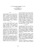
). The relaxation of
an excited Chl to the ground state from the
singlet state is realized in different ways:
excitation energy transfer from Chl to Chl until
the reaction centre to drive photochemical
reaction; re-emission of a photon
(fluorescence); dissipation as heat by internal
conversion; dissipation as heat in a controlled
pathway (NPQ); decay via the triplet state
(
3
Chl*) (Figure 1). A
3
Chl* can return to the
ground state by energy transfer to the O
2
in the
ground-state to generate singlet excited oxygen
(
1
O
2
*), which is an extremely damaging
reactive oxygen species. At room temperature,
Chl fluorescence originates essentially from
PSII, and the yield of fluorescence is generally
low (0.6%–3%) [25]. Non-photochemical
processes that dissipate excitation energy
(collectively called NPQ) also quench Chl
fluorescence, since dissipative pathways are in
competition each other [6]. Chl fluorescence is
indeed used to measure indirectly, but precisely,
NPQ and photochemistry.
K.T.T. Hương et al. / VNU Journal of Science: Natural Sciences and Technology, Vol. 30, No. 2 (2014) 6-22
8
Balance between energy used for
photochemical reactions and dissipative
pathways are important for plant resistance and
productivity in the natural environment where
light intensity changes continuously. Indeed,
under conditions of excess light, plants use only
a small part of the absorbed light energy for
photochemical reactions, while up to 80% of
the absorbed energy is dissipated as heat [26].
This mechanism known as non-photochemical
chlorophyll quenching is triggered to dissipate
excess absorbed light energy within the PSII
antenna system as heat, preventing
photodamage of the reaction center. Energy
dissipation is based on at least four different
mechanisms called qE, qT, qI as described by
Muller et al (2001) [6] and qZ as proposed by
Nilkens et al (2010) and Willelm et al (2011)
[28,29]. They are recently discussed by Ruban
et al (2012) [30]:
Figure 1. The use of the excitation energy of the
chlorophylls. All pathways are in competition.
Plants can control photosynthesis and NPQ. An
increased NPQ is necessary to reduce 3Chl*
formation (thus ROS formation) and can be detected
as a concomitant decrease of fluorescence emission.
* The qT is a quenching associated to state
transitions: in State II part of the major antenna
LHCII of PSII migrates to PSI, thereby
reducing the amount of excitation energy and
fluorescence of PSII. This process contributes
for a small component of NPQ (Figure 2) and
relaxes within tens of minutes [6].
* The qI is a consequence of damaged
reaction centers of PSII acting as weak energy
traps in the absence of ∆pH and is described as
photoinhibitory quenching. It shows very slow
recovery in the range of hours in the dark after a
period of illumination and it is not
photoprotective [30].
* The recently proposed qZ component is a
PsbS-independent but zeaxanthin-dependent
quenching [28] that is related to zeaxanthin-
dependent conformational changes in PSII
antenna proteins [27]. Its formation and
relaxation times are in the order of 10-15 min
and correspond to the synthesis and epoxidation
of zeaxanthin [28].
* The qE is the thylakoid energization-
dependent quenching that is rapidly inducible
and rapidly reversible and it needs the presence
of a transmembrane thylakoid proton gradient
(∆pH). Activation and relaxation is within
seconds to minutes [6,29,30]. The qE has been
shown as a very effective short-term regulatory
mechanism capable of protecting PSII in excess
light conditions and is the main component of
NPQ. For this reason, many investigations on
qE have been performed in the last decades.
However, so far the mechanism of energy
quenching is still not completely elucidated and
the mechanistic aspects are still debated and
controversial [31-34].
K.T.T. Hương et al. / VNU Journal of Science: Natural Sciences and Technology, Vol. 30, No. 2 (2014) 6-22
9
Figure 2. The components of NPQ, from [6],
measured via Chl fluorescence measurement on
Arabidopsis leaves. NPQ (qE + qT + qI) is related to
the difference between Fm (maximal fluorescence of
dark-adapted plants, which do not have NPQ
activated) and Fm’ (the maximal fluorescence
during a light period). The rest of the Fm quenching
during a light phase is related to photochemical
quenching (qP). The recently proposed qZ
component is not shown, but it would contribute to
part of qE and qI shown in the figure. After
switching off the actinic light, recovery of Fm’
within a few minutes reflects relaxation of the qE
component of NPQ. F
0
represents the minimal
fluorescence of the system, related to inevitable
energy losses.
Full qE activation is known to require four
main components: i) a low lumenal pH; ii) the
protein PsbS as sensor of lumenal pH; iii) the
xanthophyll cycle (in particular zeaxanthin
synthesis in high light); iv) the presence of
some Lhcb proteins (PSII antenna complexes).
These components interact each other in some
way and if one is lacking, qE is decreased.
In the following session, we will discus the
functional role of PsbS and zeaxanthin in
energy quenching.
3. The conformation and location of PsbS
Properties of the PsbS protein have been
analyzed in many plant species as Arabidopsis
[9], maize [35], spinach [36], rice [18,23,37],
tomato [38], Marchantia polymorpha [39],
tobacco [40] and it has been concluded that this
protein is accumulated in all land plants [41].
Nevertheless, it does not seem accumulated in
the unicellular green alga Chlamydomonas
reinhardtii under many growth conditions and
in other unicellular green algae [41]. In these
organisms, it seems that the LHCSR proteins,
which also belong to the Lhc superfamily,
replace PsbS for photoprotection by NPQ
[42,43]. In the moss Physcomitrella patens both
PsbS and LHCSR are found and participate in
NPQ [44].
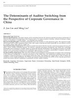
Figure 3. Topology of PsbS from [11] with indicated
the two protonable lumenal glutamates (E122/E226).
In plants, PsbS was firstly isolated in
spinach as a 22 kDa protein by Kim and co-
workers [36]. It was found having a precursor
sequence of 274-residue originated from a
single-copy gene [45] and was called CP22
(Chlorophyll binding Protein of 22 KDa).
Although PsbS is a member of the Lhc
superfamily, which is composed by three
helices membrane proteins, PsbS is predicted
with four transmembrane helices [13,46]. Some
glutamate and aspartate residues are present in
the two lumenal loops in symmetrical position
[47].
K.T.T. Hương et al. / VNU Journal of Science: Natural Sciences and Technology, Vol. 30, No. 2 (2014) 6-22
10
The biochemical, biophysical, and
physiological properties of the PsbS protein
were studied in vitro and in vivo in plants
carrying a modified PsbS obtained by mutating
these lumen-exposed glutamate/aspartate
residues. Li and coworkers have used a site-
directed mutagenesis approach to change one
single glutamate in glutamine (EQ) or aspartate
in asparagine (DN) or both the symmetrical
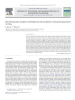
residues at the same time [11]. Results showed
that qE is reduced 50% in the single mutants
E122Q and E226Q as compared with the
control and to the level of the PsbS-KO mutant
(npq4.1) in the double mutant E122Q-E226Q
(Figure 4) [11]. PsbS is a DCCD (N,N′-
dicyclohexylcarbodiimide) binding protein
[11,47]. DCCD binds proton-active residues in
hydrophobic environments and is an inhibitor
of qE [48]. DCCD binding in plant carrying
mutated PsbS was about 50% of the control in
single mutants (E122Q or E226Q) and
undetectable in the double mutant carrying
glutamines at the place of glutamates (E122Q-
E226Q) [11]. Thus these two glutamates
(Figure 3) are strongly indicated as the residues
responsible for pH sensitivity of PsbS [11,47].
PsbS is a 2-fold symmetrical protein and these
two glutamates E122 and E226 seems to act
independently and addictively in qE (Figure 4)
[11,47,49]. Since DCCD binding to both
glutamates is efficient only at low pH [11], it is
very likely that a conformational change of
PsbS after protonation brings these residues in a
hydrophobic environment, necessary to activate
PsbS and qE.
In intact chloroplasts and whole plants,
PsbS seems to exist in dimeric or monomeric
form depending on lumenal pH: the monomer is
present at acidic pH and the dimer at alkaline
pH. The dimer-to-monomer conversion is
reversibly induced by light, which causes
lumenal acidification by the electron transport
chain [50]. PsbS conformational switch has
been suggested to contribute in the
reorganization of PSII supercomplex
[16,22,51,52] necessary for the NPQ activation
induced by variations in light intensity.
Even if it is clear that PsbS is mainly
located in the grana membranes, the precise
location of PsbS is still enigmatic. Different
studies have been performed to find PsbS
location, but results obtained are controversial.
In spinach, Kim and coworker suggested that
this protein is associated with the oxygen-
evolving complex, although it is not needed for
oxygen evolution function [13,36]. In
accordance with this suggestion [53] reported
that PsbS is found in PSII preparations depleted
of LHC. It has been suggests that PsbS could
localise near minor antennas in the PSII-LHCII
supercomplex [54].
Figure 4. Effect of the mutations of the two
glutamates E122 and E226 on NPQ, from [20].
On the contrary, using cryoelectron
microscopy and single particle analysis in
spinach, Nield and colleagues observed that
PsbS protein is not located within the PSII-
LHCII supercomplex, but it can be located in
K.T.T. Hương et al. / VNU Journal of Science: Natural Sciences and Technology, Vol. 30, No. 2 (2014) 6-22
11
the LHCII-rich regions that interconnect the
supercomplex [55]. This is supported by other
researches on purified PSII particles [56]. It had
been also reported that PsbS can associate with
PSII core in dimeric form in the dark and with
LHCII antenna in monomeric form upon
illumination [35] and the monomeric form
would be the active form for qE. It was found to
be present in numerous sucrose gradient
fractions containing PSII supercomplex, but not
bound to PSII [56]. Using immunoprecipitation
studies, Teardo and co-authors reported that
PsbS is associated with numerous thylakoid
complexes including trimeric LHCII, CP29, PSI
and ATP synthase [57]. However, the “sticky”
behavior of PsbS [47,56] and the fact that PsbS
was found to interact with several thylakoid
complexes [57] on which it has no function (as
PSI and ATP synthase), suggest that artificial
aggregation during immunoprecipitation are
possible. Thus, a conclusive answer for PsbS
localization is still not available.
PsbS was shown capable to enhance the
dynamic of thylakoid membrane and its
sensitivity to detergents [22]. It has been
reported that PsbS can catalyze the dissociation
of the PSII-LHCII supercomplex leading to a
reorganization of the PSII supercomplexes,
which seem a fundamental step for triggering
energy quenching in high light
[16,22,51,52,58,59,60]. Thereby, after
protonation and conformational change, PsbS
would dissociate LHCII complexes from PSII
core and induce aggregation of LHCII, which
would cause energy quenching in the antenna
(see below).
4. Is PsbS a pigment binding protein?
PsbS capability to bind pigments is another
question that has been discussed for longtime.
To be the quencher site, PsbS needs to bind
pigment. On the contrary, if no pigment is
bound to this protein, this implies that PsbS can
only be the sensor of low lumenal pH and
would transfer the signal to the PSII-LHCII
complex in some way.
PsbS has some sequence similarity to the
Lhc chlorophyll-binding proteins of PSII [36].
Funk and colleagues reported that the PsbS is
able to bind chlorophylls [46,61]. However,
they also reported that PsbS, differently from
other chlorophyll-binding proteins, is stable in
the absence of pigments [8], in accordance to a
previous report [45]. PsbS pigment binding
ability was also analyzed by experiences of
purification from thylakoids and by
reconstitution experiments of the overexpressed
protein in E. coli in presence of pigments (Chls
and Cars): in no case PsbS was purified or
refolded with some pigments bound,
accordingly to the lack of most of the pigment
binding sites present in the other Lhc proteins
[20,47]. Results from Aspinall-O'Dea and co-
authors, indicating zeaxanthin binding to PsbS
in vitro [62], were found an experimental
artifact [20]. In normal light conditions, the
pigment and photosynthetic protein content do
not change in the npq4.1 mutant of Arabidopsis
(lacking PsbS) as compared to the wild type
[63].
In conclusion present knowledge strongly
suggest that native PsbS protein does not bind
chlorophylls or carotenoids, differently from
others Lhc proteins, which maintain full
pigment binding in the same conditions [47].
Alternatively, the binding of xanthophylls
(especially zeaxanthin and lutein) to PsbS could
be weak or only transient under qE condition
[64].
K.T.T. Hương et al. / VNU Journal of Science: Natural Sciences and Technology, Vol. 30, No. 2 (2014) 6-22
12
5. Relation between PsbS and zeaxanthin in
NPQ formation
The xanthophylls cycle consists in the
reversible deepoxidation of violaxanthin into
zeaxanthin via antheraxanthin by the action of
the violaxanthin deepoxidase enzyme (VDE)
and zeaxanthin epoxidase (ZE) [65]. Under
conditions of excess light, zeaxanthin
accumulates thanks to the action of the VDE,
which is activated by the low lumenal pH
generated by photochemical reactions [65]. In
this condition, zeaxanthin binds one or more
proteins of the PSII-LHCII macromolecular
complex [66,67] and the PsbS protein is
protonated, thus activating qE [11]. In the
absence of PsbS (npq4 mutant), NPQ is largely
reduced. In the presence of PsbS, but in the
absence of zeaxanthin (in the npq1 mutant plant
blocked in the xanthophyll cycle in high light),
NPQ is reduced by ~50-70% with respect to
wild type, but less than in the npq4 mutant
[9,20]. This indicates that the function of PsbS
in qE activation is dominant compared to that
one of zeaxanthin in the presence of ∆pH.
Indeed, the absence of both PsbS and
zeaxanthin show the same qE reduction as in
the case of the single PsbS-KO mutant (Figure
5) [9]. It is suggested that zeaxanthin cannot
perform its qE function if PsbS is absent, while
PsbS can still induce qE without zeaxanthin.
However, reports of [27,28] indicated that
PsbS-independent/zeaxanthin-dependent NPQ
components would exist.
Figure 5. Non photochemical quenching phenotypes
of npq4-1 (no PsbS), npq1-2 (no zeaxanthin), the
double mutant and wild type plants. White bars
above graphs indicate periods of illumination with
high light (1250 µmol photons m
-2
s
-1
); black bars
indicate darkness, from [9].
Transgenic plants over accumulating PsbS
(L17 mutant of Arabidopsis) can enhance NPQ
in the presence or absence of zeaxanthin
[10,11,68,69]. A ∆F682 fluorescence signal in
the difference spectrum between the quenched
and unquenched states showed that a negative
fluorescence peak at 682 nm is formed
independently from zeaxanthin and is due to
PsbS-specific conformational changes in the
quenching site for qE [70]. Moreover, Johnson
and colleagues observed that NPQ in npq4
Arabidopsis leaves blocked in zeaxanthin
formation by infiltration of DTT (dithiothreitol,
inhibitor of violaxanthin de-epoxidase) was
reduced compared with untreated leaves, but it
was found to be not significantly different from
DTT-infiltrated wild type leaves [17]. This
suggests that PsbS can act independently from
zeaxanthin in energy quenching activation, and
zeaxanthin can activate qE independently from
PsbS and enhance PsbS-dependent NPQ. It is
evident that their interaction can strongly
enhance photoprotection capacity in plants
[11,20,24,28,30,69,71,72]. These suggest that it
exist a synergistic effect between PsbS and
zeaxanthin in NPQ formation.
K.T.T. Hương et al. / VNU Journal of Science: Natural Sciences and Technology, Vol. 30, No. 2 (2014) 6-22
13
6. Where is the quenching site?
It has been proposed that PsbS is the site of
energy quenching [73]. However, to day, this
proposition is unlike, because PsbS would not
be able to bind pigments, as discussed before.
Furthermore, qE seems activated also in the
absence of PsbS, but on a longer time scale
[19,24]. Hence it is very probable that PsbS
does not quench directly singlet excited
chlorophyll state [47], despite its key role in qE
[9].
Previous research showed that minor
antenna proteins, as CP26 and CP29, can bind
DCCD [74,75], thus they can be protonated by
low lumen pH as the PsbS protein. Using
genetic approaches such as antisense or knock-
out techniques to manipulate Lhcb content, it
was found that the absence of CP26 has little
effect on qE [76,77], elimination of CP29
decreases qE more than CP26 absence [60,76]
and deletion of CP24 leads to the strongest
decrease of qE [60,77,78]. However, in plants
lacking both CP29 and CP24, qE shows a
smaller reduction as compared to the single
koCP24 mutant [71,79], and similar results
were found for the koCP24/CP26 double
mutant [60,77]. A deep investigation indicated
that qE decrease in the koCP24 is not due to the
presence of the quencher site in this subunit, but
it is due the particular organisation of the
complexes in the membranes and the reduced
capacity of electron transport and thus ΔpH
creation [77]. It is therefore unlikely that the
quenching site is localized only in minor
antenna complexes [30].
Indeed major antenna LHCII is also an
important candidate to be the quencher. In
lhcb1-2 antisense plants, the capacity for non-
photochemical quenching was reduced, but not
completely deleted [71,80]. However, in
Arabidopsis T-DNA koLhcb3 plants, the
absence of Lhcb3, which is compensated by
increased amounts of Lhcb1 and Lhcb2, did not
result in any significant alteration of qE [81]. In
conclusion, there are strong indications that the
quenching site is not associated to one single
subunit [30].
Using ultrafast fluorescence techniques on
intact leaves, Holzwarth and coworker proposed
that there are two independent NPQ quenching
sites in vivo, which depend differently on the
actions of PsbS and zeaxanthin. One site is
formed in the functionally dissociated major
light-harvesting complex LHCII and depends
strictly on the PsbS protein, while the second
site localize in the minor antennae of PSII and
depends on the presence of zeaxanthin [34].
Both qE components would arise from a
quenching mechanism based on a
conformational change within the PSII antenna,
optimized by Lhcb subunit-subunit interactions
and tuned by the synergistic effects of PsbS and
xanthophylls [71]. The second site is in
agreement with the allosteric model of
zeaxanthin in qE proposed by [82,83]. A model
for PsbS action in qE is presented in the
following section.
7. Action mechanism of PsbS in photoprotection
The role of PsbS on PSII-LHCII supercomplex
reorganization for qE activation
The largest purifiable PSII supercomplex
consists of two PSII cores (C2), two copies of
CP29, CP26 and CP24, two strongly bound
LHCII trimers (S2) and two trimers bound with
moderate strength (M2), and it is called
C2S2M2 [84]. It has been suggested for a long
time that the structural changes within the grana
membrane, where PSII supercomplexes
K.T.T. Hương et al. / VNU Journal of Science: Natural Sciences and Technology, Vol. 30, No. 2 (2014) 6-22
14
localize, could provide a physiological
mechanism for regulating the partitioning of
energy between utilization in photosynthesis
and dissipation by NPQ [26,85].
Recently, it has been proposed that in the
quenched state, the PSII-LHCII supercomplex
is reorganized by dissociation of PSII core
complex and antenna and/or clustering of PSII
core units and LHCII antenna aggregates
[52,85], in a process controlled by PsbS
[22,30,34,51,52,86].
Using electron microscopy and fluorescence
spectroscopy analysis on thylakoids prepared
from wild type, PsbS-deficient and PsbS
overexpressing Arabidopsis plants, Kiss and
colleagues observed that reorganization of PSII-
LHCII during thylakoid re-stacking could be
regulated by the level of PsbS. The Mg
2+
requirement in this process was negatively
correlated with the level of PsbS [22].
Moreover, the increase of the amplitude of the
psi-type CD signal originating from features
associable to the PSII-LHCII organization is
also correlated to the PsbS level [22].
It was also found that the content of PsbS
would regulate the PSII organisation in the
grana membrane [16]. Indeed, it was observed
that PSII units assembled into semicrystalline
arrays in grana membranes are higher in the
absence of PsbS, lower in wild type and not
found in membrane enriched in PsbS (L17
mutant) [16]. Therefore in the presence of PsbS,
thylakoid membranes would become more
dynamic and in its absence the association of
the supercomplexes would be stronger [16,22].
PsbS would therefore regulate the interaction
between LHCII and PSII and/or between PSII
complexes in the grana membranes organisation
[16,22].
Consistently with these findings, it was also
reported a PsbS-dependent change in the
distance between PSII core complexes observed
by electron microscopy, implying a
reorganization of the PSII-LHCII
macrostructure occurring during illumination
[51]. This was supported by biochemical
analysis showing that a part of the C2S2M2
supercomplex, consisting of the LHCII M-
trimer, CP24, and CP29 (B4C subcomplex), is
dissociated by light treatment and dependent on
the presence of PsbS [51].
In addition, using freeze-fracture electron
microscopy, combined with laser confocal
microscopy employing the fluorescence
recovery after photobleaching technique in
intact spinach chloroplasts, Johnson and
coworker proposed that the formation of the
photoprotective state requires a structural
reorganization of the photosynthetic membrane
involving dissociation of LHCII from PSII and
its aggregation [52]. The structural changes,
which occur rapidly and reversibly, are
manifested by a reduced mobility of Lhc
antenna chlorophyll proteins [52]. The LHCII
aggregates may cause specific changes in the
LHCII pigment population able to regulate
energy flow. This hypothesis is supported by
spectroscopic analyses on purified LHCII in the
quenched and unquenched states indicating a
conformational change between these two states
[32,58]. This supercomplex reorganization
could be related to the two quenchings Q1 and
Q2 proposed by [34].
K.T.T. Hương et al. / VNU Journal of Science: Natural Sciences and Technology, Vol. 30, No. 2 (2014) 6-22
15
Figure 6. Model describing the reorganization of the
PSII-LHCII supercomplexes under the combined
action of PsbS, zeaxanthin and lumen pH, from [30].
Aggregates of LHCII would be formed in high light
conditions and would dissipate excitation energy as
heat.
Johnson and colleagues [52] suggested that
the structural rearrangement lead to the
formation of internal dissipative pigment
interactions, and energy quenching occurs in
accordance with the xanthophyll-chlorophyll
models proposed by [33,87,88,89] or
chlorophyll-chlorophyll quenching model [90].
Therefore today, the results on the
molecular basis for quenching mechanisms
support the hypothesis that reversible and
flexible reorganisation of PSII-LHCII
supercomplexes triggers energy quenching
formation (Figure 6) promoted by PsbS under
the control of low lumen pH
[16,22,29,30,52,58,59,86].
Low lumen pH as a signal for photoprotection
During the photosynthetic process, a ∆pH in
the thylakoid lumen is generated from the water
photooxydation reaction in the oxygen evolving
complex and during electron transfer at the
level of Cyt b
6
f complex. Besides activating
ATP synthase for ATP synthesis, ∆pH is
indispensable for the qE component of NPQ.
The ∆pH regulation of energy quenching is a
flexible and rapid regulation of PSII activity.
Low lumenal pH activates NPQ via PsbS
protonation [9,11,20], which causes its
conformational change [11,35], and through the
activation of the xanthophylls cycle [91,92].
In addition, it was found that both PsbS-
dependent and PsbS-independent NPQ depend
on lumen pH. In wild type leaves infiltrated
with nigericin, a protonophore that dissipates
the ∆pH, NPQ decreases strongly even in the
presence of PsbS [52,93]. On the contrary, in
the absence of PsbS, NPQ shows a strong
increase when ΔpH is enhanced by a
diaminodurene treatment [19]. Transient qE,
particularly visible on dark adapted plants in the
first minutes after switching on a low light, is
also dependent on the PsbS protein and is
determined by a transient low lumenal pH due
to a delay in the activation of the Calvin cycle
that causes proton accumulation [94].
Figure 7. Model explaining the action of PsbS and
the xanthophylls cycle in pH-dependent energy
quenching. The proposed pKa of LHCII is ~4.0
[95,96], too low for qE activation by physiological
lumen pH values of ~5.8 [97]. However when PsbS
and VDE, which would have a pK for their
activation of ~6.0 [98], bind protons, together they
would trigger the aggregation of LHCII increasing
the hydrophobicity of the environment of the qE-
active residues and shifting the pKa of LHCII to
~6.0, thus activating qE at physiological lumen pH
values, from [30].
K.T.T. Hương et al. / VNU Journal of Science: Natural Sciences and Technology, Vol. 30, No. 2 (2014) 6-22
16
In short, the initiation of qE involves two
lumen pH-dependent processes, the activation
of the xanthophylls cycle and the protonation of
PsbS. It is proposed that PsbS could interact
with xanthophylls cycle for regulation of PSII
antenna reorganization by regulating the pK for
qE activation [19]. In the absence of zeaxanthin
and PsbS, the pH necessary for PSII-LHCII
reorganisation would be very low and thus qE
activated to a minor extent at a given pH. In
presence of PsbS/zeaxanthin, the pK of qE
would increase, allowing a faster and more
efficient thermal dissipation during the light
induced decrease of lumenal pH [30]. It is also
proposed that the LHCII complex, which
presents several acidic residues exposed to the
lumen, may be protonated in qE condition.
Protonation would be necessary for their
“aggregation” and energy quenching. PsbS and
zeaxanthin could have a role by increasing the
pKa of some important residues of LHCII. In
their absence, pKa would be very low and
LHCII would remain unprotonated even at high
light (low lumenal pH), with little activation of
the energy quenching (Figure 7) [30,52].
8. Role physiological of PsbS in plant
photosynthesis regulation
Photosynthesis regulation is a complex
network of mechanisms necessary to adapt
photosynthesis to different environmental
conditions and finally optimise plant fitness.
Photosynthesis can be regulated at many levels,
such as at a macroscopic level (leaf movement),
microscopic level (chloroplast movement) or
molecular level (long-term responses such as
regulations of gene expression to optimise
metabolism and photosynthesis and, of course,
short-term responses modulating photochemistry)
[2,3]. Since photochemical reactions are at the
beginning of the whole metabolism in
photosynthetic organism, numerous researches
investigate all steps of photosynthesis to fully
understand this fundamental process and its
regulation and finally to provide strategies to
improve natural or create artificial
photosynthesis.
However so far, photosynthesis
improvements have been obtained mainly at a
plant level (leaf shape and orientation) rather
than at a molecular level, and the possibilities to
improve photosynthetic efficiency are recently
discussed by different authors
[29,99,100,101,102]. Since the very first
photosynthetic events are the light harvesting
and excitation energy transfer to reaction
centres, and the main regulation at this level is
NPQ, these topics have been at the centre of
many researches to understand the mechanistic
aspects (see [30] for a recent review), as well
the physiological importance. The de-excitation
of singlet excited Chls by heat dissipation
(NPQ) is fast (in the order of few seconds).
Thus, it was assumed that such a
photoprotective mechanism is important to
control PSII photoinhibition under high light to
dissipate excess absorbed energy and under
variable light to rapidly match available
excitation energy and photosynthetic capability.
The importance of PsbS on plant fitness was
indeed demonstrated under variable light
[103,104,105], while under constant high light
conditions other photoprotective mechanisms
can compensate, at least partially, for the lack
of PsbS [14,103].
Plants acclimated to different light
conditions showed that the amount of PsbS is
adjusted to some extent to the intensity of the
light and it is lowered when plants are grown
under low light compared to high light
[44,106,107]. Similarly, an investigation on the
K.T.T. Hương et al. / VNU Journal of Science: Natural Sciences and Technology, Vol. 30, No. 2 (2014) 6-22
17
presence of the PsbS protein in different green
organisms suggested that deep-water algae
accumulate less PsbS than sun-exposed algae
[41]. This suggest that, if competition for
excitation energy between NPQ and
photochemical activity is not significant under
saturating light, it could be important in low
light for photosynthesis optimization and PsbS
down-regulation would be important for
optimal photochemistry. Indeed, it was
observed that the L17 mutant overexpressing
PsbS, which has a higher NPQ capacity, shows
a decreased growth in low light [108]. This
would be in some way similar to what found for
the npq2 mutant of Arabidopsis, which
accumulates zeaxanthin even at low light. This
mutant would waste useful energy in low light
due to some unnecessary NPQ activation,
leading to a reduced growth as compared to
wild type plants [27]. PsbS accumulation is not
abolished even at low light, probably because
plants evolved in natural environment where
conditions can rapidly change. PsbS presence
could reduce plant performances in low light,
but this would be a price to pay to stand with
variable illuminated habitats. Thus, it might be
possible to optimize photosynthesis in nature
light condition by PsbS overexpression [2] and
in not natural and controlled environments
where light is strongly liming by elimination of
its present in plant. Since plants have evolved in
natural environments, the photosynthetic
apparatus may not be well adapted for the
optimised conditions encountered in certain
agricultural systems. Regulations necessary to
improve stress resistance and plant survival in
nature might reduce photosynthesis potential.
Thus, improvement of photosynthesis might be
possible (see [29,99,101] for some reviews).
Acknowledgments
We acknowledge support from the 322
project of the Vietnamese Government and
LGBP, Aix Marseille University, France and
Vietnam Forestry University.
References
[1] K.K. Niyogi, Photoprotection revisited: Genetic
and molecular approaches, Annu.Rev.Plant
Physiol.Plant Mol.Biol. 50 (1999) 333.
[2] Z. Li, S. Wakao, B.B. Fischer, K.K. Niyogi,
Sensing and responding to excess light, Annu
Rev Plant Biol 60 (2009) 239.
[3] P.A. Davis, R.P. Hangarter, Chloroplast
movement provides photoprotection to plants
by redistributing PSII damage within leaves,
Photosynth Res (2012).
[4] C.A. Wraight, A.R. Crofts, Energy-dependent
quenching of chlorophyll alpha fluorescence in
isolated chloroplasts, Eur J Biochem 17 (1970) 319.
[5] G.H. Krause, C. Vernotte, J.M. Briantais,
Photoinduced quenching of chlorophyll
fluorescence in intact chloroplasts and algae,
Biochim.Biophys.Acta 679 (1982) 116.
[6] P. Muller, X.P. Li, K.K. Niyogi, Non-
photochemical quenching. A response to excess
light energy, Plant Physiol 125 (2001) 1558.
[7] P. Horton, A.V. Ruban, Molecular design of the
photosystem II light-harvesting antenna:
photosynthesis and photoprotection, J.Exp.Bot.
56 (2005) 365.
[8] C. Funk, I. Adamska, B.R. Green, B.
Andersson, G. Renger, The nuclear-encoded
chlorophyll-binding photosystem II-S protein is
stable in the absence of pigments, J.Biol.Chem.
270 (1995) 30141.
[9] X.P. Li, O. Bjorkman, C. Shih, A.R. Grossman,
M. Rosenquist, S. Jansson, K.K. Niyogi, A
pigment-binding protein essential for regulation
of photosynthetic light harvesting, Nature 403
(2000) 391.
[10] X.P. Li, P. Muller-Moule, A.M. Gilmore, K.K.
Niyogi, PsbS-dependent enhancement of
feedback de-excitation protects photosystem II
from photoinhibition, Proc Natl Acad Sci U S A
99 (2002) 15222.
[11] X.P. Li, A.M. Gilmore, S. Caffarri, R. Bassi, T.
Golan, D. Kramer, K.K. Niyogi, Regulation of
photosynthetic light harvesting involves
K.T.T. Hương et al. / VNU Journal of Science: Natural Sciences and Technology, Vol. 30, No. 2 (2014) 6-22
18
intrathylakoid lumen pH sensing by the PsbS
protein, J Biol Chem 279 (2004) 22866.
[12] U. Ljungberg, H.E. Akerlund, B. Andersson,
Isolation and characterization of the 10-kDa and
22-kDa polypeptides of higher plant
photosystem 2, Eur J Biochem 158 (1986) 477.
[13] S. Kim, E. Pichersky, C.F. Yocum, Topological
studies of spinach 22 kDa protein of
Photosystem II, Biochim Biophys Acta 1188
(1994) 339.
[14] T. Golan, P. Muller-Moule, K.K. Niyogi,
Photoprotection mutants of Arabidopsis thaliana
acclimate to high light by increasing
photosynthesis and specific antioxidants, Plant
Cell Environ 29 (2006) 879.
[15] L. Kalituho, T. Grasses, M. Graf, J. Rech, P.
Jahns, Characterization of a nonphotochemical
quenching-deficient Arabidopsis mutant
possessing an intact PsbS protein, xanthophyll
cycle and lumen acidification, Planta 223
(2006) 532.
[16] S. Kereiche, A.Z. Kiss, R. Kouril, E.J.
Boekema, P. Horton, The PsbS protein controls
the macro-organisation of photosystem II
complexes in the grana membranes of higher
plant chloroplasts, FEBS Lett 584 (2010) 759.
[17] M.P. Johnson, A.V. Ruban, Arabidopsis plants
lacking PsbS protein possess photoprotective
energy dissipation, Plant J 61 (2010) 283.
[18] S. Hubbart, O.O. Ajigboye, P. Horton, E.H.
Murchie, The photoprotective protein PsbS
exerts control over CO(2) assimilation rate in
fluctuating light in rice, Plant Journal 71 (2012)
402.
[19] M.P. Johnson, A.V. Ruban, Restoration of
rapidly reversible photoprotective energy
dissipation in the absence of PsbS protein by
enhanced DeltapH, J Biol Chem 286 (2011)
19973.
[20] G. Bonente, B.D. Howes, S. Caffarri, G.
Smulevich, R. Bassi, Interactions between the
photosystem II subunit PsbS and xanthophylls
studied in vivo and in vitro, J Biol Chem 283
(2008) 8434.
[21] G. Bonente, M. Ballottari, T.B. Truong, T.
Morosinotto, T.K. Ahn, G.R. Fleming, K.K.
Niyogi, R. Bassi, Analysis of LhcSR3, a protein
essential for feedback de-excitation in the green
alga Chlamydomonas reinhardtii, PLoS Biol 9
(2011) e1000577.
[22] A.Z. Kiss, A.V. Ruban, P. Horton, The PsbS
protein controls the organization of the
photosystem II antenna in higher plant
thylakoid membranes, Journal of Biological
Chemistry 283 (2008) 3972.
[23] I. Kasajima, K. Ebana, T. Yamamoto, K.
Takahara, M. Yano, M. Kawai-Yamada, H.
Uchimiya, Molecular distinction in genetic
regulation of nonphotochemical quenching in
rice, Proc Natl Acad Sci U S A 108 (2011)
13835.
[24] A.V. Ruban, E.H. Murchie, Assessing the
photoprotective effectiveness of non-
photochemical chlorophyll fluorescence
quenching: A new approach, Biochim Biophys
Acta 1817 (2012) 977.
[25] G.H. Krause, E. Weis, Chlorophyll fluorescence
and photosynthesis: The basics, Ann.Rev.Plant
Physiol. 42 (1991) 313.
[26] P. Horton, A.V. Ruban, R.G. Walters,
Regulation of Light harvesting in Green Plants,
Annu.Rev.Plant Physiol.Plant Mol.Biol. 47
(1996) 655.
[27] L. Dall'Osto, S. Caffarri, R. Bassi, A
mechanism of nonphotochemical energy
dissipation, independent from PsbS, revealed by
a conformational change in the antenna protein
CP26, Plant Cell 17 (2005) 1217.
[28] M. Nilkens, E. Kress, P. Lambrev, Y.
Miloslavina, M. Muller, A.R. Holzwarth, P.
Jahns, Identification of a slowly inducible
zeaxanthin-dependent component of non-
photochemical quenching of chlorophyll
fluorescence generated under steady-state
conditions in Arabidopsis, Biochim Biophys
Acta 1797 (2010) 466.
[29] C. Wilhelm, D. Selmar, Energy dissipation is an
essential mechanism to sustain the viability of
plants: The physiological limits of improved
photosynthesis, J Plant Physiol 168 (2011) 79.
[30] A.V. Ruban, M.P. Johnson, C.D. Duffy, The
photoprotective molecular switch in the
photosystem II antenna, Biochim Biophys Acta
1817 (2012) 167.
[31] N.E. Holt, D. Zigmantas, L. Valkunas, X.P. Li,
K.K. Niyogi, G.R. Fleming, Carotenoid cation
formation and the regulation of photosynthetic
light harvesting, Science 307 (2005) 433.
[32] A.V. Ruban, R. Berera, C. Ilioaia, I.H.M. van
Stokkum, J.T.M. Kennis, A.A. Pascal, H. van
Amerongen, B. Robert, P. Horton, R. van
Grondelle, Identification of a mechanism of
photoprotective energy dissipation in higher
plants, Nature 450 (2007) 575.
[33]
S. Bode, C.C. Quentmeier, P.N. Liao, N. Hafi,
T. Barros, L. Wilk, F. Bittner, P.J. Walla, On
the regulation of photosynthesis by excitonic
interactions between carotenoids and
chlorophylls, Proc Natl Acad Sci U S A 106
(2009) 12311.
K.T.T. Hương et al. / VNU Journal of Science: Natural Sciences and Technology, Vol. 30, No. 2 (2014) 6-22
19
[34] A.R. Holzwarth, Y. Miloslavina, M. Nilkensb,
P. Jahnsb, Identification of two quenching sites
active in the regulation of photosynthetic light-
harvesting studied by time-resolved
fluorescence, Chemical Physics Letters 483
(2009) 262.
[35] E. Bergantino, A. Segalla, A. Brunetta, E.
Teardo, F. Rigoni, G.M. Giacometti, I. Szabo,
Light- and pH-dependent structural changes in
the PsbS subunit of photosystem II, Proc Natl
Acad Sci U S A 100 (2003) 15265.
[36] S. Kim, P. Sandusky, N.R. Bowlby, R.
Aebersold, B.R. Green, S. Vlahakis, C.F.
Yocum, E. Pichersky, Characterization of a
spinach psbS cDNA encoding the 22 kDa
protein of photosystem II, FEBS Lett 314
(1992) 67.
[37] T. Iwasaki, Y. Saito, E. Harada, M. Kasai, K.
Shoji, M. Miyao, N. Yamamoto, Cloning of
cDNA encoding the rice 22 kDa protein of
Photosystem II (PSII-S) and analysis of light-
induced expression of the gene, Gene 185
(1997) 223.
[38] M. Wallbraun, S. Kim, B.R. Creen, B.
Piechulla, E. Pichersky, Nucleotide Sequence of
a Tomato psbS Gene, Plant Physiol Biochem
106 (1994 ) 1703.
[39] R. Harrer, R. Bassi, M.G. Testi, C. Sch„fer,
Nearest-neighbor analysis of a photosystem II
complex from Marchantia polymorpha
L.(liverwort), which contains reaction center
and antenna proteins, Eur.J.Biochem. 255
(1998) 196.
[40] A.D. Heiber, O. Kawabata, H.Y. Yamamoto,
Significance of the lipid phase in the dynamics
and functions of the xanthophyll cycle as
revealed by PsbS overexpression in tobacco,
Plant Cell Physiol 45 (2005) 92.
[41] G. Bonente, F. Passarini, S. Cazzaniga, C.
Mancone, M.C. Buia, M. Tripodi, R. Bassi, S.
Caffarri, The occurrence of the psbS gene
product in Chlamydomonas reinhardtii and in
other photosynthetic organisms and its
correlation with energy quenching, Photochem
Photobiol 84 (2008) 1359.
[42] G. Peers, T.B. Truong, E. Ostendorf, A. Busch,
D. Elrad, A.R. Grossman, M. Hippler, K.K.
Niyogi, An ancient light-harvesting protein is
critical for the regulation of algal
photosynthesis, Nature 462 (2009) 518.
[43] G. Bonente, S. Pippa, S. Castellano, R. Bassi,
M. Ballottari, Acclimation of Chlamydomonas
reinhardtii to different growth irradiances, J
Biol Chem 287 (2012) 5833.
[44] C. Gerotto, A. Alboresi, G.M. Giacometti, R.
Bassi, T. Morosinotto, Role of PSBS and
LHCSR in Physcomitrella patens acclimation to
high light and low temperature, Plant Cell
Environ 34 (2011) 922.
[45] N. Wedel, R. Klein, U. Ljungberg, B.
Andersson, R.G. Herrmann, The single-copy
gene psbS codes for a phylogenetically
intriguing 22 kDa polypeptide of photosystem
II, FEBS Lett 314 (1992) 61.
[46] C. Funk, W.P. Schr”der, B.R. Green, G.
Renger, B. Andersson, The Intrinsic 22 kDa
Protein Is a Chlorophyll-Binding Subunit of
Photosystem II, FEBS Lett. 342 (1994) 261.
[47] P. Dominici, S. Caffarri, F. Armenante, S.
Ceoldo, M. Crimi, R. Bassi, Biochemical
properties of the PsbS subunit of photosystem II
either purified from chloroplast or recombinant,
Journal of Biological Chemistry 277 (2002)
22750.
[48] A.V. Ruban, R.G. Walters, P. Horton, The
Molecular Mechanism of the Control of
Excitation Energy Dissipation in Chloroplast
Membranes - Inhibition of DeltapH- Dependent
Quenching of Chlorophyll Fluorescence by
Dicyclohexylcarbodiimide, FEBS Lett 309
(1992) 175.
[49] X. Li, A. Phippard, J. Pasari, K. Niyogi,
Structural-functional analysis of Photosystem II
subunit S (PsbS) in vivo, Functional Plant
Biology 29 (2002) 1131
[50] E. Bergantino, A. Brunetta, E. Touloupakis, A.
Segalla, I. Szabo, G.M. Giacometti, Role of the
PSII-H subunit in photoprotection: novel
aspects of D1 turnover in Synechocystis 6803, J
Biol Chem 278 (2003) 41820.
[51] N. Betterle, M. Ballottari, S. Zorzan, S. de
Bianchi, S. Cazzaniga, L. Dall'osto, T.
Morosinotto, R. Bassi, Light-induced
dissociation of an antenna hetero-oligomer is
needed for non-photochemical quenching
induction, J Biol Chem 284 (2009) 15255.
[52] M.P. Johnson, T.K. Goral, C.D. Duffy, A.P.
Brain, C.W. Mullineaux, A.V. Ruban,
Photoprotective energy dissipation involves the
reorganization of photosystem II light-
harvesting complexes in the grana membranes
of spinach chloroplasts, Plant Cell 23 (2011)
1468.
[53] D.F. Ghanotakis, C.M. Waggoner, N.R.
Bowlby, D.M. Demetriou, G.T. Babcock, C.F.
Yocum, Comparative structural and catalytic
properties of oxygen- evolving photosystem II
preparations, Photosynth.Res. 14 (1987) 191.
K.T.T. Hương et al. / VNU Journal of Science: Natural Sciences and Technology, Vol. 30, No. 2 (2014) 6-22
20
[54] E. Thidholm, V. Lindstrom, C. Tissier, C.
Robinson, W.P. Schroder, C. Funk, Novel
approach reveals localisation and assembly
pathway of the PsbS and PsbW proteins into the
photosystem II dimer, Febs Letters 513 (2002)
217.
[55] J. Nield, C. Funk, J. Barber, Supermolecular
structure of photosystem II and location of the
PsbS protein, Philos.Trans.R.Soc.Lond B
Biol.Sci. 355 (2000) 1337.
[56] S. Caffarri, R. Kouril, S. Kereiche, E.J.
Boekema, R. Croce, Functional architecture of
higher plant photosystem II supercomplexes,
Embo J 28 (2009) 3052.
[57] E. Teardo, P.P. de Laureto, E. Bergantino, F.
Dalla Vecchia, F. Rigoni, D. Szabo, G.M.
Giacometti, Evidences for interaction of PsbS
with photosynthetic complexes in maize
thylakoids, Biochimica Et Biophysica Acta-
Bioenergetics 1767 (2007) 703.
[58] A.A. Pascal, Z.F. Liu, K. Broess, B. van Oort,
H. van Amerongen, C. Wang, P. Horton, B.
Robert, W.R. Chang, A. Ruban, Molecular basis
of photoprotection and control of
photosynthetic light-harvesting, Nature 436
(2005) 134.
[59] P. Horton, M.P. Johnson, M.L. Perez-Bueno,
A.Z. Kiss, A.V. Ruban, Photosynthetic
acclimation: does the dynamic structure and
macro-organisation of photosystem II in higher
plant grana membranes regulate light harvesting
states?, Febs J 275 (2008) 1069.
[60] Y. Miloslavina, S. de Bianchi, L. Dall'Osto, R.
Bassi, A.R. Holzwarth, Quenching in
Arabidopsis thaliana mutants lacking
monomeric antenna proteins of photosystem II,
J Biol Chem 286 (2011) 36830.
[61] C. Funk, G. Renger, I. Adamska, B. Andersson,
P. Schr”der, The PS II-S polypeptide. An
atypical CAB protein, From Light to Biophere
Vol.I, Kluwer, Dordrecht, 1995, pp. 339-342.
[62] M. Aspinall-O'Dea, M. Wentworth, A. Pascal,
B. Robert, A. Ruban, P. Horton, In vitro
reconstitution of the activated zeaxanthin state
associated with energy dissipation in plants,
Proc Natl Acad Sci U S A 99 (2002) 16331.
[63] X.P. Li, A.M. Gilmore, K.K. Niyogi, Molecular
and global time-resolved analysis of a psbS
gene dosage effect on pH- and xanthophyll
cycle-dependent nonphotochemical quenching
in photosystem II, J Biol Chem 277 (2002)
33590.
[64] T. Barros, A. Royant, J. Standfuss, A. Dreuw,
W. Kuhlbrandt, Crystal structure of plant light-
harvesting complex shows the active, energy-
transmitting state, Embo J 28 (2009) 298.
[65] B. Demmig-Adams, Carotenoids and
photoprotection in plants: A role for the
xanthophyll zeaxanthin, Biochim.Biophys.Acta
1020 (1990) 1.
[66] A.V. Ruban, P. Horton, The xanthophyll cycle
modulates the kinetics of nonphotochemical
energy dissipation in isolated light-harvesting
complexes, intact chloroplasts, and leaves of
spinach, Plant Physiol 119 (1999) 531.
[67] R. Bassi, S. Caffarri, Lhc proteins and the
regulation of photosynthetic light harvesting
function by xanthophylls, Photosynthesis
Research 64 (2000) 243.
[68] S. Crouchman, A. Ruban, P. Horton, PsbS
enhances nonphotochemical fluorescence
quenching in the absence of zeaxanthin, FEBS
Lett 580 (2006) 2053.
[69] A. Zia, M.P. Johnson, A.V. Ruban,
Acclimation- and mutation-induced
enhancement of PsbS levels affects the kinetics
of non-photochemical quenching in Arabidopsis
thaliana, Planta 233 (2011).
[70] I.S. Zulfugarov, A. Tovuu, B. Dogsom, C.Y.
Lee, C.H. Lee, PsbS-specific zeaxanthin-
independent changes in fluorescence emission
spectrum as a signature of energy-dependent
non-photochemical quenching in higher plants,
Photochem Photobiol Sci 9 (2010) 697.
[71] M.P. Johnson, M.L. Perez-Bueno, A. Zia, P.
Horton, A.V. Ruban, The zeaxanthin-
independent and zeaxanthin-dependent qE
components of nonphotochemical quenching
involve common conformational changes within
the photosystem II antenna in Arabidopsis,
Plant Physiol 149 (2009) 1061.
[72] P. Jahns, A.R. Holzwarth, The role of the
xanthophyll cycle and of lutein in
photoprotection of photosystem II, Biochim
Biophys Acta 1817 (2012) 182.
[73] K.K. Niyogi, X.P. Li, V. Rosenberg, H.S. Jung,
Is PsbS the site of non-photochemical
quenching in photosynthesis?, J Exp Bot 56
(2005) 375.
[74] R.G. Walters, A.V. Ruban, P. Horton, Higher
plant light-harvesting complexes LHCIIa and
LHCIIc are bound by dicyclohexylcarbodiimide
during inhibition of energy dissipation,
Eur.J.Biochem 226 (1994) 1063.
[75] R.G. Walters, A.V. Ruban, P. Horton,
Identification of proton-active residues in a
higher plant light- harvesting complex,
Proc.Natl.Acad.Sci.USA 93 (1996) 14204.
K.T.T. Hương et al. / VNU Journal of Science: Natural Sciences and Technology, Vol. 30, No. 2 (2014) 6-22
21
[76] J. Andersson, R.G. Walters, P. Horton, S.
Jansson, Antisense inhibition of the
photosynthetic antenna proteins CP29 and
CP26: Implications for the mechanism of
protective energy dissipation Plant Cell 13
(2001) 1193.
[77] S. de Bianchi, L. Dall'Osto, G. Tognon, T.
Morosinotto, R. Bassi, Minor antenna proteins
CP24 and CP26 affect the interactions between
photosystem II Subunits and the electron
transport rate in grana membranes of
Arabidopsis, Plant Cell 20 (2008) 1012.
[78] L. Kovacs, J. Damkjaer, S. Kereiche, C. Ilioaia,
A.V. Ruban, E.J. Boekema, S. Jansson, P.
Horton, Lack of the light-harvesting complex
CP24 affects the structure and function of the
grana membranes of higher plant chloroplasts,
Plant Cell 18 (2006) 3106.
[79] P. Jahns, G.H. Krause, Xanthophyll cycle and
energy-dependent fluorescence quenching in
leaves from pea plants grown under intermittent
light, Planta 192 (1994) 176.
[80] J. Andersson, M. Wentworth, R.G. Walters,
C.A. Howard, A.V. Ruban, P. Horton, S.
Jansson, Absence of the Lhcb1 and Lhcb2
proteins of the light-harvesting complex of
photosystem II - effects on photosynthesis,
grana stacking and fitness, Plant Journal 35
(2003) 350.
[81] J.T. Damkjaer, S. Kereiche, M.P. Johnson, L.
Kovacs, A.Z. Kiss, E.J. Boekema, A.V. Ruban,
P. Horton, S. Jansson, The photosystem II light-
harvesting protein Lhcb3 affects the
macrostructure of photosystem II and the rate of
state transitions in Arabidopsis, Plant Cell 21
(2009) 3245.
[82] P. Horton, A.V. Ruban, M. Wentworth,
Allosteric regulation of the light-harvesting
system of photosystem II, Philos Trans R Soc
Lond B Biol Sci 355 (2000) 1361.
[83] M.L. Perez-Bueno, M.P. Johnson, A. Zia, A.V.
Ruban, P. Horton, The Lhcb protein and
xanthophyll composition of the light harvesting
antenna controls the DeltapH-dependency of
non-photochemical quenching in Arabidopsis
thaliana, FEBS Lett 582 (2008) 1477.
[84] J.P. Dekker, E.J. Boekema, Supramolecular
organization of thylakoid membrane proteins in
green plants, Biochim.Biophys.Acta 1706
(2005) 12.
[85] P. Horton, A.V. Ruban, D. Rees, A.A. Pascal,
G. Noctor, A.J. Young, Control of the light-
harvesting function of chloroplast membranes
by aggregation of the LHCII chlorophyll-
protein complex, FEBS Lett 292 (1991) 1.
[86] Y. Miloslavina, A. Wehner, P.H. Lambrev, E.
Wientjes, M. Reus, G. Garab, R. Croce, A.R.
Holzwarth, Far-red fluorescence: a direct
spectroscopic marker for LHCII oligomer
formation in non-photochemical quenching,
FEBS Lett 582 (2008) 3625.
[87] P. Horton, M. Wentworth, A. Ruban, Control of
the light harvesting function of chloroplast
membranes: The LHCII-aggregation model for
non-photochemical quenching, Febs Letters 579
(2005) 4201.
[88] T.K. Ahn, T.J. Avenson, M. Ballottari, Y.C.
Cheng, K.K. Niyogi, R. Bassi, G.R. Fleming,
Architecture of a charge-transfer state
regulating light harvesting in a plant antenna
protein, Science 320 (2008) 794.
[89] P.N. Liao, C.P. Holleboom, L. Wilk, W.
Kuhlbrandt, P.J. Walla, Correlation of Car S(1)
> Chl with Chl > Car S(1) energy transfer
supports the excitonic model in quenched light
harvesting complex II, J Phys Chem B 114
(2010) 15650.
[90] M.G. Muller, P. Lambrev, M. Reus, E.
Wientjes, R. Croce, A.R. Holzwarth, Singlet
energy dissipation in the photosystem II light-
harvesting complex does not involve energy
transfer to carotenoids, Chemphyschem 11
(2010) 1289.
[91] A.M. Gilmore, Mechanistic aspects of
xanthophyll cycle-dependent photoprotection in
higher plant chloroplasts and leaves,
Physiological Plantarum 99 (1997) 197.
[92] K.K. Niyogi, A.R. Grossman, O. Bjorkman,
Arabidopsis mutants define a central role for the
xanthophyll cycle in the regulation of
photosynthetic energy conversion, Plant Cell 10
(1998) 1121.
[93] P. Joliot, G.N. Johnson, Regulation of cyclic
and linear electron flow in higher plants, Proc
Natl Acad Sci U S A 108 (2011) 13317.
[94] L. Kalituho, K.C. Beran, P. Jahns, The
transiently generated nonphotochemical
quenching of excitation energy in Arabidopsis
leaves is modulated by zeaxanthin, Plant
Physiol 143 (2007) 1861.
[95] A.V. Ruban, A.J. Young, A.A. Pascal, P.
Horton, The effects of illumination on the
xanthophyll composition of the photosystem II
light-harvesting complexes of spinach thylakoid
membranes, Plant Physiol. 104 (1994) 227.
[96] H.E. Akerlund, B. Andersson, A. Persson, P.A.
Albertsson, Isoelectric points of spinach
thylakoid membrane surfaces as determined by
cross partition, Biochim Biophys Acta 552
(1979) 238.
K.T.T. Hương et al. / VNU Journal of Science: Natural Sciences and Technology, Vol. 30, No. 2 (2014) 6-22
22
[97] D.M. Kramer, C.A. Sacksteder, J.A. Cruz, How
acidic is the lumen, Photosynth. Res. 60 (1999)
151.
[98] P. Jahns, D. Latowski, K. Strzalka, Mechanism
and regulation of the violaxanthin cycle: the
role of antenna proteins and membrane lipids,
Biochim Biophys Acta 1787 (2009) 3.
[99] X.G. Zhu, S.P. Long, D.R. Ort, Improving
photosynthetic efficiency for greater yield,
Annu Rev Plant Biol 61 (2010) 235.
[100] E.H. Murchie, M. Pinto, P. Horton, Agriculture
and the new challenges for photosynthesis
research, New Phytol 181 (2009) 532.
[101] E.H. Murchie, K.K. Niyogi, Manipulation of
photoprotection to improve plant
photosynthesis, Plant Physiol 155 (2011) 86.
[102] J. Beckmann, F. Lehr, G. Finazzi, B. Hankamer,
C. Posten, L. Wobbe, O. Kruse, Improvement
of light to biomass conversion by de-regulation
of light-harvesting protein translation in
Chlamydomonas reinhardtii, J Biotechnol 142
(2009) 70.
[103] C. Kulheim, J. Agren, S. Jansson, Rapid
regulation of light harvesting and plant fitness
in the field, Science 297 (2002) 91.
[104] N.M. Krah, B.A. Logan, Loss of psbS
expression reduces vegetative growth,
reproductive output, and light-limited, but not
light-saturated, photosynthesis in Arabidopsis
thaliana (Brassicaceae) grown in temperate light
environments, Am J Bot 97 (2010) 644.
[105] S. Hubbart, O.O. Ajigboye, P. Horton, E.H.
Murchie, The photoprotective protein PsbS
exerts control over CO(2) assimilation rate in
fluctuating light in rice, Plant J 71 (2012) 402.
[106] I.S. Zulfugarov, O.K. Ham, S.R. Mishra, J.Y.
Kim, K. Nath, H.Y. Koo, H.S. Kim, Y.H.
Moon, G. An, C.H. Lee, Dependence of
reaction center-type energy-dependent
quenching on photosystem II antenna size,
Biochim Biophys Acta 1767 (2007) 773.
[107] M. Ballottari, L. Dall'Osto, T. Morosinotto, R.
Bassi, Contrasting behavior of higher plant
photosystem I and II antenna systems during
acclimation, J Biol Chem 282 (2007) 8947.
[108] T. Roach, A. Krieger-Liszkay, The role of the
PsbS protein in the protection of photosystems I
and II against high light in Arabidopsis thaliana,
Biochim Biophys Acta 1817 (2012) 2158.
Chức năng của protêin PsbS trong điều chỉnh quang hợp
ở thực vật
Khương Thị Thu Hương
1,2,3,4
, Robaglia Christophe
2,3,4
,
Caffarri Stefano
2,3,4
1
Trường Đại học Lâm nghiệp Việt Nam
2
Đại học Aix Marseille, LGBP, Marseille, Pháp
3
CEA, DSV, Viện
Sinh học Môi trường và Công nghệ Sinh học, Marseille, Pháp
4
CNRS, UMR7265 Sinh học Thực vật và Vi sinh vật Môi trường, Marseille, Pháp
Tóm tắt: Quá trình quang hợp chuyển hóa năng lượng ánh sáng mặt trời thành năng lượng hóa
học trong các hợp chất hữu cơ cung cấp cho hầu hết các dạng sống trên trái đất. Khi năng lượng ánh
sáng hấp thụ vượt quá khả năng quang hợp của cây, nó có thể chuyển thành ROS (Reactive Oxygen
Species) gây nguy hiểm cho cây. Trong điều kiện đó, cây phát triển một cơ chế tỏa ra năng lượng ánh
sáng dư thừa ở dạng nhi
ệt được gọi là NPQ (Non Photochemical Quenching), để chống lại ROS bảo
vệ cho cây. Trong bài tổng quan này, chúng tôi giới thiệu về một protêin của hệ quang hóa II, protêin
PsbS được biết có vai trò chìa khóa trong sự hoạt hóa NPQ. Sự tăng hay giảm NPQ tỉ lệ với hàm
lượng protêin PsbS trong lá cây. Các nghiên cứu trước đây chỉ ra rằng PsbS hoạt động như một cảm
biến độ pH thấp của lumen trên màng thylakoid, một yếu tố quan trọng để hoạt hóa NPQ. Protêin PsbS
còn được đề xu
ất kiểm soát sự tổ chức lại cấu trúc màng grana để họat hóa NPQ trong điều kiện ánh
sáng cao. Protêin này có vẻ như không kết hợp với sắc tố quang hợp vậy nên nó không phải là điểm
dập tắt năng lượng, nhưng nó có quan hệ với zeaxanthin để kích hoạt đầy đủ NPQ. Vì vậy, PsbS có vai
trò rất quan trọng trong tính chống chịu ánh sáng cao của thực vật. Nghiên cứu protêin này mở ra một
triển vọng đ
iều chỉnh sự sử dụng năng lượng ánh sáng hấp thụ trong quang hợp để tăng năng xuất cây trồng.
Từ khóa: Ánh sáng, NPQ, protêin PsbS, quang hợp, ROS.