Triệt phá đường dẫn truyền phụ vùng dưới vách bằng sóng RF (ablation of posteroseptal AP)
Bạn đang xem bản rút gọn của tài liệu. Xem và tải ngay bản đầy đủ của tài liệu tại đây (2.99 MB, 57 trang )
Triệt phá đường dẫn truyền phụ
vùng dưới vách bằng sóng RF
(Ablation of Posteroseptal AP)
Bs. Phạm Trường Sơn
Bệnh viện trung ương Quân đội 108
Viện tim mạch
Posteroseptal AP :Inferoseptal AP (inferior to the true septal)
The most complex: Pyramid space, confluence of 4 chamber
- Superior: Central fibrous
- Anterior:Ventricular septum
- Posterior: left and Right Atria
- TV is 5 mm to MV: + right atrium- left Ventricle space (thin tissue: AP inside)
+ CS os: at the superior (ablation site)
4 courses: Right to right, Right to left (common)
Left to left, CS to left , CS to Diverticulum
Ablated at: the MV ring , TV ring, inside CS
ECG characteristic
1/ Negative delta waves in leads III, aVF (less common)
positive delta waves in I and aVL
2/Retrograde P’ waves :
- Negative in the leads II, III, aVF
- Positive in AVR ,AVL
3/ Slow and decremental retrograde conduction: incessant
tachycardia.
- Permanent junctional reciprocating tachycardia(PJRT)
1. During AV reentrant tachycardia, the earliest atrial activation usually
is recorded from
+ The ostium of the coronary sinus
+ The proximal coronary sinus within 2 cm
(those beyond 1–2 cm are considered as left free-wall pathways: left-
sided endocardial approach)
2. Functional left bundle branch block during tachycardia results in
either no change or only a nominal prolongation of the VA interval
(10 to 30 msec)
- Functional right bundle branch block usually does not affect the VA
interval.
EP Studying:
Differentiation with SS-AVNRT (long VA time)
- Advanced A during His refractoriness
- Terminate tachy by PVC not conduct to A
- Parahisian pacing: No change Stim to A
- VA (V pacing and Tachy)< 85ms
- VOD (PPI – CL) <115 ms
- VA pacing(at V base)<(at V apex)
Left sided aproach
ECG
1/ Positive delta wave at V1, some study show no role
2/ R/S ratio ≥1 in lead V1 is the most predictive location
3/ If QRS transition between V1 and V2, the R-wave ( lead I) < S-
wave + 1.0 mV is indicative of left-sided AP.
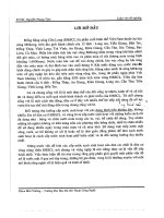
The 12-lead electrocardiogram of a patient with manifest right
posteroseptal accessory pathway. R/S ratio < 1 in lead V1
The 12-lead electrocardiogram of a patient with manifest left
posteroseptal . R/S ratio
≥
1 in lead V1
EPS
1. Earliest atrial activation during AVRT was in the middle CS:
2. The prolongation (10–30ms increase) of the VA interval in response to the
development of left bundle-branch block during orthodromic AVRT :
3. Long-RP AVRT defining a right endocardial AP.
4. The difference of VA intervals during AVRT, measured at the His catheter and the
site of earliest atrial activation in the CS:
-If ≥25 ms: a left endocardial AP in 89% of patients
-If
<
25 ms in 95% of patients with a right endocardial or epicardial AP.
- These mapping criteria above are not necessarily predictive of where the
effective ablation site is located.
- Dhala et al: Overall success rate for the right-sided approach alone was 94%
regardless of the above-mentioned electrophysiologic criteria
+ The right posteroseptum or the proximal coronary sinus: 50 %
+ The posteroseptal region of the tricuspid valve : 30%
+ Within the terminal 1 cm of the coronary sinus including its ostium: 16 %
- Explained : many posteroseptal APs are “right atrio-left ventricular”fibers : right
atrial approach
The most common reasons for prolonged or failed attempts at ablation:
-Difficulties with catheter manipulation :48%
+Inability to reach the appropriate AP site
+Catheter instability
+ Inadequate tissue contact
-Inaccurate mapping: 26%
+AP at a site away from the mapped Area
-The presence of epicardial APs
-40% of all patients referred after an unsuccessful ablation procedure
- The CS method used as a primary method had an efficacy rate of 56%
-The prevalence of an epicardial AP was 4%-25%. The largest series by Sun
et al
.:
+ CS-associated AP in 171 pt (36%)
- Arruda et al :24% of the patients with posteroseptal APs required RF application in a
venous branch or anomaly of the CS
+(22%) in CS diverticulum and (70%) in the middle cardiac vein
- Require: longer procedure times, longer radiation exposure, more RF, complication
- Embryologically, the CS develops:
+ As a remnant of sinus venosus musculature and as a continuation of RA
+ 40 mm of the CS is surrounded: striated muscle, with connections to LA
- When connected with the epicardial surface of the ventricle: epicardial AP
-Multiple left atrial myocardium connections to V:
+Ablation at the Atrial site often produces : change in the activation sequence, should
target at single V insertion.
- Extensions of the CS myocardial coat to the : 5-20 mm deep into the vein
+The middle cardiac vein (MCV): 82%
+ Posterior coronary vein (PCV):11%
+Both vein: 5%
-Requires ablation of the ventricular end at the insertion of the MCV or PCV into the CS
Diagnosis
-Negative delta wave in lead II : highly specific finding for epicardial
+Highest sensitivity (87%)
+Low specificity (20-70%)
-Positive delta in aVR
+ Highest specificity (99%)
+ Moderate sensitivity (70%)
-Deep S in V6
+ Se: 70%
+ Sp:87%
- Combination of 2 mentioned criteria: Highly accurate predictor
-The differentiation between epicardial and left endocardial Ap: difficult.
- Delta wave is negative in lead II and positive in AVR.
-AP can be localized to CS
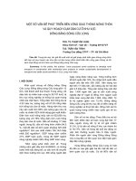
The 12-lead electrocardiogram of a patient with manifest epicardial
posteroseptal accessory pathway. Positive delta wave in lead aVR,
and deep S-wave in V6.
Target
1. Earliest ventricular activation during atrial pacing or earliest atrial activation during
ventricular pacing/orthodromic reciprocating tachycardia.
-Not ideal. For example, downward slanted posteroseptal pathway, the earliest atrial
activation may be close to the compact AV node.
2.“Fragmented” or double potentials at the site of earliest atrial activation in the CS:
during retrograde conduction
-While further away from this site the potentials frequently fused producing a single
patter
- Antz clarify the double potential at CS.
+One is sharp potential representing CS musculature
+The other is rouded, recorded along the floor of the CS and usually ( leftward activation
of the CS musculature).
3. The ideal site for successful ablation: at a site where the
accessory pathway courses epicardially (accessory pathway
potentials)
-A strong relation between AP potential amplitude and success
+The large amplitude of AP potential indicates close proximity to
the AP
-Differentiate with the other signal from:
the atrial electrogram, ventricular electrogram, His bundle
electrogram .
- Pacing at a more rapid rate causes block in the accessory pathway and
subsequently block in the AV node, as well.
-The A and V electrogram is seen to be easily dissociated from the accessory
pathway potential (Ap)
RF delivery inside CS
- Power <30 W, irrigated <15 w
-Avoided in distal small branches of CS:
-Slow flow Lack of cooling high temperature
low power delivery, venous occlusion.
-Intramural thrombosis of the CS has been reported.
- Solution :
+
Larger catheter electrodes or fluid irrigation can be used