SELF ASSEMBLY AND DRUG DELIVERY IN AMPHIPHILIC PEPTIDES MICROSCOPIC INSIGHTS FROM COARSE GRAINED SIMULATIONS
Bạn đang xem bản rút gọn của tài liệu. Xem và tải ngay bản đầy đủ của tài liệu tại đây (8.84 MB, 170 trang )
SELF-ASSEMBLY AND DRUG DELIVERY IN
AMPHIPHILIC PEPTIDES: MICROSCOPIC INSIGHTS
FROM COARSE-GRAINED SIMULATIONS
NARESH THOTA
(M.Tech., IIT Roorkee)
A THESIS SUBMITTED
FOR THE DEGREE OF DOCTOR OF PHILOSOPHY
DEPARTMENT OF CHEMICAL AND BIOMOLECULAR
ENGINEERING
NATIONAL UNIVERSITY OF SINGAPORE
2015
To My Parents, Teachers
&
Almighty God
Declaration
I hereby declare that the thesis is my original work and it has been written by me
in its entirety.
I have duly acknowledged all the sources of information which have been used in
this thesis.
This thesis has also not been submitted for any degree in any university
previously.
_______________________
Naresh Thota
May 2015
i
Acknowledgements
First of all, I would like to express my sincere gratitude to my supervisor A/Prof.
Jiang Jianwen for his constant guidance and support throughout my tenure of
graduate studies. His technical advice and continuous motivation towards research
inspired me to work diligently in achieving my targets in a punctual manner. I am
very thankful to his guidance and support especially during the initial period of
my research. The support he has shown on me during my ankle sprain injury was
especially unforgettable. His guidance and suggestions will be definitely helpful
to achieve my professional and personal aspirations. I am fortunate to work in his
research group with highly technical and friendly environment.
I am thankful to my lab mates for their helping nature and discussions in
the lab. Specially, I want to thank Dr. Hu Zhongqiao and Dr. Luo Zhonglin for
their help during the initial set up of my simulations. I am happy about working
with other colleagues Dr. Anjaiah Nalaparaju, Dr. Chen Yifei, Dr. Fang Weijie,
Dr. Krishna Mohan Gupta, Ms. Zhang Kang in the group.
I would like to thank the internal and external examiners for spending
precious time in examining my thesis and providing valuable comments. I am
thankful to A/Prof. Yang Kun-Lin and A/Prof. Chen Shing Bor for being the
panel examiners in my oral qualifying examination and thesis advisory
committee. Their suggestions and comments were helpful in improving my
research. I would also be grateful to the Department staff, including Vanessa,
Sandy, Kwee Mei, Boey for their help during department administrative and
ii
laboratory works. The scholarship provided by the National University of
Singapore and the Ministry of Education, Singapore was really helpful for my
study and research.
I want to express special thanks to my roommates Srinath, Vamsi krishna,
Naveen, Sanjeeva, Upendar Rao, Venkateshwara Reddy, Anil, Gopal, Shiva,
Vinay for their help and special care, which were always positive, supportive and
made me to stay healthily. I also want to thank my friends Chandu, Prakash, Ravi
kiran, Siva, Praveen tej, Balaji for their positive words whenever I talked to them.
I cannot forget to mention about my parents on this occasion whose love,
affection, care and support made me to reach till this stage of my life. I specially
want to mention my mother’s patience and help for my homework during school
days. The discipline, hard work, patience and punctuality taught by my father
made me strong to face all the circumstances with enough strength. I am happy to
mention about my sister Kamala for her support and care throughout my life. I
convey my gratitude to my uncle, aunts, siblings and cousins for sharing my
happiness and sorrows with them. I would like to thank each and every teacher in
my life because of their contributions in building my career.
Finally, I want to thank his almighty God for giving this life, good health
and strength. It would have been a dream to finish the PhD program without his
blessings and kindness on me.
Naresh Thota
iii
Table of Contents
Acknowledgements i
Table of Contents iii
Summary vi
List of Tables ix
List of Figures x
Abbreviations xvi
List of Symbols xvii
Chapter 1. Introduction 1
1.1 Background 1
1.2 Amino Acids 3
1.3 Applications 7
1.3.1 Antimicrobial Activities 7
1.3.2 Nano Fabrication 8
1.3.3 Drug and Gene Delivery 10
1.3.4 Cosmetic and Skin Care Applications 11
1.3.5 Other Applications 11
1.4 Objectives and Scope of the Thesis 13
Chapter 2. Literature Review 15
2.1 Surfactant-Like Peptides 15
2.2 Lipid-Based Peptides 20
2.3 Amphiphilic Peptides 25
Chapter 3. Simulation Methodology 30
3.1 MARTINI Model 30
3.2 Molecular Dynamics Simulation 34
iv
Chapter 4. Self-Assembly of Short Amphiphilic Peptides F
m
D
n
and F
m
K
n
36
4.1 Introduction 36
4.2 Models and Methods 38
4.3 Results and Discussion 43
4.3.1 F
m
D and F
m
K Peptides 44
4.3.2 F
3
K
n
and F
6
K
n
Peptides 47
4.4 Conclusions 57
Chapter 5. Self-Assembly of Amphiphilic Peptide (AF)
6
H
5
K
15
59
5.1 Introduction 59
5.2 Models and Methods 61
5.3 Results and Discussion 64
5.3.1 Effect of Box Size 64
5.3.2 Effect of Peptide Concentration 72
5.4 Conclusions 76
Chapter 6. Self-Assembly of FA32 Derivatives: Roles of Hydrophilic and
Hydrophobic Residues 78
6.1 Introduction 78
6.2 Models and Methods 80
6.3 Results and Discussion 82
6.3.1 Length of Hydrophilic Residues 82
6.3.2 Replacement of Ala by Phe Residues 89
6.3.3 Length of Hydrophobic Residues 92
6.4 Conclusions 97
v
Chapter 7. Ibuprofen Loading and Release in FA32and its Derivatives 99
7.1 Introduction 99
7.2 Models and Methods 102
7.3 Results and Discussion 106
7.3.1 IBU Loading in FA32 107
7.3.2 IBU Loading in F
12
H
5
K
15
and F
16
H
5
K
15
114
7.3.3 IBU Release 116
7.4 Conclusions 119
Chapter 8. Effects of Peptide Sequence on Self-Assembly and Ibuprofen
Loading 121
8.1 Introduction 121
8.2 Models and Methods 122
8.3 Results and Discussion 124
8.3.1 Effect of Peptide Sequence on Assembly 124
8.3.2 Effect of Peptide Sequence on IBU Loading 129
8.4 Conclusions 131
Chapter 9. Conclusions and Recommendations 132
9.1 Conclusions 132
9.2 Recommendations for Future Studies 135
Bibliography 138
Journal Publications 149
Conference Presentations 150
vi
Summary
Amphiphilic peptides are biodegradable and biocompatible, important
characteristics for ideal drug carriers. They can form nano-sized micelles with
hydrophobic cores allowing for encapsulation of hydrophobic drugs, and thus
provide an effective protection against hydrolysis and degradation. In addition,
the size, stability, permeability and elasticity of the micelles can be fine-tuned by
tailoring peptide sequence, length, solution conditions, etc. The micelles may
undergo structural transition triggered by pH variation or other stimuli leading to
drug release. Therefore, amphiphilic peptides have received considerable interest
for drug delivery. Nevertheless, there is no theoretical guidance currently
available on the rational selection and design of amphiphilic peptides to achieve
optimal drug delivery.
Through molecular dynamics simulation, the objective of this thesis is to
quantitatively understand the self-assembly behavior of amphiphilic peptides from
a microscopic scale, elucidate the detailed process of drug loading and release,
and provide bottom-up guidelines towards the intelligent design of new
amphiphilic peptides for drug delivery. The main contents of the thesis contain
four parts.
(1) Self-assembly of short amphiphilic peptides F
m
D
n
and F
m
K
n
is examined.
Within s-scale simulation, FD and FK only form loose polymeric clusters. Upon
increasing the length of Phe residues in F
m
D and F
m
K (m = 2 to 4), larger and
more stable micelles are formed. F
m
K and F
m
D prefer to assemble into quasi-
vii
spherical and sheet-like micelles, respectively. For F
3
K
n
(n = 2 to 8) and F
6
K
n
(n =
4 to 12), the assembly capability reduces leading to smaller micelles when the
length of Lys residues increases. For the formation of quasi-spherical micelles
with distinct core/shell structure, the optimal ratio of hydrophobic/hydrophilic
residues is found to be 3/4 for both F
3
K
n
and F
6
K
n
.
(2) A relatively longer amphiphilic peptide FA32 [(AF)
6
H
5
K
15
] is studied.
Spherical micelles are formed, with Ala and Phe in hydrophobic core, Lys in
hydrophilic shell and His at core/shell interface. The assembly process and
microscopic structures are analyzed in terms of the number of clusters, the radii of
micelle, core and shell and the density profiles of residues. It is found that the
micellar structures and microscopic properties are essentially independent of the
size of simulation box. With increasing concentration, quasi-spherical micelles
change to elongated shape and micelle size generally increases.
(3) The effects of hydrophilic and hydrophobic chain lengths on self-assembly
are studied. With increasing length of hydrophilic Lys residues in (AF)
6
H
5
K
n
(n =
10, 15, 20 and 25), the assembly capability is reduced by forming smaller micelles
or the presence of individual peptide chains. Upon replacing Ala by more
hydrophobic Phe in A
m
F
n
H
5
K
15
(m + n = 12), larger micelles are formed. With
increasing length of hydrophobic Phe residues in F
n
H
5
K
15
(n = 4, 8, 12 and 16),
micelle size increases and the morphology shifts from spherical to fiber-like.
(4) A model hydrophobic drug, ibuprofen (IBU), is investigated for loading
and release in FA32, F
12
H
5
K
15
and F
16
H
5
K
15
. Upon the loading of IBU in FA32,
quasi-spherical core/shell structured micelles are formed. IBU is predominantly
viii
located in hydrophobic core and covered by Phe and Ala residues, while Lys is in
the hydrophilic shell. With increasing concentration of IBU, the radii of micelle
and core increase. In F
16
H
5
K
15
, however, the loading of IBU leads to a well-
structured nanofiber. The release of IBU from FA32 micelles is slower than from
F
16
H
5
K
15
nanofiber, suggesting the former is better in controlled release.
Furthermore, the effects of peptide sequence on IBU loading are investigated in
(AF)
6
H
5
K
15
, H
5
(AF)
6
K
15
, H
5
K
5
(AF)
6
K
10
and (AF)
3
H
5
K
15
(AF)
3
. It is revealed that
peptide sequence has an insignificant effect on drug loading.
From this thesis, microscopic insights into the self-assembly of amphiphilic
peptides, and the loading and release of drug are provided. Equilibrium and
dynamic properties are obtained from a molecular level. Key governing factors
such as chain length, sequence and hydrophobicity have been identified. The
bottom-up guidelines are useful towards the development of new amphiphilic
peptides for high-efficacy drug delivery.
ix
List of Tables
Table 1.1. Representations and physical properties of 20 amino acids 6
Table 2.1. Surfactant-like peptides. 19
Table 2.2. Lipid-based peptides 24
Table 2.3. Amphiphilic peptides 28
Table 3.1. LJ interaction matrix. 32
Table 3.2. Parameters σ and ε of LJ potential in the MARTINI model.
136
33
Table 4.1. Simulation conditions 42
Table 4.2. Number of micelles, peptides per micelle, radii of micelle, core and
shell. 50
Table 4.3. Interaction energies (kJ/mol) at free and aggregated states. 53
Table 5.1. Three different box sizes. 63
Table 5.2. Nine different peptide concentrations in 18 nm box. 63
Table 5.3. Number of micelles, peptides per micelle, R
micelle
, R
core
,
and R
shell
in
three box sizes. 66
Table 5.4. Number of micelles, peptides per micelle, R
micelle
, R
core
,
and R
shell
in 18
nm box. 74
Table 6.1. Number of micelles, peptides per micelle, R
micelle
, R
core
and R
shell
. 87
Table 7.1. Number of micelles for IBU loading in FA32 with different initial
positions. 108
Table 7.2. Number of micelles, peptides per micelle, R
micelle
, R
core
and R
shell
for
IBU loading in FA32.
a
from Chapter 6. 110
x
List of Figures
Figure 1.1. Different morphologies formed by amphiphilic molecules (a)
micelle
22
(b) vesicle
23
(c) nanofiber.
20
2
Figure 1.2. Representation of peptide bond formation between two amino acids. 4
Figure 1.3. Structures and classifications of 20 amino acids.
26
4
Figure 1.4. Schematic illustrations of actions of A
9
K leading toward bacterial
membrane permeation and disruption. (a) A
9
K molecules self-assemble into
nanorods (red) with the positive charges outside the rod. (b) A
9
K molecules flap
on to outer membrane surface through charge affinity and may become inserted in
the membrane through hydrophobic effect. (c) They can then flip to insert into the
inner leaf of the membrane and make a “through barrel” or micelles to cause
leakage or lysis. (d) Nanorods might also associate with the cell membrane
surface directly through charge interaction and (e) become inserted subsequently
due to different effects including electrostatic and hydrophobic interactions.
17
8
Figure 1.5. Proposed plausible self-assembly process of the nanodonut structure.
(A) Randomly oriented and distributed peptides at low concentration. (B) Micelle
formation above the CAC concentration. (C) Fusion or elongation of the micelles
for the formation of a nanopipe. (D) Bending of the nanopipe for the formation of
a nanodonut structure.
38
9
Figure 1.6. (a) Images of DOX loaded P
FD
-5 hydrogel shaped on a glass slide by
a syringe. (b) DOX loaded hydrogel in a well prior to the addition of medium
(left) and fragmented hydrogel on the sixth day in medium (right) showing the
colored DOX released to the medium.
42
11
Figure 1.7. Casting of silver nanowires with the peptide nanotubes. (A) The
nanowires were formed by the reduction of silver ions within the tubes, followed
by enzymatic degradation of the peptide mold. (B) TEM analysis (without
staining) of peptide tubes filled with silver nanowires. (C and D) TEM images of
silver nanowires that were obtained after the addition of the proteinase K enzyme
to the nanotube solution.
48
12
Figure 3.1. Coarse-grained representation of amino acids based on the MARTINI
model.
137
31
Figure 4.1. Atomistic and coarse-grained models of Phe (a, d), Asp (b, e) and Lys
(c, f), respectively. Color codes for (a), (b) and (c): N, blue; O, red; C, cyan and
H, white. 39
xi
Figure 4.2. Atomistic representations of peptides F
m
D
n
and F
m
K
n
. N: blue, O: red,
C: cyan, and H: white 40
Figure 4.3. CG representations of F
m
D
n
and F
m
K
n
peptides using the MARTINI
model. F: yellow, D and K: red 41
Figure 4.4. Final snapshots for F
m
D and F
m
K (m = 1, 2, 3 and 4) at 2500 ns. Phe:
yellow, Asp and Lys: red. Water and ions are not shown for clarity. 44
Figure 4.5. Energy minimized structures of F
3
K, F
4
K, F
3
D and F
4
D. 45
Figure 4.6. Number of clusters versus time for F
m
D and F
m
K (m = 2, 3 and 4). . 46
Figure 4.7. Final snapshots for F
3
K
n
(n = 2, 3, 4, 5, 6 and 8) at 2500 ns. Phe:
yellow, Lys: red. Water and ions are not shown for clarity. 47
Figure 4.8. Number of clusters versus time for F
3
K
n
(n = 2, 3, 4, 5, 6 and 8). 48
Figure 4.9. Radii of micelle (R
micelle
), core (R
core
) and shell (R
shell
) for F
3
K
2
,
F
3
K
4
and F
3
K
6
. 49
Figure 4.10. Distributions of R
micelle
for F
3
K
n
(n = 2, 3, 4, 5, 6 and 8) 51
Figure 4.11. Density profiles for F
3
K
2
, F
3
K
4
and F
3
K
6
. The micelles contain 58,
10 and 5 peptides, respectively. 52
Figure 4.12. Final snapshots for F
6
K
n
(n = 4, 6, 8, 10 and 12) at 5000 ns. Phe:
yellow, Lys: red. Water and ions are not shown for clarity. 53
Figure 4.13. Number of clusters versus time for F
6
K
n
(n = 4, 6, 8, 10 and 12). 54
Figure 4.14. Radii of micelle (R
micelle
), core (R
core
) and shell (R
shell
) for F
6
K
4
,
F
6
K
8
and F
6
K
12
. 55
Figure 4.15. Distributions of R
micelle
for F
6
K
n
(n = 4, 6, 8, 10 and 12). 56
Figure 4.16. Density profiles for F
6
K
4
, F
6
K
8
and F
6
K
12
. The micelles contain 72,
16 and 11 peptides, respectively. 57
Figure 5.1. (a) Atomistic representation of FA32. N: blue, O: red, C: cyan, and H:
white. (b) CG representation of FA32 using the MARTINI model. Ala and Phe:
yellow, His: blue, and Lys: red. (c) Aggregated structure of FA32 62
Figure 5.2. Initial and final snapshots of (a) 10 peptides in an 11 nm box (b) 26
peptides in a 15 nm box (c) 44 peptides in an 18 nm box. Ala and Phe: yellow,
His: blue, Lys: red. Water and Cl
ion are not shown for clarity. 65
Figure 5.3. Snapshots for 44 peptides in 18 nm box at different time intervals. . 67
xii
Figure 5.4. Number of clusters versus time for 10, 26 and 44 peptides in 11, 15
and 18 nm boxes, respectively. 68
Figure 5.5. Radii of micelle (R
micelle
), core (R
core
) and shell (R
shell
) for (a) 10
peptides in 11 nm box, (b) 26 peptides in 15 nm box, and (c) 44 peptides in 18 nm
box 69
Figure 5.6. Distributions of R
micelle
for (a) 10 peptides in 11 nm box (b) 26
peptides in 15 nm box (c) 44 peptides in 18 nm box. 70
Figure 5.7. Density profiles of micelles for 26 peptides in 15 nm box. The
micelles contain 10, 8, and 8 peptides in (a), (b), and (c), respectively. 71
Figure 5.8. Final snapshots for different peptide concentrations in 18 nm box.
From (a) to (i), N
p
= 12, 18, 24, 30, 36, 42, 48, 54, and 60, respectively. 73
Figure 5.9. Number of clusters for 18, 36, and 60 peptides in 18 nm box. 73
Figure 5.10. Radii of micelle (R
micelle
), core (R
core
), and shell (R
shell
) for (a) 18, (b)
36 and (c) 60 peptides in 18 nm box. 74
Figure 5.11. Distributions of R
micelle
for (a) 18 (b) 36 (c) 60 peptides in 18 nm
box 75
Figure 5.12. Density profiles of micelles for 18, 36, and 60 peptides in 18 nm
box. The micelles contain 7, 10, and 12 peptides in (a), (b), and (c), respectively.
76
Figure 6.1. Coarse-grained models of FA32 derivatives. Ala and Phe: yellow,
His: blue, and Lys: red. 81
Figure 6.2. Snapshots for (AF)
6
H
5
K
10
, (AF)
6
H
5
K
15
, (AF)
6
H
5
K
20
and (AF)
6
H
5
K
25
at different time intervals. Water and Cl
ions are not shown for clarity. 83
Figure 6.3. Number of clusters versus time for (AF)
6
H
5
K
10
, (AF)
6
H
5
K
15
,
(AF)
6
H
5
K
20
and (AF)
6
H
5
K
25
. 85
Figure 6.4. Radii of micelle (R
micelle
), core (R
core
) and shell (R
shell
) for (AF)
6
H
5
K
10
,
(AF)
6
H
5
K
15
, (AF)
6
H
5
K
20
and (AF)
6
H
5
K
25
. 86
Figure 6.5. Distributions of R
micelle
for (AF)
6
H
5
K
10
, (AF)
6
H
5
K
15
, (AF)
6
H
5
K
20
and
(AF)
6
H
5
K
25
. 88
Figure 6.6. Density profiles for (AF)
6
H
5
K
15
, (AF)
6
H
5
K
20
and (AF)
6
H
5
K
25
. The
micelles contain 10, 8 and 6 peptides in (a), (b) and (c), respectively. 89
Figure 6.7. Final snapshots for (AF)
6
H
5
K
15
, (AF
3
)
3
H
5
K
15
and F
12
H
5
K
15
. 89
xiii
Figure 6.8. Number of clusters for (AF)
6
H
5
K
15
, (AF
3
)
3
H
5
K
15
and F
12
H
5
K
15
. 90
Figure 6.9. Radii of micelle (R
micelle
), core (R
core
) and shell (R
shell
) for (AF)
6
H
5
K
15
,
(AF
3
)
3
H
5
K
15
and F
12
H
5
K
15
. 91
Figure 6.10. Distributions of R
micelle
for (AF)
6
H
5
K
15
, (AF
3
)
3
H
5
K
15
and F
12
H
5
K
15
.
91
Figure 6.11. Density profiles for (AF)
6
H
5
K
15
, (AF
3
)
3
H
5
K
15
and F
12
H
5
K
15
. The
micelles contain 5, 6 and 15 peptides in (a), (b) and (c), respectively. 92
Figure 6.12. Snapshots for F
4
H
5
K
15
, F
8
H
5
K
15
, F
12
H
5
K
15
and F
16
H
5
K
15
at different
time intervals. 93
Figure 6.13. Number of clusters versus time for F
4
H
5
K
15
, F
8
H
5
K
15
, F
12
H
5
K
15
and
F
16
H
5
K
15
. 94
Figure 6.14. Radii of micelle (R
micelle
), core (R
core
) and shell (R
shell
) for F
4
H
5
K
15
,
F
8
H
5
K
15
, F
12
H
5
K
15
and F
16
H
5
K
15
. 95
Figure 6.15. Distributions of R
micelle
for F
4
H
5
K
15
, F
8
H
5
K
15
, F
12
H
5
K
15
and
F
16
H
5
K
15
. 96
Figure 6.16. Density profiles for F
4
H
5
K
15
, F
8
H
5
K
15
and F
16
H
5
K
15
. The micelles
contain 9, 11 and 10 peptides in (a), (b) and (c), respectively. 97
Figure 7.1. Atomistic and coarse-grained structures of (a-b) (AF)
6
H
5
K
15
(c-d)
F
12
H
5
K
15
(e-f) F
16
H
5
K
15
and (g-h) IBU. In (a), (c), (e) and (g): O, red; N, blue; C,
cyan and H, white. In (b), (d) and (e): Ala and Phe, yellow; His, blue; Lys, red.
103
Figure 7.2. Radial distribution functions between different groups of IBU in
atomistic model. 104
Figure 7.3. Radial distribution functions between different groups of IBU in P1,
P2 and P3 model. 104
Figure 7.4. Radial distribution functions between different groups of IBU at
different bond lengths. 105
Figure 7.5. Snapshots for IBU loading in FA32 at D/P = 0.15, 0.20 and 0.25. Ala
and Phe: yellow, His: blue, Lys: red, IBU: green. Water and Cl
ions are not
shown for clarity. 107
Figure 7.6. Number of clusters versus time for IBU loading in FA32 at D/P =
0.15, 0.20 and 0.25. 108
xiv
Figure 7.7. Radii of micelle (R
micelle
), core (R
core
) and shell (R
shell
) for IBU-loaded
FA32 micelles at D/P = 0.15, 0.20 and 0.25. 110
Figure 7.8. Distributions of R
micelle
for IBU-loaded FA32 micelles at D/P = 0.15,
0.20 and 0.25. 111
Figure 7.9. Density profiles for IBU-loaded FA32 micelles at D/P = 0.15, 0.20
and 0.25. The micelles contain 16, 31 and 25 peptides in (a), (b) and (c),
respectively. 112
Figure 7.10. Final snapshots for IBU loading in FA32 at different D/P. 112
Figure 7.11. Radii of micelle (R
micelle
), core (R
core
) and shell (R
shell
) for IBU-
loaded FA32 micelles at D/P = 0.10, 0.30, 0.50 and 0.70. 113
Figure 7.12. Density profile for IBU-loaded FA32 micelle at D/P = 0.70. The
micelle contains 50 peptides. 114
Figure 7.13. Final snapshots for IBU loading in FA32, F
12
H
5
K
15
and F
16
H
5
K
15
at
D/P = 0.25. 115
Figure 7.14. Density profiles for IBU-loaded F
16
H
5
K
15
nanofiber. The inset
denotes the cross-section view. 116
Figure 7.15. IBU release from FA32 micelles. 117
Figure 7.16. IBU release from F
16
H
5
K
15
nanofiber. 118
Figure 7.17. Cumulative IBU release from FA32 micelles and F
16
H
5
K
15
nanofiber. 119
Figure 8.1. Coarse-grained models of FA32-I: (AF)
6
H
5
K
15
, FA32-II: H
5
(AF)
6
K
15
,
FA32-III: H
5
K
5
(AF)
6
K
10
and FA32-IV: (AF)
3
H
5
K
15
(AF)
3
. Ala and Phe: yellow,
His: blue, and Lys: red. 123
Figure 8.2. Snapshots for (AF)
6
H
5
K
15
, H
5
(AF)
6
K
15
, H
5
K
5
(AF)
6
K
10
and
(AF)
3
H
5
K
15
(AF)
3
at different time intervals. Water and Cl
ions are not shown.
125
Figure 8.3. Number of clusters versus time for (AF)
6
H
5
K
15
, H
5
(AF)
6
K
15
,
H
5
K
5
(AF)
6
K
10
and (AF)
3
H
5
K
15
(AF)
3
. 126
Figure 8.4. Radii of micelle (R
micelle
), core (R
core
) and shell (R
shell
) for (AF)
6
H
5
K
15
,
H
5
(AF)
6
K
15
, H
5
K
5
(AF)
6
K
10
, and (AF)
3
H
5
K
15
(AF)
3
. 127
Figure 8.5. Distributions of R
micelle
for (AF)
6
H
5
K
15
, H
5
(AF)
6
K
15
, H
5
K
5
(AF)
6
K
10
,
and (AF)
3
H
5
K
15
(AF)
3
. 128
xv
Figure 8.6. Density profiles for (AF)
6
H
5
K
15
, H
5
(AF)
6
K
15
and H
5
K
5
(AF)
6
K
10
micelles. The micelles contain 5, 10 and 10 peptides in a, b and c, respectively.
129
Figure 8.7. Snapshots for (AF)
6
H
5
K
15
, H
5
(AF)
6
K
15
, H
5
K
5
(AF)
6
K
10
and
(AF)
3
H
5
K
15
(AF)
3
loaded with IBU at different time intervals. 130
Figure 8.8. Number of clusters versus time for IBU loading in FA32-I, FA32-II,
FA32-III and FA32-IV at D/P = 0.25. 131
xvi
Abbreviations
PEO Poly ethylene oxide
PLAA Poly l-amino acids
CAC Critical aggregation concentration
CMC Critical micelle concentration
MD Molecules dynamics
PAs Peptide amphiphiles
AFM Atomic force microscopy
DLS Dynamic light scattering
DNA Deoxyribonucleic acid
RES Reticuloendothelial system
IBU Ibuprofen
DOX Doxorubicin
PTX Paclitaxel
CPT Camptothecin
SASA Solvent accessible surface area
CG Coarse-grained
COM Center of mass
VMD Visual molecular dynamics
DPD Dissipative particle dynamics
PAE Poly(β-amino ester)
PEG Poly ethylene glycol
D/P Drug to peptide ratio
xvii
List of Symbols
u
i
potential energy of atom i
m
i
mass of atom i
a
i
acceleration of atom i
v
i
velocity of atom i
U
LJ
LJ potential
U
el
electrostatic potential
U
b
bond-stretching potential
U
a
bond-bending potential
U
d
proper torsional potential
U
id
improper torsional potential
K
b
, K
a
force constants of bond-stretching and bending potentials
K
d
, K
id
force constants of proper and improper torsional potentials
d
ij
bond distance between atoms i and j
ijk
angle by atoms i, j and k
ijkl
torsional angle by atoms i, j, k
and l
ij
,
ij
collision diameter and well depth for atoms i and j
q
i
atomic charge of atom i
ε
0
vacuum permittivity
r distance
g(r) radial distribution functions at distance r
Chapter 1. Introduction
1
Chapter 1. Introduction
1.1 Background
Conventional cancer treatments are limited to surgery, chemotherapy and
radiation. Surgery is to remove tumors in human body. Chemotherapy uses
medicines to destroy cancer cells. In radiation treatment, high-energy radiations
are used to kill or decline the growth of cancer cells.
1
Apart from these methods,
additional techniques such as transplantation and gene therapy are in pre-clinical
development stage.
2
While killing cancer cells, chemotherapy and radiation also
kill healthy cells, which is a main concern in cancer treatment. To overcome this
limitation, some alternatives such as targeted and sustained drug delivery have
been proposed. Targeted delivery can effectively inhibit the growth of cancer cells
and causes less damage to healthy cells, and sustained release offer several
advantages like less frequency administration, reduction of side effects and better
compliance.
3
In targeted and sustained delivery, drug carrier materials play an indispensable
role. Over the last two decades, numerous experimental studies have been
reported on developing advanced materials as drug carriers.
4-6
Initially, low
molecular weight surfactants were used for encapsulation of drugs.
7-9
However,
surfactants have less micellization capacity compared to block copolymers and
drug-loaded surfactants tend to rapidly dissociate drug into blood (kinetically
unstable).
10
Consequently, copolymers have received considerable attention for
drug delivery, for example, poly ethylene oxide (PEO) and poly l-amino acids
Chapter 1. Introduction
2
(PLAA).
4,6,11
By changing polymer structure, different carriers could be derived
with improved properties in terms of drug loading, sensitivity to local
environment, release and kinetic stability. However, some polymers are cytotoxic
to host cells
12,13
and cannot be clinically used. Ideal carriers for drug delivery
should possess certain characteristics such as nontoxic, non-immunogenic,
biocompatible, biodegradable and kinetically stable.
11
In this context, amphiphilic
peptides have emerged as “smart” materials for drug delivery. They were tested
for delivering drug or gene or both, and better therapeutic effects were found on
cancer cells or genetic disorders.
14
In addition, a wide variety of peptides were
examined for assembly
15
, drug delivery
16
, gene delivery
14
, anti-microbial
activity
17
. Every year, about 17 new peptides enter into clinical studies and about
140 peptides are currently in the development stage.
5
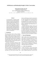
Amphiphilic peptides are composed of hydrophilic and hydrophobic blocks
(residues). By self-assembly, they can form various morphologies such as
micelles
18
, vesicles
19
, fibers
20
and hydrogels,
21
as illustrated in Figure 1.1.
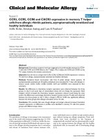
Figure 1.1. Different morphologies formed by amphiphilic molecules (a)
micelle
22
(b) vesicle
23
(c) nanofiber.
20
(a)
(b)
(c)
Chapter 1. Introduction
3
The morphologies formed are dependent on the ratio of hydrophobic to
hydrophilic blocks, peptide sequence, concentration, and other factors. Generally,
hydrophilic blocks favor to form micelles, hydrophobic blocks would produce
nano-particles, and blocks with intermediate hydrophobicity could tend to form
vesicles.
4,19,24
The morphologies formed by peptides can encapsulate drug molecules and
deliver drug to cancer cells. Their size is usually less than 100 nm, which is an
advantage to hide from reticuloendothelial system (RES) of human body.
11
In
addition, the hydrophilic shell can keep the structures untraceable during blood
circulation.
25
More importantly, their size, stability, permeability and elasticity
can be fine-tuned by tailoring peptide sequence, length, solution conditions, etc.
With 20 naturally occurring amino acids, it can be envisioned that tremendously
large number of peptides would be explored.
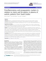
1.2 Amino Acids
Peptides are composed of amino acids as the basic building blocks connected by
peptide bonds (-CO-NH-). Each amino acid has a central α-carbon atom attached
with four different groups including a basic amino group (-NH
2
), an acidic group
(-COOH), a hydrogen atom (-H) and a functional side chain (-R). As illustrated in
Figure 1.2, a peptide bond is formed between the carbon atom of carboxyl group
in one amino acid and the nitrogen atom of amine group in the other amino acid.
Peptides usually consist of 2-50 amino acids, and long chains of peptides are
known as proteins. There are 20 naturally occurring amino acids (see Figure 1.3)
utilized in the synthesis of peptides and proteins in biological cells.
26
Therefore,
Chapter 1. Introduction
4
almost unlimited number of peptides can be formed with various arrangements
and combinations of 20 amino acids.
Figure 1.2. Representation of peptide bond formation between two amino acids.
Figure 1.3. Structures and classifications of 20 amino acids.
26
-H
2
O
Chapter 1. Introduction
5
Depending on the side chain functional groups, amino acids possess different
properties. Table 1.1 lists the physical properties (e.g. pKa and hydropathy index).
Different classifications exist for the 20 amino acids based on the functionality of
side chain, polarity, essential or non-essential, etc. Among these, hydrophobicity
and polarity-based classifications are the most commonly used. Specifically,
amino acids are classified into hydrophobic, hydrophilic, charged and others. The
hydrophobic amino acids are further classified into aliphatic (A, I, L, M and V)
and aromatic (F, W and Y); the hydrophilic amino acids (S, N, T and Q) possess
hydrogen bonding capability; the charged amino acids are either positively
charged (H, R and K) or negatively charged (D and E); the remaining (C, P and
G) belong to the others.
Another classification is based on polarity, including polar charged, polar
uncharged and nonpolar types.
27
The polar charged amino acids (K, R, H, D and
E) have two subtypes namely acidic (negatively charged D, E) and basic
(positively charged K, R and H); the polar uncharged include S, T, N, Q, Y and C;
nonpolar type are G, A, V, L, I, M, P, F and W. One more type of classification is
based on nutritional supplement to human body by internal metabolism (non-
essential) or external supplements (essential). Out of 20 amino acids, human body
can produce 11 that are typically non-essential, the remaining 9 have to be
procured by external supplements such as food and known as essential amino
acids (I, L, K, M, F, T, W and V).