Characterization of novel anticoagulants from hematophagous arthropods
Bạn đang xem bản rút gọn của tài liệu. Xem và tải ngay bản đầy đủ của tài liệu tại đây (3.51 MB, 176 trang )
CHARACTERIZATION OF NOVEL ANTICOAGULANTS
FROM HEMATOPHAGOUS ARTHROPODS
TAN WEI LING, ANGELINA
(B.Sc. (Hons.), National University of Singapore)
A THESIS SUBMITTED
FOR THE DEGREE OF DOCTOR OF PHILOSOPHY
NUS GRADUATE SCHOOL FOR INTEGRATIVE
SCIENCES AND ENGINEERING
NATIONAL UNIVERSITY OF SINGAPORE
2014
DECLARATION
I hereby declare that this thesis is my original work and it has been written by
me in its entirety. I have duly acknowledged all the sources of information which
have been used in the thesis.
This thesis has also not been submitted for any degree in any university
previously.
_____________________________________
Tan Wei Ling, Angelina
8
th
September 2014
ACKNOWLEDGMENTS
I would like to thank my PhD supervisor, Prof. R. Manjunatha Kini, for
his support during my entire PhD candidature. He was very enthusiastic and
welcomed me into his lab when I first approached him in my first year.
Throughout the years, he has always taught us not be limited by boundaries
and walls, and to break the wall when it gets in our way! His pep talks and
stories were a constant motivation during this whole time.
I would like to express my gratitude to our collaborators from the Slovak
Academy of Sciences, Bratislava - RNDr. Mirko Slovak and RNDr. Maria
Kazimirova, who has been the source of ticks for all these experiments. Thank
you for your hard work and efforts and always striving to meet our endless
requests for the ticks. Thank you for your hospitality when I was at your institute
and showing me how tick collection is performed in the wild. It was indeed an
eye-opening hands-on trip. Another collaborator of ours - Dr. Jose Ribeiro,
thank you for taking time out to personally teach me how raw reads are
processed and all the bioinformatics involved. Thank you for patiently
answering my endless questions regarding the tables, and all the calculations.
I would also like to thank members of Protein Science Laboratory who
have worked with me and have brought many joys, whether during benchwork,
lab meetings, nonsense in the student room or chai pani session etc. Special
thanks to Cho Yeow, my senior, who got me acquainted with the world of ticks
and blood coagulation. Buddies Shiyang, Amrita, Girish and Sheena, you guys
never fail to brighten my day in the lab, and I am glad for the strong friendship
forged after all these years together. To Sindhuja and Janaki, thank you for
supporting me by providing a listening ear whenever I needed to “release
tension”. I will always remember all the silly things we have done outside of the
lab. Lastly, thanks to all else who worked along me in the lab and made the lab
a pleasant environment to work in: Ryan, Bhaskar, Bidhan, Summer, Angie,
Chen Wan, Ritu, Norrapat, Feng Jian, Varuna, and all the honours students
who have spent time with us. To Bee Ling and Liyuan, our lab officers, thank
you for supporting our lab, and always helping us out with a big smile on your
faces. Thank you for helping us process our orders timely without fail, keeping
the lab orderly and sane, and always entertaining all sorts of nonsense from us.
I would like to thank my PhD buddies – Veronica and Carol. I would
never have lasted through without both of you guys. Thank you for your
constant support, encouragement, feedbacks and suggestions. Thank you also
for applying the right pressure on me, not too much but not too little as well, and
always at the right time.
I would also like to thank NGS for awarding me with the PhD
scholarship, allowing me this opportunity to further my studies in NUS. Thank
you for providing the funding for overseas attachments. A big thank you to Irene
who has always cheered me on whenever I showed up at the NGS office.
A special thanks to my family members, who have always supported
the decisions I have made in life. This whole period wasn’t easy for me, Mom
and Dad, thank you for cutting me some slack when I was not having the best
of days. To my Chieh, thank you for your support, encouragements, and thank
you for letting me feel I am actually doing something of worth. I know I have
ii
always fanned you off when you have asked me to teach you a thing or two
about anticoagulants, and I hereby promise I’ll teach you now. Thank you
Gerald, for being the best chauffeur in the world, allowing me to have that extra
sleep in the mornings on the way to school.
Last but definitely not the least, a loving thanks to my fiancé, Hansel.
Thank you for going through thick and thin, ups and down together with me,
Thank you for being patient with me, being understanding, and being my pillar
of support.
iv
TABLE OF CONTENTS
ACKNOWLEDGMENTS i
TABLE OF CONTENTS v
Summary viii
List of Tables x
List of Figures xii
List of Abbreviations xiv
Chapter 1: Introduction 1
1.1 Hemostasis 3
1.1.1 Overview 3
1.1.2 Vasoconstriction 3
1.1.3 Platelet aggregation 4
1.1.4 Blood coagulation 6
1.1.4.1 Initiation 6
1.1.4.2 Amplification 8
1.1.4.3 Propagation 8
1.1.5 Fibrinolysis 8
1.2 Hematophagous animals 10
1.2.1 Anticoagulants from hematophagous animals 11
1.2.1.1 Thrombin inhibitors 12
1.2.1.2 FXa inhibitors 17
1.2.1.3 Extrinsic Tenase Complex Inhibitors 20
1.2.1.4 Intrinsic Tenase Complex Inhibitors 21
1.3 Ticks 22
1.3.1 Feeding behaviours of ticks 22
1.3.2 Sexual reproduction in ticks 23
1.4 Aim and scope of the thesis 25
Chapter 2: Materials and Methods 28
2.1 Salivary glands and extracts 30
2.2 Purification and anticoagulation activity testing 31
2.2.1 Protein quantification 31
2.2.2 Enzymatic inhibition assays 32
2.2.3 Purification of crude salivary extracts 33
2.2.3.1 Size exclusion chromatography 33
2.2.3.2 FXa affinity chromatography 33
2.2.3.3 Reverse phase HPLC 34
2.3 Transcriptomics 34
2.3.1 cDNA library construction 34
2.3.2 Transcriptome assembly and bioinformatics 35
2.3.3 Sequence analyses 36
2.4 Proteomics 36
2.4.1 Tryptic Digestion 36
2.4.2 SDS-PAGE 37
2.4.3 Sample clean-up 38
2.4.4 Mass spectrometry 38
2.5 Quantitation of differential expression 39
2.5.1 RNA Isolation and first-strand cDNA synthesis 39
2.5.2 Primer design 40
2.5.3 Polymerase chain reaction amplification 42
2.5.4 DNA gel electrophoresis 42
2.5.5 DNA sequencing 43
2.5.6 Quantitative Real Time – Polymerase Chain Reaction 44
Chapter 3: Results 48
3.1 Anticoagulant activity of ticks 50
3.1.1 Protein quantification of crude salivary gland extracts 50
3.1.2 Activity of crude salivary gland extracts 50
3.1.3 Purification of salivary gland extracts 51
3.2 Sialome of R. pulchellus 55
3.2.1 Transcriptomes of R. pulchellus 58
3.2.1.1 Public sequence disclosure 60
3.2.2 Proteomes of R. pulchellus 61
3.2.3 Housekeeping proteins 63
3.2.4 Putative secreted proteins 68
3.2.4.1 Enzymes 89
3.2.4.2 Proteinase inhibitor domains 92
3.2.4.3 Immunity-related proteins 94
vi
3.2.4.4 Antimicrobial peptides 95
3.2.4.5 Tick-specific protease inhibitors 95
3.2.4.6 Glycine-rich proteins and mucins 96
3.2.4.7 Lipocalins 97
3.2.4.8 Ixodegrins 98
3.2.4.9 DA-p36 family 99
3.2.4.10 Evasins 99
3.2.4.11 Immunoglobulin G binding proteins 99
3.2.4.12 Tick-specific, unknown function 100
3.2.5 Transposable elements 103
3.3 Gender-dependent expression of Bilaris proteins 104
3.3.1 Subclasses of R. pulchellus Bilaris proteins 104
3.3.2 Differential expression of bilaris proteins 108
3.3.3 Relative abundance of bilaris proteins 111
Chapter 4: Discussion 118
4.1 Anticoagulant activities of R. pulchellus 120
4.2 Sialome of R. pulchellus 122
4.3 Reproduction of ticks 127
4.4 Bilaris proteins 128
Chapter 5: Conclusion and Future Perspectives 133
5.1 Conclusion 135
5.2 Future perspectives 137
5.2.1 Functional studies on bilaris proteins 137
5.2.2 Functional studies on monolaris protein 138
5.2.3 Time-dependent expression of salivary proteins 139
References 141
Appendices 153
Supplemental Files …………………………………………………… …… DVD
Summary
Ticks are hematophagous arthropods that rely exclusively on blood for
their survival. During feeding, ticks inject into their hosts, a complex salivary
cocktail that induces vasodilation, and impedes platelet aggregation, blood
clotting and host immunity, thus overcoming host responses. These
pharmacological mediators may also enhance the efficiency of pathogen
transmission. Although both male and female ticks feed on blood, the manner
that they feed off their host differs in length of time and volume taken up.
Firstly, to investigate the difference in salivary composition between
male and female Rhipicephalus pulchellus, we profiled the salivary gland
extracts in terms of its anticoagulant properties. While the female salivary
glands extracts displayed excellent inhibition towards key blood coagulation
factors FXa and thrombin, that of males showed poor inhibition properties.
Further, we also established that the salivary protein content between the two
genders differs.
In order to obtain information on the salivary transcriptome of R.
pulchellus, we sequenced two cDNA libraries from pools of adult males and
females salivary glands at different feeding time points, using the Illumina
HiSeq protocol. De novo assembly of a total of 241,229,128 paired-end reads
lead to the extraction of 50,460 coding sequences (CDS). In addition, we
generated the proteomes of male and female R. pulchellus separately, which
yielded a total of 454 and 2,063 proteins, respectively, which were identified by
one or more peptides with at least 95% confidence.
viii
A comparison between the male and female tick sialome revealed male-
and female-specific transcripts. From the proteome, 169 and 1,777 proteins
were found exclusively in males and females respectively. We hypothesize that
certain classes of proteins which were highly expressed in the male glands may
be involved in reproduction as males use their mouthparts to introduce their
spermatophores into the females’ genital pore during copulation. In addition.
we have analyzed Kunitz-type serine protease inhibitors in detail and we report
five new subclasses of bilaris proteins. qPCR data suggests that male and
female R. pulchellus selectively express certain subclasses of these proteins.
The analyses of the sialomes of male and female ticks independently
allow us to understand the various strategies used by each gender which
enables them to feed successfully off their hosts. It has opened up opportunities
to discover new salivary proteins and determine candidate male salivary
proteins that may assist reproduction. Knowledge of the salivary protein
repertoire of ticks may also lead to vaccine targets to disrupt feeding and/or
parasite transmission as well as lead to the discovery of novel pharmacological
agents.
List of Tables
Table 1. SDS-PAGE composition 37
Table 2. List of primers for qPCR 41
Table 3. PCR reaction mix 42
Table 4. Cycle sequencing reaction mix 43
Table 5. Protein quantification 50
Table 6: Functional classification of extracted coding sequences (CDS)
from the sialotranscriptome of R. pulchellus. 58
Table 7: Functional classification of proteins identified from the proteome
of R. pulchellus. 61
Table 8: Functional classification of extracted coding sequences (CDS)
from the putative housekeeping class from the sialotranscriptome
of R. pulchellus. 66
Table 9: Functional classification of proteins from the putative
housekeeping class from the proteome of R. pulchellus. 67
Table 10. Functional classification of extracted coding sequences (CDS)
from the putative secreted class from the sialotranscriptome of
adult Rhipicephalus pulchellus ticks. 69
Table 11. Functional classification of extracted coding sequences (CDS)
from the putative secreted class from the proteome of adult
Rhipicephalus pulchellus ticks. 74
Table 12: Number of gender-biased CDS from the secretory class found
in the transcriptome 80
x
Table 13: Number of gender-biased proteins from the secretory class
found in the proteome 84
Table 14. Total number of sex-biased CDS in the R. pulchellus
transcriptome 124
Table 15. Total number of gender-biased proteins from the R. pulchellus
proteome. 125
List of Figures
Figure 1.1 Platelet activation 5
Figure 1.2 Blood coagulation cascade 7
Figure 3.1. Anticoagulant activity of crude R. pulchellus salivary gland
extracts. 51
Figure 3.2. Anticoagulant profile of R. pulchellus SGEs. 52
Figure 3.3. Anticoagulant profile of D. reticulatus SGEs. 54
Figure 3.4. Reverse phase chromatography of FXa inhibitors from D.
reticulatus on C18 column. 55
Figure 3.5. FXa-affinity column purification of DRFXaI-3. 57
Figure 3.6. Components of R. pulchellus transcriptome 59
Figure 3.7. Components of R. pulchellus proteome 62
Figure 3.8 Differential expression of secretory proteins 78
Figure 3.9. Classification of gender-biased CDS in R. pulchellus
transcriptome. 83
Figure 3.10. Classification of gender-biased proteins in R. pulchellus
proteome. 88
Figure 3.11. Five subclasses of bilaris proteins. 106
Figure 3.12. Sequence alignment of bilaris subclases. 107
Figure 3.13. Phylogenetics and associated number of reads of bilaris CDS
109
Figure 3.14. Expression difference of bilaris proteins between male and
female R. pulchellus. 110
Figure 3.15. PCR screening of qPCR primers. 112
Figure 3.16. Quantitative PCR on selected bilaris proteins. 114
xii
Figure 3.17. DNA gel electrophoresis of bilaris proteins. 116
Figure 3.18. APTT assay of expressed RpSigp-759502 117
Figure 3.19. Sequence alignment of bilaris proteins. 131
List of Abbreviations
Single and three letter abbreviations of amino acids were followed as per the
IUPAC-IUBMB Joint Commission on Biochemical Nomenclature.
Chemicals and reagents
ACN Acetonitrile
APS Ammonium persulfate
BSA Bovine serum albumin
CaCl
2
Calcium chloride
CNBr Cyanogen bromide
dNTPs Deoxyribonucleotide triphosphate
DTT Dithiothreitol
EDTA Ethylenediamine tetraacetic acid
FA Formic acid
FXa Factor Xa
HCl Hydrochloric acid
NaCl Sodium chloride
PBS Phosphate buffered saline
SDS Sodium dodecyl sulfate
S2222 Benzoyl-IIe-Glu (Glu-γ -methoxy)-Gly-Arg-p-nitroanilide
(pNA) hydrochloride (HCl)
S2238 H-D-Phe-pipecolyl (Pip)-Arg-pNA•2HCl
TBS Tris buffered saline
TCEP Tris(2-carboxyethyl)phosphine
TEMED N,N,N’,N’-Tetramethylethylenediamine
TF Tissue factor
TFA Trifluoroacetic acid
Tris Tris(hydroxymethyl)-aminomethane
xiv
Units
Å Angstrom
Bp Base-pair
Da Daltons
h Hour
kbp Kilo base-pair
kDa Kilo daltons
M Molar
mg Milli-gram
min Minute
ml Milli-litre
mM Milli-molar
mm Milli-metre
ng Nano-gram
nl Nano-litre
nM Nano-molar
nm Nano-metre
rpm Revolutions per minute
µg Micro-gram
µl Micro-litre
µM Micro-molar
µm Micro-metre
V Volt
⁰C Degree Celsius
% Percent
Others
ADP Adenosine diphosphate
AFXa Anticoagulant-factor Xa
APTT Activated partial thromboplastin time
BPTI Bovine pancreatic trypsin inhibitor
CDD Conserved domain database
CDS Coding sequence
EST Expressed sequence tags
FXaI FXa-inhibitor
GP Glycoprotein
HPLC High performance liquid chromatography
IG Immunoglobulin
IGFBP Insulin growth factor binding proteins
iTRAQ Isobaric tags for relative and absolute quantitation
ML MD-2-related lipid-recognition
MS Mass spectrometry
ORF Open reading frame
PAR Protease-activated receptors
PCR Polymerase Chain Reaction
qRT-PCR Quantitative Real Time – Polymerase Chain Reaction
RQ Relative quantitation
SDS-PAGE Sodium dodecyl sulfate polyacrylamide gel electrophoresis
SG Salivary glands
SGE Salivary gland extracts
TAP Tick anticoagulant peptide
TE Transposable elements
TEP Thioester containing proteins
TIL Trypsin inhibitor-like
t-PA Tissue-type plasminogen activator
vWF von Willebrand factor
xvi
CHAPTER 1
Introduction
Chapter 1: Introduction
1
Chapter 1: Introduction
2
Chapter 1: Introduction
1.1 Hemostasis
1.1.1 Overview
The cardiovascular system is one of the most important organ system
and is crucial for the survival of an organism. Within this system, blood
circulates and transports nutrients, oxygen and other compounds to all parts of
the body. In vertebrates, it is a closed and high-pressured system. Thus, when
there is a breach, if large enough, it can be life-threatening. It is hence critical
for the body to seal this breach to prevent excessive loss of blood. Hemostasis
is a process that maintains the integrity of this system when damage occurs. It
is regulated by three basic mechanisms, namely vasoconstriction, platelet
aggregation and blood coagulation.
1.1.2 Vasoconstriction
When injury to the blood vessels occur, the constriction of the blood
vessels is the first response. Endothelial dilating agents, such as nitric oxide,
adenosine and prostacyclins, which are present under normal conditions, are
reduced. In addition, adenosine diphosphate (ADP), serotonin and
thromboxane are released and they act on the vascular smooth muscle cells to
trigger constriction of the vessels (Becker et al., 2000). Endothelin, a potent
vasoconstrictor, is also produced and released by endothelial cells. This first
phase of hemostasis aims to reduce and even stop the flow of blood.
3
Chapter 1: Introduction
1.1.3 Platelet aggregation
The second phase of hemostasis is the aggregation of blood platelet
cells. Endothelial cells normally produce nitric oxide and prostacyclin I
2
which
suppresses platelets adhesion and aggregation. During vessel injury when the
endothelium is disrupted, collagen is exposed to the circulating blood, thus
triggering the activation of platelets. Figure 1.1 illustrates a platelet and its
receptors and agonists that binds to it. Collagen binds to the glycoprotein (GP)
VI on the platelet while collagen-bound von Willebrand factor (vWF) binds to
GPIb/V/IX and integrin α
IIb
β
3
, which is the most important adhesive receptor for
platelet aggregation (Jennings, 2009; Versteeg et al., 2013). Integrin α
2
β
1
, also
plays a role in platelet adhesion and anchoring, supporting platelet interaction
via other platelet receptors (Clemetson and Clemetson, 2001; Nieswandt et al.,
2011). The above mediates the adhesion of the platelets to the site of injury.
Thereafter, platelet activation causes the release of ADP from granules with the
platelets and signals the aggregation of other free platelets to the site of injury
(Versteeg et al., 2013). During this process of platelet activation, the platelets
undergo a change of shape to become more rounded.
A second pathway, triggered by tissue-factor and independent of
collagen, can also initiate platelet activation. This pathway is dependent on
thrombin, and is part of the blood coagulation cascade which will be elaborated
in detail in the next section. Thrombin, which is activated through this pathway,
interacts with the protease-activated receptors (PAR) on the surface of platelets
thereby activating them (Furie and Furie, 2008). This results in the release of
ADP, serotonin and thromboxane A
2
, which further activates other platelets
(Brass, 2003).
4
Chapter 1: Introduction
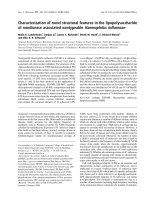
Figure 1.1 Platelet activation. Platelets have various receptors for recruitment
and activation for platelet plug formation. Upon activation, the granules release
agonists such as ADP, serotonin and TXA
2
which further amplifies and
propagate the activation of other platelets.
ADP
PAR
PGD
PGI
GPVI
FcRγ
Thrombin
TXA2
ADP
Serotonin
vWF
PAF
TXA
2
α
IIb
β
3
GPIb/V/IX
vWF
α
2
β
I
5