DEVELOPMENT AND APPLICATION OF NEW APPROACHES FOR STUDIES OF INFLUENZA INDUCED INFLAMMATION AND DNA DAMAGE
Bạn đang xem bản rút gọn của tài liệu. Xem và tải ngay bản đầy đủ của tài liệu tại đây (3.59 MB, 16 trang )
RESEARCH ARTICLE
Influenza infection induces host DNA damage and dynamic DNA
damage responses during tissue regeneration
Na Li
1,3
•
Marcus Parrish
2
•
Tze Khee Chan
1,4
•
Lu Yin
1
•
Prashant Rai
1,3
•
Yamada Yoshiyuki
1
•
Nona Abolhassani
2
•
Kong Bing Tan
5
•
Orsolya Kiraly
1
•
Vincent T. K. Chow
3
•
Bevin P. Engelward
2
Received: 30 September 2014 / Revised: 18 February 2015 / Accepted: 2 March 2015
Ó Springer Basel 2015
Abstract Influenza viruses account for significant mor-
bidity worldwide. Inflammatory responses, including
excessive generation of reactive oxygen and nitrogen spe-
cies (RONS), mediate lung injury in severe influenza
infections. However, the molecular basis of inflammation-
induced lung damage is not fully understood. Here, we
studied influenza H1N1 infected cells in vitro, as well as
H1N1 infected mice, and we monitored molecular and cel-
lular responses over the course of 2 weeks in vivo. We show
that influenza induces DNA damage to both, when cells are
directly exposed to virus in vitro (measured using the comet
assay) and also when cells are exposed to virus in vivo
(estimated via cH2AX foci). We show that DNA damage, as
well as responses to DNA damage persist in vivo until long
after virus has been cleared, at times when there are in-
flammation associated RONS (measured by xanthine
oxidase activity and oxidative products). The frequency of
lung epithelial and immune cells with increased cH2AX foci
is elevated in vivo, especially for dividing cells (Ki-67-
positive) exposed to oxidative stress during tissue regen-
eration. Additionally, we observed a significant increase in
apoptotic cells as well as increased levels of DNA double
strand break (DSB) repair proteins Ku70, Ku86 and Rad51
during the regenerative phase. In conclusion, results show
that influenza induces DNA damage both in vitro and
in vivo, and that DNA damage responses are activated,
raising the possibility that DNA repair capacity may be a
determining factor for tissue recovery and disease outcome.
Keywords Nuclear foci Á Immunofluorescence Á
Repair deficiency Á Acute infection
Abbreviations
edA 1, N
6
-Etheno-2
0
-deoxyadenosine
edG 1, N
2
-Etheno-2
0
-deoxyguanosine
8-OH-dG 8-Hydroxy-deoxyguanosine
8-OH-G 8-Hydroxyguanosine
AEII Alveolar epithelial type II cells
ATM Ataxia telangiectasia mutated
ATR ATM- and Rad-3 related
BER Base excision repair
BALF Bronchoalveolar lavage fluid
CCSP Club cell secretary protein
DDR DNA damage response
DSBs DNA double-strand breaks
DNA-PKcs DNA-dependent protein kinase
catalytic subunit
Electronic supplementary material The online version of this
article (doi:10.1007/s00018-015-1879-1) contains supplementary
material, which is available to authorized users.
& Bevin P. Engelward
1
Singapore-MIT Alliance for Research and Technology, 1
CREATE Way, #03-10/11 Innovation Wing, #03-12/13/14
Enterprise Wing, Singapore 138602, Singapore
2
Department of Biological Engineering, Massachusetts
Institute of Technology, 77 Massachusetts Ave., 16-743,
Cambridge, MA 02139, USA
3
Department of Microbiology, National University of
Singapore, 5 Science Drive 2, Blk MD4, Level 3, Singapore
117545, Singapore
4
Department of Pharmacology, Yong Loo Lin School of
Medicine, National University Health System, Clinical
Research Center, MD11, 10 Medical Drive, Level 5, #05-09,
Singapore 117597, Singapore
5
Department of Pathology, Yong loo Lin School of Medicine,
National University Health System and National University
of Singapore, Lower Kent Ridge Road, Singapore 119074,
Singapore
Cell. Mol. Life Sci.
DOI 10.1007/s00018-015-1879-1
Cellular and Molecular Life Sciences
123
HA Hemagglutinin
HR Homologous recombination
MDCK Madin–Darby canine kidney
MOI Multiplicity of infection
NHEJ Non-homologous end joining
NS1 Non-structural protein 1
PI3K-like kinases Phosphatidylinositol-3-kinase-like
kinases
Pro-SPC Pro-surfactant protein C
RONS Reactive oxygen and nitrogen species
SSBs DNA single strand breaks
XO Xanthine oxidase
Introduction
Influenza A viruses are a group of respiratory pathogens that
pose significant health burden worldwide. It has been shown
that damage to lung tissue is not only a result of virus-in-
duced cytopathy, but also due to cytotoxic effects of aberrant
and excessive inflammation [1, 2]. Inflammation-induced
reactive oxygen and nitrogen species (RONS) constitute one
of the key contributors of pathogenicity in severe influenza A
viral infections [3–5]. However, the underlying mechanisms
of RONS-induced pathogenesis are not fully understood.
RONS exposure leads to DNA lesions, which can promote
mutations and cell death [6, 7]. Hence, we hypothesize that
oxidative DNA damage is induced by influenza-induced
inflammation, which may contribute to cytotoxicity in vivo.
Inflammation induces many types of base lesions [e.g.,
8-hydroxy-deoxyguanosine (8-OH-dG) and 8-ni-
troguanosine] [6, 8], from which DNA strand breaks can
arise via chemical reactions, or via enzymatic processes
associated with DNA repair or replication fork breakdown
[9, 10]. In the presence of DNA damage, cells respond by
eliciting DNA damage responses (DDR), which include
activation of cell cycle arrest, DNA repair, senescence or
cell death, depending on the cell type and severity of DNA
damage [11, 12]. DDR is orchestrated by many events
including post-translational modification of chromatin,
which can mediate signal transduction and assembly of
repair proteins at the site of DNA strand breaks [13, 14].
Phosphorylation of H2AX histones at Ser-139 (cH2AX) is
a well-studied example of chromatin modification that
occurs following formation of DNA double-strand breaks
(DSBs), via the activity of phosphatidylinositol-3-kinase-
like kinases (PI3K-like kinases), such as ataxia telangiec-
tasia mutated (ATM) kinase and DNA-dependent protein
kinase catalytic subunit (DNA-PKcs). Signal amplification
causes the phosphorylation of H2AX proteins to spread
along approximately two megabases around the site of each
DSB, to yield cH2AX foci that are visible and quantifiable
by immunofluorescence microscopy [15, 16]. Interestingly,
cH2AX foci can also be triggered by stalled replication
fork via ATM- and Rad-3-related (ATR) kinase-dependent
phosphorylation. These stalled replication forks can be
associated with cH2AX and can breakdown to form phy-
sical DSBs [17, 18]. Therefore, phosphorylated cH2AX
foci indicates the presence of biologically significant DNA
damage, and serves as an excellent approach for investi-
gating DSBs and DNA damage-induced by replicative
stress during influenza infection.
Importantly, the biological importance of cH2AX lies in
its involvement in recruiting DNA repair proteins and
maintenance of cell cycle arrest to facilitate repair of DSBs
[19]. Two dominant DSB repair pathways are evolved to
counteract the detrimental effects of DSBs, namely non-
homologous end joining (NHEJ) and homologous recom-
bination (HR). NHEJ is a rapid joining process that does
not require a homologous DNA template. HR is a pathway
that enables retrieval of genetic information at the site of
the DSB by homology searching, strand invasion and repair
synthesis [20]. Both HR and NHEJ require concerted in-
volvement of many DNA repair proteins [19], and defects
in DSB repair can contribute to chromosomal breakage and
large scale sequence rearrangements that promote cyto-
toxicity and mutagenesis, respectively [21,
22]. Here, we
hypothesize that influenza infection induces DNA damage,
and that DNA damage responses modulate cytotoxicity and
tissue damage in infected mice.
In this study, we used the PR8 mouse model of influenza
A (H1N1) virus infection to explore the impact of influenza
infection and inflammation on DNA damage and DNA
damage responses. By studying chromatin phosphorylation
as a measure of DNA damage, we show that the level of
DNA damage increases following influenza infection, and
we provide data that supports a role for replication fork
breakdown as a driver of DNA strand breaks. Importantly,
we observed a significant increase in the levels of proteins
involved in DSB repair, namely Ku70, Ku86, Rad51 and
PCNA, especially during the tissue regenerative phase,
suggesting that DNA repair was induced following infec-
tion. Together, these studies raise the possibility that DNA
damage and DNA repair modulate the severity of influen-
za-induced cytotoxicity, thereby affecting tissue damage
and regeneration, and ultimately disease outcome.
Materials and methods
Cell culture, infection and immunofluorescence
Madin–Darby canine kidney (MDCK) cells were cultured
on gelatinized coverslips overnight, and subsequently in-
fected with PR8 influenza at multiplicity of infection
N. Li et al.
123
(MOI) of one, diluted in 2 mg/mL bovine serum albumin
(BSA) (Sigma) and 2 lg/mL tosyl phenylalanyl chlor-
omethyl ketone treated (TPCK-) trypsin (Sigma) in
minimum essential medium (MEM) (Invitrogen) for 3, 6, 9,
or 12 h. Non-treated cells were incubated with 2 mg/mL
BSA and 2 lg/mL TPCK-trypsin in MEM for 12 h. Cells
were then fixed and incubated with 2 lg/ml mouse anti-
cH2AX (Millipore) overnight at 4 °C. Stained cells were
then incubated with FITC-conjugated anti-mouse antibody
(Santa-Cruz), mounted with ProLong Antifade containing
DAPI (Invitrogen) and imaged with a Nikon 80i upright
microscope under 609 magnification. At least ten images
were taken per time-point in a blinded fashion. To quantify
cH2AX-positive cells, images were ‘‘blinded’’ and counted
manually for DAPI-positive nuclei. At least 100 cells were
counted for each sample, with the exception of three
samples for which 82–99 cells were quantified. Nuclei
harboring 5 or more cH2AX foci were considered positive
for cH2AX. Three independent biological replicates were
performed for each condition and time-point.
CometChip for high-throughput comet assays
of influenza-infected cells
CometChip was fabricated using a polydimethylsiloxane
(PDMS, Dow Corning) mold as described previously [23,
24]. Briefly, molten 1 % normal melting point agarose
(Invitrogen) was applied to a sheet of GelBond film
(Lonza), and allowed to gel with the PDMS mold on top.
Removal of the PDMS mold revealed a *300 lm thick gel
with arrayed microwells. The microwell gel was then
clamped between a glass plate and either a bottomless
24-well or 96-well titer plate (Greiner BioOne) to create
the CometChip. Cells were added to each well of the
CometChip, and allowed to settle by gravity in complete
growth media at 37 °C, 5 % CO
2
. Excess cells were aspi-
rated after 15 min and the bottomless plate was removed to
capture the arrayed cells in a layer of 1 % low melting
point agarose (Invitrogen).
After encapsulation in agarose, the bottomless plate
was re-aligned to the original position on the CometChip.
Wells were infected with 50 lLofPR8influenzavirusat
MOI of *1 in virus medium (0.2 % bovine serum albu-
min, 2 lg/mL TPCK-trypsin in minimum essential
medium) at 37 °C. Negative controls were treated with
50 lL of virus medium under the same conditions. After
1 h, the bottomless plate was removed, and all wells were
incubated with 0.2 % bovine serum albumin and 2 lg/mL
TPCK-trypsin in Opti-MEM at 37 °C. At 3, 6 and 9 h
after influenza exposure, at least three influenza virus-
infected wells were processed according to either the al-
kaline or neutral comet assay described in Supplementary
methods and materials.
Fluorescence imaging and comet analysis
After electrophoresis, alkaline comet and neutral comet
gels were neutralized in 0.4 M Tris, pH 7.5 (2 9 15 min)
and stained with SYBR Gold (Invitrogen). Images were
captured using an automated epifluorescent microscope,
and analyzed using custom software written in MATLAB
(The Mathworks) [23].
Mouse model and infection
9–12 weeks old C57Bl6Ntac mice (InVivos) were infected
with a sublethal dose (12–15 PFU) of H1N1 Influenza
A/Puerto Rico/8/34 (PR8) by intratracheal instillation,
while uninfected controls were instilled with same volume
of sterile PBS. Procedures were performed in accordance to
guidelines and protocols approved by Institutional Animal
Care and Use Committee (IACUC). Left lungs were fixed
in 10 % neutral buffered formalin and paraffin-embedded.
Alternatively, they were embedded in optimal cutting
temperature compound and frozen for histology. Right
lungs were frozen in liquid nitrogen or lavaged with 1 ml
ice-cold PBS to collect bronchoalveolar lavage fluid
(BALF).
Lung homogenization and virus titration
Apical and cardiac lobes were homogenized with 300 llof
PBS with Stainless steel beads (Qiagen) and Qiagen Tis-
sueLyser (max oscillation speed, 2 min, 4 °C). Lung
homogenate was spun down at 3000 rcf for 10 min at 4 °C
and stored at -80 °C. Virus titration with MDCK cells was
performed based on previous publication [25]. Plaque
forming units (PFU) were normalized to protein concen-
tration of lung homogenate estimated with a Bradford
assay (for more details of plaque assay, please see Sup-
plementary methods and materials).
Haematoxylin and eosin (H&E) staining
and histopathologic analyses
Paraffin-embedded lung section (5 lm) was stained with
hematoxylin and eosin (H&E) as described previously with
minor modifications [26]. Histopathologic analyses of
H&E stained sections were performed by an experienced
pathologist. A total of 3–5 sections were analyzed per time-
point.
Evaluation of oxidative stress
Lung homogenates processed at various time-points were
diluted 1009–4009 with cold PBS. Xanthine oxidase
quantification was performed with the diluted lung
Influenza infection induces host DNA damage
123
homogenates using a Xanthine oxidase Fluorometric assay
kit (Caymen) based on manufacturer’s protocol. To mea-
sure oxidative damage to nucleic acids, BALF was
collected and centrifuged, and the supernatant was ana-
lyzed with a DNA/RNA Oxidative Damage EIA Kit
(Caymen) that measures the levels of free 8-OH-dG and
8-hydroxyguanosine (8-OH-G) (for quantifications of
modified DNA bases and etheno adducts, please see Sup-
plementary methods and materials).
Immunofluorescence
Paraffin sections were boiled in Target retrieval solution
(Dako) for 30 min, blocked and permeabilized with 10 %
Donkey serum in PBS with 0.3 % Triton-X 100 for 1 h at
room temperature, and then incubated overnight at 4 °C
with 20 lg/ml of anti-cH2AX (Cell Signaling), 1 lg/ml of
anti-Club cell secretory protein (CCSP; Santa-Cruz), 1 lg/
ml of anti-pro-surfactant protein C (SPC; Santa-Cruz) and/
or 1 lg/ml of antibodies against H1N1 non-structural
protein 1 (NS1; Santa-Cruz) in staining buffer (5 % donkey
serum and 0.3 % Triton-X 100 in PBS). Sections were
washed and incubated with 5 lg/ml of Alexafluor dyes-
conjugated secondary antibodies (Molecular probes) for
1 h at room temperature on the following day, followed by
mounting with ProLong gold antifade reagent (Life tech-
nologies). To co-stain for Ki-67 and cH2AX, antigen-
retrieved lung sections were first incubated with 1 lg/ml
anti-Ki-67 (DAKO) for 5 h at room temperature, and in-
cubated with secondary antibodies before tissues were
further probed for cH2AX overnight at 4 °C. Cryosections
(10 lm) were fixed in 4 % PFA for 10 min and stained
with 1 lg/ml of anti-CD3 (eBioscience) and 20 lg/ml of
anti-cH2AX (Cell Signaling) based on the protocol de-
scribed above, except that incubation of primary antibodies
were shortened to 1 h at room temperature (for Terminal
deoxynucleotidyl transferase dUTP nick end labeling
(TUNEL) and quantification, please see Supplementary
methods and materials).
Microscopy
All sections were imaged at 209 magnification with Mirax
Midi slide scanner or at 409 magnification with Zeiss LSM
700 confocal microscope (Carl Zeiss) at a thickness of
3 lm. Bronchial epithelium were identified by positive
CCSP staining and pseudostratified columnar tissue struc-
ture. Almost all bronchi and bronchioles were captured
from each lung section. To collect images for lung
parenchyma (CCSP-negative) and pro-SPC-positive cells,
10 random regions were captured per lung section. Laser
channel for cH2AX was switched off when random fields
were selected to prevent bias.
Manual and semi-automated quantification
of cH2AX-positive cells
Ten images of bronchioles, pro-SPC cells and lung
parenchyma were blindly selected and counted for each
mouse. To quantify nuclei in the lung parenchyma, DAPI-
stained nuclei were counted using Imaris version 7.6.5. At
least 1000 cells in the lung parenchyma were counted for
each mouse.
Nuclei of bronchial epithelium and pro-SPC cells were
counted manually. Bronchioles were first identified by the
presence of CCSP staining in the lumen lined by pseu-
dostratified columnar epithelium. All pseudostratified
columnar cells in the bronchioles were then counted
manually regardless of CCSP expression. At least 400
bronchiolar epithelial cells were counted for each mouse.
Pro-SPC-positive cells were quantified by counting nuclei
surrounded by pro-SPC staining. More than 100 cells were
counted for most mice, except 5 mice where 53–90 cells
were counted as there were fewer pro-SPC-positive cells in
the captured images. To prevent bias, the fluorescence
channel for cH2AX was switched off while manually
counting the number of nuclei. Counted nuclei were la-
beled using the manual spot function on Imaris to identify
counted cells. For DSB analysis, cells harboring 5 or more
foci were considered positive for cH2AX. Cells with pan-
nuclear cH2AX were quantified separately.
To determine the relationship between cell division and
DSB formation, 15 random images were acquired for lung
sections co-stained with Ki-67 and cH2AX. The number of
nuclear Ki-67-positive cells in each 0.1 mm
2
lung area, and
the proportion of cH2AX-positive cells among the Ki-67
positive population were enumerated manually for each
image.
Flow cytometry of BALF cells
Bronchoalveolar lavage fluid cells (1 right lung lavaged
with 1 ml PBS) were pelleted and incubated with 1 ml
ACK lysis buffer (Life Technologies) for 5 min at room
temperature. Cells were then stained with two panels of
fluorophore-conjugated antibodies. Panel 1 consisted of
anti-CD45-APC, anti-Siglec F-PE, anti-CD11b-PE-Cy7,
anti-CD11c-Pacific Blue and anti-GR-1-PerCp Cy5.5.
Panel 2 is comprised of anti-CD45-PE-Cy7, anti-CD3-
APC, anti-CD4-PerCP Cy5.5, anti-CD8a-Pacific Blue and
anti-CD19-FITC. Cells were stained in PBS with 1 % BSA
for 30 min at room temperature, and the populations of
alveolar macrophages (Siglec F?/CD11c?), eosinophils
(Siglec F?/CD11c-), neutrophils (Siglec F-/GR1?/
CD11b?), CD8?T cells (CD3?/CD8a?) and CD4? T
cells (CD3?/CD4?) were quantified based on their surface
N. Li et al.
123
markers [27–29]). All antibodies were purchased from BD
Pharmingen, eBiosciences or Miltenyi biotech. Stained
cells were analyzed with BD LSRFortessa (BD Bioscience)
and FlowJo.
cH2AX staining of BALF cells
Bronchoalveolar lavage fluid cells were spun onto poly-
L-
Lysine slides with a Cytospin 3 cytocentrifuge (Thermo Sci-
entific), fixed with 4 % PFA for 10 min at room temperature,
washed thrice with PBS and blocked/permeabilized with
blocking solution (3 % BSA with 0.1 % Triton-X 100 in PBS)
for 1 h at room temperature. Cells were incubated with 5 lg/
ml of anti-cH2AX (Cell signaling) and 5 lg/ml of anti-F4/80
(Biolegend) in blocking solution for 1 h at room temperature,
washed and stained with 10 lg/ml of Alexafluor dyes-conju-
gated secondary antibodies (Molecular probes) for 45 min at
room temperature, followed by staining with DAPI for
15 min.
Western blotting
Middle and inferior lobes were homogenized with 29
Laemmli sample buffer with DTT and boiled. Protein
concentration was estimated with DC Protein reagent
(Biorad) based on manufacturer’s protocol and diluted to
the same concentration for each batch of mice. Anti-
bodies used included anti-Hemagglutin (HA;
Sinobiological Inc.), anti-cH2AX (Millipore), anti-
Rad51, anti-Ku86 (Santa-Cruz), anti-Proliferating cell
nuclear antigen (PCNA; Santa-Cruz), anti-Ku70 (Cell
Signaling), anti-cleaved capsase 3 (abcam) and anti-b
actin (Sigma). Each blot contained samples from dif-
ferent mice, and seven blots were analyzed for each
protein. Blots were exposed on film and analyzed using
myImageAnalysis version 1.1 (Thermo Scientific).
Bands were selected automatically by myImageAnalysis
software using ‘‘Auto-Analyze Find Bands’’ function.
Only HA and cleaved caspase 3 bands were selected
manually since control samples did not have distinct
bands detectable by myImageAnalysis. In this case, the
selected band widths were the same for every lane in
each blot. Band intensity (volume of band) was quanti-
fied and normalized to uninfected controls and the
housekeeping b-actin protein.
Statistical analysis
Quantification data were analyzed with Student’s t test or
Mann–Whitney U test and western blot analyses were
performed with Wilcoxon signed ranked test using
Graphpad prism unless otherwise stated in the figure
legends.
Results
Influenza infection of cultured cells leads
to an increase in cH2AX foci
We first set out to investigate whether influenza infection
of cultured cells leads directly to DNA damage. For these
studies, MDCK cells were infected with H1N1 virus at a
MOI of 1, fixed at the indicated times, and examined by
immunofluorescence to detect cH2AX (Fig. 1a). The fre-
quency of cells with significant increased DNA strand
breaks was quantified by counting cH2AX-positive cells
that harbor 5 or more cH2AX foci. More than twice as
many cells were cH2AX-positive as early as 3 hpi, com-
pared to uninfected control. The number of cH2AX-
positive cells decreased thereafter, but remained sig-
nificantly higher than uninfected control even after 12 hpi
(Fig. 1b). This result suggests that viral infection induces
DNA strand breaks, at least during the early stage of
infection.
c
ab
d
Uninf.
6 hpi
3 hpi
12 hpi
H2AX (g)
DAPI (b)
Uninf. 3 6 9
0
20
40
60
Time Post Infection (h)
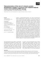
Fig. 1 H1N1 infection of MDCK cells induces DNA damage and
cH2AX foci formation. a MDCK cells infected with PR8 virus at
MOI 1. cH2AX [green fluorescence (g)] at 3, 6 and 12 h post-
infection (hpi) and uninfected controls (Uninf.). (DAPI-stained nuclei
in blue; b blue). Images are representative of three independent
experiments. Scale bar 20 lm. b Percentages of cH2AX-positive
cells (C5 foci per cell). The frequency of cH2AX-positive cells is
significantly higher at 3, 9 and 12 hpi compared to uninfected
controls. c Detection of single strand breaks, abasic sites and alkali
sensitive sites using the alkaline comet assay. d Detection of double-
strand breaks with neutral comet assay. (for b–d, results show
mean ± SD for three independent experiments; *p \ 0.05 for paired
two-tailed student’s t test compared to uninfected controls)
Influenza infection induces host DNA damage
123
To learn more about the potential for influenza to induce
DNA strand breaks, we performed a comet assay, a method
that is well established for directly measuring physical
DNA single stranded lesions and DSBs [23, 24]. The un-
derlying principle of the comet assay is that damaged DNA
migrates more readily when electrophoresed in comparison
to undamaged DNA [30]. We first studied DNA single
strand breaks (SSBs), abasic sites and alkali-labile sites in
MDCK cells using the alkaline comet assay. We observed a
similar trend as compared to the cH2AX assay, wherein
there is a significantly higher percentage of DNA in the
comet tail (percent tail DNA) at 3 hpi compared to unin-
fected controls (Fig. 1c). Similarly, the neutral comet
assay, which detects DSBs, shows that the comet tail length
of influenza-infected cells is significantly higher at 3 hpi
compared to uninfected control in each experiment
(Fig. 1d), suggesting that DSBs are elevated in cells at least
during early hours of infection. The result that 6 and 9 hpi
are not significantly higher than uninfected controls may be
explained by repair of damage, as well as the detection
limits for the neutral comet assay, which requires a mini-
mum of about 40–50 DSBs for detection [16, 31, 32]. In
contrast, cH2AX foci labeled by immunofluorescence give
rise to a signal sufficient for detecting a single DSB [16,
33]. Given that analysis of fluorescent cH2AX foci can be
applied to study DNA damage in fixed tissues, it is thus
used here as an indicator of DNA damage.
Viral load peaks before cellular infiltration
Influenza pathogenesis has long been known to result from
a combination of viral infection and host responses [34]. To
learn about the impact of influenza on DNA damage and
DDRs, we took advantage of a mouse model wherein
C57Bl/6 mice were infected sub-lethally with PR8 virus. In
this model, we found that the viral titer was highest at
5 days post-infection (dpi), and at 9 dpi, median viral titer
was reduced by approximately 100 fold. By 13 dpi, no
virus was detected indicating that PR8 had been cleared
(Fig. 2a). In parallel, significant weight loss among in-
fected mice began at 5 dpi, reached minimum around
9 dpi, and gradually returned to baseline thereafter, sug-
gesting recovery after viral clearance (Suppl. Fig. 1). In
contrast with viral load, which peaked on 5 dpi, whole lung
images stained with H&E (Fig. 2b) show that the density of
infiltrating cells in the lungs was more pronounced from 9
to 17 dpi, suggesting that lung inflammation did not
completely resolve for more than 2 weeks following
infection.
To study the kinetics of immune responses, we analyzed
the immune cell populations among cells in BALF. BALF
cells have been shown to roughly correlate with pathologic
changes in the lung interstitium, thereby providing a means
of sampling the types of cells present in the lungs [35]. Flow
cytometric analysis revealed that total BALF cells increased
with time (Suppl. Fig. 2a), among which CD45-positive
leukocytes peaked at 9 dpi (Fig. 2c). Further analyses indi-
cates that innate cells involved in oxidative burst (namely
infiltrating neutrophils, followed by alveolar macrophages),
were the dominant cell types on 5 dpi, while CD4? and
CD8a? T cells of adaptive immunity were more prevalent at
9 dpi (Fig. 2d). Interestingly, eosinophils, which can con-
tribute to respiratory burst and are commonly associated with
parasites and allergy [36], were a relatively minor proportion
of immune cells, but increased at 13 dpi (Suppl. Fig. 2b).
Consistent with previous studies [24, 25], and with histo-
logical verification by an experienced pathologist, the flow
cytometry shows evidence of a contribution by adaptive
immunity later during disease progression (7–13 dpi). In-
terestingly, histological analysis also clearly indicates the
presence of regenerating lung epithelial cells during the late
time-points (from 13 to 17 dpi, when lymphocytic infiltra-
tion was still prominent) (Suppl. Fig. 2c). Taken together,
these observations demonstrate that immune responses per-
sist after active viral replication, and through until the onset
of tissue regeneration.
Oxidative stress is elevated following infection
To evaluate the kinetics of oxidative stress during influenza
infection, we measured the levels of xanthine oxidase (XO)
and 8-OH-G, which are a reflection of increased RONS
production in the lungs. XO, a superoxide producing en-
zyme that contributes to tissue damage during influenza
infection [5] was significantly increased in the mouse
model on 5, 9, and 13 dpi (Fig. 3a). The highest level of
XO was measured at 9 dpi, which corresponded with
substantial decline in viral load. In addition, 8-OH-G in
cell-free BALF gradually increased after infection, reach-
ing significant levels at 13 dpi (Fig. 3b). 8-OH-G
(including 8-OH-dG) could arise from free guanosine being
oxidized in the extracellular matrix or from accumulation
of 8-OH-G released by dead cells into the extracellular
matrix, both suggesting higher oxidative stress. The ob-
servations that XO and 8-OH-G are elevated demonstrate
that oxidative stress is induced in the lungs after influenza
infection, when the viral load is suppressed.
Host responses induce DNA damage in lung
epithelium after influenza infection
Based upon the observation that there is an increase in
oxidative stress following influenza infection, we asked if
DNA strand breaks occur during the course of infection by
quantifying cells that have increased cH2AX. Whole lung
lysate was first analyzed for influenza antigen HA and
N. Li et al.
123
ab
cd
Uninf. 5
7
9
13 17
Days Post Infection
Days Post Infection
0 5 7 9 13 0 5 7 9 13 0 5 7 9 13 0 5 7 9 13
0
1
2
3
4
5
AM
Neut CD4T
CD8T
AM
Neut
CD4T
CD8T
PFU / mg Protein
Fig. 2 Significant lung inflammation and pathology persist after peak
viral load. a Viral load peaked at 5 days post-infection (dpi). The
number of infectious virus particles (PFU/mg of protein) in lung
homogenate was enumerated by a plaque assay. Median viral load
peaks at 5 dpi and was reduced by *10 fold on 7 dpi, and by *100
fold on 9 dpi (compared to 5 dpi). No viral plaques were detected for
uninfected controls or on 13 dpi. b Cellular infiltration continues after
viral clearance. Whole lung sections were stained with H&E to
evaluate the extent of immune cell infiltration. Regions of high cell
infiltration are associated with darker purple staining due to higher
density of nuclei. Increasing staining density from 5 to 17 dpi is
indicative of increased cellular infiltration. Images are representative
of 8–11 mice. c Quantification of total CD45? leukocyte (results
reflect kinetics of immune cell infiltration into the lungs after
infection). d BALF cell populations are consistent with a transition
from innate to adaptive inflammatory responses. BALF cells lavaged
from right lungs of mice were stained for cell type specific markers
and analyzed by flow cytometry. AM alveolar macrophages, Neut
Neutrophils, CD4 T CD4? T cells, CD8 T CD8? T cells. For 0 dpi,
mice were mock instilled with PBS. (for a, c, d, median is indicated
by the solid line and each symbol represents one animal; *p \ 0.05
compared to uninfected controls for two-tailed Mann–Whitney test;
n = 6–7 mice per time-point)
ab
8-OH-G (pg/ml x 10
4
)
Fig. 3 Oxidative stress increases following infection. a Lung ho-
mogenate was analyzed for XO levels. XO levels significantly
increased from 5 to 13 dpi compared to uninfected controls, and were
highest on 9 dpi. (n = 6–7 mice per time-point) b Free 8-hydrox-
yguanosine (8-OH-G) in bronchoalveolar lavage fluid (BALF) is
higher post-infection. Median 8-OH-G concentration was significant-
ly higher than controls on 13 dpi. (for a, b, median is indicated by the
solid line and each symbol represents one animal; *p \ 0.05
compared to uninfected controls for two-tailed Mann–Whitney test;
n = 3–4 per time-point)
Influenza infection induces host DNA damage
123
phosphorylated cH2AX by western blot (Fig. 4a). Relative
intensities of HA and cH2AX bands were quantified and
normalized to b-actin, and the levels of HA and cH2AX
relative to uninfected controls is shown in Fig. 4b, c. While
HA was significantly elevated at 5–7 dpi (Fig. 4b), total
cH2AX in lung lysate was statistically higher than
uninfected controls from 7 to 17 dpi (Fig. 4c), suggesting
an induction of DNA damage both during and after the
phase of active influenza infection in lung cells.
To understand the spatiotemporal relationships among
DNA damage, infection and inflammation, we evaluated
the frequency of cH2AX-positive cells in specific cell
H2AX
HA
Days Post Infection
Uninf. 3 5 7 9 13 17
β-actin
a
DAPI (blue)
H2AX (yellow)
CCSP (red)
Uninfected
5 dpi
9 dpi
13 dpi
bc
de
h
Uninfected
DAPI (blue)
H2AX (yellow)
Pro-SPC (red)
5 dpi
9 dpi 13 dpi
fg
5 dpi
9 dpi
Uninfected
13 dpi
DAPI (blue)
H2AX (yellow)
NS1 (purple)
Uninf. 5 9 13
0
5
10
15
Days Post Infection
5 Foci Pan-nuclear
Uninf. 5 9 13
0
5
10
15
20
5 Foci Pan-nuclear
Days Post Infection
Fig. 4 Analysis of cH2AX in lungs during the course of disease.
a Western analysis of cH2AX and HA in lung lysates shows peak
viral load on 5 and 7 dpi and increased cH2AX at 5–17 dpi. Results
shown are representative of 7 independent experiments. b Den-
sitometry of HA by western. (for statistical analysis, n = 7; *p \0.05
for Wilcoxon signed rank test). c Densitometry of cH2AX by western.
Statistical analysis as per part b. d cH2AX foci formation increased in
bronchial epithelial cells after infection. Lung sections were co-
stained with club cell secretary protein (CCSP). PR8 infection
resulted in increased cells with five or more cH2AX foci (white
arrow; magnified in inset) as well as pan-nuclear cH2AX staining
(orange arrow). Scale bar 50 lm. Images are representative of 8
animals per time-point. e Number of bronchiolar epithelial cells with
C5 cH2AX foci was highest at 9 dpi. Pseudostratified columnar
bronchiolar epithelial cells with C5 cH2AX foci (cH2AX-positive)
and pan-nuclear cH2AX was quantified (see ‘‘Materials and meth-
ods’’). The median percentages of cH2AX-positive cells were
significantly higher than uninfected controls on 5, 9 and 13 dpi, and
highest on 9 dpi. Solid lines indicate median, blue circles show data
for cells with C5 cH2AX foci, and red circles show data for pan-
nuclear cH2AX. (For statistical analysis, n = 8 mice per time-point;
*p \ 0.05 compared to uninfected controls for two-tailed Mann–
Whitney test). f cH2AX formation in cells counter stained for pro-
SPC-expressing alveoli type II (AEII) cells. Image is representative of
8 mice per time-point. g
Frequency of pro-SPC? cells with more than
five cH2AX foci. Statistical analysis as per part e. h Increased
cH2AX foci formation in both infected and uninfected cells. cH2AX
foci were observed among cells positive for NS1 (orange arrows)as
well as cells that are not positive for NS1 (white arrows and inset)at5
and 9 dpi. Scale bar 20 lm
N. Li et al.
123
types at various times. First, cH2AX-positive (C5 foci)
cells in the bronchiolar epithelium were quantified as de-
scribed in the Methods section. Results show that induction
of cH2AX foci (white arrow) in bronchiolar epithelium
was evident by 5 dpi after influenza infection (Fig. 4d).
Additionally, the frequency of cH2AX-positive bronchial
cells was highest at 9 dpi and remained significantly higher
than uninfected controls at 13 dpi, when virus has been
cleared (Fig. 4e). Examination of alveolar epithelial type II
cells (AEII) using antibodies against pro-SPC (Fig. 4f)
further showed that despite an increasing trend in DNA
damage levels in AEII cells from 5 dpi onwards, the fre-
quency of cH2AX-positive AEII cells was only statistically
higher than uninfected controls at 13 dpi when viral
clearance had already occurred (Fig. 4g). Together, the
induction of cH2AX foci in airway and alveolar cells is
consistent with DNA damage in lung epithelium after the
phase of active viral infection.
Given that influenza infection in vitro causes single- and
double-stranded lesions in the DNA of cultured MDCK
cells, at least during the early time-point post-infection, we
next investigated the extent to which DNA damage occurs
in directly infected cells versus uninfected cells in vivo.
Antibody against the influenza NS1 protein (which is only
expressed in infected cells) was used to distinguish be-
tween infected and uninfected bystander cells in lung
tissues. Results show that cH2AX foci were observed in
both NS1? (infected) and NS1- (uninfected) bronchiolar
epithelial cells (Fig. 4h), as well as in lung parenchymal
cells (Suppl. Fig. 3) at 5 and 9 dpi. While no intracellular
NS1 staining was found in lung sections at 13 dpi (con-
sistent with the data in Fig. 2a), there were evidently higher
levels of cH2AX-positive cells at 13 dpi compared to un-
infected controls. Taken together, the presence of cH2AX
foci in NS1-negative cells during viral replication and after
viral clearance, suggests that although influenza viruses can
directly cause DNA damage in infected cells, other factors
also contribute to DNA damage in uninfected cells during
influenza pneumonia in vivo.
In addition to the presence of cells with punctate cH2AX,
we also observed pan-nuclear staining in cells of infected
lungs (orange arrows; Fig. 4d). After infection, cells with
pan-nuclear cH2AX co-localized to the same regions as
caspase 3 positive cells in successive lung sections (data not
shown), suggesting that cells with pan-nuclear cH2AX may
be apoptotic. These data are consistent with a previous study
showing that cH2AX forms a ring structure in the nuclei of
pre-apoptotic cells, followed by global cH2AX distribution in
the nuclei during the course of apoptosis [37]. However, it is
also possible that some portion of pan-nuclear cH2AX
phosphorylation is due to the presence of unrepaired complex
DNA lesions, as has been shown previously [38].
DNA damage occurs in immune cell populations
Immune cells are themselves exposed to RONS generated
during inflammation. Hence, we evaluated whether in-
flammation affects the genomic DNA of immune cells
during influenza infection. Lung parenchyma, which was
highly infiltrated with immune cells after infection, had
significantly more cH2AX-positive cells than uninfected
lung parenchyma (Fig. 5a), especially at later time-points
(9 and 13 dpi; Fig. 5b), raising the possibility that immune
cells also experience DNA damage.
To learn about DNA damage in different immune cells,
we analyzed co-localization of cH2AX and immune cell
type specific markers. Immunofluorescence staining of
immune cells demonstrates that cH2AX phosphorylation
occurs in various immune cell populations. For example,
we found that among BALF cells positive for cH2AX,
many are polymophonuclear cells (Fig. 5c) and F4/80?
macrophages (Fig. 5d). In addition, at 9 dpi, when the
frequency of cH2AX-positive cells was highest in infil-
trated lung parenchyma, many CD3? T cells in lungs were
also stained positive for cH2AX foci (Fig. 5e). Thus,
cH2AX foci formation in multiple resident and infiltrating
cell populations is consistent with extensive DNA damage
in many cell types during the course of disease.
Given that programmed cell death can be a consequence of
unrepairable DNA damage, we evaluated the kinetics of apop-
tosis in whole lungs using TUNEL staining (Suppl. Fig. 4a and
4b) and cleaved caspase 3 by western blot analysis (Suppl.
Fig. 4c and 4d). Results indicate that apoptotic markers peaked
at 9 dpi whichcoincides with the kinetics of induction of cH2AX
foci. These results raise the possibility that DNA damage,
especially from 5 to 9 dpi, contributes to apoptosis both in in-
fected lung epithelium and in damaged immune cells.
Influenza infection elevates DNA damage in dividing
cells
It is known that the predominant forms of DNA damage
generated by endogenous stresses are single strand lesions,
such as base damage, abasic sites and SSBs [39, 40]. While
DNA strand breaks can arise directly via the cleavage of
DNA backbone by RONS, strand breaks can also arise via
DNA lesions that stall replication forks and generate phy-
sical DSBs during replication fork collapse [41]. Our
findings show that the frequencies of cH2AX-positive cells
were generally higher during 9–13 dpi in lung epithelial
cells compared to 5 dpi or uninfected mice. Interestingly,
similar mouse models demonstrate that epithelial cells
undergo cell division and replacement following influenza-
induced lung injury after *7 dpi [42, 43]. These obser-
vations are consistent with the possibility that RONS and
Influenza infection induces host DNA damage
123
DNA synthesis during cell division may work synergisti-
cally to cause DNA damage by replication fork breakdown.
To explore the possibility that DNA damage in lungs
was promoted by cell division during influenza infection,
lung sections were co-stained for cH2AX and Ki-67, a
cell proliferative marker (Fig. 6a). We first quantified the
number of Ki-67 cells in random regions of lung sec-
tions and found that, consistent with previous reports,
there is an overall increase in Ki-67-positive cells fol-
lowing infection, especially during the later time-points
of 9 and 13 dpi (Fig. 6b). We then calculated the fre-
quency of cH2AX-positive cells (C5 foci) among the
Ki-67-positive cells, and observed an increase in
cH2AX-positive cells that are undergoing cell division,
especially on 13 dpi (Fig. 6c), suggesting that events
that occur after infection accentuate DNA damage
among proliferating cells. Taken together, these results
reveal that DNA damage is promoted in dividing cells
after infection, especially during the tissue regeneration
phase; consistent with our hypothesis that replication
fork breakdown results from RONS-induced DNA le-
sions in dividing cells.
Interestingly, ELISA and mass spectrometry analysis of
purified genomic DNA showed no elevation in the levels of key
damaged bases, including 8-OH-dG, 1, N
6
-Etheno-2
0
-deox-
yadenosine (edA), 1, N
2
-Etheno-2
0
-deoxyguanosine (edG) and
Hypoxanthine (Suppl. Fig. 5a-e). The observation that there is
not a change in the steady state levels of base lesions does not
preclude the possibility that conditions lead to damaged bases.
This is due to the fact that DNA glycosylases efficiently remove
damaged bases as part of the base excision repair (BER) path-
way. Thus, induced damage may not exceed the capacity of
glycosylases to remove the damage, leading to no overall
change in the levels of damaged bases in the genome. Never-
theless, many previous studies show that there can be conditions
of imbalanced BER, wherein downstream BER enzymes are
unable to keep up with DNA glycosylases [44–46]. This can
lead to an increase in the overall levels of SSBs, which can be
converted to DSBs if closely opposed or if encountered by a
replication fork [47–50]. Indeed the observation that influenza
leads to an increase in the levels of single strand lesions in vitro
(as measured by the alkaline comet assay, Fig. 1c) is consistent
with an associated increase in cH2AX foci, suggestive of
conversion of SSBs into DSBs.
Influenza infection modulates the levels of DNA
repair proteins
DNA repair processes are an essential defense against
DNA damage-induced cell death, and may be important in
preventing further tissue injury. We therefore explored the
possibility that DNA repair enzyme levels are induced by
influenza, with particular focus on proteins involved in
DSB repair pathways, NHEJ and HR. We observed that a
key NHEJ pathway protein, Ku70, is reduced during active
influenza infection (Fig. 7a), reaching statistical
9 dpi
13 dpi
H2AX (yellow)
DAPI (blue)
Uninfected
5 dpi
ab
5 dpi
9 dpi
Macrophages
PMNs
H2AX (yellow)
DAPI (blue)
DAPI (blue)
H2AX (yellow)
F4/80 (green)
Uninfected
9 dpi
DAPI (blue)
H2AX (yellow)
CD3 (red)
d
ce
Fig. 5 Increased formation of cH2AX foci in immune cells after
infection. a Increased nuclear-cH2AX in infiltrated lung parenchyma
after influenza infection. Infiltrated lung parenchyma (CCSP-nega-
tive) was evaluated for cH2AX status (cells with C5 foci are
designated as being cH2AX-positive). Examples of cells that are
positive for cH2AX are indicated by the white arrows and are shown
in the inset images. Scale bar 50 lm. Image is representative of 8
mice per time-point. b cH2AX-positive cells in lung parenchyma
were highest on 9 dpi. The percentages of cH2AX-positive cells and
pan-nuclear cH2AX were quantified. Analysis shows *p \ 0.05 as
compared to uninfected controls according to two-tailed Mann–
Whitney test (n = 8 animals per time-point). c Polymorphonuclear
cells (PMNs) and d macrophages in bronchoalveolar fluid were
cH2AX-positive. cH2AX foci were detected among c PMNs that
were identified via their multi-lobe nuclei, and d macrophages that
stained positive with anti-F4/80. (Scale bar shows 10 lm; n = 4
mice per time-point.) e cH2AX foci were induced in CD3-positive T
cells. Co-staining for CD3 and cH2AX shows that cH2AX foci were
induced in T cells. Image is representative of 4 mice on 9 dpi, and 2
mice for uninfected controls
N. Li et al.
123
significance at 5 and 7 dpi compared to uninfected con-
trols. Ku86 (Ku80 in human cells) is also reduced at 7 dpi
in three out of seven mice (Fig. 7a), though no statistical
significance is observed. However, following 7 dpi, both
Ku70 and Ku86 levels increased, and are significantly
higher than uninfected control at 17 dpi (Fig. 7b, c).
Although the significance of Ku70 and Ku86 reduction at 5
and 7 dpi is unclear, the observation that these proteins
increase during tissue regeneration (13–17 dpi) is consis-
tent with a role for NHEJ in protecting cells against
inflammation-induced DSBs during the late recovery phase
of infection (17 dpi).
In addition to NHEJ proteins, we interrogated the
changes in the levels of Rad51, a protein that is critical for
HR. We found that Rad51 is consistently upregulated after
infection from 5 to 17 dpi (Fig. 7d, e). Concomitantly,
there is also an increase in PCNA, which is consistent with
an elevation in overall cell proliferation after influenza
infection (Fig. 7d, f). Rad51 expression increases in human
and CHO cells during the S and G2 phases of the cell cycle
[51], possibly to facilitate HR activity that repairs DSBs
predominantly in the presence of newly synthesized sister
chromatids [20, 52]. Nevertheless, the levels of Rad51 and
PCNA are not always concordant suggesting that expres-
sion of Rad51 may be affected by tissue stress, not just cell
proliferation. Overall, the increase in proteins involved in
NHEJ and HR during tissue recovery phase may reflect
increased DNA repair capacity, which potentially con-
tributes to restoration of lung homeostasis following
influenza infection.
Discussion
Severe influenza infection is associated with inflammatory
illness, gross lung damage, and in some cases, mortality. In
addition to exposure to inflammation-induced RONS, in-
fluenza infection has also been shown to more directly
elicit oxidative stress [53, 54], which is thought to be a key
contributor of cytotoxicity. While oxidative damage to
DNA has been long associated with malignancies and
chronic inflammatory disorders [55, 56], the impact of in-
flammation on DNA is less well understood under acute
inflammatory conditions, such as during influenza infec-
tion. By analyzing DNA repair foci (as indicated by
cH2AX), results here show that there is a significant in-
crease in DNA strand breaks in host cells after influenza
infection, both in vitro and in vivo. Extending upon reports
that DNA is damaged during influenza infection in vitro
[57, 58], the studies presented here show that DNA damage
not only occurs early in viral infection, but persists until
long after the virus has cleared. Additionally, analysis of
specific cell types shows that both lung epithelial cells and
immune cells suffer DNA damage during the regenerative
a
c
b
5 dpi
9 dpi
Uninfected
13 dpi
DAPI (blue)
Ki-67 (pink)
H2AX (yellow)
Uninf. 5 9 13
0
10
20
30
40
Days Post Infection
Ce
l
l
C
oun
t
/
0
.1
mm
2
Fig. 6 Increased DNA damage in proliferating cells after infection.
a Lung sections were co-stained for cH2AX and Ki-67 at indicated
time-points. Examples of Ki-67-positive cells that possess cH2AX
foci are indicated by the white arrows and are shown in the inset
images. Image is representative of 8 mice per time-point. Scale
40 lm. b Cell proliferation increased after infection. The frequency
of Ki-67-positive cells increased in lung tissue after PR8 infection,
especially during later time-points of 9 and 13 dpi. c Frequency of
proliferating cells experiencing DNA damage increased after infec-
tion. The percentage of cH2AX-positive cells (C5 foci) among total
Ki-67-positive cells in lung sections was quantified. [For b, c, solid
lines indicate median and each open circle represents an animal.
Analysis shows *p \ 0.05 as compared to uninfected controls
according to two-tailed Mann–Whitney test (n = 8 animals per
time-point)]
Influenza infection induces host DNA damage
123
phase of infection. Indeed, results show that dividing cells
are particularly vulnerable to DNA damage, which is
consistent with replication fork arrest or breakdown upon
encounter with RONS-induced DNA damage (created ei-
ther directly or as downstream intermediates during
excision repair). Results shown here thus suggest a possible
role for DNA repair in modulating disease outcome.
Following infection, the process of influenza replication
can induce intracellular ROS in host cells [59, 60], which
could contribute to oxidative damage to the host genome.
Indeed, ectopic expression of influenza matrix (M2) protein
alone in human lung epithelial cell lines (A549 and H441)
is sufficient to elevate intracellular and mitochondrial re-
active oxygen species [53]. While direct induction of ROS
by viral infection may be important in the disease process,
in this study, we observed that viral replication is ki-
netically separable from cH2AX foci induction. Indeed,
integrated results from measures of inflammatory cell in-
filtration, RONS-induced damage to macromolecules, and
molecular responses to DNA damage together call atten-
tion to the importance of the immune response in the
induction of DNA damage in lung epithelial and infiltrating
immune cells.
In the PR8 model of influenza studied here, at times
when virus is nearly eliminated (e.g., *9 dpi and later), we
observed concurrent epithelial cell division (Ki-67-positive
cells), increased cH2AX foci, highly elevated levels of XO,
and an increase in CD8 ? T cells. These results show that
cell division occurs concurrently with increased RONS
produced during inflammation. In addition to RONS se-
creted by inflammatory cells, host cells may also incur
ROS stress due to inflammatory mediators such as TNF-a
and granzyme A (secreted by CD8? T cells) that promote
intracellular oxidative stress [61–63]. The coincidence of
unresolved inflammation and DNA synthesis may account
for the observed increase in DNA damage in dividing lung
epithelial cells. Interestingly, we observed an earlier peak
in DNA damage levels in bronchiolar epithelial cells
(9 dpi) compared to AEII (13 dpi). The recovery of
alveolar epithelial cells is slower compared to bronchiolar
epithelial club cells during influenza infection [42]. Hence,
delayed cell division of AEII cells relative to bronchiolar
epithelial cells may explain the delay in the kinetics of
cH2AX foci among AEII cells.
Failure to effectively repair DNA damage during in-
flammation may delay tissue recovery. Indeed, a recent
study shows that animals that are deficient in DNA repair
have an increased susceptibility to inflammation-induced
cytotoxicity in the colon [64]. Analogously, pulmonary
inflammation during influenza pneumonia may contribute
Ku86
Ku70
Uninf. 3 5 7 9 13 17
Days Post Infection
β-actin
β-actin
Rad51
PCNA
Uninf. 3 5 7 9 13 17
Days Post Infection
ad
b
c
e
f
Uninf. 3 5 7 9 13 17
0
1
2
3
Days Post Infection
Fig. 7 Western analysis of
DNA repair proteins involved in
NHEJ and HR. a Western
analysis of the NHEJ proteins,
Ku86 and Ku70. b,
c Quantification of the levels of
Ku86 (b) and Ku70 (c). Levels
of Ku86 trend upward and are
statistically significantly higher
on 17 dpi. Although Ku86 is
reduced at 7 dpi in three out of
seven mice, the reduction is not
statistically significant. Ku70 is
at a slightly lower level than
controls on 5 and 7 dpi. Similar
to Ku86, levels of Ku70 rise and
are significantly higher than
controls by 17 dpi. d Western
analysis of Rad51 and PCNA.
Representative results are
shown from among seven
independent experiments. e,
f Quantification of the levels of
Rad51 (e) and PCNA (f). For
statistical analysis, *p \ 0.05
according to the Wilcoxon Sign
Rank test. For all results, n = 7
mice per time-point and
quantitative data are from seven
independent experiments
N. Li et al.
123
to a poorer prognosis if damaged DNA is not efficiently
repaired. While results shown here point to a role for DNA
repair in preventing cytotoxicity caused by RONS-induced
fork breakdown in dividing cells, it is also possible that
DNA repair plays a role in suppressing RONS-induced
toxicity in non-dividing cells. Specifically, complex DNA
lesions (sites with two or more DNA lesions in close
proximity) can develop into gross chromosomal aberra-
tions, detectable when such cells divide [65]. Following
influenza infection, injured lung epithelium was shown to
undergo cell proliferation and hyperplasia in the midst of
inflammation [43, 66]. Hence, the ability to prevent or
repair DNA lesions before or during DNA replication can
potentially play an important role in determining disease
outcome of influenza infection.
In the PR8 mouse model of influenza, we observed dy-
namic changes in the expression levels of NHEJ proteins. In
response to DSBs, NHEJ proteins, Ku70 and Ku86,
translocate to the sites of DSBs to form the Ku heterodimeric
complex. Ku complex then protects exposed DNA ends and
recruits DNA-PKcs for downstream NHEJ processing [67].
Deficiency in Ku can lead to DNA degradation at DSBs and
increased frequencies of deletions and translocations [68].
Ku deficiency has also been shown to render cells more
sensitive to DNA damage-induced apoptosis [69, 70]. Here,
results show that the levels of Ku70 and Ku86 are increased
during the recovery phase of influenza infection (17 dpi),
which is consistent with a possible role for NHEJ in recovery
of lung tissue after influenza infection.
In contrast to NHEJ, HR is a relatively error-free DSB
repair pathway that is active during S/G2, when sister
chromatids are available for participation in repair. Results
here show that the levels of Rad51, an essential component
of HR, increased during the course of influenza infection.
Many in vitro studies point to relocalization of Rad51
rather than an increase in protein levels in response to
genomic stress. Nevertheless, it remains possible that
in vivo physiological conditions, such as inflammatory
stress, lead to a generalized increase in Rad51 protein
levels. In addition, it has been shown that in fibroblasts,
Rad51 overexpression alone induces redistribution of
Rad51 as foci in nucleus [71]. More importantly, cells with
overexpression of Rad51 are protected from DSBs, chro-
mosome aberrations, and apoptosis when exposed to
genotoxic exposures [71, 72]. Taken together, the obser-
vation that there are increased levels of Rad51 in response
to influenza is consistent with the possibility that Rad51
enhances DNA repair capacity, protecting cells from
genotoxic stress during tissue regeneration.
Results here demonstrate that T cells possess cH2AX
foci in inflamed lungs. It has been shown that T lym-
phocytes can undergo RAG-dependent DNA cleavage
during V(D)J recombination, which leads to transient
DSBs and cH2AX foci. However, it is also reported that
usually fewer than three cH2AX foci are formed in im-
mature thymocytes, which falls below the criteria for
being cH2AX-positive (defined as a cells with C5foci)
[73]. Additionally, V(D)J recombination centers are
usually restricted to lymphoid organs, such as bone
marrow, thymus, lymph nodes and spleen [74]. Mature T
cells, which are present in the lungs, have not been
showntoharboranincreaseincH2AX foci. While it is
possible that T cells harbor DNA damage in the inflamed
lungs, this damage is consistent with T cells being ex-
posed to high levels of RONS. Additionally, since T
cells can undergo clonal expansion in the lungs [24], it is
likely that DNA replication forks break down upon en-
counter with RONS associated SSBs, leading to DSBs.
Clearly, future studies are needed to clarify the role of
DDR on the function, survival, and clearance of immune
cells. Such studies have the potential to provide addi-
tional insights into the underlying molecular processes
that govern inflammation-induced influenza
pathogenesis.
In response to influenza infection, lung tissue becomes
heavily infiltrated by immune cells, which outnumber lung
epithelial cells during peak inflammation. Rodrigue-Ger-
vais et al. [75] have shown that in C57Bl/6 infected with
PR8, during peak inflammation, more than 60 % of whole
lungs are leukocytes (CD45 positive), wherein the re-
mainder are CD45 negative cells, which include epithelial,
endothelial and mesenchymal cells. Granulocytes and
mononuclear cells exposed to PMA, LPS and interferon-c
undergo oxidative burst, and has been shown to cause DNA
base damages, cH2AX foci formation, and ATM phos-
phorylation [76, 77]. Thus, normal responses of immune
cells to inflammatory conditions are potentially DNA
damaging, which may in turn modulate cell fate.
In conclusion, this study shows that DNA damage is
induced in cells exposed to influenza both in vitro and
in vivo, and the in vivo kinetics are consistent with dual
roles for direct induction of DNA damage, as well as DNA
damage caused by host inflammatory responses. The ob-
servation that there are DDRs during influenza infection
may reflect a more generic phenomenon in infectious dis-
eases that are associated with robust acute inflammatory
responses. In this regard, this study forms a framework for
future investigation of the clinical significance of DNA
damage, not just for influenza infections, but also in the
context of other acute infectious diseases, for which the
role of DNA repair is increasingly recognized. Finally,
inefficient DNA repair has been shown to sensitize cells
and tissues to DNA damage, potentiating tissue injury.
Given that individuals vary in their DNA repair capacity,
this study raises the possibility that DNA repair may play a
role in disease susceptibility. Taken together, by addressing
Influenza infection induces host DNA damage
123
a previously understudied area of research, this work opens
doors to further investigation into the role of DNA damage
and repairs during severe influenza, and may allude to
novel opportunities for ameliorating severe influenza in-
fection as well as other acute microbial diseases.
Acknowledgments We thank M. C. Phoon and S. H. Lau for
propagating influenza virus and technical assistance. This study was
supported by the Singapore National Research Foundation (NRF) and
administered by the Singapore–MIT Alliance for Research and
Technology. The views expressed herein are solely the responsibility
of the authors and do not necessarily represent the official views of
NRF.
Conflict of interest The authors declare no competing interests.
References
1. Kobasa D, Jones SM, Shinya K, Kash JC, Copps J, Ebihara H,
Hatta Y, Kim JH, Halfmann P, Hatta M, Feldmann F, Alimonti
JB, Fernando L, Li Y, Katze MG, Feldmann H, Kawaoka Y
(2007) Aberrant innate immune response in lethal infection of
macaques with the 1918 influenza virus. Nature
445(7125):319–323. doi:10.1038/nature05495
2. Walsh KB, Teijaro JR, Wilker PR, Jatzek A, Fremgen DM, Das
SC, Watanabe T, Hatta M, Shinya K, Suresh M, Kawaoka Y,
Rosen H, Oldstone MB (2011) Suppression of cytokine storm
with a sphingosine analog provides protection against pathogenic
influenza virus. Proc Natl Acad Sci USA 108(29):12018–12023.
doi:10.1073/pnas.1107024108
3. Snelgrove RJ, Edwards L, Rae AJ, Hussell T (2006) An absence
of reactive oxygen species improves the resolution of lung in-
fluenza infection. Eur J Immunol 36(6):1364–1373. doi:10.1002/
eji.200635977
4. Vlahos R, Stambas J, Bozinovski S, Broughton BR, Drummond
GR, Selemidis S (2011) Inhibition of Nox2 oxidase activity
ameliorates influenza A virus-induced lung inflammation. PLoS
Pathog 7(2):e1001271. doi:10.1371/journal.ppat.1001271
5. Akaike T, Ando M, Oda T, Doi T, Ijiri S, Araki S, Maeda H
(1990) Dependence on O2- generation by xanthine oxidase of
pathogenesis of influenza virus infection in mice. J Clin Investig
85(3):739–745. doi:10.1172/JCI114499
6. Lonkar P, Dedon PC (2011) Reactive species and DNA damage
in chronic inflammation: reconciling chemical mechanisms and
biological fates. Int J Cancer J Int Cancer 128(9):1999–2009.
doi:10.1002/ijc.25815
7. Cabon L, Galan-Malo P, Bouharrour A, Delavallee L, Brunelle-
Navas MN, Lorenzo HK, Gross A, Susin SA (2012) BID reg-
ulates AIF-mediated caspase-independent necroptosis by
promoting BAX activation. Cell Death Differ 19(2):245–256.
doi:10.1038/cdd.2011.91
8. Cooke MS, Evans MD, Dizdaroglu M, Lunec J (2003) Oxidative
DNA damage: mechanisms, mutation, and disease. FASEB J Off
Publ Fed Am Soc Exp Biol 17(10):1195–1214. doi:10.1096/fj.02-
0752rev
9. Charbon G, Bjorn L, Mendoza-Chamizo B, Frimodt-Moller J,
Lobner-Olesen A (2014) Oxidative DNA damage is instrumental
in hyperreplication stress-induced inviability of Escherichia coli.
Nucleic Acids Res 42(21):13228–13241. doi:10.1093/nar/
gku1149
10. Simonelli V, Narciso L, Dogliotti E, Fortini P (2005) Base ex-
cision repair intermediates are mutagenic in mammalian cells.
Nucleic Acids Res 33(14):4404–4411. doi:10.1093/nar/gki749
11. Chen X, Chen J, Gan S, Guan H, Zhou Y, Ouyang Q, Shi J (2013)
DNA damage strength modulates a bimodal switch of p53 dy-
namics for cell-fate control. BMC Biol 11:73. doi:10.1186/1741-
7007-11-73
12. von Zglinicki T, Saretzki G, Ladhoff J, d’Adda di Fagagna F,
Jackson SP (2005) Human cell senescence as a DNA damage
response. Mech Ageing Dev 126(1):111–117. doi:10.1016/j.mad.
2004.09.034
13. Paull TT, Rogakou EP, Yamazaki V, Kirchgessner CU, Gellert
M, Bonner WM (2000) A critical role for histone H2AX in re-
cruitment of repair factors to nuclear foci after DNA damage.
Curr Biol CB 10(15):886–895
14. Wang J, Gong Z, Chen J (2011) MDC1 collaborates with TopBP1
in DNA replication checkpoint control. J Cell Biol
193(2):267–273. doi:10.1083/jcb.201010026
15. Rogakou EP, Pilch DR, Orr AH, Ivanova VS, Bonner WM (1998)
DNA double-stranded breaks induce histone H2AX phosphory-
lation on serine 139. J Biol Chem 273(10):5858–5868
16. Rothkamm K, Lobrich M (2003) Evidence for a lack of DNA
double-strand break repair in human cells exposed to very low
X-ray doses. Proc Natl Acad Sci USA 100(9):5057–5062. doi:10.
1073/pnas.0830918100
17. Ward IM, Chen J (2001) Histone H2AX is phosphorylated in an
ATR-dependent manner in response to replicational stress. J Biol
Chem 276(51):47759–47762. doi:10.1074/jbc.C100569200
18. Ewald B, Sampath D, Plunkett W (2007) H2AX phosphorylation
marks gemcitabine-induced stalled replication forks and their
collapse upon S-phase checkpoint abrogation. Mol Cancer Ther
6(4):1239–1248. doi:10.1158/1535-7163.MCT-06-0633
19. Podhorecka M, Skladanowski A, Bozko P (2010) H2AX phos-
phorylation: its role in DNA damage response and cancer
therapy. J Nucleic Acids 2010:920161. doi:10.4061/2010/920161
20. Mao Z, Bozzella M, Seluanov A, Gorbunova V (2008) DNA
repair by nonhomologous end joining and homologous recom-
bination during cell cycle in human cells. Cell Cycle
7(18):2902–2906
21. Richardson C, Jasin M (2000) Frequent chromosomal transloca-
tions induced by DNA double-strand breaks. Nature
405(6787):697–700. doi:10.1038/35015097
22. Helleday T, Lo J, van Gent DC, Engelward BP (2007) DNA
double-strand break repair: from mechanistic understanding to
cancer treatment. DNA Repair 6(7):923–935. doi:10.1016/j.
dnarep.2007.02.006
23. Wood DK, Weingeist DM, Bhatia SN, Engelward BP (2010)
Single cell trapping and DNA damage analysis using microwell
arrays. Proc Natl Acad Sci USA 107(22):10008–10013. doi:10.
1073/pnas.1004056107
24. Weingeist DM, Ge J, Wood DK, Mutamba JT, Huang Q, Row-
land EA, Yaffe MB, Floyd S, Engelward BP (2013) Single-cell
microarray enables high-throughput evaluation of DNA double-
strand breaks and DNA repair inhibitors. Cell Cycle
12(6):907–915. doi:
10.4161/cc.23880
25. Li N, Yin L, Thevenin D, Yamada Y, Limmon G, Chen J, Chow
VT, Engelman DM, Engelward BP (2013) Peptide targeting and
imaging of damaged lung tissue in influenza-infected mice. Fu-
ture Microbiol 8(2):257–269. doi:10.2217/fmb.12.134
26. Fischer AH, Jacobson KA, Rose J, Zeller R (2008) Hematoxylin
and eosin staining of tissue and cell sections. CSH Protoc
2008:pdb prot4986. doi:10.1101/pdb.prot4986
27. Zaynagetdinov R, Sherrill TP, Kendall PL, Segal BH, Weller KP,
Tighe RM, Blackwell TS (2013) Identification of myeloid cell
subsets in murine lungs using flow cytometry. Am J Respir Cell
Mol Biol 49(2):180–189. doi:10.1165/rcmb.2012-0366MA
N. Li et al.
123
28. Han HaZ S (2013) Bronchoalveolar Lavage and Lung Tissue
Digestion. Bio-protocol 3(16):e859
29. Buchweitz JP, Karmaus PW, Harkema JR, Williams KJ,
Kaminski NE (2007) Modulation of airway responses to influenza
A/PR/8/34 by Delta9-tetrahydrocannabinol in C57BL/6 mice.
J Pharmacol Exp Ther 323(2):675–683. doi:10.1124/jpet.107.
124719
30. Collins AR (2004) The comet assay for DNA damage and repair:
principles, applications, and limitations. Mol Biotechnol
26(3):249–261. doi:10.1385/MB:26:3:249
31. Olive PL, Banath JP (2006) The comet assay: a method to
measure DNA damage in individual cells. Nat Protoc 1(1):23–29.
doi:10.1038/nprot.2006.5
32. Kawaguchi S, Nakamura T, Yamamoto A, Honda G, Sasaki YF
(2010) Is the comet assay a sensitive procedure for detecting
genotoxicity? J Nucleic Acids 2010:541050. doi:10.4061/2010/
541050
33. Ismail IH, Wadhra TI, Hammarsten O (2007) An optimized
method for detecting gamma-H2AX in blood cells reveals a
significant interindividual variation in the gamma-H2AX re-
sponse among humans. Nucleic Acids Res 35(5):e36. doi:10.
1093/nar/gkl1169
34. Peiris JS, Hui KP, Yen HL (2010) Host response to influenza
virus: protection versus immunopathology. Curr Opin Immunol
22(4):475–481. doi:10.1016/j.coi.2010.06.003
35. Yoshii C et al (1998) Relationship between inflammatory cells in
bronchoalveolar lavage fluid and pathologic changes in the lung
interstitium. Respiration 65(5):386–392
36. Petreccia DC, Nauseef WM, Clark RA (1987) Respiratory burst
of normal human eosinophils. J Leukoc Biol 41(4):283–288
37. Solier S, Pommier Y (2009) The apoptotic ring: a novel entity
with phosphorylated histones H2AX and H2B and activated DNA
damage response kinases. Cell Cycle 8(12):1853–1859
38. Meyer B, Voss KO, Tobias F, Jakob B, Durante M, Taucher-
Scholz G (2013) Clustered DNA damage induces pan-nuclear
H2AX phosphorylation mediated by ATM and DNA-PK. Nucleic
Acids Res 41(12):6109–6118. doi:10.1093/nar/gkt304
39. Lindahl T (1993) Instability and decay of the primary structure of
DNA. Nature 362(6422):709–715. doi:10.1038/362709a0
40. Vilenchik MM, Knudson AG (2003) Endogenous DNA double-
strand breaks: production, fidelity of repair, and induction of
cancer. Proc Natl Acad Sci USA 100(22):12871–12876. doi:10.
1073/pnas.2135498100
41. Harper JV, Anderson JA, O’Neill P (2010) Radiation induced
DNA DSBs: contribution from stalled replication forks? DNA
Repair 9(8):907–913. doi:10.1016/j.dnarep.2010.06.002
42. Yin L, Xu S, Cheng J, Zheng D, Limmon GV, Leung NH, Ra-
japakse JC, Chow VT, Chen J, Yu H (2013) Spatiotemporal
quantification of cell dynamics in the lung following influenza
virus infection. J Biomed Opt 18(4):046001. doi:10.1117/1.JBO.
18.4.046001
43. Zheng D, Limmon GV, Yin L, Leung NH, Yu H, Chow VT, Chen
J (2013) A cellular pathway involved in Clara cell to alveolar
type II cell differentiation after severe lung injury. PLoS One
8(8):e71028. doi:10.1371/journal.pone.0071028
44. Hofseth LJ, Khan MA, Ambrose M, Nikolayeva O, Xu-Welliver
M, Kartalou M, Hussain SP, Roth RB, Zhou X, Mechanic LE,
Zurer I, Rotter V, Samson LD, Harris CC (2003) The adaptive
imbalance in base excision-repair enzymes generates mi-
crosatellite instability in chronic inflammation. J Clin Invest
112(12):1887–1894. doi:10.1172/JCI19757
45. Harrison JF, Hollensworth SB, Spitz DR, Copeland WC, Wilson
GL, LeDoux SP (2005) Oxidative stress-induced apoptosis in
neurons correlates with mitochondrial DNA base excision repair
pathway imbalance. Nucleic Acids Res 33(14):4660–4671.
doi:10.1093/nar/gki759
46. Cabelof DC, Raffoul JJ, Nakamura J, Kapoor D, Abdalla H,
Heydari AR (2004) Imbalanced base excision repair in response
to folate deficiency is accelerated by polymerase beta haploin-
sufficiency. J Biol Chem 279(35):36504–36513. doi:10.1074/jbc.
M405185200
47. Sedletska Y, Radicella JP, Sage E (2013) Replication fork col-
lapse is a major cause of the high mutation frequency at three-
base lesion clusters. Nucleic Acids Res 41(20):9339–9348.
doi:10.1093/nar/gkt731
48. Kozmin SG, Sedletska Y, Reynaud-Angelin A, Gasparutto D,
Sage E (2009) The formation of double-strand breaks at multiply
damaged sites is driven by the kinetics of excision/incision at
base damage in eukaryotic cells. Nucleic Acids Res
37(6):1767–1777. doi:10.1093/nar/gkp010
49. Kidane D, Murphy DL, Sweasy JB (2014) Accumulation of
abasic sites induces genomic instability in normal human gastric
epithelial cells during Helicobacter pylori infection. Oncogenesis
3:e128. doi:10.1038/oncsis.2014.42
50. Ebrahimkhani MR, Daneshmand A, Mazumder A, Allocca M,
Calvo JA, Abolhassani N, Jhun I, Muthupalani S, Ayata C,
Samson LD (2014) Aag-initiated base excision repair promotes
ischemia reperfusion injury in liver, brain, and kidney. Proc Natl
Acad Sci USA 111(45):E4878–E4886. doi:10.1073/pnas.
1413582111
51. Chen F, Nastasi A, Shen Z, Brenneman M, Crissman H, Chen DJ
(1997) Cell cycle-dependent protein expression of mammalian
homologs of yeast DNA double-strand break repair genes Rad51
and Rad52. Mutat Res 384(3):205–211
52. Wong EA, Capecchi MR (1987) Homologous recombination
between coinjected DNA sequences peaks in early to mid-S
phase. Mol Cell Biol 7(6):2294–2295
53. Lazrak A, Iles KE, Liu G, Noah DL, Noah JW, Matalon S (2009)
Influenza virus M2 protein inhibits epithelial sodium channels by
increasing reactive oxygen species. FASEB J Off Publ Fed Am
Soc Exp Biol 23(11):3829–3842. doi:10.1096/fj.09-135590
54. Buffinton GD, Christen S, Peterhans E, Stocker R (1992) Ox-
idative stress in lungs of mice infected with influenza A virus.
Free Radical Res Commun 16(2):99–110
55. Jackson SP, Bartek J (2009) The DNA-damage response in hu-
man biology and disease. Nature 461(7267):1071–1078. doi:10.
1038/nature08467
56. Meira LB, Bugni JM, Green SL, Lee CW, Pang B, Borenshtein
D, Rickman BH, Rogers AB, Moroski-Erkul CA, McFaline JL,
Schauer DB, Dedon PC, Fox JG, Samson LD (2008) DNA
damage induced by chronic inflammation contributes to colon
carcinogenesis in mice. J Clin Invest 118(7):2516–2525. doi:10.
1172/JCI35073
57. Vijaya Lakshmi AN, Ramana MV, Vijayashree B, Ahuja YR,
Sharma G (1999) Detection of influenza virus induced DNA
damage by comet assay. Mutat Res 442(1):53–58
58. Khanna M, Ray A, Rawall S, Chandna S, Kumar B, Vijayan VK
(2010) Detection of influenza virus induced ultrastructural
changes and DNA damage. Indian J Virol Off Organ Indian Virol
Soc 21(1):50–55. doi:10.1007/s13337-010-0004-1
59. Ling JX, Wei F, Li N, Li JL, Chen LJ, Liu YY, Luo F, Xiong HR,
Hou W, Yang ZQ (2012) Amelioration of influenza virus-induced
reactive oxygen species formation by epigallocatechin gallate
derived from green tea. Acta Pharmacol Sin 33(12):1533–1541.
doi:10.1038/aps.2012.80
60. Lee JS, Hwang HS, Ko EJ, Lee YN, Kwon YM, Kim MC, Kang
SM (2014) Immunomodulatory activity of red ginseng against
influenza A virus infection. Nutrients 6(2):517–529. doi:10.3390/
nu6020517
61. Wheelhouse NM, Chan YS, Gillies SE, Caldwell H, Ross JA,
Harrison DJ, Prost S (2003) TNF-alpha induced DNA damage in
primary murine hepatocytes. Int J Mol Med 12(6):889–894
Influenza infection induces host DNA damage
123
62. Suematsu N, Tsutsui H, Wen J, Kang D, Ikeuchi M, Ide T,
Hayashidani S, Shiomi T, Kubota T, Hamasaki N, Takeshita A
(2003) Oxidative stress mediates tumor necrosis factor-alpha-
induced mitochondrial DNA damage and dysfunction in cardiac
myocytes. Circulation 107(10):1418–1423
63. Martinvalet D, Dykxhoorn DM, Ferrini R, Lieberman J (2008)
Granzyme A cleaves a mitochondrial complex I protein to initiate
caspase-independent cell death. Cell 133(4):681–692. doi:10.
1016/j.cell.2008.03.032
64. Calvo JA, Meira LB, Lee CY, Moroski-Erkul CA, Abolhassani
N, Taghizadeh K, Eichinger LW, Muthupalani S, Nordstrand LM,
Klungland A, Samson LD (2012) DNA repair is indispensable for
survival after acute inflammation. J Clin Invest
122(7):2680–2689. doi:10.1172/JCI63338
65. Asaithamby A, Hu B, Delgado O, Ding LH, Story MD, Minna
JD, Shay JW, Chen DJ (2011) Irreparable complex DNA double-
strand breaks induce chromosome breakage in organotypic three-
dimensional human lung epithelial cell culture. Nucleic Acids
Res 39(13):5474–5488. doi:10.1093/nar/gkr149
66. Castleman WL, Powe JR, Crawford PC, Gibbs EP, Dubovi EJ,
Donis RO, Hanshaw D (2010) Canine H3N8 influenza virus in-
fection in dogs and mice. Vet Pathol 47(3):507–517. doi:10.1177/
0300985810363718
67. West RB, Yaneva M, Lieber MR (1998) Productive and non-
productive complexes of Ku and DNA-dependent protein kinase
at DNA termini. Mol Cell Biol 18(10):5908–5920
68. Betermier M, Bertrand P, Lopez BS (2014) Is non-homologous
end-joining really an inherently error-prone process? PLoS Genet
10(1):e1004086. doi:10.1371/journal.pgen.1004086
69. Errami A, Smider V, Rathmell WK, He DM, Hendrickson EA,
Zdzienicka MZ, Chu G (1996) Ku86 defines the genetic defect
and restores X-ray resistance and V(D)J recombination to com-
plementation group 5 hamster cell mutants. Mol Cell Biol
16(4):1519–1526
70. Fattah KR, Ruis BL, Hendrickson EA (2008) Mutations to Ku
reveal differences in human somatic cell lines. DNA Repair
7(5):762–774. doi:10.1016/j.dnarep.2008.02.008
71. Raderschall E, Bazarov A, Cao J, Lurz R, Smith A, Mann W,
Ropers HH, Sedivy JM, Golub EI, Fritz E, Haaf T (2002) For-
mation of higher-order nuclear Rad51 structures is functionally
linked to p21 expression and protection from DNA damage-in-
duced apoptosis. J Cell Sci 115(Pt 1):153–164
72. Martin RW, Orelli BJ, Yamazoe M, Minn AJ, Takeda S, Bishop
DK (2007) RAD51 up-regulation bypasses BRCA1 function and
is a common feature of BRCA1-deficient breast tumors. Cancer
Res 67(20):9658–9665. doi:10.1158/0008-5472.CAN-07-0290
73. Chen HT, Bhandoola A, Difilippantonio MJ, Zhu J, Brown MJ,
Tai X, Rogakou EP, Brotz TM, Bonner WM, Ried T, Nussen-
zweig A (2000) Response to RAG-mediated VDJ cleavage by
NBS1 and gamma-H2AX. Science 290(5498):1962–1965
74. Abe M, Hayashida K, Takayama K, Shiku H (1991) V(D)J re-
combinase activity in primary and secondary murine lymphoid
organs: assessment by a PCR assay with extrachromosomal
plasmids. Int Immunol 3(10):1025–1033
75. Rodrigue-Gervais IG, Labbe K, Dagenais M, Dupaul-Chicoine J,
Champagne C, Morizot A, Skeldon A, Brincks EL, Vidal SM,
Griffith TS, Saleh M (2014) Cellular inhibitor of apoptosis pro-
tein cIAP2 protects against pulmonary tissue necrosis during
influenza virus infection to promote host survival. Cell Host
Microbe 15(1):23–35. doi:10.1016/j.chom.2013.12.003
76. Tanaka T, Halicka HD, Traganos F, Darzynkiewicz Z (2006)
Phosphorylation of histone H2AX on Ser 139 and activation of
ATM during oxidative burst in phorbol ester-treated human
leukocytes. Cell Cycle 5(22):2671–2675
77. deRojas-Walker T, Tamir S, Ji H, Wishnok JS, Tannenbaum SR
(1995) Nitric oxide induces oxidative damage in addition to
deamination in macrophage DNA. Chem Res Toxicol
8(3):473–477
N. Li et al.
123