TITANIUM DIOXIDE NANOMATERIALS EFFECTS ON ENDOTHELIAL CELL BARRIER INTEGRITY a CASE STUDY OF NANOMATERIALS INTERACTION WITH BIOLOGICAL SYSTEM
Bạn đang xem bản rút gọn của tài liệu. Xem và tải ngay bản đầy đủ của tài liệu tại đây (5.25 MB, 164 trang )
TITANIUM DIOXIDE NANOMATERIALS EFFECTS
ON ENDOTHELIAL CELL BARRIER INTEGRITY:
A CASE STUDY OF NANOMATERIALS
INTERACTION WITH BIOLOGICAL SYSTEM
MAGDIEL INGGRID SETYAWATI
(B. Eng., Sepuluh Nopember Institute of Technology)
(M. Sc. (Eng), National Taiwan University of Science and Technology)
A THESIS SUBMITTED
FOR THE DEGREE OF DOCTOR OF PHILOSOPHY
DEPARTMENT OF CHEMICAL AND BIOMOLECULAR
ENGINEERING
NATIONAL UNIVERSITY OF SINGAPORE
2014
DECLARATION
I hereby declare that the thesis is my original work and it has been written by
me in its entiretY.
I have duly acknowledged all the sources of information which have been used
in the thesis.
I
This thesis has also not been submitted for any degree in any university
previously
a
\
MAGDIEL INGGRID SETYAWATI
15 November.2014
ACKNOWLEDGEMENT
“Blessed are those that can give without remembering and receive without forgetting.”
Author Unknown
This thesis would not have been possible without the kind help and support from the great
many people.
I am greatly indebted to Assistant Professor Leong Tai Wei David who has been a great
supervisor and mentor to me. Despite his hectic schedule, he is always available for
discussion. His advices, constructive suggestions and valuable discussions equipped me to
pursue excellence in research and complete this thesis work.
I would like to thank my thesis committee members, Associate Professor Ting Yen Peng,
Professor Feng Shi Shen and Assistant Professor Xie Jianping, for their support, proposed
ideas, and constructive suggestions.
I am grateful for the support rendered by Associate Professor Tan Nguan Soo and Dr Chong
Han Chung. Their expertise and continuous support have made the in vivo study possible.
I owe my gratitude to my friends in Leong’s lab (Dr. Tay Chor Yong, Ms. Chia Sing Ling,
Ms. Wanru Fang, Goh Sherli, Neo Min Jun, Marcella Giovanni, Rajaletchumy Veloo Kutty,
Nandita Menon) because of whom my graduate experience has been one that I will cherish
forever.
Special thanks to Marie Francene Cutiongco and Priscilia Limadinata. Their timely help and
friendship shall never be forgotten.
I also extend my thanks to the staffs in the Department of Chemical and Biomolecular
Engineering, Mdm. Siew Woon Chee, Dr. Yang Liming, Ms. Vanessa Chan, who were
always ready to give their kind help whenever required.
I dedicated this thesis to my family who always believe in me and cheer me on in all my
endeavors. Their unflagging love, advices, and prays have been the constant source of
strength and encouragement for me.
Soli Deo Gloria!
Magdiel Inggrid Setyawati
15 November 2014
ii
TABLE OF CONTENT
DECLARATION ................................................................................................................ i
ACKNOWLEDGEMENTS ................................................................................................ ii
TABLE OF CONTENTS .................................................................................................... iii
SUMMARY ........................................................................................................................ vi
LIST OF TABLES .............................................................................................................. viii
LIST OF FIGURES ............................................................................................................ ix
LIST OF ILLUSTRATIONS .............................................................................................. xi
LIST OF ABBREVIATIONS ............................................................................................. xii
Chapter 1: Introduction
1.1. Background................................................................................................................ 2
1.2. Hypothesis and Objectives ........................................................................................ 3
1.3. Organization .............................................................................................................. 4
Chapter 2: Literature Review
2.1. Nanoparticles ............................................................................................................. 6
2.1.1. Physical and chemical properties of titanium dioxide nanoparticles
(TiO 2 -NPs) ....................................................................................................... 7
2.1.2. Applications of TiO 2 -NPs ................................................................................ 9
2.2. TiO 2 -NPs and human exposure: potential TiO 2 -NPs release throughout their life
cycle ........................................................................................................................ 12
2.3. TiO 2 -NPs and human exposure: uptake route and distribution in human body........ 16
2.3.1. Inhalation of TiO 2 -NPs .................................................................................... 17
2.3.2. Ingestion of TiO 2 -NPs ..................................................................................... 18
2.3.3. Dermal penetration of TiO 2 -NPs .................................................................... 19
2.3.4. Blood circulation as TiO 2 -NPs distribution route............................................ 20
2.3.5. Biopersistence and excretion of TiO 2 -NPs ...................................................... 21
2.4. TiO 2 -NPs induced biological response ..................................................................... 22
2.4.1. Cytotoxicity...................................................................................................... 23
2.4.2. Genotoxicity .................................................................................................... 24
2.4.3. Oxidative stress ................................................................................................ 25
2.4.4. Correlation of physicochemical properties of TiO 2 -NPs and the elicited
biological responses ......................................................................................... 27
2.5. Blood vessels and the endothelial cell barrier ........................................................... 33
2.6. Maintenance of endothelial cell monolayer integrity ................................................ 36
iii
2.6.1. Adherens Junctions (AJs)................................................................................ 37
2.6.2. Tight Junctions (TJs)........................................................................................ 39
2.6.3. Gap junctions ................................................................................................... 39
2.7. Endothelial cell barrier leakiness .............................................................................. 40
2.8. TiO 2 -NPs and endothelial cells interaction: the knowledge gap ............................... 41
2.9. Problem statement and scope of study ...................................................................... 42
Chapter 3: Materials and Methods .................................................................................
3.1. Materials .................................................................................................................... 45
3.1.1. Cells ................................................................................................................. 45
3.1.2. Animals ............................................................................................................ 45
3.1.3. Chemicals ......................................................................................................... 45
3.1.4. Antibodies ........................................................................................................ 46
3.1.5. Buffers .............................................................................................................. 47
3.2. Method ....................................................................................................................... 48
3.2.1. Cell culture ....................................................................................................... 48
3.2.2. TiO 2 -NPs characterization ............................................................................... 48
3.2.3. Preparation of TiO 2 -NPs suspension ............................................................... 48
3.2.4. Immunofluorescence staining .......................................................................... 49
3.2.5. Permeability Transwell® Assay ...................................................................... 50
3.2.6. Reactive oxygen species (ROS) level measurement ........................................ 50
3.2.7. Cell viability measurement .............................................................................. 51
3.2.8. Protein extraction and immunoblotting............................................................ 51
3.2.9. Preparation of FITC-TiO 2 -NPs ........................................................................ 52
3.2.10. Confocal imaging of internalized TiO 2 -NPs ................................................. 52
3.2.11. Quantification of internalized of TiO 2 -NPs .................................................. 53
3.2.12. TiO 2 -NPs pulldown ....................................................................................... 53
3.2.13. Preparation of mouse IgG-conjugated TiO 2 -NPs......................................... 54
3.2.14. Proximity Ligation Assay (PLA)................................................................... 55
3.2.15. Immunoblotting detection of VE-cadherin phosphorylation ......................... 57
3.2.16. Immunoprecipitation ..................................................................................... 57
3.2.17. Immunoblotting determination of VE-cadherin internalization and
degradation .................................................................................................... 57
3.2.18. Immunofluorescence detection of VE-cadherin internalization .................... 58
3.2.19. ROCK inhibition assay .................................................................................. 59
3.2.20. Animal handling ............................................................................................ 59
3.2.21. In vivo subcutaneous vascular leakiness assay .............................................. 59
3.2.22. In vivo murine melanoma-lung metastasis model ......................................... 59
3.2.23. RNA extraction and real time qPCR ............................................................. 60
3.2.24. Histology scoring........................................................................................... 61
3.2.25. Statistical analysis ......................................................................................... 62
Chapter 4: Nanomaterials induced endothelial cells leakiness
4.1. Results ....................................................................................................................... 64
4.1.1. TiO 2 -NPs characterization ............................................................................... 64
iv
4.1.2. TiO2-NPs induce the in vitro endothelial cells leakiness ................................. 68
4.1.3. NanoEL is independent of apoptosis ............................................................... 72
4.1.4. NanoEL is independent of oxidative stress ...................................................... 75
4.1.5. NanoEL is independent of cellular uptake ....................................................... 77
4.2. Discussion.................................................................................................................. 80
4.3. Summary.................................................................................................................... 81
Chapter 5: Mechanism of nanomaterials induced endothelial cell leakiness
5.1. Results ....................................................................................................................... 83
5.1.1. TiO2-NPs directly bind to VE-cadherin ........................................................... 84
5.1.2. TiO2-NPs induce the declustering of homophilically interacted
VE-cadherin ..................................................................................................... 88
5.1.3. TiO2-NPs trigger activation of VE-cadherin pathway ..................................... 89
5.1.4. TiO2-NPs induce internalization and degradation of VE-cadherin .................. 93
5.1.5. TiO2-NPs trigger activation of actin remodeling to induce NanoEL ............... 97
5.2. Discussion.................................................................................................................. 99
5.3. Summary.................................................................................................................... 100
Chapter 6: In vivo validation of nanomaterials induced endothelial cell leakiness
6.1. Results ...................................................................................................................... 102
6.1.1. TiO2-NPs cause endothelial cell leakiness in subcutaneous blood vessels ...... 102
6.1.2. TiO2-NPs cause endothelial cell leakiness in a mouse lung
metastasis model .............................................................................................. 104
6.2. Discussion ................................................................................................................... 111
6.3. Summary ..................................................................................................................... 112
Chapter 7: Conclusions and Recommendations
7.1. Conclusions ............................................................................................................... 114
7.2. Future Perspectives .................................................................................................... 118
REFERENCES .................................................................................................................. 120
APPENDIX
Appendix A Supplementary information ............................................................................ 134
Appendix B List of Publications ......................................................................................... 140
Appendix C List of Awards ................................................................................................ 142
Appendix D Copyrights ...................................................................................................... 144
v
SUMMARY
The exponential increase in nanomaterials (NMs) production and application has
triggered concerns on the potential effect of these NMs to human health. This concern is not
unwarranted as NMs, due to their small size, could persist in tissues. Moreover, their small
size allows them to interact with cells or any other biological entities in the human body.
Efforts to identify this potential interaction between NMs and any biological entities have
been made, nevertheless most studies are dedicated on the human major organs such as lung
and kidney but not the blood vessel network despite its pervasive critical function in human
body. They act as conduits for the blood cells, nutrients, hormones and wastes circulation in
and out of the human body. These pervasive conduits are known to be lined with a single cell
layer of endothelial cells which regulate the solute exchange between the blood stream and
the surrounding tissue. This makes endothelial cells to be the most likely cells that encounter
the NMs circulating in human body. Undoubtedly, there is a need to investigate the
interaction that occurs between endothelial cells and the NMs. Thus far, most research has
been dedicated on the NMs’ cytotoxicity and inflammation inducement on endothelial cells.
However, little work with the emphasis on understanding the interaction that manifest on
function impairment of the endothelial cell barrier has been done.
This study aims to elucidate the interaction between NMs and endothelial cells with
the emphasis on the mechanism which leads to the impairment of the endothelial cell barrier.
This novel interaction was studied by employing human microvascular endothelial cells
(HMVECs) and titanium dioxide nanoparticles (TiO 2 -NPs) as endothelial cells and NMs
models, respectively. It is observed that TiO 2 -NPs, but not their microparticles counterpart,
could induce intercellular gaps between adjoining endothelial cells. This phenomenon was
coined as nanomaterials induced endothelial cells leakiness (NanoEL). NanoEL could be
triggered in dose dependent manner within a short exposure time of 30 minutes. NanoEL was
1
observed to be independent from the known nanotoxicity events such as apoptosis and
oxidative stress. From our NMs tracking analysis, NanoEL was observed to be activated
through some extracellular trigger, as evidenced by the majority of the TiO 2 -NPs which had
not been endocytosed by the cells at the onset of NanoEL.
A mechanistic study was conducted in order to understand how NanoEL was
triggered. It was found that the NanoEL was initiated by the physical interaction of TiO 2 -NPs
with endothelial cells adherens junction (AJ) protein, VE-cadherin, which is responsible to
maintain the integrity of endothelial cells barrier. This led to the disruption of VE-cadherin
homophilic interactions and activated an aberrant downstream signal transduction. It was
found that the VE-cadherin lost its interaction with its anchoring proteins, β-catenin and
p120, leading to its endocytosis and degradation. In addition, cell cytoskeleton rearrangement
process was activated, which led to cell retraction and eventually brought about NanoEL.
The in vitro findings of NanoEL effect triggered by TiO 2 -NPs were validated by the
in vivo study. It was observed that subcutaneous injection of TiO 2 -NPs could cause leakiness
in the surrounding subcutaneous blood vessels in mice. In addition, TiO 2 -NPs induced blood
vessel leakiness promoted the melanoma-to-lung metastasis both in acute and sub-chronic
exposure scenario.
Overall, the study’s findings have revealed a new NMs’ toxic effect that is apparently
non-cytotoxic but profoundly changes the normal functioning of endothelial cells. Most
importantly, this study uncovers a novel non-receptor mediated mechanism which allows
NMs to trigger intracellular signaling cascade through their physical binding with the AJ
proteins, VE-cadherin.
2
LIST OF TABLES
Table 2.1: Selected publications on TiO 2 -NPs induced biological responses in the
lung model .......................................................................................................................... 28
Table 2.2: Selected publications on TiO 2 -NPs induced biological responses in the
nervous system model ......................................................................................................... 29
Table 2.3: Selected publication on TiO 2 -NPs induced biological responses in the
dermal model ...................................................................................................................... 29
Table 2.4: Selected publications on TiO 2 -NPs induced biological responses in the
gastrointestinal model. ........................................................................................................ 30
Table 2.5: Selected publications on TiO 2 -NPs induced biological responses in the
liver model .......................................................................................................................... 30
Table 2.6: Selected publications on TiO 2 -NPs induced biological responses in the
kidney model ....................................................................................................................... 31
Table 2.7: Selected publications on TiO 2 -NPs induced biological response in the
cardiovascular model .......................................................................................................... 31
Table 2.8: Selected publications on TiO 2 -NPs induced biological responses in the
hematopoietic model ........................................................................................................... 32
Table 3.1: Concentration conversion of TiO 2 -NPs used in the study................................. 49
Table 3.2: Real time qPCR primer sequences .................................................................... 61
Table 4.1: Summary of hydrodynamic characterization of TiO 2 -NPs ............................... 67
viii
LIST OF FIGURES
Figure 2.1: Pervasive use of NPs in modern lifestyle products .......................................... 7
Figure 2.2: Forecast of TiO 2 -NPs production in U. S. ....................................................... 10
Figure 2.3: Potential human exposure to TiO 2 -NPs ........................................................... 13
Figure 2.4: Possible entry route and translocation of TiO 2 -NPs in human body ............... 16
Figure 2.5: Predicted inhaled nanoparticle distribution in the human lung ........................ 17
Figure 2.6: The threats of reactive oxygen species (ROS) in cells ..................................... 26
Figure 2.7: Paracellular and transcellular route of solute transport across the
microvascular endothelial cell barrier ................................................................................ 34
Figure 2.8: Formation of intercellular junctions on endothelial cell barrier ....................... 37
Figure 2.9: Adherens junctions in endothelial cell barrier .................................................. 38
Figure 4.1: Characterization of TiO 2 -NPs .......................................................................... 65
Figure 4.2: TiO 2 -NPs induce in vitro endothelial cells leakiness as observed with
immunofluorescence technique .......................................................................................... 69
Figure 4.3: TiO 2 -NPs induce dose dependent in vitro endothelial cells leakiness as
observed with Transwell permeability assay ...................................................................... 71
Figure 4.4: TiO 2 -NPs, SiO 2 -NPs and Ag-NPs induce dose dependent in vitro
endothelial cell leakiness as observed with Transwell permeability assay......................... 72
Figure 4.5: NanoEL is independent of apoptosis ................................................................ 74
Figure 4.6: NanoEL is independent of ROS formation ...................................................... 76
Figure 4.7: NanoEL is independent of TiO 2 -NPs endocytosis .......................................... 79
Figure 5.1: TiO 2 -NPs directly bind to homophilic VE-cadherin in the AJ as observed
with TiO 2 -NPs pull-down assay ......................................................................................... 85
Figure 5.2: TiO 2 -NPs directly bind to homophilic VE-cadherin in the AJ as observed
with TiO 2 -NPs in situ proximity ligation assay (PLA) ...................................................... 87
Figure 5.3: TiO 2 -NPs cause the disruption of VE-cadherin clusters .................................. 88
Figure 5.4: TiO 2 -NPs induce phosphorylation of VE-cadherin ......................................... 91
Figure 5.5: TiO 2 -NPs treatment induces release of p120 and β-catenin from
VE-cadherin ........................................................................................................................ 92
Figure 5.6: TiO 2 -NPs induce internalization of VE-cadherin ............................................ 95
Figure 5.7: TiO 2 -NPs induce degradation of VE-cadherin ................................................ 96
ix
Figure 5.8: TiO 2 -NPs induce actin remodelling ................................................................. 98
Figure 6.1: TiO 2 -NPs promote in vivo endothelial cell leakiness in subcutaneous
skin model .......................................................................................................................... 103
Figure 6.2: Superficial observation of the lung shows TiO 2 -NPs capability to promote
in vivo endothelial cell leakiness in an acute TiO 2 -NPs exposure melanoma to lung
metastasis model ................................................................................................................. 105
Figure 6.3: qPCR shows TiO 2 -NPs capability to promote in vivo endothelial cell
leakiness in an acute TiO 2 -NPs exposure melanoma to lung metastasis model............... 106
Figure 6.4: Histology analysis of the lung sections shows TiO 2 -NPs capability to
promote in vivo endothelial cell leakiness in an acute TiO 2 -NPs exposure melanoma to
lung metastasis model ......................................................................................................... 107
Figure 6.5: Superficial observation of the lungs shows the capability of TiO 2 -NPs and
not TiO 2 -MPs to promote in vivo endothelial cell leakiness in a sub-chronic TiO 2 -NPs
exposure lung metastasis mouse model .............................................................................. 108
Figure 6.6: qPCR shows the capability of TiO 2 -NPs and not TiO 2 -MPs to promote in vivo
endothelial cell leakiness in a sub-chronic TiO 2 -NPs exposure lung metastasis
mouse model ...................................................................................................................... 109
Figure 6.7: Histology analysis of lung sections shows the capability of TiO 2 -NPs and
not TiO 2 -MPs to promote in vivo endothelial cell leakiness in a sub-chronic TiO 2 -NPs
exposure lung metastasis mouse model .............................................................................. 110
Figure 7.1: Proposed mechanism of TiO 2 -NPs induced endothelial leakiness (NanoEL) . 117
x
LIST OF ILLUSTRATIONS
Scheme 3.1: Schematic presentation of TiO 2 -NPs assisted protein precipitation .............. 54
Scheme 3.2: Schematic illustrating mouse IgG-conjugation to TiO 2 -NPs......................... 55
Scheme 3.3: Schematic illustrating PLA assay with mouse IgG-TiO 2 -NPs ...................... 56
Scheme 3.4: Image based analysis of lung section for tumor infiltration degree
determination ..................................................................................................................... 62
xi
LIST OF ABBREVIATIONS
Ag-MPs
silver microparticles
Ag-NPs
silver nanoparticles
ANGPTL4
Angiopoietin-like 4 protein
AJs
adherens junctions
APTES
aminopropyltriethoxysilane
BSA
bovine serum albumin
BER
base excision repair
CVD
chemical vapor disposition
DCFH-DA
dichlorofluorescin diacetate
EBD
Evans blue dye
EDTA
ethylenediaminetetraacetic acid
FBS
fetal bovine serum
FITC
fluorescein isothiocyanate
FITC-TiO 2 -NPs
FITC conjugated TiO 2 -NPs
HEPES
4-(2-hydroxyetyl)-1-piperazineethanesulfonic acid
HMVECs
human microvascular endothelial cells
HRP
horseradish peroxidase
IP
Immunoprecipitation
MβCD
methyl-β-cyclodextrin
MDC
monodansylcadaverine
NanoEL
nanomaterials induced endothelial cells leakiness
NER
nucleotide excision repair
NMs
nanomaterials
PBS
phosphate buffered saline
PECAM-1
platelet endothelial cell adhesion molecule-1
xii
PLA
proximity ligation assay
qPCR
quantitative polymerase chain reaction
RIPA
radio immunoprecipitation Assay
ROCK
Rho-associated protein kinase
SDS
sodium dodecyl sulfate
SDS – PAGE
SDS – polyacrylamide gel electrophoresis
SOD1
superoxide dismutase 1
SiO 2 -MPs
silicon dioxide microparticles
SiO 2 -NPs
silicon dioxide nanoparticles
TBST
tris buffered saline with Tween 20
TE
tris-EDTA
TiO 2 -MPs
titanium dioxide microparticles
TiO 2 -NPs
titanium dioxide nanoparticles
TJs
tight junctions
VEGF
vascular endothelial growth factor
VEGFR-2
vascular endothelial growth factor receptor 2
Y658
Tyrosine 658
Y731
Tyrosine 731
xiii
Chapter 1
Introduction
Chapter 1: Introduction
1.1. Background
Nanotechnology, with its capability to produce precise nano-sized materials, has
influenced human life tremendously. For example, in biomedical field NMs were deliberately
introduced to detect and treat human diseases (Brigger et al., 2002; Jain and Stylianopoulos,
2010). Yet the biggest NM utilization is in consumer products (PEN, 2011; Setyawati et al.,
2013a). The increase of human exposure to NMs, either deliberately or unintentionally, has
incited many to question the safety of these NMs. This has subsequently prompted the
investigation of these NMs interaction with human cells. Much progress has been made in
studying the interaction of these NMs with various human cell models, manifested in
traditional toxicity readouts like cell death, DNA damage and oxidative stress (Tay et al.,
2014b; Wu et al., 2012; Zhao et al., 2013). However, among the vast collective knowledge of
nano-induced toxicity, only a few offers understanding of the interaction NMs with
endothelial cells of the blood vessel.
The understanding of endothelial cell interaction with NM is pivotal due to the
following reasons. First, the pervasive blood vessel networks in human body are the main
conduits for compound distribution. Intravascular injection, despite all its downside is still the
most preferable route of introduction for many nanomedicine (Howard et al., 2014). In
addition, NMs in consumer products could unintentionally enter the human body through
inhalation and ingestion, get distributed through the blood circulation and finally accumulate
in various major organs (Davis et al., 2010; Hagens et al., 2007). The role of blood vessels as
conduits to circulate many compounds, including NMs makes endothelial cells, which form
the inner lining (Alberts et al., 2002), to be the most likely cells to encountered by and
interact with the circulating NMs. Moreover, endothelial cells hold an important role of
forming a barrier that regulates the exchange between the blood stream and the surrounding
tissue (Alberts et al., 2002; Dejana, 2004). Given the importance of its function, endothelial
2
Chapter 1: Introduction
cell barrier integrity is regulated tightly and the impairment of this barrier has been
implicated in many known pathophysiological conditions such as metastatic tumor
development, hypertension and atherosclerosis (Cai and Harrison, 2000). Considering the
importance of the endothelial cell barrier function and the high probability of its interaction
with NMs, it is then vital to understand the nature of the interaction between NMs and
endothelial cells. Understanding the nature of this interaction is important not only to give a
holistic view of current NMs design but also to enable the design of safer NMs in the future.
TiO2-NPs were employed as model NMs to study the interaction of NMs with
endothelial cells. TiO2-NPs were chosen in this study due to their high utilization in the
biomedical field as well as their prevalent application in many consumer products (PEN,
2011; Setyawati et al., 2013a; Yin et al., 2013). Compared to other metal or metal oxide
materials such as silver and zinc oxide, TiO2-NPs are relatively non-cytotoxic (Setyawati et
al., 2013a), allowing us to gauge subtle interactions between NPs and endothelial cells, which
are normally obscured under more pronounced cytotoxicity readouts.
1.2. Hypothesis and Objectives
It is hypothesized that NMs circulating in the blood circulation interact with the
endothelial cells that line the blood vessel and exert damaging effect to the endothelial cells
which is manifested in the functional impairment of the endothelial cells barrier. This study
aims to investigate the said interaction between NMs and endothelial cells with the emphasis
on the functional impairment of the endothelial cell barrier. In addition, this study aims to
elucidate the mechanism behind the observed functional impairment and validate the findings
in the animal model. This interaction was studied by employing HMVECs and TiO2-NPs as
models for endothelial cells and NMs, respectively.
3
Chapter 1: Introduction
1.3. Organization
This thesis consists of seven chapters. Following this chapter, the literature review
(Chapter 2) sums up the latest findings of TiO2-NPs applications, potential release and
exposure to human. In addition, the known interaction between TiO2-NPs with human cells in
general and their interaction with endothelial cells in particular are described. Chapter 3
comprises of the experimental methodologies, approaches and analyses employed in this
study. Chapter 4 shows the evidences of the disruption in the endothelial cell barrier as a
damage arising from TiO2-NPs interaction with endothelial cells. Chapter 5 describes the
mechanistic study to understand the interaction between TiO2-NPs and endothelial cells.
Chapter 6 contains the in vivo validation of the effect of TiO2-NPs interaction with
endothelial cells. Lastly, Chapter 7 summarizes overall findings followed by the future
outlook from this thesis.
4
Chapter 2
Literature Review
Chapter 2: Literature Review
This chapter presents the literature review pertinent to studies on the interaction
between TiO2-NPs and endothelial cells, particularly on the manifestation of endothelial cell
monolayer permeability as the outcome. Studies that highlight the prevalence of TiO2-NPs in
biomedical field and consumer products and the possible entry routes of TiO2-NPs into the
human body are reviewed. In addition, studies that support NMs deposition in various major
organs and NMs induction of increased endothelial cell barrier permeability are discussed to
highlight the rationale of the present study.
2.1. Nanoparticles
Human life has rapidly progressed beyond imagination within the space of a few
decades. One of the recognized powers behind this rapid progress is the technological
prowess to manipulate materials on small dimensions. The science that allows this capability
to take place is known as nanotechnology. The Greek term “nano”, meaning “dwarf”,
denotes one billionth meter, reflecting the object of minute proportions under the purview of
this technology, the nanoparticles (NPs) (Joachim, 2005). These NPs are highly attractive due
to their enhanced physicochemical properties which have never been witnessed before in
their bulk counterpart (Johnston et al., 2009). Taking full advantage of their enhanced
properties, NPs have been utilized in many fields, ranging from cutting edge applications in
electronics (Konstantatos and Sargent, 2010), drug delivery (Brigger et al., 2002; Irvine,
2011) and over-the counter consumer products and household wares (Figure 2.1) (Augustin
and Sanguansri, 2009; Bowman et al., 2010; PEN, 2011; Setyawati et al., 2013a).
6
Chapter 2: Literature Review
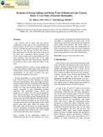
Figure 2.1: Pervasive use of NPs in modern lifestyle products. Reproduced with
permission from (Setyawati et al., 2013a). Copyright 2013, WILEY-VCH Verlag GmbH &
Co. KGaA, Weinheim.
It is necessary to note that in this thesis, the term NPs refers specifically to engineered
NPs synthesized in a controlled setting, as opposed to the free generation of NPs in
environment (e.g. carbon particulates generated from engine combustion).
2.1.1. Physical and chemical properties of TiO2-NPs
Titanium is the ninth most abundant element in the earth’s crust. Titanium (Ti) does not
naturally exist in its metallic state, due to its great affinity to oxygen and hydrogen. One of
the most common natural forms of Ti is titania, better known as titanium dioxide (TiO2).
TiO2 is mostly found in the form of a white, odorless, noncombustible powder. It possess a
molecular weight of 79.9 g/mol, boiling point of 2972°C, melting point 1843°C and relative
density of 4.26 g/cm2 (Shi et al., 2013). In contrast, TiO2-NPs are not present naturally on
7
Chapter 2: Literature Review
earth, but synthesized to arrive at their nano dimension. To date, chemical vapor disposition
(CVD) and flame hydrolysis methods are widely used to produce TiO2-NPs. Using CVD, a
volatile mixture (typically of titanium tetra-isopropoxide and argon) is converted to a
nonvolatile solid and deposited on a substrate. The volatile compound is generated by many
methods such as plasma, high temperature, and pressure (Li et al., 2003). In flame hydrolysis,
an inert gas carries TiCl4 into a flame and produces HCl as well as various sizes of TiO2-NPs
with high purity. This flame hydrolysis method was reported to produce Aeroxide P25 TiO2NPs, which is used as the model NM in this study (Mulenweg, 2004).
TiO2 naturally exists in three crystal forms: rutile, anatase, and brokite. However, TiO2NPs reactivity is mainly affected by their minute size rather than their crystallinity. Lin et al.
(2006) have shown that the decrease of particle size of TiO2 to approximately 29 nm resulted
in the decrease of the band gap of the material. This decrease in the TiO2 band gap led to
enhanced photocatalytic performance, as smaller band gap allows the material to utilize lower
energy photons more efficiently than materials with bigger band gap (Lin et al., 2006).
Moreover, the smaller the particle size, the higher surface area available for photon
absorption and catalytic reaction, further enhancing TiO2-NPs reactivity (Li et al., 2012). This
reactivity of TiO2-NPs can be witnessed in their capability to photocatalyze the formation of
reactive oxygen species (ROS) on the surface. Incident light that carry photons with energy
higher than the band gap will be absorbed by the TiO2-NPs and used to promote electron (e‒)
movement from the valence band to the conduction band. Holes (h+) are created on the
valence band that is left behind by the excited electrons (Li et al., 2012). The electrons in the
conduction band show high reducing power, reducing oxygen to produce superoxide anion
(O2●‒) (Li et al., 2012). In contrast, the holes in the valence band exhibit great oxidizing
power against adsorbed hydroxyl ions to generate hydroxyl radicals (●OH) (Li et al., 2012).
In aqueous environment, these radical species could evolve to other type of radicals such as
8
Chapter 2: Literature Review
hydrogen peroxide and peroxy radicals. The major reaction of reactive oxygen species (ROS)
formation could be seen as follows:
TiO2 + h → h+ + e‒
(1)
H2O + h+ → ●OH + H+
(2)
O2 + e‒ → O2●‒
(3)
O2●‒ + H+ → H2O●
(4)
2 H2O● → H2O2 + O2
(5)
H2O2 + O2●‒ → ●OH+ OH‒ + O2
(6)
2.1.2. Applications of TiO2-NPs
Predictably, the enhanced physical and chemical properties of TiO2-NPs when compared to
their bulk particle counterpart are notable to many technology developers. To date, more
TiO2-NPs have been used to produce high value commercial products leading to high demand
of TiO2-NPs production. In 2005, the annual TiO2-NPs global production was estimated to be
close to 2,000 MT with total market value of USD 70 million (Dransfield, 2005). The annual
TiO2-NPs global production has increased since then, reaching 10,000 MT/year in 2011
(Davis et al., 2010) and was projected to increase exponentially and reach the level of 2.5
million MT/year by 2025 in the U.S. alone (Figure 2.2) (Robichaud et al., 2009).
Most of the TiO2-NPs being produced ends up in various kinds of consumer products
as white pigment. Many food products, like gum, icing, cookies, and candies utilize the TiO2NPs for its white pigment (Chaudhry et al., 2008; Scotter, 2011; Smolander and Chaudhry,
2010). In addition, TiO2-NPs are employed to whiten skim milk (Shi et al., 2013). In recent
publications, 20 food products, like chewing gum, candy, pastry and chocolate were found to
contain 0.1- 12 µg of Ti per mg of tested food products (Peters et al., 2014; Weir et al., 2012),
suggesting the presence of TiO2-NPs in these food products. Further size analysis showed
9
Chapter 2: Literature Review
that these food products contain TiO2 particles in the range of 30-400 nm, with more than one
third having sizes less than 100 nm (Weir et al., 2012).
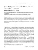
Figure 2.2: Forecast of TiO2-NPs production in U. S. The forecast suggest exponential
increase of TiO2-NPs production due to their high demand in the industrial sector. MT =
Metric tons. Reproduced with permission from (Zhang et al., 2012). Copyright 2012, Elsevier
B.V.
Moreover, TiO2-NPs are also added in food-contact materials, namely in the polymerbased food packaging and glass and metal used in food processing. Owing to its capability to
absorb UV light, TiO2-NPs are incorporated to polymers film to enhance the light-barrier
properties and prevent photo degradation of many food-packaging materials (Chaudhry and
Groves, 2010; Chaudhry et al., 2008). Another benefit for adding TiO2-NPs to food
packaging are derived from their excellent photocatalytic property. UV-activated TiO2-NPs
have been used to inactivate E. coli and Salmonella (Kim et al., 2003; Kim et al., 2009),
providing antimicrobial protection necessary to increase the quality and the shelf life of the
food (Díaz-Visurraga et al., 2010; Smolander and Chaudhry, 2010). Likewise, biofilm growth
could be prevented with application of TiO2-NPs on the steel and glass surfaces used in the
food processing step (Chorianopoulos et al., 2011).
In addition to the food industry, TiO2-NPs are widely applied as white pigment in
personal care products and cosmetics, with utilization in sunscreen products ranks as the
10
Chapter 2: Literature Review
highest among the others. The popularity of TiO2-NPs in sunscreen products due to several
reasons, namely their enhanced properties to absorb UVB (290-320 nm) while scatter UVA
(320-400 nm). This means the TiO2-NPs containing sunscreen could lend higher sun
protection factor (SPF) when compared to those utilizing the larger TiO2 microparticles (Lin
and Lin, 2011). Furthermore, TiO2-NPs scatter very little visible light, thus allowing a
transparent layer when applied on skin. This leads to greater consumer acceptance and
increased popularity of the NPs-based sunscreen products (Davis et al., 2010; Shao and
Schlossman, 1999). The popularity of TiO2-NPs utilization in sunscreen was well
documented in a recent publication by Weir et al. (2012), which showed the presence of 14 90 µg TiO2 per mg of sunscreen in 13 types of sunscreens. To date, an extensive list of
personal care products such as deodorants, toothpastes, shaving creams, anti-wrinkle creams,
moisturizers, foundations and face powders has been known to incorporate TiO2-NPs. It has
been estimated that the TiO2-NPs concentration in these products range from 0.1 to 0.5%
weight (Mulenweg, 2004; Weir et al., 2012). Additionally, TiO2-NPs are used to coat over
the counter medication such as low-dose aspirin products (81 mg aspirin dose). Zachariadis
and Sahanidou (2011) reported the TiO2-NPs content in aspirin product to be as high as 0.014
µg of TiO2/mg aspirin. A validation study done by Weir et al. (2012) found that safety coated
aspirin contains 0.017 – 10 µg of TiO2 /mg of aspirin.
Due to their enhanced catalytic properties, TiO2-NPs are also used to treat waste water
which contaminated with hazardous industrial waste, arsenics. Photocatalytic action of TiO2NPs converts the arsenite (AsIII) to arsenate (AsV), which is insoluble in water, allowing ease
of removal from waste water (Dutta et al., 2005; Ferguson et al., 2005; Pena et al., 2006).
Other utilization of TiO2-NPs photocatalytic properties in consumer products could be found
in self-cleaning tiles, self-cleaning windows, self-cleaning textiles, and anti-fogging window
(Shi et al., 2013).
11