Antibacterial and antifouling polymer coatings for prevention of catheter associated infections 2
Bạn đang xem bản rút gọn của tài liệu. Xem và tải ngay bản đầy đủ của tài liệu tại đây (6.42 MB, 184 trang )
ANTIBACTERIAL AND ANTIFOULING POLYMER
COATINGS FOR PREVENTION OF
CATHETER-ASSOCIATED INFECTIONS
DING XIN
NATIONAL UNIVERSITY OF SINGAPORE
2014
ANTIBACTERIAL AND ANTIFOULING POLYMER
COATINGS FOR PREVENTION OF
CATHETER-ASSOCIATED INFECTIONS
DING XIN
(B.Eng., XI’AN JIAOTONG UNIVERSITY)
A THESIS SUBMITTED
FOR THE DEGREE OF DOCTOR OF PHILOSOPHY
NUS GRADUATE SCHOOL FOR INTEGRATIVE
SCIENCES AND
ENGINEERING
NATIONAL UNIVERSITY OF SINGAPORE
2014
ii
Acknowledgements
This thesis is impossible without the support of many people over the
past four years. Here, I would like to express my sincere gratitude to these
lovely people.
First of all, I would like to thank my supervisor Dr. Yi Yan Yang for her
guidance and support throughout my Ph.D. study in the past four years. Her
passions, optimism, attitudes towards academic research and invaluable
suggestions inspired me all the way. I would also like to thank our collaborators,
Dr. James L. Hedrick from IBM Almaden Research Centre for his helpful
discussion and inputs in the manuscripts. Thanks to Associated Professor Yen
Wah Tong and Assistant Professor Rachel Ee for being in my Thesis Advisory
Committee and giving me a lot of
valuable suggestions.
I would like to thank my labmates in the Nanomedicine Group of the
Institute of Bioengineering and Nanotechnology (IBN) for their constant help
on experiments and discussions on my projects. I would especially thank Dr.
Chuan Yang for synthesizing all the polymers used in this study and Dr.
Shaoqiong Liu for her great inputs in hydrogel work.
I would like to acknowledge NUS Graduated School of Integrative
Sciences and Engineering (NGS) for supporting me with the scholarship and
IBN for the financial support of my PhD research work. I also would like to
thank all the staffs in NGS and IBN for their helps.
iii
Last but not the least, I would like to express my deepest gratitude to my
parents and my girlfriend for their endless love and understanding during my
graduate study.
iv
Table of Contents
Declaration i
Acknowledgements ii
Table of Contents iv
Summary vii
List of Tables x
List of Figures xi
List of Schemes xv
List of Abbreviations xvi
Chapter 1. Introduction 1
1.1 Catheter-associated infections (CAIs) 1
1.2 Bacterial biofilm 3
1.2.1 Bacteria-surface interaction and biofilm formation 3
1.2.2 Strategies for prevention and eradication of bacterial biofilm 5
1.3 Antibacterial coatings 7
1.3.1 Coatings delivering antibacterial agents 7
1.3.2 Surfaces immobilized with antibacterial agents 10
1.4 Antifouling coatings 18
1.4.1 PEG-based antifouling coatings 18
1.4.2 Zwitterionic polymer coatings and other antifouling coatings 22
1.5 Mussel-inspired adhesive coatings using dopamine/polydoamine (PDA) 25
1.6 Gaps, objectives and scope 31
1.7 References 35
Chapter 2. Antibacterial and Antifouling Catheter Coatings Using Surfaces Grafted with
PEG-b-Cationic Polycarbonate Diblock Copolymers 47
2.1 Background 47
2.2 Materials and methods 50
2.2.1 Materials 50
2.2.2 Polymer synthesis 50
2.2.3 Preparation of silicone rubber 51
2.2.4 Polymer coating on silicone rubber surface 52
2.2.5 X-ray photoelectron spectroscopy (XPS) measurements 52
2.2.6 Static contact angle measurements 52
2.2.7 Quartz crystal microbalance with dissipation (QCM-D)
measurements 53
2.2.8 Colony assay 54
2.2.9 Antifouling activity analysis by XTT reduction assay 55
2.2.10 LIVE/DEAD Baclight bacterial viability assay 55
2.2.11 Evaluation of biofilm formation by scanning electron microscopy
(SEM) 56
2.2.12 Analysis of platelet adhesion 57
2.2.13 Static hemolysis assay 57
v
2.3 Results and discussion 58
2.3.1 Polymer synthesis and characterization 58
2.3.2 Characterization of polymer coatings 59
2.3.3 Antibacterial activity of polymer coatings against S. aureus 64
2.3.4 Antifouling activity of polymer coatings against S. aureus 66
2.3.5 Antibacterial and antifouling activities against MRSA 68
2.3.6 Prevention of biofilm formation 71
2.3.7 Static blood compatibility 72
2.4 Conclusion 75
2.5 References 75
Chapter 3. Antimicrobial and Antifouling Hydrogels Formed In Situ from Polycarbonate
and Poly (ethylene glycol) via Michael Addition 80
3.1 Background 80
3.2 Materials and methods 82
3.2.1 Materials 82
3.2.2 Polymer synthesis 82
3.2.3 Preparation of PEG-CPC hydrogels via Michael addition 83
3.2.4 Hydrogel characterization 84
3.2.5 Surface coating on silicone rubber 84
3.2.6 Confocal laser scanning microscopy (CLSM) 84
3.2.7 Hemolysis assay 85
3.3 Results and discussion 86
3.3.1 Polymer synthesis and characterization 86
3.3.2 Hydrogel characterization 87
3.3.3 Antibacterial activities of hydrogels 88
3.3.4 Antibacterial and antifouling activities of coatings with hydrogels 91
3.3.5 Biocompatibility of hydrogels 94
3.4 Conclusion 95
3.5 References 96
Chapter 4. Brush-Like Polycarbonates Containing Dopamine, Cations and PEG Providing
Broad-Spectrum Antibacterial and Antifouling Surface via One-Step Coating 98
4.1 Background 98
4.2 Materials and methods 100
4.2.1 Materials 100
4.2.2 Polymer synthesis and characterization 101
4.2.3 Polymer coating on silicone rubber and silicone catheter 101
4.2.4 Coating morphology and thickness analysis by scanning electron
microscopy 102
4.2.5 Coating stability study under a simulated blood flow condition using
X-ray photoelectron spectroscopy (XPS) 102
4.2.6 Quartz crystal microbalance with dissipation (QCM-D)
measurements 103
4.2.7 Colony assay 104
4.2.8 Testing of zone of inhibition 104
vi
4.2.9 LIVE/DEAD bacterial viability assay 105
4.2.10 Hemolysis assay 105
4.2.11 Protein adsorption 105
4.2.12 Analysis of platelet adhesion 106
4.3 Results and discussion 106
4.3.1 Polymer synthesis and characterization 106
4.3.2 Coating characterization and mechanism of thin film formation 108
4.3.3 Antibacterial activities of polymer coatings 117
4.3.4 Antifouling activities of polymer coatings 120
4.3.5 Effects of quaternization agents 122
4.3.6 Long-term stability 128
4.3.7 Hemocompatibility 130
4.4 Conclusion 132
4.5 References 134
Chapter 5. Conclusion and Future Perspectives 136
1.1 Conclusion 136
1.2 Future perspectives 139
1.3 References 142
APPENDICES 143
Appendix A: Synthetic procedures and characterization of PEG-b-cationic
polycarbonate diblock copolymers 143
Appendix B: Evaluation methods of hydrogels formed in situ from polycarbonate
and PEG via Michael addition 148
Appendix C: Synthetic procedures and characterization of polymers containing
dopamine, cations and PEG 154
Appendix D: List of Publications and Presentations 164
vii
Summary
Catheter-associated infections (CAIs) as one of the most common
medical devices-associated infections (MDAIs) have caused significant
morbidity, mortality and costs. Bacterial biofilm formation on catheter
surfaces is the major cause for the CAIs, and also leads to failure of
conventional antibiotics treatment. To prevent the bacterial biofilm formation,
antibacterial and antifouling coatings as two of the most promising strategies
to kill bacteria and prevent bacterial adhesion have been applied. However, the
need for a facile, nontoxic and effective anti-infective coating on catheter
surfaces is still pressing. The objective of this study was to design
biocompatible polymer coatings with antibacterial and antifouling activities,
and to fabricate them in a facile manner. It is postulated that the
aforementioned coatings can be achieved by incorporating mussel-inspired
dopamine/polydopamine (PDA), cations and poly (ethylene glycol) (PEG) as
adhesive, antibacterial and antifouling moieties in coating systems. To assess
this hypothesis, this study was aimed to:
(1): Design diblock copolymers of PEG-b-cationic polycarbonates and
coat these copolymers onto silicone rubber surfaces by a two-step process: (1)
attaching a reactive PDA layer onto catheter surface and (2) grafting the
copolymers onto the PDA layer via the Michael addition reaction between the
thiol group in the copolymers and oxidized catechol group in PDA. It was
viii
demonstrated that these polymer coatings exhibited excellent antibacterial
activity against notorious bacteria S. aureus including methicillin-resistant S.
aureus (MRSA), and efficiently prevented their fouling on surfaces.
Furthermore, these coatings inhibited biofilm formation without causing
significant hemolysis, blood protein adsorption or platelet adhesion.
(2): Broaden antibacterial spectrum and enhance antifouling activities of
the aforementioned copolymer coatings by designing a PEG-based
antimicrobial hydrogel coating on silicone surfaces. Thiol-containing
tetra-sulfhydryl PEG was first coated on PDA treated silicone surfaces,
followed by in situ hydrogel formation via Michael addition by adding
tetra-acrylate PEG conjugated with PEG-b-cationic polycarbonates. This
hydrogel displayed excellent antibacterial activity against both Gram-positive
bacteria S. aureus and Gram-negative bacteria E. coli through a contact-based
killing mechanism. In addition, the antifouling activity of this hydrogel to
prevent bacterial adhesion was superior to hydrogel with only the PEG
network.
(3): Further simplify the coating process by chemically incorporating
dopamine into the copolymers of PEG and cations. These brush-like
copolymers that contain an optimized number of dopamine were readily
coated onto silicone surfaces via a one-step immersion. The coating
mechanism of the polymers containing dopamine was explored through
comparing polymers with different compositions. By adjusting hydrophobicity
ix
of quaternization agent of the polymers, polymer coatings with
broad-spectrum antibacterial activity against both Gram-positive and
Gram-negative bacteria were obtained. Importantly, these polymer coatings
were stable under simulated blood flow condition, and their antibacterial and
antifouling activities remained even after being in contact with bacterial
suspension for over two weeks. Moreover, these coatings were
hemocompatible.
In conclusion, the findings of this thesis supported the hypothesis that
polymer coatings with the incorporation of dopamine/PDA, cations and PEG
can be easily fabricated and possess excellent antibacterial and antifouling
activities with good biocompatibility, providing a potential solution for
prevention of CAIs. (496 words)
x
List of Tables
Table 2.1 Static contact angles of uncoated, PDA-, polymer 1-, 2- and
3-coated silicone rubbers.
Table 2.2 Nitrogen content of surfaces before and after coatings examined by
XPS (%).
Table 3.1 Physicochemical and antimicrobial properties of cationic
polycarbonates (CPCs).
Table 3.2 Physical and antimicrobial properties of hydrogels.
Table 4.1 Minimal inhibitory concentrations (MICs, mg/L) of various
polymers against S. epidermidis and S. aureus.
Table 4.2 Minimal inhibitory concentrations (MICs, mg/L) of polymer 4b, 4b’
and 4b’’ against S. aureus and E. coli.
xi
List of Figures
Figure 1.1 The biofilm formation: initial attachment (surface conditioning,
reversible attachment and irreversible attachment), the growth of complex
biofilms, and detachment of bacteria.
Figure 1.2 Different strategies developed to prevent the MDAIs.
Figure 1.3 The membrane target of cationic antimicrobial agents of
multicellular organisms and the basis of specificity.
Figure 1.4 Two approaches (“grafting to” and “grafting from”) to immobilize
polymeric QACs. (A) Physical adsorption of diblock copolymer on surface
(grafting to approach); (B) grafting of polymers with functional group on
modified surface (grafting to approach); (C) polymer brushes grown via
surface-initiated polymerization (grafting from approach).
Figure 1.5 Possible reactions between oxidized catechols and amines, thiols or
imidazoles.
Figure 2.1 XPS characterization of polymer coatings. XPS wide-scan spectra
of uncoated and polymer-coated silicone rubber surfaces (A1);
High-resolution N1s spectra of PDA-coated (A2) and polymer 2-coated
silicone rubber surfaces (A3).
Figure 2.2 Polymer coating characterized by quartz crystal microbalance. (A)
Frequency shift (∆f) and dissipation shift (∆D) of the 3
rd
overtone as a
function of time after polymer 2 coating at various concentrations; (B)
Hydrated thickness of the polymer coating as a function of polymer
concentration.
Figure 2.3 Antibacterial and antifouling activities of polymer coatings against
S. aureus. The bacteria colonies in solution (A) and bacterial metabolic
activity (B) on the uncoated surface and surfaces coated with various polymers
at different concentrations after 8 and 24 h of incubation. 1, 2, and 3
correspond to polymer concentrations of 0.075, 0.75 and 1.88 mM
respectively.
Figure 2.4 Live/dead cell staining on the uncoated surface and the surfaces
coated with PDA, PEG and polymer 2 after 4 and 24 h of incubation with S.
aureus.
xii
Figure 2.5 Antibacterial and antifouling activities of polymer coatings against
MRSA. The colonies of the bacteria in solution (A) and bacterial metabolic
activity (B) on the uncoated surface and surfaces coated with various polymers
at different concentrations specified after 8 h of incubation.
Figure 2.6 SEM images of the uncoated surface and the surfaces coated with
PDA, PEG and polymer 2 after 7 days of incubation with S. aureus.
Figure 2.7 Real-time frequency shift (∆f) and dissipation shift (∆D) of the
QCM-D as a function of time in the presence of BSA.
Figure 2.8 SEM images of platelet adhered on the uncoated silicone rubber
surface (A) and the surface coated with polymer 2 (B). The insertion in image
(A) is magnified platelet image on the uncoated silicone rubber surface.
Figure 2.9 Hemolysis evaluation of the uncoated surface and surfaces coated
with PDA and polymer 2 at various concentrations.
Figure 3.1 Growth inhibition of TCP (control), PEG gel (control) and gel 4
against Gram-positive bacteria S. aureues (a), Gram-negative bacteria E. coli
(b) and fungi C. albicans (c). (Higher OD reading indicating more microbes in
growth medium).
Figure 3.2 Antibacterial activities of silicone surfaces with and without
hydrogel coating. (a) Confocal images of surfaces incubated with S. aureus for
24 h: (a1) Pristine silicone surface, (a2) silicone surface coated with PEG
hydrogel, (a3) silicone surface coated with gel 4. Inserts are 3D overlay
images. (b) Number of bacterial cells (S. aureus and E. coli) in bacterial
suspension on gel 4-coated silicone surfaces and pristine silicone surface after
incubation with bacteria for 24h. Solid circle indicating no CFUs found.
Figure 3.3 SEM images of S. aureus (a-c) and E. coli (d-f) after contacting
with PEG hydrogel (b, e) and gel 4 (c, f) for 2 hours. TCP (a, d) was used as
controls.
Figure 3.4 Agar plate assay to test contact killing activities. Agar plates with
gel 4 (left) and PEG hydrogel (right) in the centre of the plates incubated with
S. aureus for 2 weeks.
Figure 3.5 Hemolytic activity of hydrogels with various formulations.
Figure 4.1 Characterization of polymer-coated silicone. (A) Colour changes of
surfaces, (B) contacting angles, (C) high-resolution N1s XPS spectra and (D)
surface morphology examined by SEM.
xiii
Figure 4.2 Transmittance of copolymer solution in function of temperature. (A)
polymer 2a, (B) polymer 3a, (C) polymer 3b, (D) polymer 4a and (E) polymer
4c.
Figure 4.3 Transmittance of polymer 4b solution in function of temperature.
The photograph in the left is the clear micelle solution, and the one in the right
is the turbid vesicle solution.
Figure 4.4 Transmission electron microscopy (TEM) observations of different
nanostructures of polymer 4b in solution at different conditions. Polymer 4b at
room temperature (A), polymer kept at 70 ºC for 4 h (B) and polymer kept at
70 ºC for 8h (C).
Figure 4.5 SEM images of polymer 4b coating at different coating time.
Column A and B are the images with low (1k) and high (10k) magnification
respectively, and column C are the cross-section images.
Figure 4.6 Polymer coating characterized by quartz crystal microbalance with
dissipation. Frequency shift (blue curve) and dissipation shift (red curve) of
the 3rd overtone in function of time as temperature increased from room
temperature to 65 ºC and then maintained at 65 ºC till the end of the
measurement.
Figure 4.7 Antibacterial activities of polymer coatings against S. aureus and S.
epidermidis. Bacteria colonies in bacterial suspension on the uncoated surface
and surfaces coated with various polymers after 24 h of incubation. Solid
circles indicate no colonies found.
Figure 4.8 Zone of inhibition observed for the pristine silicone surface (A)
and surfaces coated with polymer 4a (B), 4b (C) and 4c (D) with incubation of
S. aureus for 24h.
Figure 4.9 Antifouling activities of polymer coatings against S. aureus and S.
epidermidis. Bacterial metabolic activities on the uncoated surface and
surfaces coated with various polymers after 24 h of incubation.
Figure 4.10 Live/dead cell staining of the uncoated silicone surface and the
surfaces coated with various polymers after 24 h of incubation with S. aureus.
Figure 4.11 Surface characterization of pristine silicone (A), polymer 4b (B),
4b’ (C) and 4b’’ (D) coatings by high-resolution N (1s) XPS spectra, contact
angle, and SEM.
xiv
Figure 4.12 Surface morphology and cross-section SEM images of 4b’’
coatings with different thickness by changing the concentration of polymer 4b’’
(a) 0.1 mM; (b) 0.5 mM; (c) 1.0 mM; (d) 1.5 mM.
Figure 4.13 Antibacterial activities of polymer 4b’’ coating against
Gram-positive S. aureus with different coating thickness.
Figure 4.14 Effect of quaternizing agents on antibacterial and antifouling
activities of polymer coatings against Gram-positive bacteria S. aureus and
Gram-negative bacteria E. coli. (A) Colony-forming units (CFUs) of S. aureus
and E. coli in the bacterial suspension after 24 h of incubation with the pristine
and polymer-coated silicone surfaces; (B) Confocal images of S. aureus and E.
coli on the pristine and polymer-coated after 24 h of incubation. E. coli cells
were stained using LIVE/DEAD BacLight Bacterial Viability Kits. The green
and red regions represent live and dead cells, respectively; (c) The number of
live and dead E. coli cells on the untreated and polymer-coated silicone
surfaces.
Figure 4.15 Stability of polymer 4b’’ coating. (A) Photographs of an untreated
silicone catheter and 4b’’-coated catheter surfaces before and after flushing by
mimic blood flow for 7 days. (B) XPS spectra of the untreated catheter and
4b’’-coated catheter surfaces before and after flushing. (C) High-resolution
N(1s) XPS spectrum of polymer 4b’’ coating after flushing.
Figure 4.16 Long-term antibacterial and antifouling activities of the 4b’’
coating (A) Colony-forming units (CFUs) of S. aureus in the solution after
being in contact with the untreated silicone and the silicone coated with 4b’’ at
1.5 mM over different periods of time. A fresh bacterial suspension (10
5
CFU/mL) was added to the surfaces every 24 h. Solid circles indicate no
colony found; (B) Confocal images of S. aureus on the untreated silicone
surface and the surface coated with 4 b’’ at 1.5 mM after 14 days.
Figure 4.17 Hemolysis evaluation of the silicone rubber surface coated with
polymer 4b’’ at various concentrations.
Figure 4.18 Hemocompatibility of polymer 4b’’ coating. (A) Evaluation of
bovine serum albumin (BSA) adsorption on 4b’’ coating by Micro BCA™
protein assay; SEM images of platelet adhesion on pristine silicones surface
(B) and polymer 4b’’ coated surface (C).
xv
List of Schemes
Scheme 1.1 Proposed mechanism of PDA formation by self-polymerization of
dopamine.
Scheme 1.2 The main aim of this thesis and the coating strategies applied in
this study.
Scheme 2.1 Synthesis of aminated polycarbonates (A) and polymer coating
process (B).
Scheme 3.1 Synthetic schemes of PEG-CPC hydrogel formation (a) and
hydrogel coated onto silicone surface (b).
Scheme 4.1 (A) Synthesis procedures and structures of polycarbonates for
catheter coatings; (B) The mussel-inspired adhesive catecholamine. (C)
Schematic demonstration of one-step coating on silicone surface using
polymers containing adhesive, antibacterial and antifouling moieties.
Scheme 4.2 Illustration of the proposed mechanism of polymer 4b thin film
formation.
xvi
List of Abbreviations
1-ODT
1-octadecanethiol
4-MBA
4-methylbenzyl alcohol
A. baumannii
Acinetobacter baumannii
AMPs
Antimicrobial peptides
ATRP
Atom transfer radical polymerization
BCA
Bicinhoninic acid
BSA
Bovine serum albumin
CAIs
Catheter-associated infections
C. albicans
Candida albicans
CFUs
Colony-forming units
CLSM
Confocal laser scanning microscopy
CMCS
Carboxymethyl chitosan
C. neoformans
Cryptococcus neoformans
CPC
Cationic polycarbonate
C. tropicalis
Candida tropicalis
DBU
1,8-Diazabicyclo[5,4,0]undec-7-ene
DI
De-ionized
DOPA
3,4-dihydroxyphenylalanine
E. coli
Escherichia coli
E. faecalis
Enterococcus faecalis
ePTFE
Expanded polytetrafluoroethylene
FE-SEM
Field emission scanning electron microscope
Fmoc/tBu
Fluorenylmethyloxycarbonyl/tert-Butyl esters
GLP
Good laboratory practice
GPC
Gel permeation chromatograph
HS-PGE-CPC
Thiol-terminated diblock copolymers of PEG and
cationic polycarbonate
LD
50
Median lethal dose
MDAIs
Medical device-associated infections
Mefp-5
Mytilus edulis foot protein-5
MHB
Mueller Hinton Broth
MICs
Minimal inhibitory concentrations
mPEG-SH
Thiol-terminated methoxy-poly (ethylene glycol)
MRSA
Methicillin-resistant S. aureus
MSSA
Methicillin-susceptible S. aureus
MTC
Methylcarboxytrimethylene carbonate
MTC-BnCl
MTC monomer containing benzyl chloride
MTC-OEt
MTC monomer containing ethoxy
MTC-OPrBr
MTC monomer containing propyl bromide
MTC-PEG
MTC monomer containing PEG
xvii
OD
Optical density
PAAm
Polyacrylamide
P. aeruginosa
Pseudomonas aeruginosa
PBS
Phosphate-buffered saline
PCBMA
Poly (carboxybetaine methacrylate)
PDA
Polydopamine
PDI
Polydispersity index
PEG
Poly (ethylene glycol)
PEGMA
Poly (ethylene glycol methacrylate)
PEI
Polyethylene imine
PEO
Poly (ethylene oxide)
PLL
Poly (L-lysine)
PRP
Platelet-rich plasma
PSBMA
Poly (sulfobetaine methacrylate)
PTFE
Poly (tetrafluoroethylene)
PU
Polyurethane
QACs
Quaternary ammonium compounds
QCM-D
Quartz crystal microbalance with dissipation
ROP
Ring opening polymerization
SAM
Self-assembly monolayer
S. aureus
Staphylococcus aureus
SEM
Scanning electron microscopy
SDS
Sodium dodecyl sulfate
S. epidermidis
Staphylococcus epidermidis
S. salivarius
Streptococcus salivarius
TCP
Tissue culture plate
TEM
Transmission electron microscopy
TMA
Trimethylamine
TSB
Tryptic soy broth
TU
N-(3,5-trifluoromethyl)phenyl-N’-
cyclohexylthiourea
UVO
Ultraviolet-ozone
UV-Vis
Ultraviolet-visible
VRE
Vancomycin-resistant enterococcus
XPS
X-ray photoelectron spectroscopy
XTT
2,3-bis (2-methoxy-4-nitro-5-sulfo- phenyl)-2H-
tetrazolium-5-carboxanilide
1
Chapter 1. Introduction
In an aging society, various medical devices are increasingly used to
improve patients’ quality of life and also to extend their life expectancy.
However, medical device-associated infections (MDAIs) are responsible for a
large portion of nosocomial infections, which have thus and undermined the
purposes of applications of these medical devices.
1, 2
In particular,
catheter-associated infections (CAIs) are one of the most common and serious
MDAIs. These infections are usually caused by the formation of bacterial
biofilm on catheter surfaces.
3, 4
To prevent biofilm formation and occurrences
of CAIs, facile, non-toxic and effective coatings are urgently needed. In this
chapter, an overview of catheter-associated infections and various strategies
for prevention of CAIs will be discussed.
1.1 Catheter-associated infections (CAIs)
Catheters are tubular medical devices that are inserted into human body
mainly to administer drugs and fluids for patients. More specifically,
intravascular catheters are used to deliver fluids or drugs into bloodstream, and
urinary catheters are used for drainage of waste fluids.
5
In the modern medical
care system, the insertion of these catheters has, at times, become inevitable.
And yet, catheter insertion could result in serious bacterial infections.
6, 7
In the
USA alone, more than 5 million intravascular catheters are used each year and
2
CAIs have been reported in up to 8% of all inserted catheters, resulting in
considerable morbidity and mortality.
8, 9
Additional financial costs attributable
to CAIs can reach USD30,000 for each episode of infection, along with
prolonged hospitalization.
9, 10
In Europe, it was reported that more than 60%
of nosocomial infections are related to the usage of intravascular catheters.
5
The CAIs are caused mainly by the adhesion of microbes and the
subsequent formation of a microbial biofilm which is a community of
microbes and the extracellular polymeric substances secreted by the microbes
on the device surfaces.
3, 11
The biofilm then acts like a microbe reservoir to
release microbes into the human bodies resulting in CAIs. In terms of
intravascular-associated bloodstream infections, the bacteria most often
isolated from the infected catheters are Gram positive S. epidermidis and
followed by S. aureus.
6
The most common bacteria found in infected urinary
catheters are E. faecalis and E. coli.
5
The interactions between bacteria and device surfaces play a critical role
during the development of CAIs.
1, 11
In order to gain an understanding of CAIs
and design an effective anti-infective coating system on catheter surfaces,
bacterial biofilm development on surfaces and the effects of surface properties
on bacteria attachment and biofilm formation will be reviewed in the next
section.
3
1.2 Bacterial biofilm
1.2.1 Bacteria-surface interaction and biofilm formation
Over the past few decades, various materials have been fabricated for
biomedical applications, especially medical devices. In particular, polymers
(e.g. polyethylene, polyurethane, silicone rubber and PET) are widely applied
in medical devices such as catheters and vascular grafts. However, due to the
hydrophobic nature of these materials, device surfaces favour bacterial
attachment and subsequent biofilm formation.
6
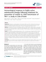
In general, bacterial biofilm
formation as shown in Figure 1.1 comprises of five main steps: initial
attachment, irreversible attachment, initial growth, final growth of complex
biofilm and detachment of bacteria.
3, 4
When medical devices come in contact
with bodily fluids, organic or inorganic nutrients such as proteins, they can
quickly form a conditioning layer that facilitates bacterial attachment and
provides nutrients for the attached bacteria. After surface conditioning,
bacteria could attach themselves on surfaces of the catheter and form a
complex biofilm. The bacteria could also detach from the biofilm and be
released into bodily fluids, leading to bloodstream or urinary tract infections.
These infections are extremely difficult to eradicate due to the presence of the
bacterial biofilm. Once biofilm is formed on the surface, bacteria in the
biofilm could have altered their phenotype, showing different growth rates and
gene expression compared to that of planktonic bacteria. This alteration could
4
make some antibiotics that target specific genes ineffective. In addition,
extracellular polymeric substances secreted by bacteria in the biofilm envelop
the bacteria, and these substances act as a physical barrier to further protect
bacteria from antibiotic attack and the innate immune response.
4, 6
Biofilm has
become a hallmark of MDAIs, as biofilm phenotypes could exhibit more than
1,000 times greater minimum inhibitory concentrations (MIC) of clinical
antibiotics in comparison to the non-biofilm strains.
12-15
Due to the virulence
of bacterial biofilm, biofilm-related infections such as blood stream infections
and urinary tract infections are extremely difficult to treat. Therefore, it is
quite important to prevent biofilm formation. To be more specific, the
prevention of initial bacterial attachment is extremely desirable for medical
device surfaces.
Figure 1.1 The biofilm formation: initial attachment, irreversible attachment,
initial growth, the growth of complex biofilms, and detachment of bacteria.
(Reprinted with permission from Ref.
3
)
5
1.2.2 Strategies for prevention and eradication of bacterial biofilm
To address the problem of CAIs, great efforts have been devoted to
developing various strategies to eradicate formed biofilm or to prevent biofilm
formation on device surfaces. To eradicate biofilm, one of the most common
approaches used in hospitals is to remove infected catheters. This is necessary
if the microorganisms isolated from devices are highly virulent and difficult to
treat, but frequent removal of device could pose more risks to patients, in
addition to increasing patients’ financial cost. Alternatively, salvaging of the
device with antimicrobial agents like antibiotics is also a popular option.
However, the presence of biofilm could makes antimicrobial treatment
ineffective and overuse of antibiotics may induce drug-resistant bacterial
strains.
16
Thus, eradication of biofilm and curing the infection is relatively
difficult, while prevention of the infection is easier and more practical. The
most important and simplest strategy to prevent CAIs is hand and skin
antisepsis as well as standardization of aseptic care,
17-19
but it is difficult to
meet the high requirement of aseptic care, especially for developing countries.
In addition, usage of antibiotic-lock or ethanol-lock is recommended for
catheters,
18
but these techniques may result in the emergence of more
superbugs or systematic toxicity.
16, 20
Therefore, there is a pressing need to
develop effective and facile approaches to prevent CAIs. Surface modification
of medical devices has been frequently reported as a promising approach to
prevent biofilm formation.
11, 18
6
The bacterial attachment is critically influenced by surface properties of
the device, such as surface morphology, hydrophobicity/hydrophilicity and
surface chemistry. It was reported that bacterial attachment was successfully
inhibited by using smooth surfaces rather than rough or porous surfaces.
21
In
addition, hydrophilic surfaces were reported to decrease bacterial attachment
in contrast to hydrophobic surfaces.
22
Cationic surfaces were also found to kill
the bacteria in contact with the surfaces and prevent bacterial fouling on
surfaces.
23-25
Therefore, approaches by modulating surface properties with
different coating materials and techniques to inhibit bacterial biofilm
formation are very promising, and some of these strategies are summarized in
Figure 1.2.
26
Particularly, antifouling coatings and antibacterial coatings are
prevalent in the field for the prevention of CAIs and other MDAIs.
The purpose of this study is to modify device surfaces by coating them
with antibacterial and antifouling polymers in a facile manner for the
prevention of bacterial adhesion and subsequent CAIs. To better understand
this concept, a detailed review on current antibacterial and antifouling coatings
will be given in the following sections.