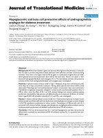
Part i synthesis and biological evaluation of phosphoglycolipid PGL1 analogues part II synthesis and biological evaluation of andrographolide analogues
Bạn đang xem bản rút gọn của tài liệu. Xem và tải ngay bản đầy đủ của tài liệu tại đây (10.61 MB, 335 trang )
PART I.
SYNTHESIS AND BIOLOGICAL EVALUATION OF
PHOSPHOGLYCOLIPID PGL1 ANALOGUES.
PART II.
SYNTHESIS AND BIOLOGICAL EVALUATION OF
ANDROGRAPHOLIDE ANALOGUES.
HADHI WIJAYA
NATIONAL UNIVERSITY OF SINGAPORE
2014
PART I.
SYNTHESIS AND BIOLOGICAL EVALUATION OF
PHOSPHOGLYCOLIPID PGL1 ANALOGUES.
PART II.
SYNTHESIS AND BIOLOGICAL EVALUATION OF
ANDROGRAPHOLIDE ANALOGUES.
HADHI WIJAYA
(BSc. (Hons.) NUS)
A THESIS SUBMITTED FOR THE DEGREE OF DOCTOR OF
PHILOSOPHY
DEPARTMENT OF CHEMISTRY
NATIONAL UNIVERSITY OF SINGAPORE
2014
ACKNOWLEDGEMENTS
First and foremost I would like to express my sincere gratitude to my
supervisor, A/P Lam Yulin, who has given me the opportunity to join her
research lab as a graduate student. I would like to thank her for her guidance
provided to me throughout my PhD studies. She has always been patient and
understanding by giving me sufficient time and advice to overcome difficulties
that I encountered over the course of my research.
I would also like to thank our collaborator Prof. Wu Shih-Hsiung, Prof.
Hua Kuo-Feng, Dr. Yang Feng-Ling, Prof. Yao Shao Qin, Dr. Li Lin, Ms. Sun
Chenyang, Prof. Wong Wai-Hsiu and Ms. Loh Xinyi. They have provided
valuable ideas and are responsible for the biological studies discussed in this
thesis.
I would like to express my sincere appreciation to Mdm. Han Yanhui and
Dr. Wu Ji’En from NMR lab, Mdm. Wong Lai Kwai, Mdm. Lai Hui Ngee and
Dr. Liu Qiping from Mass spec lab as well as Ms. Tan Geok Kheng and Ms.
Hong Yimian from X-ray lab for their assistance in the compounds
characterization.
To all past and present members of A/P Lam lab, Dr. Kong Kah Hoe, Dr.
Fang Zhanxiong, Dr. Che Jun, Dr. Wong Lingkai, Dr. Samanta Sanjay, Dr.
Woen Susanto, Lin Xijie, Alan Sim, Cliff Anderson, Ang Wei Jie, Poh Zhong
Wei, Ng Cheng Yang, Ran Jiangkun, Niu Zilu, Gan Chin Heng, Linus Lim
Wei Jie and Chng Yong Sheng, I would like to say thank you for your advice
and help.
iii
I would like to thank my family for their continuous support. Without them,
I would not have the opportunity to further my study in Singapore.
Last but not least, I woud like to express my gratitude to my partner Ms.
Pulvy Iskandar. I would like to thank her for her encouragement, motivation,
patience and understanding. For the past fifteen years, she has always been
there for me, giving me the strength and courage to face all the challenges and
obstacles that I encountered in my life.
iv
TABLE OF CONTENTS
DECLARATION i
ACKNOWLEDGEMENTS ii
TABLE OF CONTENTS iv
SUMMARY vii
LIST OF TABLES ix
LIST OF FIGURES xi
LIST OF SCHEMES xv
LIST OF ABBREVIATIONS xvii
CHAPTER 1. INTRODUCTION 1
1.1 Overview 1
1.2 Definition, Classification and sources of Natural Products 2
1.3 Natural Products with Anti-inflammatory Activity 8
1.4 Synthesis of Natural Product and Analogues 12
1.5 Purpose of Research Work in this Thesis 14
1.6 References 15
CHAPTER 2. SYNTHESIS AND BIOLOGICAL 19
EVALUATION OF PHOSPHOGLYCOLIPID PGL1 ANALOGUES
2.1 Introduction 19
2.2 Results and Discussions 24
2.2.1 Retrosynthesis 24
2.2.2 Synthesis of glycosyl donor 2-3 25
2.2.3 Synthesis of glycerate 2-4 28
2.2.4 Synthesis of H-phosphonate 2-2 30
v
2.2.5 Synthesis of glycolipid 2-29 and 2-35 38
2.2.6 Synthesis of glycosyl donor 2-41 42
2.2.7 Synthesis of PGL1 analogues 43
2.3 Biological Results 50
2.4 Conclusion 54
2.5 References 55
CHAPTER 3. SYNTHESIS OF PROBES FOR ACTIVITY 58
BASED PROTEIN PROFILING OF POTENTIAL CELLULAR
TARGETS OF ANDROGRAPHOLIDE
3.1 Introduction 58
3.2 Results and Discussions 63
3.2.1 Design of probes 63
3.2.2 Synthesis of probes 67
3.3. Biological Results 73
3.3.1 Cell Proliferation Assay and Western Blott Analysis 73
of STAT 3 Phosphorylation in HepG2 cell line
3.3.2 In situ protein profiling in HepG2 cell line with AP1 75
3.3.3 In situ protein profiling of all probes in HepG2 cell lines 76
3.3.4 In situ protein profiling by AP1 in different cell lines 77
3.3.5 Pull down and Target Validation 78
3.3.6 Fluorescence enzymatic assay 80
3.3.7 Cellular imaging with APNP, AP1NP and AP2NP 82
3.4 Conclusion 84
3.5 References 86
vi
CHAPTER 4. SYNTHESIS AND BIOLOGICAL EVALUATION 90
OF ANDROGRAPHOLIDE ANALOGUES AS POTENTIAL
INHIBITORS OF NF-B.
4.1 Introduction 90
4.2 Results and Discussion 93
4.2.1 Synthesis of Analogues 93
4.2.2 Biological Results 98
4.3 Conclusion 103
4.4 References 104
APPENDICES
Appendix A. Experimental Procedures and Characterization 106
Data for Compounds 2
Appendix B. Experimental Procedures and Characterization 210
Data for Compounds 3
Appendix C. Experimental Procedures and Characterization 234
Data for Compounds 4
Appendix D. NMR Spectra of Some Intermediates and Analogues 251
vii
SUMMARY
Natural products play an important role in drug discovery. Numerous life
saving medicine and medical breakthrough have been associated and
intrinsically linked to natural products. Today, half of the drugs approved for
marketing are of natural origin or derived from natural products. Currently,
less than 10% of world biodiversity has been explored for medicinal purposes.
Many more biologically active compounds from nature have yet to be
discovered and developed into useful drugs for the treatment of various
diseases. In this thesis, the synthesis and biological evaluation of analogues of
two natural products are presented.
In Chapter 2, the synthesis and biological evaluation of phosphoglycolipid
PGL1 analogues are described. The synthetic route towards PGL1 was
successfully established and a library consisting of 21 analogues was prepared.
The analogues were initially evaluated for their immunostimulation activity.
However, no positive results were obtained. This could be due to various
factors such as incorrect chiral centre of the glycerol moiety or incorrect fatty
acid chains (length and branching). The analogues were then evaluated for
their inhibition activity agains TNF- and IL-6 instead. Two of the analogues,
PGL1j and PGL1s showed significant inhibition against IL-6.
In Chapter 3, the synthesis of andrographolide probes as well as Activity
Based Protein Profiling (ABPP) of potential andrographolide targets are
described. The probes were designed based on previous structure-activity
viii
relationship study and anti-inflammatory mechanism of andrographolide.
Protein profiling and subsequent target validation confirmed p50 as the protein
target in HepG2 cell. Pull down/LC-MS/MS analysis identified NAMPT as
one of the potential targets in A549 cell. The fluorophore containing probes
were successfully utilized in live cell imaging which produced fluorescent
signal upon reacting with the targets. The imaging result were also consistent
with the pull down and Western blot analysis.
Finally, the synthesis and biological evaluation of andrographolide
analogues for inhibition of NF-B are described in Chapter 4. The synthesis
involved modification of the lactone ring of andrographolide. Biological
evaluation revealed that one of the analogues, 4-7c showed a concentration-
dependent inhibition of NF-B in A549 cell.
ix
LIST OF TABLES
Table 1.1. Different classes of natural products 3
Table 2.1. Analogues of 2-22 29
Table 2.2. Analogues of 2-4 29
Table 2.3. Analogues of 2-24 30
Table 2.4. Analogues of 2-6 31
Table 2.5. Analogues of 2-27 33
Table 2.6. Analogues of 2-28 34
Table 2.7. Analogues of 2-2 35
Table 2.8. Reaction conditions for the glycosylation of 2-3a 38
Table 2.9. Reaction conditions for the glycosylation of 2-3b 39
Table 2.10. Analogues of 2-43 46
Table 2.11. Analogues of PGL1 47
Table 2.12. Cytotoxicity of PGL1 analogues (concentration of 52
12.5 M to 200M) in J774A.1 macrophages
Table 2.13. Cytotoxicity of PGL1 analogues (concentration of 52
3.25 M to 200M) in J774A.1 macrophages
Table 2.14. Inhibition of TNF- and IL-6 by PGL1 analogues 53
(at concentration of 12.5 µM) in J774A.1 macrophages
x
Table 2.15. Inhibition of TNF- and IL-6 by PGL1 analogues 53
(at concentration of 3.25 µM)) in J774A.1 macrophages
Table 3.1. Protein candidates identified from in situ pull down 80
experiment in A549 cells
xi
LIST OF FIGURES
Figure 1.1. Approved Anticancer Drugs, 1981 – 2010 2
Figure 1.2. Classification of Natural Products 4
Figure 1.3. Structure of paclitaxel 1-1 5
Figure 1.4. Structure of artemisinin 1-2 5
Figure 1.5. Structure of penicillin 1-3 6
Figure 1.6. Structure of bryostatin 1-4 7
Figure 1.7. Structure of captopril 1-5 7
Figure 1.8. Structure of teprotide 1-6 8
Figure 1.9. Examples of Natural products with anti-inflammatory 11
activity
Figure 1.10. Diverted Total Synthesis of Migrastatin Ether 13
Figure 1.11. Semi-synthesis of Paclitaxel 14
Figure 2.1. General structure of Gram-negative LPS 19
Figure 2.2. Structure of Lipid A of E. coli 21
Figure 2.3. Glycosphingolipid from sphingomonas 21
Figure 2.4. Structures of PGL1 and PGL2 22
Figure 2.5. HMBC correlation of monoester 2-26 (proton H
a
) 36
Figure 2.6. Overlapped HSQC/HMBC Spectrum (zoom in) 37
of compound 2-26
xii
Figure 2.7. Overlapped HSQC/HMBC Spectrum of compound 2-26 37
Figure 2.8. HMBC correlation of compound 2-42 (proton H
d
) 48
Figure 2.9. HSQC spectrum of 2-42 48
Figure 2.10. Dihedral angle between proton at C1 and proton at 48
C2 for and glycosides
Figure 2.11. HMBC spectrum of 2-42; Correlation between H
d
49
and carbon of amide carbonyl
Figure 2.12. HMBC spectrum of 2-42; Correlation between H
d
50
and anomeric carbon
Figure 3.1. Representative structure of an ABPP probe 59
Figure 3.2. Gel-based activity-based protein profiling (ABPP) 59
Figure 3.3. Tag-free ABPP using bio-orthogonal reactions 60
Figure 3.4. ABPP of parthenolide using biotinylated derivative 61
Figure 3.5. Structural features of andrographolide 62
Figure 3.6. Structures of andrographolide probes 64
Figure 3.7. Structure of fluorophore-containing andrographolide 65
probes
Figure 3.8. Cell proliferation assay of andrographolide and probes 74
Figure 3.9. Western blot analysis of STAT3 phosphorylation in 75
HepG2 cell
Figure 3.10. In situ protein profiling in HepG2 cell with AP1 75
xiii
Figure 3.11. In situ protein profiling of all probes and 76
andrographolide in HepG2 cell
Figure 3.12. In situ protein profiling of AP1 in different cell lines 77
Figure 3.13. In-gel fluorescence scanning and Western blot analysis 78
(anti p50) of HepG2 cell.
Figure 3.14. In-gel fluorescence scanning and Western blot analysis 79
(anti-p50, anti-tubulin, anti-PDI) of A549 cell
Figure 3.15. Concentration (reacted for 3 h) and time 80
(with 1M of GSH) dependent fluorescence assays
of APCM
Figure 3.16. Fluorescence activated cell sorting of live HepG2 cell 81
incubated with APCM and APNP
Figure 3.17. One-photon excited fluorescence images of HepG2 (a) 83
and A549 (b) cells upon treatment with APNP, AP1NP
and AP2NP (10.0 µM)
Figure 4.1. Activation pathway (canonical) of NF-B. 91
Figure 4.2. Different natural products that inhibit NF-κB 92
Figure 4.3. X-ray crystal structure of 4-10 97
Figure 4.4. SEAP reporter assay (inhibition of NF-B) for 99
4-5b, 4-5d, 4-5e, 4-7b
Figure 4.5. SEAP reporter assay (inhibition of NF-B) for 100
4-5a, 4-5c, 4-7a, 4-7c and 4-11
xiv
Figure 4.6. SEAP reporter assay (inhibition of NF-B) for 100
andrographolide
Figure 4.7. SEAP reporter assay (inhibition of NF-B) for 4-12 101
Figure 4.8. Flow cytometric analysis of cytotoxicity of 102
andrographolide, 4-7c and 4-12 against A549 cell
xv
LIST OF SCHEMES
Scheme 2.1. Retrosynthesis of PGL1 24
Scheme 2.2. Synthesis of glycosyl donor 2-3a 26
Scheme 2.3. Synthesis of glycosyl donor 2-3b 27
Scheme 2.4. Synthesis of amine 2-7a and 2-7b 28
Scheme 2.5. Synthesis of amine 2-7d 28
Scheme 2.6. Synthesis of glycerate 2-4 29
Scheme 2.7. Synthesis of carboxylic acid 2-6 30
Scheme 2.8. Synthesis of phospholipid 2-2 32
Scheme 2.9. Glycosylation of gylcosyl donor 2-3a 38
Scheme 2.10. Glycosylation of glycosyl donor 2-3b 38
Scheme 2.11. Synthesis of glycolipid 2-35 39
Scheme 2.12. Glycosylation mechanism of glycosyl donor with 41
participating neighboring group
Scheme 2.13. Synthesis of diazo transfer reagent 2-36 42
Scheme 2.14. Synthesis of glycosyl donor 2-41 43
Scheme 2.15. Synthesis of PGL1 45
Scheme 2.16. Solvent participation in glycosylations 45
Scheme 3.1. Mechanism of inhibition of NF-B by andrographolide 64
xvi
Scheme 3.2. Proposed mechanism of self-reporting property of 66
probe APNP
Scheme 3.3. Synthesis of AP1 and AP2 68
Scheme 3.4. Synthesis of AP3 69
Scheme 3.5. Synthesis of NC1 70
Scheme 3.6. Synthesis of APCM and APNP 71
Scheme 3.7. Synthesis of AP1CM and AP1NP 71
Scheme 3.8. Synthesis of AP2CM and AP2NP 72
Scheme 4.1. Synthesis of 4-1a and 4-1b 93
Scheme 4.2. Synthesis of ylide 4-3 93
Scheme 4.3. Synthesis of analogues 4-5a, 4-5b, 4-5c and 4-5d 94
Scheme 4.4. Synthesis of analogue 4-5e 95
Scheme 4.5. Synthesis of analogues 4-7a, 4-7b and 4-7c 95
Scheme 4.6. Synthesis of analogue 4-11 96
Scheme 4.7. Synthesis of analogue 4-12 97
xvii
LIST OF ABBREVIATIONS
ABPP activity based protein profiling
Ac
2
O acetic anhydride
ACN acetonitrile
AcOH acetic acid
AcSH thioacetic acid
aq aqueous
BAIB bis(acetoxy)iodobenzene
BF
3
.OEt
2
boron trifluoride diethyl etherate
Bn benzyl
BnBr benzyl bromide
CCl
3
CN trichloroacetonitrile
CDI 1,1’-carbonyldiimidazole
DBU 1,8-Diazabicyclo[5.4.0]undec-7-ene
DCC N,N’-dicyclohexylcarbodiimide
DCM dichloromethane
DHP dihydropyran
DIAD diisopropyl azodicarboxylate
DMAP 4-Dimethylaminopyridine
DMF N-N-dimethylformamide
DMSO dimethylsulfoxide
EDC N’-ethylcarbodiimide
EI electron impact
ELISA enzyme-linked immunosorbent assay
ESI electron spray ionization
xviii
Et ethyl
Et
3
N triethylamine
Et
2
O diethylether
EtOAc ethyl acetate
HMBC heteronuclear multiple bond correlation
HOBt hydroxybenzotriazole
HRMS high resolution mass spectroscopy
HSQC heteronuclear single quantum coherence
IL-1 interleukin 1
IL-6 interleukin 6
K
2
CO
3
potassium carbonate
LCMS-IT-TOF liquid chromatography mass spectrometer-ion trap-
time of flight
LDA lithium diisopropylamine
LDL low-density lipoprotein
LPSs lipopolysaccharides
Me methyl
M.W. microwave irradiation
NaOMe sodium methoxide
NF-B nuclear factor kappa-light-chain-enhancer of activated
B cells
NIS N-iodosuccinimide
NMR nuclear magnetic resonance
PAGE polyacrylamide gel electrophoresis
PAMPs pathogen-associated molecular patterns
xix
PDC pyridinium dichromate
PGLs phosphoglycolipids
Ph phenyl
PhSH thiophenol
PMA phosphomolybdic acid
PPh
3
triphenylphosphine
PRRs pattern recognition receptors
r.t. room temperature
SDS sodium dodecyl sulfate
TBAF tetrabutylammonium fluoride
TBDMS tert-butyldimethyslsilyl ethers
TEMPO (2,2,6,6-tetramethylpiperidin-1-yl)oxidanyl
TFA trifluoroacetic acid
THF tetrahydrofuran
TLC thin-layer chromatography
TMSOTf trimethylsilyl trifluoromethanesulfonate
TNF tumor-necrosis factor
TOCSY total correlation spectroscopy
TrCl triphenylmethyl chloride
1
CHAPTER 1. INTRODUCTION
1.1 Overview
Natural products play an important role in drug discovery. Numerous life
saving medicines and medical breakthroughs have been associated and
intrinsically linked to natural products. These compounds are particularly
evident in the development of treatment for infectious diseases, cancers,
hypercholesterolemia and immunological disorders.
1
Humans have utilized natural products for medicinal purposes for
thousands of years. The earliest records can be traced back to as early as 2900
– 2600 BC,
2
which documented the use of approximately 1000 plant-derived
substances in Mesopotamia,
3
In addition, ancient civilizations such as the
Chinese, Egyptian, Indians, Greeks and Romans were reputed for their use of
mostly plant based natural products for the treatment of various diseases and
illneses.
4
Natural products continue to inspire the development of therapeutic
treatment today. This is demonstrated by number of approved anti-cancer
drugs (Figure 1.1),
5
From 1981 to 2010, up to 50% of the approved drugs were
natural products or of natural product origin.
Despite their abundance in nature, currently only less than 10% of the
world’s biodiversity has been explored for their potential therapeutic
applications.
6
The nature pool thus presents many exciting opportunities for
new compounds to be discovered and applied for various medicinal purposes.
2
Figure 1.1. Approved Anticancer Drugs, 1981 – 2010.
5
(“B”: biological,
usually large protein, “N”: natural product, “NB”: natural product “Botanical”,
“ND”: derived from Natural product, usually semi-synthetic, “S”: totally
synthetic drug, “S*”: made by total synthesis, natural product pharmacophore,
“NM”: natural product mimic, “v”: vaccine.
1.2 Definition, Classification and Sources of Natural Products
There are different ways in which a natural product can be defined. The
general definition of a natural product is a substance isolated from a living
organism in nature. In the organic chemistry context, natural products are
purified organic compounds, which are produced by the pathways of primary
or secondary metabolism.
7
For a medicinal chemist, natural product means
secondary metabolites produced by a living organism.
8
Although there is no universal classification available due to the highly
diverse structure, function and biosynthesis, natural products are generally
categorized into the following classes: terpenoids and steroids, nonribosomal
3
polypeptides, alkaloids, enzyme cofactors, fatty acids and polyketides (Figure
1.2).
9
The following are the descriptions (Table 1.1)
9
for the different classes
of natural products:
CLASS
DESCRIPTION
EXAMPLE
Terpenoid
a class of natural product, which
contains isoprene (2-methyl-1,3-
butadiene) unit as the basic
building block
Steroid
A modified terpenoid, which has
tetracyclic carbon skeleton
Non
ribosomal
polypeptide
peptide-like compounds
synthesized by nonribosomal
peptide synthetases without direct
RNA transcription
Alkaloids
a class of natural product, which
contains basic nitrogen atoms.
Enzyme
cofactor
non-protein component of
enzymes which is usually
inorganic ions or small organic
molecules
Fatty Acids
carboxylic acids that contain long
hydrocarbon chain (saturated or
unsaturated).
Polyketides
a class of natural product, which
contains alternating carbonyl and
methylene group.
Table 1.1. Different classes of natural products.
9
2,6-Dimethyloctane
O O
O
COOH
Zhankuic acid A
O
N
O
NH
2
OHN NHOO
O
N
O
N
O
N
HN
O
H
N
O
O
O
N
H
O
N
O
N
O
Actinomycin D
N
O
O
OH
O
H
H
Cryptocin
NHHN
S
O
H H
COOH
Biotin
O
HO
trans-Oleic acid
HO
O O
OH O
Curvulin
4
Figure 1.2. Classification of Natural Products.
9
Natural products can be extracted from various sources including plants,
microbes, marine species, venoms and toxins. An example of a clinically
useful drug derived from plants is paclitaxel (Figure 1.3). It was isolated from
the Pacific yew tree Taxus brevifolia in 1971 by Wall and Wani.
10
This
anticancer compound was subsequently developed commercially by Bristol-
Myers Squibb (BMS) and sold under the trademark name Taxol. Paclitaxel is
used to treat a large variety of cancers such as lung, ovarian, breast, head and
neck. The anticancer property of paclitaxel is related to its ability to bind to
the beta-tubulin subunits of microtubules.
11
This binding process results in the
inhibition of cell division and eventually cell death, which serves to kill the
rapidly dividing tumor cells.
Another natural product derived from plant origin is artemisinin 1-2
(Figure 1.4). This compound was isolated from the leaves of Artemesia
annua. Artemesia has been used as traditional Chinese medicine for over 2000


