Protein s nitrosylation and its relevance to redox control of cell signaling
Bạn đang xem bản rút gọn của tài liệu. Xem và tải ngay bản đầy đủ của tài liệu tại đây (16.66 MB, 213 trang )
PROTEIN S-NITROSYLATION
AND ITS RELEVANCE TO REDOX CONTROL OF
CELL SIGNALING
KYAW HTET HLAING
(M.B.B.S, UM 2)
A THESIS SUBMITTED
FOR THE DEGREE OF DOCTOR OF PHILOSOPHY
NUS GRADUATE SCHOOL FOR
INTEGRATIVE SCIENCES AND ENGINEERING
NATIONAL UNIVERSITY OF SINGAPORE
2012
DECLARATION
I hereby declare that this thesis is my original work and it has
been written by me in its entirety. I have duly
acknowledged all the sources of information which have been
used in the thesis.
This thesis has also not been submitted for any degree in any university previously.
Kyaw Htet Hlaing
24 Dec 2012
!
i!
Acknowledgements
I wish to express my deepest gratitude to my supervisor, Associate Professor
Marie-Véronique Clément, Department of Biochemistry, for introducing me into the
field of “Redox Control of Cell Signaling”, and guiding me along the arduous journey
of my Ph.D. study. I am truly grateful for her warm encouragement and constant
optimism in the face of “reality of day-to-day life of a graduate student” over the
years. This thesis has not been complete without her unending support and kind
understanding. I also like to thank my TAC members, Dr Andrew Jenner and
Professor Kini R Manjunatha, for their comments, useful advice and feedbacks
throughout my study.
My heart-felt thanks to my lab members for listening to both of my happy and
frustrating stories. Spending time together with them has made my life in the lab most
enjoyable. I want to thank Luo Le in particular for taking time to read the draft of my
thesis and giving me useful feedback. Also my special thank to Ms Lee Mui Khin for
keeping things in order and making sure that I always get what I need in time.
Lastly, my deepest gratitude to my family for their encouragement and support
all along. I wish to express my special thank to my older sister, Ms Wint Wint Htet
Hlaing, for helping me out financially when in need and motivating me when
confronted with various setbacks during my study.
!
ii!
Contents
Acknowledgements i
Contents ii
Summary vii
List of Figures ix
List of Tables xiii
Abbreviations xiv
CHAPTER 1: INTRODUCTION 1
1.1 BIOCHEMISTRY OF FREE RADICALS 1
1.2 SOURCES AND FORMATION OF REACTIVE OXYGEN AND
NITROGEN SPECIES 2
1.2.1 Superoxide 3
1.2.2 Hydrogen Peroxide and Hydroxyl Radical 6
1.2.3 Nitric Oxide and its derivatives 6
1.3 EFFECTS OF REACTIVE OXYGEN AND NITROGEN SPECIES ON
CELLULAR STRACTURE AND SIGNALING 9
1.3.1 Cellular Toxicity 9
1.3.2 Physiological Function: Redox Signaling 10
1.4 MECHANISMS OF REDOX-BASED REGULATION OF CELL
SIGNALING: FUNCTIONAL CONSEQUENCES OF OXIDATION OF
“REACTIVE CYSTEINE” 14
1.4.1 Inhibition of Activity 15
1.4.2 Activation of Protein Functions 16
!
iii!
1.4.3 Multimerization of Subunits 17
1.4.4 Release of Regulatory Proteins 17
1.4.5 Oxidation of Transcription Factors 18
1.5 TYPES OF REVERSIBLE CYSTEINE OXIDATION 19
1.6 DIFFERENTIAL REDOX-MODIFICATION AND FUNCTIONAL
CONSEQUENCES 21
1.7 REDOX-MODIFICATION: PHYSIOLOGICAL SIGNALING VERSUS
CELLULAR TOXICITY 22
1.8 PROTEIN S-NITROSYLATION 24
1.8.1 Factors influencing protein S-nitrosylation 25
1.9 ABERRATION OF REDOX SIGNALING AND CARCINOGENESIS
31
1.10 RATIONALE OF THESIS 36
CHAPTER 2: MATERIALS AND METHODS 39
2.1 MATERIALS 39
2.1.1 Chemicals 39
2.1.2 Antibodies 41
2.1.3 Cell Lines and Cell Culture 42
2.2## METHODS# # # # # # # # # 43#
2.2.1 Whole Cell Lysate Preparation 43
2.2.2 Sodium Dodecyl sulphate polyacrylamide gel electrophoresis
(SDS-PAGE) and Western Immunoblotting 43
2.2.3 Transient Transfection 45
2.2.4 siRNA Transfection 45
!
iv!
2.2.5 Detection of S-nitrsoylated and Oxidized PTEN by
Oxidation/Reduction Assay 46
2.2.6 Biotin Switch Technique (BST) 47
2.2.6.1 Detection of Total Protein and PTEN S-nitrosylation 47
2.2.6.2 Detection of Total Protein and PTEN Oxidation 48
2.2.7 Lucigenin Chemiluminiscence Assay for Detection of Intracellular
Superoxide 49
2.2.8 Fluorescence Flow Cytometry Assay for Detection of Intracellular
Hydrogen Peroxide, Nitric Oxide and Calcium 50
2.2.9 Statistical Analysis 51
CHAPTER 3: RESULTS 52
3.1 INCREASE IN INTRACELLULAR O
2
˙
-
INDUCES GENERALIZED
PROTEIN S-NITROSYLATION 52
3.1.1 Serum withdrawal causes a reduction in basal production of
intracellular O
2
˙
-
53
3.1.2 Pharmacological inhibition of Cu-Zn SOD leads to an increase in
intracellular O
2
˙
-
without concurrent rise in H
2
O
2
level 54
3.1.3 Detection of protein S-nitrosylation 57
3.1.3.1 Oxidation/reduction assay 57
3.1.3.2 Biotin Switch Technique 58
3.1.4 Both pharmacological inhibition and siRNA gene silencing of
Cu-Zn SOD induce protein S-nitrosylation 64
3.2 PHYSIOLOGICALLY RELEVENT CONCENTRATIONS OF H
2
O
2
INDUCES PROTEIN S-NITROSYLATION WHEREAS HIGH
!
v!
CONCENTRATION OF H
2
O
2
CAUSES NON-SNO OXIDATIVE
MODIFICATIONS 67
3.3 PROTEIN S-NITROSYLATION INDUCED BY GROWTH FACTORS
76
3.4 OXIDATIVE MODIFICATION OF TUMOR SUPPRESSOR PTEN BY
ROS AND GROWTH FACTORS 83
3.5 PROCESS OF PROTEIN S-NITROSYLATION 88
3.5.1 Intracellular NO˙ is decreased with an increase in O
2
˙
-
generation
whereas it is actively synthesized by H
2
O
2
and growth factors 88
3.5.2 Identification of S-nitrosylation species for oxidants- and growth
factors-induced S-nitrosylation 92
3.5.3 Peroxynitrite: oxidation vs nitration 99
3.5.4 Role of calcium in protein S-nitrosylation caused by ROS and
PDGF 103
3.5.5 GSNOR inhibition enhances protein S-nitrosylation 107
3.5.6 Inhibition of O
2
˙
-
production enhances protein S-nitrosylation through
an increase in intracellular NO˙ 111
3.6 PROTEIN S-NITROSYLATION IN SIGNAL TRANSDUCTION 113
3.6.1 Scavenging PNOO˙ prevents PDGF activation of Akt kinase whereas
GSNOR inhibition enhances it 113
3.6.2 O
2
˙
-
/ NO˙ Balance in Signal Transduction 115
3.6.3 ONOO
-
mediates Akt activation by O
2
˙
-
and low concentration of
H
2
O
2
119
3.7 S-NITROSYLATION AND TUMOR MAINTENANCE 121
3.7.1 Maintenance of protein S-nitrosylation in the absence of serum is
!
vi!
associated with sustained signal transduction in precancerous and cancer
cells. 121
3.7.2 Protein de-nitrosylation in cancer 126
CHAPTER#4:#DISCUSSION# # # # # 129
4.1 S-NITROSYLATION IS THE COMMON MECHANISM OF PROTEIN
OXIDATION USED BY O
2
˙
-
AND PHYSIOLOGICALLY RELEVANT
CONCENTRATION OF H
2
O
2
129
4.1.1 O
2
˙
-
and SNO Modification 129
4.1.2 H
2
O
2
and SNO Modification 130
4.1.3 Redox Signaling: O
2
˙
-
vs H
2
O
2
131
4.2 REDOX SIGNALING BY GROWTH FACTORS IS THROUGH
S-NITROSYLATION 132
4.2.1 PTEN: an example of oxidative modification of protein upon
growth factor induction of cell proliferation 133
4.3 PEROXYNITRITE: A POTENTIAL PHYSIOLOGICALLY
RELEVANT S-NITROSYLATING INTERMEDIATE 134
4.4 O
2
-
AND NO˙: STRIKING THE RIGHT BALANCE FOR SIGNAL
TRANSDUCTION 145
4.5 PROTEIN S-NITROSYLATION AND ROS-DRIVEN
CARCINOGENESIS 149
4.6. CONCLUSION 152
References 155
Publication and Presentation 195
!
vii!
Summary
Discovery of the function of oxidants as signaling molecules marks the
beginning of the field of redox control of cell signaling. Understanding the
mechanism of how free radicals regulate signaling is critical to distinguish between
normal physiology and cellular toxicity both caused by reactive species. It is now
known that free radicals influence various cellular processes by altering the function
of critical proteins as a result of reversible oxidation of “reactive cysteine” within the
proteins. Different types of oxidative modification such as S-nitrosylation, S-
glutathionylation, di-sulphide bond formation, sulphenic acid formation, have been
proposed to mediate redox control of cell signaling. However, physiological relevance
of these modifications is somehow missing. Furthermore, there has been a debate
about relative importance of O
2
˙
-
versus H
2
O
2
in mediating enhanced cell
proliferation. Following up on our previous study that demonstrates that O
2
˙
-
activates
survival kinase Akt through S-nitrosylation of the tumor suppressor PTEN, our
current study deciphers the mechanistic aspect of how oxidative signal by O
2
˙
-
is
transformed into nitrosative signal. We also provide evidence that physiologically
relevant concentration of H
2
O
2
predominately induces protein S-nitrosylation over
non-SNO modifications. We demonstrate that protein S-nitrosylation induced by O
2
˙
-
and H
2
O
2
is both mediated by common S-nitrosylating species, ONOO
-
although the
pathways to formation of ONOO
-
are different in each case.
Moreover, we show that oxidation of proteins that occurs following incubation
with PDGF, EGF and 10% FBS is by protein S-nitrosylation. Particularly in the case
of PDGF, the growth factor does not generate a high level intracellular H
2
O
2
regardless of concentration of PDGF used and it consistently induces protein S-
!
viii!
nitrosylation. Again, we find that the relevant S-nitrosylating species that mediates
growth factors-induced protein S-nitrosylation is ONOO
-
. Removal of ONOO
-
prevents protein S-nitrosylation as well as activation of Akt induced by O
2
˙
-
, H
2
O
2
and
PDGF demonstrating protein S-nitrosylation is of relevance to redox control of cell
signaling.
We also highlight the consequences of disturbing O
2
˙
-
/NO˙ balance in cell
signaling. On one hand, removal of NO˙ is effective in preventing S-nitrosylation but
it increases the levels of intracellular O
2
˙
-
and H
2
O
2
potentially causing oxidative
stress with damaging consequences. On the other hand, we demonstrate the
ineffectiveness of removing O
2
˙
-
alone to stop pro-survival signaling as the latter
could continue by ONOO
-
-independent but NO˙-dependent S-nitrosylation.
Lastly, we show that increased ROS and RNS production in breast cancer cell
line (MCF7) correlate with sustained protein S-nitrosylation and Akt activation in the
absence of serum. However, the prevalence of this finding still has to be tested in
other types of cancers. We also find that protein S-nitrosylation and Akt activation in
MCF7 is very stable requiring further studies on identifying the factors contributing to
this stability.
!
!
!
!
!
!
!
!
ix!
List of Figures
Figure 1: NOX2 (gp91phox) activation and generation of O
2
˙- in phagocytes 5
Figure 2: Consequences of oxidation of reactive cysteines within the target proteins
15
Figure 3: Schematic representation of various types of reversible cysteine oxidations
20
Figure 4: A continuum of redox-based modifications. 23
Figure 5: Compartmentalization of cellular NO˙ source and its targets 27
Figure 6: Mechanism of enzyme-mediated protein de-nitrosylation 30
Figure 7: Serum withdrawal results in a decrease in base level O
2
˙- production 53
Figure 8: Inhibition of Cu-Zn SOD by 1mM DDC caused an increase in intracellular
O
2
˙- production but a decrease in intracellular H
2
O
2
56
Figure 9: Overview of oxidation/reduction assay 58
Figure 10: Overview of biotin switch technique 59
Figure 11: Both NO˙ donor and tranS-nitrosylating agent induce protein S-
nitrosylation in mouse embryonic fibroblasts. 62
Figure 12: Pharmacological inhibition of Cu-Zn SOD induces protein S-nitrosylation
65
Figure 13: siRNA gene silencing of Cu-Zn SOD led to an increase O2˙- in
production, a decrease in H
2
O
2
level and induction of protein S-nitrosylation 66
Figure 14: Exogenous H
2
O
2
treatment increases intracellular H2O2 level but has no
effect on O
2
˙- production. 70
Figure 15: Protein S-nitrosylation occurs predominately at low concentrations of
H
2
O
2
although H
2
O
2
causes protein oxidation in a dose dependent manner 72
Figure 16: High concentration of H
2
O
2
is toxic to the cells. 74
!
x!
Figure 17: 50uM H
2
O
2
induces protein S-nitrosylation in a time dependent manner.
75
Figure 18: ROS production during growth factors signaling. 78
Figure 19: Growth factors induced protein S-nitrosylation 80
Figure 20: Protein S-nitrosylation is maintained in the presence of high concentration
of PDGF treatment. 82
Figure 21: Slow release NO˙ donor, Deta-NONOate and tranS-nitrosylating agent,
CysNO cause S-nitrosylation of PTEN 84
Figure 22: Exposure of cells to O
2
˙-, H
2
O
2
and PDGF all S-nitrosylate tumor
suppressor, PTEN in a time dependent manner. 85
Figure 23: S-nitrosylation of PTEN predominately occurs at low concentrations of
H2O2 while it is equally induced by all concentrations of PDGF treatment. 88
Figure 24: Increase in intracellular O
2
˙- is associated with decrease in intracellular
NO˙ level whereas exogenous H
2
O
2
treatments cause increased production of
intracellular nitric oxide. 90
Figure 25: PDGF, EGF and 10% FBS all increase intracellular NO˙. 91
Figure 26: Intracellular NO˙ is essential for protein S-nitrsylation induced by O
2
˙-,
H2O2 and PDGF. 93
Figure 27: Total protein and PTEN S-nitrosylation induced by O
2
˙-, low
concentration H
2
O
2
and PDGF may depend on formation of peroxynitrite. 97
Figure 28: L-NMMA reduces basal production of NO˙ in MEF cells and prevents
new production of NO˙ stimulated by low concentration of H
2
O
2
and PDGF. 98
Figure 29: Low concentrations of exogenous ONOO- induce total protein and PTEN
S-nitrosylation whereas at high concentration, it causes non-SNO oxidative
modifications and 3-NT formation. 102
!
xi!
Figure 30: 50uM H
2
O
2
causes an increase in intracellular Ca
2+
whereas 10ng
PDGF has on effect on intracellular Ca
2+
level. 104
Figure 31: Intracellular release of Ca
2+
enhances protein S-nitrosylation through
activation of NOS. 106
Figure 32: Enhancement of protein S-nitrosylation by GSNOR inhibition is through
an increase in intracellular NO˙. 108
Figure 33. A GSNOR inhibitor, C3 enhances total protein and PTEN snitrosylation
induced by H
2
O
2
and PDGF. 110
Figure 34: Inhibition of O
2
˙- generation enhances protein S-nitrosylation 112
Figure 35: Scavenging ONOO- prevents Akt-phosphorylation by PDGF whereas
GSNOR inhibition enhances it. 114
Figure 36: NO˙ scavenging increases intracellular ROS level that maintains Akt
phosphorylation 117
Figure 37: Inhibition of O
2
˙- alone does not affect Akt activation by PDGF but
simultaneous removal of NO˙ prevents it. 118
Figure 38: ONOO- attenuates Akt activation by O
2
˙- and low concentration of H
2
O
2
120
Figure 39: Protein S-nitrosylation is maintained in the absence of serum in MEF
PTEN knocknout cell line. 123
Figure 40: Increased S-nitrosylation in MCF7 breast cancer cells 124
Figure 41: Akt phosphorylation is maintained in the absence of serum in MEF K/O
and MCF7 cell lines. 125
Figure 42: FeTPPS de-nitrosylate proteins and dephosphorylate Akt in MEF WT but
not in MCF7. 128
!
xii!
Figure 43: Proposed pathway for the formation of ONOO- upon an increase in
intracellular O
2
˙ 136
Figure 44: Low concentration of H
2
O
2
decrease intracellular Cu-Zn SOD activity 137
Figure 45: Proposed pathway of ONOO- formation by exogenous H
2
O
2
138
Figure 46: Proposed pathway for ONOO- formation by growth factors 140
Figure 47: The interplay of NO˙, O
2
˙-, ONOO-, and NO
2
- 141
Figure 48: Proposed pathways for ONOO- formation and S-nitrosylation 144
Figure 49: Schematic representation of the impact of O
2
˙- and NO˙ balance in cell
signaling. 147
!
xiii!
List of Tables
Table 1: Reactive Oxygen Speciess and Reactive Nitrogen Speciess 2
Table 2: Human NOX/DUOX enzymes 4
Table 3: Major Reactive Nitrogen Speciess in Biological System 8
Table 4: List of Ligands inducing ROS production 12
Table 5: Enzymes that reduce reversible cysteine oxidation 20
!
!
!
!
!
!
!
!
!
!
!
!
xiv!
Abbreviations
Akt Protein kinase B
Ang II Angiotensin II
Biotin-HPDP N-6-Biotinamido-hexyl-3!-2!-Pyridyldithio-Propionamide
c-PTIO 2-4-Carboxyphenyl-4,4,5,5-tetramethylimidazoline-1-oxyl-3-
oxide potassium salt
Cu-Zn SOD Copper Zinc Superoxide dismutase
Cys Cysteine
DAF 4-amino-5-methylamino-2',7'-difluorofluorescein diacetate
DCFDA 5-(and-6)-chloromethyl-2´,7´-dichlorodihydrofluorescein
diacetate acetyl ester
DDC Diethyldithiocarbamate
DETA NONOate 1-2-2-Aminiethyl-N-2-Ammonioethyl-Amino-Diazen-1-ium-
1,2-diolate
DMF Dimethylformamide
DMSO Dimethylsulfoxide
DPI Diphenyleneiodonium chloride
DTT Dithiothreitol
EDTA Ethylenediamine tetraacetic acid
EGF Epidermal growth factor
FBS Fetal Bovine Serum
FeTPPS 5,10,15,20-Tetrakis (4-sulfonatophenyl)prophyrinato iron (III),
chloride
GSH Reduced glutathione
GSSG Glutathione disulfide
!
xv!
Hepes 4-(-2-hydroxyethyl)-1- piperazineethanesulfonic acid
H
2
O
2
Hydrogen peroxide
KO cell MEF PTEN-/-cell
L-NMMA N
G
– monomethyl – L – Arginine. Monoacetate
MAPK Mitogen-activated protein kinases xiv
MEF Mouse embryonic fibroblast
NAC N-acetylcysteine
NaNO
2
Sodium nitrite
NEM N - ethylmaleimide
NF-κB NF-kappaB
NO˙ Nitric Oxide
NO
+
Nitrosonium ion
NO
-
Nitroxyl anion
NOX NADPH oxidase
NOS Nitric Oxide Synthase
N
2
O
3
Dinitrogen trioxide
OH˙ Hydroxy radical
ONOO
-
Peroxynitrite
O
2
˙
-
Superoxide
PBS Phosphate buffered saline
PDGF Patelet-derived growth factor
PIP3 Phosphatidylinositol-3,4,5-trisphosphate
PI3-K Phosphatidylinositol 3 - kinase
PP2A Protein phosphatase 2A
PTEN Phosphatase and Tensin Homolog Deleted on Chromosome 10
!
xvi!
PTP Protein tyrosine phosphatase
RBS Reactive bromine species
RCS Reactive chlorine species
RNS Reactive nitrogen species
ROS Reactive oxygen species
RSS Reactive sulphur species
Ser Serine
SNO S-nitrosylation
S-S Di-sulphide bond formation
Thr Threonine
TNFα Tumour necrosis factor alpha
Tyr Tyrosine
VEGF Vascular endothelial growth factor
WT cell MEFWT cell
3-NT 3-nitrotyrosine
!
!
Chapter(1:(Introduction(
1(
CHAPTER 1: INTRODUCTION
!
1.1! !Biochemistry!of!Free!Ra dic als!
The history of free radicals dates back to the time when oxygen was first
recognized as a toxic gas in 1954 (Gershman R et al, 1954). Initially, it was suggested
that toxic properties of oxygen could come from its direct inhibition of essential
enzymes (Hauggard N, 1968), but subsequent findings revealed that these damaging
effects are rather due to the action of oxygen-derived radicals (Glibert DL 1981).
A free radical can be defined as any species capable of independent existence
that contains one or more unpaired electrons in atomic or molecular orbits (Halliwell
B and Gutteridge JMC, 2007). It is this unpaired electron(s) that make(s) free radicals
highly reactive, but the degree of reactivity varies widely among different radicals.
Not all free radicals derive from molecular oxygen. There are many other types of
non-oxygen derived free radicals made in living systems, namely; carbon-centre
radicals such as CCl
3
˙, most transition metal ions with exception of zinc and some
oxides of nitrogen such as NO˙ and NO
2
(Halliwell B and Gutteridge JMC, 2007).
Current nomenclature of reactive species includes reactive oxygen species
(ROS), reactive nitrogen species (RNS), reactive chlorine species (RCS), reactive
bromine species (RBS) and reactive sulphur species (RSS). Some reactive species
belong to more than one category, for example, hydrobromous acid, HOBr is
considered both as ROS and RBS, and peroxnitrite, ONOO
-
is referred to as both
ROS and RNS. Also note that “reactive species” is a collective term and they could
either be radicals or non-radicals that are oxidizing agents easily convertible to
Chapter(1:(Introduction(
2(
radicals (Halliwell B and Gutteridge JMC, 2007). Among reactive species, ROS and
RNS have the widest range of biological functions and they are the main subjects of
discussion throughout this thesis. Table 1 shows the list of ROS and RNS that are
biologically important in living organisms.
Table 1: Reactive Oxygen Species and Reactive Nitrogen Species
Reactive oxygen species Reactive Nitrogen Species
Radicals Nonradicals Radicals Nonradicals
Superoxide (O
2
˙
-
), Hydrogen peroxide (H
2
O
2
), Nitric oxide (NO˙) Nitrous acid (HNO
2
),dinitrogen trioxide/tetroxide
hydroxyl (OH˙), peroxyl, hypochlorous acid (HOCl), nitrogen dioxide (N
2
O
3
/N
2
O
4
), nitronium ion (NO
2
+
), peroxynitrite
(RO
2
˙),alkoxyl (RO˙), ozone (O
3
), singlet oxygen (NO˙
2
) (ONOO
-
), alkyl peroxynitrite (ROONO),
hydroperoxyl (HO
2
˙) (
1
ΔgO
2
), peroxynitrite nitroxyl anion (NO
-
), nitrosyl cation (NO
+
),
(ONOO
-
) nitryl chloride (NO
2
Cl)
(Adapted from Rigas B and Sun Y, 2008)
!
1.2! !Sources!and!Formation!of!R e ac tiv e !O x yg e n!an d !
! !Nitrogen!Species!
Reactive species are generated during irradiation by UV light, by X-rays and
by gamma-rays or exist as pollutants in the atmosphere. In the biological systems,
ROS are produced as by-products of mitochondria-catalyzed electron transport
reactions or intentionally generated by neutrophils and macrophages during innate
immunity (Cadenas E, 1989; Halliwell B and Gutteridge JMC, 2007).
Chapter(1:(Introduction(
3(
1.2.1 Superoxide
The first byproduct of aerobic metabolism within mitochondria is superoxide
(O
2
˙
-
). During the process of oxidative phosphorylation, a small number of electrons
leak from the mitochondrial transport chain to oxygen prematurely, forming the
oxygen free radical O
2
˙
-
. This leakage occurs mainly at complexes I and III (Cadenas
E and Davies KJ, 2000). Another important source of O
2
˙
-
production is by stimulus-
induced activation of membrane-bound enzyme systems such as the NADPH oxidase
complex (NOX). Superoxide generation by the NOX complex is deliberate and it was
best characterized in phagocytic cells such as neutrophils that undergo a series of
reactions called the respiratory burst in response to microorganisms or inflammatory
mediators (Babio BM et al, 2002). The enzyme complex consists of six subunits- two
membrane-bound components, p91phox, p22phox which together form cytochrome
b558, the enzymatic centre of the complex, and four cytosolic proteins, p47phox,
p67phox, p40phox and the small guanosine triphosphate (GTP)-binding protein Rac1
and Rac2. This enzyme system was the first to disprove the rule that O
2
˙
-
was
generated accidentally and served no particular cellular function. During the 1990s,
the similar enzyme complex systems were found in various tissues other than
phagocytes accounting for non-mitochondrial source of O
2
˙
-
production (Banfi B et al,
2003; Cheng G et al, 2001; De Deken X et al, 2000; Edens WA et al, 2001; Geiszt M
et al, 2000 & 2003; Lambeth JD et al, 2000). There are seven isoforms identified so
far but the other six isoforms produce O
2
˙
-
at a fraction (1-10%) of the level produced
in neutrophils by NOX2 (Lambeth JD, 2004 & 2007; Petry A et al, 2010). Tissue
distribution of NADPH oxidase isoforms and their known regulators are summarized
in the following table:
Chapter(1:(Introduction(
4(
Table 2: Human NOX/DUOX enzymes
(Adapted from Lambeth JD, 2004)
NOX isoforms are homologues of gp91phox subunit that accounts for ROS
generation. The regulation of gp91phox (NOX2) is well characterized (Groemping Y
et al, 2003; Huang and Kleinberg, 1999; Vignais PV, 2002) but little is known about
the regulation of other isoforms. Generally, the catalytic component of NOXs
responsible for generation O
2
˙
-
resides within the membrane structure whereas
regulatory subunits scatter in the cytosol. Upon activation, cytosolic components are
recruited to the membrane and form a mutually stabilizing complex with membrane
catalytic subunits. The sequence of events leading to full activation of NOX is given
for the prototypic isoform NOX2 in Figure 1.
Chapter(1:(Introduction(
5(
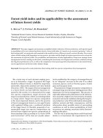
(Adapted from Lambeth, 2004)
Figure 1: NOX2 (gp91phox) activation and generation of O
2
˙
-
in phagocytes
At least three signaling cascades mediate the activation process. First, PI3K provides lipid
docks for p40phox and p47phox to station in the membrane. Second, phosphorylation of
p47phox by protein kinases such as PKC and Akt promotes its binding to p22phox by
relieving autoinhibition in p47phox. Third, activation of guanine-nucleotide exchange results
in active Rac-GTP, which then binds to p67phox for complete assembly of the holo enzyme
for generation of O
2
˙
-
.
Other intracellular sources of O
2
˙
-
generation include xanthine oxidase
(Fridovich I, 1970), NADPH cytochrome p450, lipoxygenase and cyclooxygenase
(Goeptar AR et al, 1995) and the uncoupled nitric oxide synthase (Alderton WK et al,
2001). However O
2
˙
-
generated from theses sources is associated with various
diseased conditions such as hypertension and diabetes (Dixon LJ et al, 2003 & 2005;
Jankov RP et al, 2008).
Chapter(1:(Introduction(
6(
1.2.2 Hydrogen Peroxide and Hydroxyl Radical
Hydrogen peroxide (H
2
O
2
) is produced directly in cells by several enzymes
such as glucose oxidase (Bankar SB et al, 2009), xanthine oxidase (Kelley EE et al,
2010) DUOX1, DUOX2 (Edens WA et al, 2001; Donkó A et al, 2005), and
peroxisomes (Fritz R et al, 2007). And it is also derived from two molecules of O
2
˙
-
in
a reaction called dismutation, which is accelerated by the enzyme, superoxide
dismutase (SOD).
O
2
˙
-
+ O
2
˙
-
+ 2H
+
→ H
2
O
2
+ O
2
H
2
O
2
interacts with O
2
˙
-
to generate highly reactive hydroxyl radical (OH˙) by the
iron-catalyzed Haber-Weiss reaction as follows:
Fe
3+
+ O
2
˙
-
→ Fe
2+
+ O
2
(1)
The second step is the Fenton reaction:
Fe
2+
+ H
2
O
2
→ Fe
3+
+(OH
−
+ OH˙ (2)
Net reaction:
O
2
˙
-
+ H
2
O
2
→ OH
−
+ OH˙ + O
2
(3)
In phagocytes, the enzyme myeloperoxidase produces HOCl from H
2
O
2
(Anderson
MM et al, 1999), which contributes to the inflammation of tissues during immune
defense response.
1.2.3 Nitric Oxide and its derivatives
Nitric oxide (NO˙) is a colorless gas that contains an unpaired electron on the
anti-bonding 2π orbital, and thus is a radical. Since it is soluble in organic solvents,
NO˙ can cross membranes and diffuse readily. NO˙ reacts slowly with most biological
molecules. The removal of the unpaired electron results in nitrosonium cation, NO
+
Chapter(1:(Introduction(
7(
whereas one-electron reduction gives nitroxyl anion, NO
–
. Both derivatives are more
reactive than the parent NO˙ molecule (Stamler JS et al, 1992).
NO˙ is synthesized in biological tissues by the nitric oxide synthese (NOS)
enzymes, which metabolize arginine to citrulline with the formation of NO˙ via five
electrons oxidative reaction (Andrew PJ and Mayer B, 1999; Ortiz de Montellano PR
et al, 1998). Synthesis of endogenous NO˙ is highly regulated by the activity of
isoforms of nitric oxide synthase (NOS). There are three types of NOS. Neuronal
NOS (nNOS or NOS1) and endothelial NOS (eNOS or NOS3) are constitutively
expressed in nervous system tissues and endothelia cells respectively (Bredt DS et al,
1990; Knowles RG et al, 1989; Palmer LA et al, 1988). Inducible NOS (iNOS or
NOS2) was first identified in phagocytes in response to endotoxin or cytokines
(Billiar et al, 1990; Marletta MA et al, 1988; McCall TB et al, 1989). While eNOS
and nNOS require Ca
2+
for their activation, iNOS enzymes are Ca
2+
independent and
upon induction, iNOS can generate highly localized concentration of NO˙ up to the
micromolar range (Alderton W et al, 2001; Hauschildt S et al, 1990). NO˙ can also
come directly from dietary nitrates and nitrite (Lundberg JO et al, 2009; McKnight
GM et al, 1997 & 1998). Nitrite (NO
2
-
) is the inert oxidative breakdown product of
endogenous NO˙. It can be recycled back to bioactive NO˙ in blood and tissue and
thus NO
2
˙-
is thought to serve as part of NO˙ storage system in biological systems
(Lundberg JO and Weitzberg E, 2005 & 2010). NO˙ storage system consists of free
NO˙, NO
2
˙- and NO˙ adducts such as GSNO, protein-SNO and protein-bound
dinitrosyl iron complexes. The most important NO˙ storage protein is S-nitroso-
haemoglobin (Hb-SNO) that travels throughout the body subserving NO˙ homeostasis
(Angelo M et al, 2008; Martínez MC and Andriantsitohaina R, 2009; Muller B et al,
2002).