Mechanisms and functions of lymphangiogenesis in regulating the immune response and inflammation resolution 1
Bạn đang xem bản rút gọn của tài liệu. Xem và tải ngay bản đầy đủ của tài liệu tại đây (8.4 MB, 127 trang )
!
MECHANISMS AND FUNCTIONS OF
LYMPHANGIOGENESIS IN REGULATING THE IMMUNE
RESPONSE AND INFLAMMATION RESOLUTION
TAN KAR WAI
(B.Sc (Pharm), NUS)
A THESIS SUBMITTED FOR THE DEGREE OF
DOCTOR OF PHILOSOPHY
DEPARTMENT OF MICROBIOLOGY
YONG LOO LIN SCHOOL OF MEDICINE
NATIONAL UNIVERSITY OF SINGAPORE
2011$
!
i!
Acknowledgements
I want to thank my supervisor, Dr Veronique Angeli for her guidance in the last 4 years.
You have been generous in support, immovable in faith and a beacon in stormy waters.
We had our differences, I am very grateful that you were magnanimous enough to allow
me the freedom and space to pursue my research interests.
I would also like to thank Dr Jean-Pierre Abastado for his invaluable advice and
discussions on the projects. A heartfelt shout of thanks also goes out to Dr Anne-Laure
Puaux and Dr Jo Keeble for their help and suggestions at various points in the 4 years.
I am thankful to the various friends who I have met in the last 4 years, without whom the
journey would not have been filled with great memories. Thank you Shuzhen, for the
support and corridor memories. Thank you Fiona, for being my long-suffering
accomplice in crime and food. Thank you Fei Chuin, for being putting up with my
rantings. And understanding. Thank you Hazel, for being my first friend in the program.
Thank you Victoria, for showing me that courage comes in many forms. Thank you
Jocelyn, you are the stone that I missed and that turned out to be a diamond.
I wish to thank my colleagues and friends in the VA lab, Angeline, Kim, Jun Xiang,
Michael, Serena, Lawrence, Ivan and Sandra for the help and meaningful discussions in
the 4 years. Yes, thanks JX, SY and MT for the clowning and the insanity.
I want to thank my friends outside of science for their tremendous support. You may not
understand what I am doing but you guys have not dismissed me as a geek.
Lastly, I want to dedicate this journey and thesis to my family. I want to thank my parents
who have been loving, supportive and understanding in their usual understated and
unwavering manner. And to my siblings, thank you for being different and yet the same.
Table of Contents
!
ACKNOWLEDGEMENTS i
TABLE OF CONTENTS ii
ABSTRACT ix
PUBLICATIONS xi
LIST OF TABLES xii
LIST OF FIGURES xii
LIST OF ABBREVIATIONS xvi
!
! !
CHAPTER 1. INTRODUCTION 1
1.1 The lymphatic vasculature and its functions……………………… … 1
1.2 Lymphatic vessels during development ……………………………….4
1.2.1 Morphogenesis of lymphatics during development…………………………… 4
1.3 Lymphatic vessels during inflammation……………………… …… 4
1.3.1 Molecular control of inflammation-associated lymphangiogenesis…………… 7
1.3.2 Cellular mediators of inflammatory lymphangiogenesis……………………….10
1.3.3 Biological roles of lymphatics during inflammation……………………………11
1.3.3.1 Lymphatics and immune cells trafficking……………………………………… 11
1.3.3.2 Lymphatics and inflammation resolution……………………………………… 13
1.3.3.3 Lymphatics and immune tolerance………………………………………………15
1.4 The biology of VEGFs and their receptors…………………………… 16
1.4.1 Introduction……………………………………………………………………….16
1.4.2 Molecular and functional diversity of VEGFs………………………………… 17
1.4.2.1 VEGF-A…………………………………………………………………………17
1.4.2.2 VEGF-C………………………………………………………………………….21
!
iii!
1.5 The microanatomy of the lymph node………………………………… 22
1.5.1 The building blocks of the lymph node………………………………………….22
1.5.2 Blood endothelial cells……………………………………………………………24
1.5.3 Fibroblastic reticular cells……………………………………………………… 24
1.6 Lymph node remodeling during inflammation……………………… 26
1.7 Neutrophils and immunity ……………………… 27
1.7.1 Regulation of neutrophil homeostasis during basal
conditions …………………………………………………………………………27
1.7.2 Mobilization of neutrophils from BM during stress……………………………28
1.7.3 Activation of neutrophils…………………………………………………………30
1.7.4 Neutrophils and their protein cargoes ………………….………………………30
1.7.4.1 Granules and granule proteins………………………………………………… 17
1.7.4.2 Cytokines, chemokines and angiogeneic factors……………………………… 31
1.8 Aims and rationale ……………………… 32
!
CHAPTER 2. MATERIALS AND METHODS 33
2.1 Mice……………………… ………………………………………… 33
2.2 Immunization of mice with complete Freund’s adjuvant/ keyhole
limpet hemocyanin…………………… ……………………………………33
2.3 Elicitation of chronic cutaneous hypersensitivity in mice……… … 33
2.4 Cells isolation………………………………………………… … 34
2.4.1 Isolation of stromal cells from lymph nodes…………………………………….34
2.4.2 Isolation of dendritic cells from lymph nodes…………………………….…….35
2.4.3 Isolation of cells from spleen and lymph nodes………….…………………… 35
2.4.4 Isolation of neutrophils from bone marrow……………………………… … 35
2.4.5 Isolation of peritoneal macrophages……………………………………… … 36
2.5 Neutrophil and macrophage cultures and stimulation ……… 37
!
iv!
2.6 Labelling of cells with carboxyfluorescein diacetate succinimidyl
ester (CFSE)……… 38
2.7
Adoptive cell transfers ……… 39
2.8 Culture of hybridoma cells and purification of antibodies… 39
2.9 Treatment of mice with antibodies and FTY720… 40
2.10 Dendritic cell migration assay ……… 41
2.11 Disruption of lymphatic flow through the LN ……… 41
2.12 Flow cytometry ……… 42
2.13 Immunofluorescence analysis………… 44
2.14 Hematoxylin and Eosin (H &E) staining………… …… 47
2.15 Enzyme-Linked Immunoabsorbent Assay (ELISA) … … 47
2.16 Real time-PCR……………………………… … ……… 48
2.17 Western Blot …………… ………………… …… …… 50
2.17 Statistical analysis …………… ……………… … … 52
!
CHAPTER 3. LYMPH NODE LYMPHATICS UNDERGO
DIFFERENTIAL REMODELING DURING THE COURSE
OF INFLAMMATION
53
3.1 Introduction…………………… ……………………….……… … 53
3.2 Results…………………………………………………….………… …55
3.2.1 Immunization induces prolonged inflammation and lymphatic vessel expansion
in draining lymph nodes……………………………………………………… 55
3.2.2 Lymph node lymphangiogenesis is initiated and sustained by lymphangiogenic
factors derived from the inflamed footpad and lymph node ………… 60
3.2.3 Lymph node lymphatics undergo differential remodeling during the course of
inflammation……………………………………………………………………69
3.3 Summary……………………………………….………………… ……76
!
v!
CHAPTER 4. DIFFERENTIAL LYMPHATICS
REMODELING MODULATES IMMUNE CELL
TRAFFICKING THROUGH INFLAMED LYMPH NODES
78
4.1 Introduction…………………… ……………………………… … 78
4.2 Results……………………………………………………………….…79
4.2.1 Expansion of the subcapsular sinuses augmented dendritic cell migration into
draining lymph nodes during early inflammation…………………… 79
4.2.2 Expansion of cortical and medullary sinuses during prolonged inflammation
supported naïve lymphocyte egress out of draining lymph node…… …… 81
4.2.3 Expansion of cortical and medullary sinuses supports egress of antigen-
activated and naïve T cells from stimulated lymph nodes……………….…96
4.3 Summary ……………………………………………………………101
CHAPTER 5. GROWTH OF CORTICAL AND
MEDULLARY SINUSES DURING LATE INFLAMMATION
IS DRIVEN BY A DISTINCTIVE SPATIAL-TEMPORAL
DISTRIBUTION OF VEGF-A WITHING ACTIVATED
LYMPH NODES
103
5.1 Introduction…………………… ……………………………… 103
5.2 Results…………………………………………………………… …104
5.2.1 Spatial differences in VEGF-A distribution accompany the remodeling of
cortical and medullary sinuses during prolonged inflammation …… … 104
5.2.2 Interstitial flow is required for the differential distribution of VEGF-A in
lymph node during inflammation………………………………………….… 111
5.3 Summary …………………………………………………………….116
!
vi!
CHAPTER 6. NEUTROPHILS CAN DRIVE LYMPH NODE
LYMPHANGIOGENESIS IN MICE LACKING B CELLS
118
6.1 Introduction…………………… ……………………………… 118
6.2 Results…………………………………………………………… …119
6.2.1 Lymph node lymphangiogenesis in µMT mice is greater than WT mice during
later phases of inflammation……………………………………… …… ….119
6.2.2 Lymph node lymphangiogenesis in µMT mice is accompanied by accumulation
of neutrophils and monocytes… ………………………………………….… 122
6.2.3 Neutrophils are critical for lymph node lymphangiogenesis in the absence of B
cells … ………………………………………………………………… ….… 127
6.2.4 Increased accumulation of neutrophils within µMT lymph nodes is mediated
by increased expression of neutrophil chemoattractants… ……………… ….… 131
6.2.5 Neutrophils and non-granulocytic myeloid cells cooperate to drive
lymphangiogenesis in the absence of B cells… ……………… ….… 134
6.3 Summary …………………………………………………………….137
!
CHAPTER 7. NEUTROPHILS DRIVE
LYMPHANGIOGENESIS IN THE INFLAMED PERIPHERY
OF WILD TYPE MICE
139
7.1 Introduction…………………… ……………………………… 139
7.2 Results…………………………………………………………… …140
7.2.1 Neutrophils are required for lymphangiogenesis in the inflamed periphery of
WT mice…………………….……………………………………… …… ….140
7.2.2 Neutrophils are required for lymphangiogenesis and inflammation resolution
in chronic skin inflammation…………… ……………………… …… ….153
7.3 Summary …………………………………………………………….157
!
vii!
CHAPTER 8. NEUTROPHILS MEDIATE
LYMPHANGIOGENESIS BY SECRETION OF VEGF-C
AND MMP9
158
8.1 Introduction…………………… ……………………………… 158
8.2 Results…………………………………………………………… …160
8.2.1 VEGF-A and VEGF-C are present in similar levels in immunized footpads of
NIMP-R14 and control rat IgG-treated mice …………………… …… ….140
8.2.2 Neutrophils are the main source of matrix metalloproteinase-9 (MMP-9)
within inflamed footpads and MMP-9 expression is decreased in the absence of
neutrophils …………… ………………………………………… …… ….163
8.2.3 Neutrophils secrete TIMP-1 free MMP-9 and VEGF-C in vitro following
stimulation…… ………………………………………… ………….… ….168
8.3 Summary …………………………………………………………….174
CHAPTER 9. DISCUSSION 176
9.1 Differential lymphatics remodeling regulates immune cell trafficking
through the inflamed lymph node ……………………………… 176
9.1.1 Counter-regulating lymph node expansion during inflammation.…… ….177
9.1.2 Mechanisms driving differential lymphatics remodeling in the lymph node
during inflammation.…… ….179
9.1.3 Cross-talk between lymph node stromal cells and immune cells during
inflammation may modulate lymph node remodeling.………………….….181
9.1.4 Why should we understand lymph node lymphangiogenesis? .…… ….….184
9.1.5 Future Work.…… ….185
9.2 Neutrophils as a novel and important player in
lymphangiogenesis………… ……………………………… 186
!
viii!
9.2.1 Neutrophils regulate lymphangiogenesis in inflamed sites by modulating
bioavailability of VEGF-A and producing VEGF-C.………………….….187
9.2.2 Neutrophils act as a regulator to balance angiogenesis and
lymphangiogenesis.…………………………………………………… ….191
9.2.3 Why should we understand the role that neutrophils play in driving
lymphangiogenesis.…………………………………………………… ….193
9.2.4 Future Work .…………………………………………………… ……… 196
REFERENCES 198
APPENDICES
231
Appendix 1. Buffers and media 231
Appendix 2. List of antibodies used for flow cytometry 233
Appendix 3. List of antibodies used for immunofluorescence 234
Appendix 4. List of antibodies used for western blots 234
Appendix 5. List of primers used for RT-PCR 235
!
ix!
Abstract
Research on inflammation-induced lymphangiogenesis in lymph nodes (LNs) has been
largely centered on the early phases of inflammation, subcapsular lymphatics remodeling
on the afferent side and its attendant effects on the immune response. Consequently, how
cortical and medullary sinuses, which serve as exits for lymphocytes, are remodeled
during inflammation and how this impacts the immune response and inflammation
resolution remain unexplored questions. When we induced inflammation by footpad
immunization, expansion of lymphatics within draining LNs persisted beyond 90 days.
Analysis of the lymphatic vessel network within inflamed LNs revealed that the
subcapsular sinuses and medullary and cortical sinuses were differentially expanded
during the early and late phases of inflammation, respectively. Expansion of the
subcapsular sinuses during early inflammation augmented dendritic cell migration into
inflamed LNs. During later and prolonged inflammation, the more predominant
expansion of medullary and cortical sinuses supported lymphocyte egress, restoring it to
steady state levels following an initial phase of retention. In addition, such restoration of
lymphocyte egress from inflamed LNs was demonstrated to be similar for both antigen-
activated and naïve T cells. Preferential expansion of cortical and medullary sinuses
during prolonged inflammation was a dynamic process dependent on crosstalk and
synergy between fibroblastic reticular cells (FRCs), interstitial flow and vascular
endothelial growth factor-A (VEGF-A). Our data sheds new light on the biological
significance of LN lymphangiogenesis during prolonged inflammation and further
underscores the collaborative roles of lymphatics and FRCs in modulating LN plasticity
and function.
In an earlier study, we showed that B cells were critical for initiating lymphangiogenesis
within the draining LN during early inflammation induced by footpad immunization.
!
x!
Here we discovered that during later phases of inflammation, neutrophils accumulate in
inflamed B cell deficient (µMT) LNs and can substitute for B cells in driving
lymphangiogenesis. Neutrophils do not migrate to wild type LNs during inflammation
and hence did not play a role in mediating LN lymphangiogenesis in these mice. However
neutrophils accumulate in immunized WT footpads and in sensitized skin and were found
in these primary sites of inflammation, to be critical for organizing lymphangiogenesis
and consequently, inflammation resolution. We noted that the absence of neutrophils did
not seem to alter the amount of VEGF-A and VEGF-C in the inflamed footpad. Instead,
the absence of neutrophils resulted in an obvious reduction in MMP-9, a proteolytic
enzyme involved in extracellular matrix remodeling. Subsequent experiments revealed
that neutrophils were the principal source of MMP-9 in the inflamed footpads.
Furthermore, such neutrophil-derived MMP-9 was likely to be constitutively active as it
was not associated with TIMP-1, an inhibitor of MMP activity. In vitro experiments
further revealed the unexpected finding that neutrophils are able to produce, in addition to
MMP-9, VEGF-C upon activation.
!
xi!
Publications
!
1) Expansion of cortical and medullary sinuses restrains lymph node hypertrophy during
prolonged inflammation (Journal of Immunology. In press)
Kar Wai Tan, Kim Pin Yeo, Fiona H.S. Wong, Hwee Ying Lim, Kai Ling Khoo, Jean-
Pierre Abastado, Véronique Angeli.
2) Neutrophils are novel players in driving lymphangiogenesis and inflammation
resolution (manuscript in preparation)
Kar Wai Tan, Shu Zhen Chong, Sandra Tan, Jo Keeble, Jean-Pierre Abastado,
Véronique Angeli.
!
!
xii!
!
List of tables
Table 1.1. Knockout or mutant mouse models and their phenotypes according to
stages in lymphatic vessel morphogenesis 5, 6
List of figures
Figure 1.1. Schematic overview of the structure and function of the lymphatic
vasculature 3
Figure 1.2. VEGF receptor-binding properties and signalling complexes 19
Figure 1.3. DNA, RNA and protein products of human VEGF-A families 20
Figure 1.4. Organization of the lymph nodes 23
Figure 1.5. Regulation of neutrophil homeostasis 29
Figure 3.1. LN expansion following CFA/KLH immunization 56
Figure 3.2. Immunization induced prolonged lymphangiogenesis in the
draining LNs 58
Figure 3.3. Immunization induced expansion of the BEC and FRC populations in
DLNs 59
Figure 3.4. Lymphangiogenesis in inflamed LNs is initiated and maintained by
continued proliferation of lymphatics 61
Figure 3.5. Angiogenesis in inflamed LNs is most active in the first 30 days after
immunization 62
Figure 3.6. Elevated whole LN VEGF-A and VEGF-C protein levels contrasted with
decreased whole LN mRNA expression of these factors 63
Figure 3.7. Elevated whole footpad VEGF-A and VEGF-C protein levels is
accompanied by increased whole footpad mRNA expression of these factors 64
Figure 3.8. mRNA expression of the various VEGF-A isoforms in whole LN and
footpad 65
!
xiii!
Figure 3.9. FRCs are closely associated with lymphatics and can produce VEGF-A
during inflammation to drive lymph node lymphangiogenesis 68
Figure 3.10. Strategy to distinguish subcapsular sinuses from cortical and medullary
sinuses 71, 72
Figure 3.11. Differential expansion of subcapsular, cortical and medullary sinuses in
the lymph node during different phases of inflammation 73, 74
Figure 3.12.
Quantification of total, subcapsular and cortical-medullary sinuses
density in lymph node sections 75
Figure 4.1. Subcapsular sinuses remodeling during early inflammation promotes
dendritic cells migration into inflamed LNs 80
Figure 4.2. Short term lymphocyte homing studies reveal that lymphocyte entry into
LNs at day 14 post-immunization is greater than day 4 83
Fig 4.3. Lymphocyte egress from stimulated lymph node is increased during
prolonged inflammation 85, 86
Figure 4.4. Treatment of mice with FTY720 attenuates lymphocyte egress from DLNs
at day 14 after immunization 88, 89
Figure 4.5. T cell egress during later phases of inflammation occurs through cortical
and medullary sinuses in the DLNs 92
Figure 4.6. Blocking VEGFR2 and VEGFR3 signaling abrogates
lymphangiogenesis 93
Figure 4.7. Blocking lymphangiogenesis abrogates restoration of T cell egress to
steady state levels 95
Figure 4.8. Approach to investigate egress of antigen-specific T cells compared to
their naïve counterparts 98
Figure 4.9. Expansion of cortical and medullary sinuses in LNs during inflammation
support similar egress of antigen-activated and naïve T cell 99, 100
!
xiv!
!
Figure 5.1. Spatial differences in VEGF-A distribution accompany the
differential remodeling of cortical and medullary sinuses during prolonged
inflammation 105, 106
Figure 5.2. Association of VEGF-A with the FRCs lining lymphatics 109, 110
Figure 5.3. LV surgery to disrupt lymph flow to the popliteal lymph node 112
Figure 5.4. Disrupting interstitial flow through draining LNs affected VEGF-A
localization 114, 115
Figure 6.1. LN remodeling in WT and µMT mice following CFA/ KLH footpad
immunization 120, 121
Figure 6.2. Myeloid cells and neutrophil populations in WT and µMT LNs at
baseline and day 14 after immunization as revealed by flow cytometry 123
Figure 6.3. Myeloid cells and neutrophil populations in WT and µMT LNs after
immunization 124
Figure 6.4. Myeloid cells and neutrophil populations in WT and µMT LNs at day 4
and 14 after immunization 126
Figure 6.5. Neutrophils are critical for LN lymphangiogenesis in the absence of
B cells 129, 130
Figure 6.6. mRNA expression of various neutrophil chemoattractants in WT and
µMT LNs 132
Figure 6.7. mRNA expression of Th1 (T-bet), Th2 (Gata-3), Treg (FOXP3) and Th17
(RORγ) specific transcriptional factors in WT and µMT LNs 133
Figure 6.8. Non-granulocytic myeloid cells are critical for LN lymphangiogenesis in
the absence of B cells 135, 136
Figure 7.1. Neutrophils are not critical for LN lymphangiogenesis in normal
inflammatory conditions 141, 142
Figure 7.2. Neutrophils accumulate in the inflamed footpads following CFA/ KLH
immunization 143
!
xv!
Figure 7.3. Neutrophils are critical for lymphangiogenesis in the inflamed
footpads 145, 146
Figure 7.4. Area density covered by blood vessels 148
Figure 7.5. mRNA expression of various neutrophil chemoattractants in immunized
WT footpads 149
Figure 7.6. Inhibition of lymphangiogenesis and delayed inflammation resolution
despite restoration of circulating neutrophils to basal levels 151, 152
Figure 7.7. Neutrophils are critical for lymphangiogenesis in chronic skin
inflammation 155, 156
Figure 8.1. VEGF-A is present in similar amounts in immunized footpads of mice
treated with NIMP-R14 MAb or control rat IgG 161
Figure 8.2. VEGF-C is present in similar amounts in immunized footpads of mice
treated with NIMP-R14 MAb or control rat IgG 162
Figure 8.3. MMP-9 is present in higher levels in immunized footpads of mice treated
with control rat IgG compared to NIMP-R14 MAb 165
Figure 8.4. Neutrophils are the key source of MMP-9 in immunized footpads 166
Figure 8.5. Neutrophil-derived MMP-9 is not associated with TIMP-1 167
Figure 8.6. Stimulation of neutrophils results in release of TIMP-1 free proMMP-9
and MMP-9 171
Figure 8.7. Stimulation of neutrophils results in release of VEGF-C 172, 173
Figure 9.1. A proposed model for the dynamic remodeling of LN lymphatic vessels
during inflammation 183
Figure 9.2. A proposed model for how neutrophils may work to drive
lymphangiogenesis 190
!
xvi!
List of abbreviations
APC antigen presenting cell
APC allophycocyanin
BECs blood endothelial cells
BM bone marrow
BSA bovine serum albumin
CD Cluster of Differentiation
CFA complete Freund’s adjuvant
CCL Chemokine (C-C motif) ligand
CFSE Carboxyfluorescein diacetate succinimidyl ester
CXCL Chemokine (C-X-C motif) ligand
DC dendritic cell
DLNs draining LNs
DNCB 1,2 chloro-dinitrobenzene
DPTH dibutyl phthalate
ECM extracellular matrix
EDTA ethylenediaminetetraacetic acid
ELISA Enzyme Linked Immunosorbent assay
FACS fluorescence activated cell sorting
FCS fetal calf serum
Fig Figure
FITC fluorescein-5-isothiocyanate
fMLP N-formyl-methionine- leucine-phenylalanine
FRCs fibroblastic reticular cells
!
xvii!
G-CSF granulocyte colony-stimulating factor
G-CSFR granulocyte colony-stimulating factor receptor
H&E Hematoxylin and Eosin
HBSS Hanks Buffered Saline Solution
HSPGs heparan sulphate proteoglycans
HEVs high endothelial venules
ICAM intercellular adhesion molecule
IFA incomplete Freund’s adjuvant
IFN interferon
IL interleukin
IBD inflammatory bowel disease
i.p. intra-peritoneal
KLH keyhole limpet hemocyanin
LPS lipopolysaccharide
LNs lymph nodes
LECs lymphatic endothelial cells
LT-α lymphotoxin-α
min minutes
ml millillitre
MMPs matrix metalloproteinases
OT-II specific T cell with transgenic TCR recognising MHC class II-restricted OVA 323-
339 peptide
OVA ovalbumin
mAb monoclonal antibody
MCSF macrophage-colony stimulating factor
!
xviii!
MCSF-R macrophage colony stimulating factor receptor
MACS magnetic activated cell sorting
mRNA messenger ribonucleic acid
NRPs neuropilins
OVA ovalbumin
PBS phosphate buffered saline
PE phycoerythrin
PMA phorbol 12-myristate-13-acetate
PRRs pattern recognition receptors
RA rheumatoid arthritis
RPMI Roswell Park Memorial Institute medium
RT-PCR real-time polymerase chain reaction
rpm round per minute
s.c. subcutaneous
SDS-PAGE sodium dodecyl sulphate - polyacrylamide gel electrophoresis
SMCs smooth muscles cells
SD standard deviation
TBST tris buffered saline- Tween 20 0.1%
TCR T cell receptor
Th T helper cell
TLR toll-Like receptor
TNF-α tumor necrosis factor-α
Treg regulatory T cells
VEGF vascular endothelial growth factor
VEGFR vascular endothelial growth factor receptor
!
xix!
VCAM vascular adhesion molecule
VHD VEGF homology domain
WT wild type
µl microlitre
µMT transgenic mice that are unable to express the µ heavy chain hence lacking B cells
Chapter 1
Introduction
$
!
1!
1.1 The lymphatic vasculature and its functions
Lymphatic vessels were discovered by Gaspar Asellius in 1627 at virtually the same time
William Harvey described the blood circulation. Yet the study of this ‘alternate’
circulatory system has been relatively neglected compared with the extensive study of the
blood circulation.
The lymphatic system is a characteristic feature of higher vertebrates, whose complex
cardiovascular system and large body size require the presence of a secondary vascular
system for the maintenance of fluid balance. The adult lymphatic system is composed of
peripheral capillaries, collecting vessels, lymph nodes, larger trunks and the thoracic duct.
Lymphatic vessels are present in the skin and in most internal organs except the central
nervous system, bone marrow, retina and avascular tissues such as cartilage, hair, nails,
cornea and epidermis (Tammela and Alitalo, 2010). Initial lymphatics, the absorptive part
of the lymphatic vasculature, are blind-ended vessels formed by a single layer of
lymphatic endothelial cells (LECs) devoid of pericyte coverage and possessing a
discontinuous basement membrane (Pflicke and Sixt, 2009). The initial lymphatics
connect with collecting lymphatic vessels, which are coated by perivascular smooth
muscle cells (SMC) to allow lymph propulsion and contain valves to prevent backflow
(Alitalo et al., 2005; Karpanen and Alitalo, 2008; Schulte-Merker et al., 2011; Tammela
and Alitalo, 2010). Lymphatic capillaries lack mural cells and connect to the extracellular
matrix (ECM) via anchoring filaments, which prevent the collapse of capillaries upon the
increase of interstitial pressure. In contrast to blood flow, which depends on the pumping
action of the heart and skeletal muscle contraction (venous flow), cyclical compression
and expansion of lymphatic vessels created by surrounding tissues and the spontaneous
!
2!
phasic contraction of SMCs generate lymph propulsion (Zawieja, 2009) (Figure 1.1).
Oxygen, nutrients and hormones are delivered to tissues by blood vessels and blood
capillaries are involved in the molecular exchange of these compounds with the
surrounding tissues. Blood pressure causes plasma to filtrate continuously from the
capillaries into the interstitial space. Approximately 90% of the extravasated fluid is
reabsorbed at the venous side of the capillary bed, where the colloid osmotic pressure of
the blood exceeds the blood pressure. The main function of the lymphatic vasculature is
to return the remaining 10% of this fluid back to the circulating blood. Fluid,
macromolecules and cells, such as leukocytes and antigen-presenting cells (APCs), enter
the blind-ended initial lymphatics and form the lymph. From here, lymph is transported
towards collecting lymphatic vessels and ends up in the thoracic duct, the lymphatic trunk
that runs alongside the aorta and finally connects with the subclavian vein (Alitalo et al.,
2005; Karpanen and Alitalo, 2008; Schulte-Merker et al., 2011; Tammela and Alitalo,
2010). On its way, lymph will filter through the lymph nodes (LNs). Recent studies
indicate that lymphatic vessels may also play a crucial role in the pathogenesis of
hypertension or obesity and undergo major alterations associated with circulatory factors
or fat metabolism (Harvey et al., 2005; Lim et al., 2009; Machnik et al., 2009)
!
3!
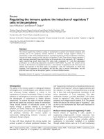
Figure 1.1. Schematic overview of the structure and function of the lymphatic vasculature. Fluid containing proteins, lipids and other
solutes leaks from blood vessels (BV), percolates through the interstitial tissue and returns to the circulation by the venous capillary bed and
lymphatic vessels (LV). Endothelial cells of lymphatic capillaries are oak shaped with overlapping scalloped edges (flaps). These flaps are
only sealed on the sides by discontinuous buttonlike junction allowing fluid entry through these flaps without disturbing cell–cell cohesion.
Lymph is subsequently transported to collecting lymphatics. Immune cells (lymphocytes [L], dendritic cells [DC]) likely enter lymphatic
capillaries through the intermingled flaps. In contrast to BV, initial lymphatics are made up of a discontinuous thin basement membrane (BM).
Anchoring filaments connect lymphatic capillaries to extracellular matrix and modulate vessel diameter by pulling adjacent endothelial cells
apart. Lymphatic endothelial cells (LEC), blood endothelial cells (BEC), fibronectin (FN), hyaluronan (HA). Figure adapted from (Paupert et
al., 2011).
!
4!
1.2 Lymphatic vessels during development
1.2.1 Morphogenesis of lymphatics during development
Experiments performed by Florence Sabin about a hundred years ago laid the
cornerstones for the widely accepted model that the mammalian lymphatic vasculature
had venous origins (Sabin, 1916). Modern day experiments performing lineage tracing by
Srinivasan et al. elegantly demonstrated that the mammalian lymphatic vasculature stem
from preexisting blood vessels (Srinivasan et al., 2007). During development, lymphatic
vascular genesis requires transdifferentiation of venous endothelial cells toward the
lymphatic endothelial phenotype, separation of blood and lymphatic vasculature,
sprouting of lymphatic vessels, and lymphatic vascular maturation. Over twenty genes
orchestrate this process in mice and these are summarized and detailed in Table 1
(Schulte-Merker et al., 2011).
1.3 Lymphatic vessels during inflammation
In adults, both blood and lymphatic endothelial cells are normally in a quiescent state, but
possess the capability to respond to a variety of stimuli. Lymphatic vessel growth also
called lymphangiogenesis, appears to follow that of the blood vessels during tissue
regeneration, wound healing, tumor growth and inflammation. New lymphatic vessels are
believed to grow primarily by sprouting from existing ones (Karpanen and Alitalo, 2008;
Tammela and Alitalo, 2010). Although the existence of bone marrow–derived
hematopoietic cells (He et al., 2004; Jiang et al., 2008; Lee et al., 2010; Religa et al.,
2005; Salven et al., 2003) or circulating macrophages (Maruyama et al., 2005) capable of
trans-differentiating into LECs has been suggested, the data has been conflicting and their
exact contribution to inflammatory lymphangiogenesis remains controversial in the field.