Structural and epitope characterization of major allergens from dust mite, BLO t 21 and DER f 7
Bạn đang xem bản rút gọn của tài liệu. Xem và tải ngay bản đầy đủ của tài liệu tại đây (4.55 MB, 159 trang )
STRUCTURAL AND EPITOPE CHARACTERIZATION OF
MAJOR ALLERGENS FROM DUST MITE,
BLO T 21 AND DER F 7
TAN KANG WEI
NATIONAL UNIVERSITY OF SINGAPORE
2011
STRUCTURAL AND EPITOPE CHARACTERIZATION
OF MAJOR ALLERGENS FROM DUST MITE,
BLO T 21 AND DER F 7
TAN KANG WEI
(B. Sc., UKM)
A THESIS SUBMITTED
FOR THE DEGREE OF DOCTOR OF PHILOSOPHY
DEPARTMENT OF BIOLOGICAL SCIENCES
NATIONAL UNIVERSITY OF SINGAPORE
2011
Acknowledgements
I am heartily thankful to my supervisor, Assoc. Prof. Dr. Henry Mok, for his encouragement,
patience, guidance and support throughout these years of my study. His critical thinking and
advices really inspired me in doing my research.
I would also like to extend my gratitude to Assoc. Prof. Dr. Chew, for being a resourceful and
understanding collaborator. Special thanks to Prof. Yang and Assoc. Prof. Dr. Sivaraman for
their sharing of ideas that have been wonderfully insightful for my studies in NMR and X-ray
crystallography.
Many thanks to Dr. Chan. Dr. Kartik, Dr. Shiva, Dr. Lin Zhi, Dr. Ong, Dr. Kumar, Dr.
Chiradeep and Dr. Jobi for their generosity in sharing invaluable experience whenever I
requested. You have been a great help throughout my candidature. Sang, Jack, Rishi, Jana,
Wentao, and everyone in SBL as well as functional genomic lab 1 and 2, my heartfelf thanks
for your delightful companionships and helpful advices for designing my experiments.
To my beloved Xin Yu, thank you for always being there for me. Your love is a great
motivation for my research that I will forever cherish. Special thanks for helping me to
proofread this thesis with admirable patience and critical comments.
My family and relatives who have been emotionally supportive from the day I stepped foot in
Singapore, I am forever indebted to you for your understanding, patience and love. Without
you, I won’t be who I am today, thank you very much.
ii
Table of Contents
Acknowledgements
……………………………………………………………… i
Table of Contents ………………………………………………………………………ii
Summary
………………………………… vii
LIST OF TABLE……………………………………………………………… ix
LIST OF FIGURES…………………………………………………………………….x
LIST OF ABBREVIATIONS…………………………………………………….……xiii
CHAPTER 1
INTRODUCTION…………………………………………………1
1. Allergy…………………………………………………………………….1
1.1 An introduction to allergy………………………………………………… 1
1.2 Mechanisms of allergy…………………………………………………… 3
1.3 Dust mite………………………………………………………………… 6
1.4 From structure determination to IgE epitope mapping 9
1.4.1 Structural biology of allergens 9
1.4.2 IgE epitope mapping of allergens 12
1.5
Specific immunotherapy………………………………………………… 15
1.7 Group 21 Allergen from dust mite 19
1.8 Group 7 Allergen from dust mite 20
1.9 Objectives and significance of this study 21
CHAPTER 2 MATERIALS & METHODS 24
2.1 Generation and subcloning of Blo t 21 and its mutants into expression vector .24
2.1.1 Bacterial host strains………………………………………………………24
2.1.2 Generation of DNA insert and Polymerase Chain Reaction 24
2.2 Generation of DNA mutant insert for site directed mutagenesis 24
2.3 Preparation of DH5-α competent cells 26
iii
2.4 Sub-cloning 27
2.5 Transformation of ligation mix into DH5-α competent cells. 27
2.6 PCR screening of transformant 28
2.7 Isolation of DNA plasmid 28
2.8 Plasmid DNA sequencing 29
2.9
Protein expression and purification 29
2.9.1 Transformation of plasmid into BL21(DE3) competent cells 29
2.9.2 Protein expression 29
2.9.3 Protein purification using nickel-affinity chromatography 30
2.9.4 Protein purification using GST affinity chromatograph 31
2.9.5 Thrombin digestion 31
2.9.6 Gel filtration FPLC (Fast Protein Liquid Chromatography) 32
2.10
Preparation of NMR sample………………………………………………32
2.11 Sodium dodecyl sulphate-polyacrylamide gel electrophoresis (SDS-PAGE) 32
2.12 Circular dichroism (CD) spectropolarimetry 33
2.12.1 Thermal denaturation experiments 33
2.13
Sequence alignment……………………………………………………….33
2.14 Nuclear magnetic resonance and structural determination 34
2.14.1 NMR chemical shift assignments 34
2.14.1.1 2D
1
H-
15
N HSQC spectrum 34
2.14.1.2 HNCACB and CBCA(CO)NH 34
2.14.1.3 C(CO)NH-TOCSY and H(CO)NH-TOCSY 35
2.14.1.4 HCCH-TOCSY 35
2.14.1.5 NOE distance restraints and hydrogen bond restraints 36
iv
2.14.1.5.1
15
N-edited NOESY 36
2.14.1.5.2
13
C-edited NOESY 37
2.15
NOE assignments and structure calculation 37
2.16 Immunoassay of Blo t 21………………………………………………….38
2.16.1 Specific IgE binding ELISA experiment 38
2.16.2 Endpoint inhibition ELISA experiment 38
2.16.3 Peptide ELISA experiment 39
2.17
Sub-cloning, expression and purification of Der f 7 39
2.18 Circular dichroism (CD) spectropolarimetry of Der f 7 41
2.18.1 Thermal denaturation experiments 41
2.18.2 Chemical denaturation experiments 41
2.19
NMR studies of Der f 7………………………………………………… 42
2.19.1 NMR chemical shift assignments 42
2.19.2 2D
1
H-
15
N HSQC spectrum 42
2.19.2.1 HNCACB and CBCA(CO)NH 42
2.19.3 Ligand binding and pH titration studies of Der f 7 43
2.19.4
15
N relaxation studies of Der f 7 43
2.20
Crystallization of Der f 7……………………………………………….….43
2.21 Data collection and structure solution of SeMet Der f 7 44
2.22 Structure-based alignment and comparison 45
2.23 Immunoassay for Der f 7 and Der p 7 45
CHAPTER 3
BLOT21: RESULTS & DISCUSSION 46
3.1 Resolving Blo t 21 Structure using NMR 46
3.1.2 2D
1
H-
15
N HSQC spectra of Blo t 21 46
v
3.2 Chemical shifts assignment of Blo t 21 47
3.2.1 Backbone and side chain assignments 47
3.2.2 Chemical shift index (CSI) 50
3.2.3 NOE assignment by CNS 51
3.3
NMR Structure of Blo t 21……………………………………………… 53
3.4 3-D structures comparison of Blo t 21 with Blo t 5 and Der p 5 56
3.5 The Allergenicity of Blot 21 Compared to Blo t 5, Der p 5 and Der f 21 58
3.6 Study on the Stability of Blo t 21, Der f 21, Blo t 5 and Der p 5 61
3.6.1 Circular Dichroism 61
3.6.2 Thermal Denaturation Experiment 62
3.7
Site-directed mutagenesis and IgE epitope mapping of Blo t 21 65
3.9 Multiple mutations of epitope residues further reduce IgE binding 73
3.10 Residue “Asp-96” - A Unique IgE Epitopes in Blo t 21? 77
3.12 Inhibition Assays………………………………………………………….80
3.12.1 End-point Inhibition assays 80
3.12.2 The effect of L73E mutation in Der p 5 83
3.12.4 Inhibition assays of Blo t 21 vs Der f 21 86
3.13
Peptide ELISA………………………………………………………… 88
3.13.1 Surface charge distribution at the putative IgE interacting site 88
3.13.2 Peptides show different IgE binding activities 90
CHAPTER 4
DER F 7: RESULTS & DISCUSSION 94
4.1 Characterization of Der f 7…………………………………………… ….94
4.2 Crystallization and Data Collection of SeMet Recombinant Der f 7 96
4.3 Crystal Structure of Der f 7………………………………………… ……98
vi
4.4 Structural homology………………………………………….………… 103
4.5 NMR Studies on Der f 7………………………………………………….106
4.6
IgE Epitope Mapping of Der f 7 109
4.6.1 Single Mutant D159A & Double Mutant L48A_F50A 111
4.6.2 Cross inhibition between Der f 7 and Der p 7 114
4.6.3 Putative IgE epitopes on Der f 7 and Der p 7 115
4.7
Ligand binding studies……………………………………………….… 118
CHAPTER 5 CONCLUSION & FUTURE WORK 122
5.1 Structural studies and imuno-characterization of Blo t 21 122
5.2 Future direction: Blo t 21 124
5.3 Crystal structure and IgE epitopes of Der f 7 125
5.4 Future direction: Der f 7 127
References
………………………………………………………………………128
Appendix I………………………………………………………………… …139
Appendix II…………………………………………………………………… 139
Appendix
III
………………………………………………………………… 1401
vii
Summary
Allergic diseases have drawn worldwide attention since its discovery for more than a
century ago. Currently, the prevalence of allergic diseases is rising steadily to an alarming
state in both developed and developing countries, taking a toll on millions of lives. These
diseases include asthma, atopic dermatitis (AD), rhinitis, anaphylaxis as well as food, drug
and insect allergy. House dust mite (HDM) stands out as one of the major causative agents of
allergic diseases owing to its ubiquitous presence in both temperate and tropical regions. To
date, more than twenty groups of allergens have been isolated from dust mites and were
shown to be highly antigenic. However, the underlying reasons of their allergenicity remain
largely unknown. Therefore, extensive immune characterization aided by sophisticated
structural studies is imperative in order to decipher the inherent features of these allergens and
to develop a hypoallergen for specific immune therapy.
This thesis aims to describe the 3D structures and the IgE epitopes of two major
allergens from dust mites, namely, Blo t 21 and Der f 7. The first part of this thesis focuses on
Blo t 21, a major allergen from Blomia tropicalis. Blo t 21 showed limited cross-reactivity
with its paralogue, Blo t 5, thus inferring that Blo t 21 should use unique epitopes to interact
with IgE antibodies. The 3D structures of Blo t 21 and Blo t 5 (PDB: 2JMH) determined by
NMR approaches shared high structural homology. However, some disparities of the local
structure could be detected. The allergenicity test on the Blo t 21 mutants using ELISA
demonstrated that residues Glu-74, Asp-79, Glu-89 and Asp-96 were the major IgE epitopes,
with residues Glu-89 and Asp-96 forming a conformational epitope. The subsequent peptide
ELISA experiments suggested the presence of a linear IgE epitope in Blo t 21, which
exhibited distinct allergenicity compared to that described in Blo t 5 previously. These data
could help to explain the limited cross reactivity between Blo t 21 and Blo t 5. The
allergenicity and cross inhibition tests conducted on the homologous proteins, Der f 21 and
Der p 5, indicated different antigenic properties as compared to Blo t 21 and Blo t 5. Further
viii
analysis implied that the primary sequence, stability and 3D structure could contribute to the
differences in these proteins. Therefore, the fundamental biophysical and structural
characterizations on these allergens should be included while mapping their IgE epitopes.
The second part of this thesis describes the crystal structure and the IgE epitope
mapping of Der f 7, a major group 7 allergen from Dermatophagoides farinae. Studies have
shown that this allergen elicits strong immune response in mite-sensitized individuals. The
crystal structure of Der f 7 is very similar to that of Der p 7, which was also solved by X-ray
crystallography method (PDB: 3H4Z). However, it was reported that these two allergens
showed dissimilar IgE binding activity, with a majority of the test subjects indicating higher
sensitivity to Der p 7. Recently, attempts to map the IgE epitopes have been reported in two
separate accounts. However, our results suggested that the proposed IgE binding residues,
Leu-48, Phe-50 and Asp-159, might not be the major IgE epitopes of Der f 7. Based on the
mapping of different residues between Der f 7 and Der p 7 on the crystal structures, we
proposed that residues Lys-25, Asp-55 and Glu-124 could be responsible for the higher IgE
binding activity of Der p 7. Nevertheless, the IgE epitopes of Der f 7 remained elusive thus
far. In addition, the data pertaining to physical characterization and ligand binding studies of
Der f 7 will be presented as well. These results may pave a way for understanding the
allergenic properties of these proteins, and aid in the development of hypoallergens suitable
for immunotherapy purposes.
ix
LIST OF TABLE
Table 1.1
Classification of dust mite allergens 9
Table 2.1
List of primers used for PCR and mutagenesis studies 26
Table 3.1
The Overall Statistics of 20 lowest-Energy Ensemble of Blo t 21 NMR
Structure.
55
Table 4.1
Data collection statistics for Der f 7 SeMet crystal
98
Table 4.2
Crystallographic data statistics for Der f 7 3D structure
99
Table 4.3
Selected DALI matches to Der f 7. 105
Table 4.4
Tm for Der p 7, Der f 7 and its mutants 121
Table 4.5
End-point inhibition assay for De f 7 and Der p 7. 129
.
x
LIST OF FIGURES
Figure 1.1
Mechanism of allergy diseases. 6
Figure 2.1
Generation of site-directed mutants 25
Figure 3.1
Two-Dimensional
1
H-
15
N HSQC of Blo t 21. 47
Figure 3.2
Sequential assignment of backbone chemical shifts of Blo t 21 49
Figure 3.3
Chemical Shift Index of Blo t 21 51
Figure 3.4
Assignment of NOESY spectrums. 53
Figure 3.5
NMR Structure of Blo t 21. 56
Figure 3.6
Superimposition of the NMR structure of Blo t 21 with Blo t 5 and
Der p 5
58
Figure 3.7
The allergenicity of Blo t 21, Der f 21, Der p 5 and Blo t 5. 60
Figure 3.8
The CD spectrum of Blo t 21, Der f 21, Blo t 5 and Der p 5. 62
Figure 3.9
The CD spectrum of Blo t 21, Der f 21, Blo t 5 and Der p 5 at
different temperatures.
63
Figure 3.10
Thermal denaturation experiment for Blo t 21, Der f 21, Der p 5 and
Blo t 5.
65
Figure 3.11
Sequence alignment of group 21 and group 5 allergens from dust
mites.
67
Figure 3.12
The prescreening to evaluate the sensitivity against wild-type Blo t
21
68
Figure 3.13
Percentage prevalence of volunteers with more than 20% reduction
in IgE binding against single mutants of Blo t 21.
68
Figure 3.14
The distribution of the residues corresponding to Glu-74, Asp-79,
Glu-84, Glu-89 and Asp-96 of Blo t 21 in the 3-D structures of Blo t
5 and Der p 5
70
Figure 3.15
Comparison of the CD spectrum of Blo t 21 and its mutants 73
Figure 3.16
Comparing the allergenicity of multiple mutants with the wild-type
Blo t 21 using ELISA experiment.
76
Figure 3.17
Specific ELISA experiment for E89A_D98A (Blo t 21) and
E91A_K98A (Blo t 5) double mutants
78
Figure 3.18
Specific ELISA experiment for E92A_E99A in Der f 21. 79
Figure 3.19
The cross-reactivity among Blo t 21, Blo t 5 and Der p 5 examined
by end-point inhibition assay.
82
xi
Figure 3.20
The CD spectrum of Der p 5 and its L73E mutant. 85
Figure 3.21
The allergenicity of Der p 5 L73E mutant compared to wild-type
Der p 5 and Blo t 5.
85
Figure 3.22
The cross-reactivity between Blo t 21 and Der f 21.
87-
88
Figure 3.23
3D distribution of charged residues at the putative IgE binding site
of Blo t 5, Blo t 21 and Der p 5.
90
Figure 3.24
Results of the ELISA experiment using peptides derived from Blo t
21, Der f 21, Blo t 5 and Der p 5.
93
Figure 4.1
SDS-PAGE and gel filtration profiles of Der f 7. 95
Figure 4.2
Circular dichroism of Der f 7 and Der p 7. 95
Figure 4.3
Mass Spectrometry of native and SeMet Der f 7 96
Figure 4.4
Crystals of recombinant Der f 7 97
Figure 4.5
Diffraction pattern of SeMet Der f 7. 97
Figure 4.6
The final model of Der f 7 crystal structure 100
Figure 4.7
Ribbon diagram of Der f 7crystal structure 101
Figure 4.8 Superimposition of Der f 7 and Der p 7. 101
Figure 4.9
Surface charge distribution of Der f 7 and Der p 7 102
Figure 4.10
Secondary structure topology of Der f 7. 102
Figure 4.11
Superimposition of Der f 7 with its homologous structures. 106
Figure 4.12
The two-dimensional
1
H
15
N-HSQC of Der f 7 with backbone
assignment.
108
Figure 4.13
Chemical Shift Index (CSI) of Der f 7. 108
Figure 4.14
Locations of residues 48, 50 and 159 in Der f 7 and Der p 7 3D
structures.
111
Figure 4.15
Specific IgE ELISA experiment comparing the allergenicity of Der f
7 and Der p 7 as well as their mutants
114
Figure 4.16
Surface diagram of Der f 7 in four different orientations. 117
Figure 4.17
Peaks perturbation in the 2D-
1
H
15
N HSQC of Der f 7 upon addition
of Polymyxin B (PB).
119
Figure 4.18
Chemical shift perturbation plot of Δδ versus residues of Der f 7 for
the PB titration experiment
120
xii
Figure 4.19
The 3D structure of Der f 7 and Der p 7 showing the possible
residues involved in PB binding.
121
LIST OF ABBREVIATIONS
Amino Acids
One letter code Three letter code Amino acid
A
Ala Alanine
C
Cys Cystein
D
Asp Aspartic acid
E
Glu Glutamic acid
F
Phe Phenylalanine
G
Gly Glycine
H
His Histidine
I
Ile Isoleucine
K
Lys Lysine
L
Leu Leucine
M
Met Methionine
N
Asn Asparagine
P
Pro Proline
Q
Gln Glutamine
R
Arg Arginine
S Ser Serine
T
Thr Threonine
V
Val Val in e
W
Trp Tyrptophan
Y
Tyr Tyrosine
xiii
Chemicals and reagents
BSA
Bovine serum albumine
CaCl
2
Calcium chloride
dATP
2’ deoxyadenosine 5’ triphosphate
dCTP
2’ deoxycytidine 5’ triphosphate
dGTP
2’ deoxyguanosine 5’ triphosphate
dNTP
Deoxynucleotide triphosphate
dTTP 2’ deoxythymidine 5’ triphosphate
IPTG
Isopropyl-D-thiogalactoside
KCl
Potassium chloride
MgCl
2
Magnesium chloride
Ni
Nickel
Ni-NTA
Nickel-Nitrilotriacetic
PBS
Phospate buffer saline
PBS-T
Phospate buffer saline with 0.05% Tween
PEG MME
Polyethylene glycol monomethyl ether
PNPP
p-Nitrophenyl phosphate disodium
SDS
Sodium dodecyl-sulphate
TEMED
N,N,N,N’-Tetramethylethylenediamine
TMB
3,3,’5,5’-Tetramethylbenzidine
Tris
Tris (hydoxymethyl)-aminomenthane
xiv
Units and measurement
bp
Base pair
Da
Dalton
Hz
Hertz
K
Kelvin
kDa
Kilo Dalton
M
Molar
pH Potential of hydrogen
ppm
Parts per million
rpm
Rotation per minute
SGD
Singapore Dollar
U
Unit (enzyme)
V
Vo l t
Others
1D/2D/3D/4D
One dimentional/Two dimensional/3D/
Four dimensional
α
alpha
β
beta
γ
gamma
δ
delta
ε
epsilon
APC
Antigen-presenting cell
CCP4
Collaborative Computational Project
Number 4
CD Circular dichroism
xv
CD4/8
Cluster of differentiation 4/8
CD23
Fc epsilon RII
CD25
alpha chain of the IL-2 receptor
CSI Chemical shift index
DNA
Deoxyribonucleic acid
ELISA
Enzyme-Linked ImmunoSorbent Assay
EST
Expressed Sequence Tag
FcεRI
IgE receptor type-I
FPLC
Fast Protein Liquid Chromatography
GST
Glutathione S-transferase
HRP
Horseradish peroxidase
HSQC
Heteronuclear Single-Quantum Correlation
IFN-γ
Interferon gamma
IgA
Immunoglobulin A
IgE
Immunoglobulin E
IgG
Immunoglobulin G
IL
Interleukin
ITAM
Immunoreceptor tyrosine-based activation
motif
LB Luria Bertani
MALDI-TOF
Matrix-assisted laser desorption/ionization
MHC
Major histocompatibility complex
NMR
Nuclear Magnetic Resonance
NOE
Nuclear Overhauser Effect
NOESY
Nuclear Overhauser Effect Spectroscopy
NPC2
Niemann Pick protein type C2
OD Optical density
PCR
Polymerase chain reaction
xvi
RIA
Radio-immuno assay
R.M.S.D.
Root mean square deviation
SeMet
Selenium methionine
SDS-PAGE Sodium Dodecyl-Sulphate Polyacrylamide
Gel
Electrophoresis
Th1
Type-1 Helper T-cells
Th2
Type-2 Helper T-cells
TNF-α
Tumor necrosis factor alpha
TOCSY
Total correlation spectroscopy
1
CHAPTER 1 INTRODUCTION
1. Allergy
1.1 An introduction to allergy
Clemens von Pirquet, an Austrian pediatrician, coined the word “Allergy” in 1906.
Originated from the Greek word 'allos', which means “change in the native state”, the term
'allergy' is now used to describe the altered immune response in a human body in response to
the supposedly innocuous foreign substances, commonly known as allergens. The immune
system in a healthy human body will function with an intricate balance to react appropriately
to the intruding foreign substances while preventing the over-reaction against self-antigens or
harmless foreign antigens. An occurrence of the excessive or uncontrolled immune response
will lead to an immune disease known as “Hypersensitivity”. Hypersensitivity disorders
include autoimmune diseases, in which the body immune system mistakes own cells or
tissues as antigens, and the diseases that result in the hyper-reactive responses against non-
harmful environmental proteins or microbes. Gell and Coombs (1963) proposed that there are
four types of hypersensitivity, distinguished by the immune-pathologenic mechanism and the
type of mediators involved (Gell and Coombs 1963). The fifth type of hypersensitivity (Type
V Hypersensitivity) was described as a rare, type 2-like hypersensitivity (Rajan 2003). Based
on these classifications, allergy is synonymous with “Type I Hypersensitivity”, in which the
immunoglobulin E (IgE) and IgG4 mediate the immune responses against foreign antigens.
Commonly mentioned disorders observed in Type I Hypersensitivity include atopy, systemic
anaphylaxis and asthma.
Atopy or atopic syndrome refers to the hereditary predisposition of an individual
toward producing specific IgE antibodies against environmental antigens and subsequent
development of immediate and acute allergic reactions (Abbas and Lichtman 2003). The
cross-linking of the allergens to the IgE antibodies bound on the surface of mast cells or
basophils triggers the release of the pro-inflammatory mediators (histamine, proteases,
2
chemokines, heparin), resulting in the clinical manifestation of diseases like atopic eczema,
asthma and allergic rhinitis (Bousquet, Holt et al. 2008).
An antigen that can trigger immediate allergic responses upon exposure is defined as
an allergen. Allergens are usually soluble proteins or chemicals that can induce the
proliferation of the IgE antibodies circulating in the atopic patients. Some common allergens’
sources include animal products, drugs, foods, insect stings, fungal and pollens. Animal
products include fur, dander, wool and the dust mite (Dermatophagoides pteronyssinus and
Dermatophagoides farinae; and storage mite Blomia tropicalis) excretion (Hurtado and Parini
1987; Fernandez, Martin-Esteban et al. 1993). Many atopic patients are known to be
hypersensitive to certain drugs like penicillin, sulfonamides and local anesthetics, which
sometimes cause complications in the medical practices. A more commonly known source of
allergen is food. Some commonly known food allergens are celery, corn, eggs, certain fruits,
seafood and nuts. Insect stings like bee venom and wasp venom are also widely known as a
major source of allergens. Several genera of fungus are implicated as major allergen sources
comprising Aspergillus, Cladosporium, Alternaria, Penicillium and Fusarium (Cromwell,
1997). Pollen allergens which are known to cause hay fever include some species of grass like
ryegrass Lolium perenne and timothy grass Phleum pratense; weeds such as ragweed
Ambrosia and nettle (Urtica dioica); as well as those from trees like birch Betula verrucosa,
alder Alnus serrulata and willow Salix fragilis (Cromwell,1997).
In the past three decades, there has been a spectacular increase in the prevalence of
asthma and allergic disease worldwide (Holgate 2004). The prevalence of asthma increased
75% from 1980 – 1994, with 160% increment in asthma rates among the children under the
age of five (Centers for Disease Control, USA, 1998). World Health Organization (WHO)
reported that in 2007, more than 300 million people suffered from asthma worldwide, with
250,000 fatalities attributed to the disease annually. Asthma and allergic diseases have caused
millions of people to suffer physical impairments and decrease in quality of life. For example,
approximately 10.1 million missed work days for adults annually in US (Akinbami 2006);
asthma was also responsible for 3,384 deaths in US (ALA age group analysis of NHIS
3
through 2005). Besides that, asthma also adds to the healthcare financial burden bore by a
nation. In Singapore, for every 10,000 students examined in 2001, 1026 male students (~10
%) and 757 (~7.5%) female students had asthma; and the incidence of asthma increased for
both genders between 1991 and 2001 (Statistics Singapore Newsletter, 2003). In US, the
annual economic cost of asthma is US$19.7 Million (ALA, USA, 2007) and according to the
survey conducted in 2009 by National Heart, Lung and Blood Institute, USA, over USD$20
billion were spent due to asthma per annum. On the other hand, around SGD$54 million were
spent yearly by asthmatic patients in Singapore (Chew, Goh et al. 1999).
Conventional clinical treatment for allergic diseases is designed to alleviate the
symptoms and to suppress the allergic inflammation. For example, antihistamines drugs,
anticholinergic agents or topical corticosteroids are commonly used to treat allergic rhinitis
(Kay, 2001). Atopic dermatitis is normally treated with antihistamines and corticosteroids to
control and suppress inflammation of the affected site (Roos, Geuer et al. 2004). Anti-
asthmatic drugs salbutamol and salmeterol are predominantly used to relieve asthmatic
symptoms and for maintenance therapy (Kon and Barnes 1997). However, relieving the
symptoms is not the most effective choice for long-term therapy. The advent of allergen-
specific immunotherapy provides a novel avenue to reverse the course of the disease and for
prolonged protection against progression of allergic diseases (Valenta 2002; Niederberger and
Valenta 2004).
1.2 Mechanisms of allergy
There are three major components involved in an allergic reaction, namely, the
allergen, the IgE and at least one type of effector cells such as mast cells, basophils or
eosinophils (Abbas and Lichtman 2003). Besides that, the immune system also requires other
cellular members such as antigen-presenting cells (APC) and lymphocyte cells to initiate and
regulate of allergic reaction and disease progression. An invading allergen is captured by APC
such as dendritic cells or cutaneous Langerhans’ cells and presented as T-cell peptide to
4
CD4+ T-cells in a major histocompatibility complex MHC class II-restricted manner (Abbas
and Lichtman, 2003 and Kay, 2001). Consequently, the CD4+ cells are primed to differentiate
into T helper 2 cells (Th2) followed by the release of Th2-type cytokines such as IL-4, IL-5,
IL-9 and IL-13 (Kay, 2001).
The APC presents the antigen in the form of peptide fragments to the T-cells. The
fragments bearing the T-cell epitopes are loaded onto the MHC and the formation of the
MHC-peptides complexes will be recognized by the T-cells. Generally, there are two classes
of MHC molecules; Class I MHC presents the peptides to the CD8+ cytolytic T-cells while
the Class II MHC presents the peptides to the CD4+ helper T-cells. The Class II MHC-
peptide complex is recognized by the T-cell receptor (TCR) located on the surface of the T-
cells. Dendritic cells, macrophages and B lymphocyte cells are the common APCs that initiate
the helper T-cells (Th). Dendritic cells and cutaneous Langerhans cells are known to present
the allergens to Th2 cells in an MHC Class II manner in cases of asthma and eczema,
respectively (Kay, 2001).
The Th2 cells will respond upon recognition of the MHC-peptide complexes by
releasing an array of cytokines. Subsequently, the proliferation of specific IgE antibodies and
the development of inflammation cells such as mast cells, basophils and eosinophils will be
implemented. The cytokines that mediate the inflammatory and immune reactions are termed
as “interleukin”. Interleukins-4 (IL-4) and IL-13 initiate the differentiation of B cells to
undergo class switching of the constant region of immunoglobulin heavy chain (C
H
)
to Fcε to
produce IgE class antibodies specifically (Valenta 2002). IL-4 and IL-9 promote the
development of mast cells, the major effector cells in releasing inflammation mediators (Kay,
2001). IL-13 plays a key role in inducing airway hyper-responsiveness, goblet cell metaplasia
and mucus hypersecretion (Wills-Karp, Luyimbazi et al. 1998), while the expansion and
recruitment of eosinophils and basophils are induced collectively by IL-4, IL-5, IL-9 and IL-
13 (Kay, 2001). The cytokines secreted by Th1 or Th2 cells act as an autocrine growth factor
to promote the proliferation of these cells while inhibiting the growth of the opposite cell type
(Fernandez-Botran, Sanders et al. 1988; Gajewski and Fitch 1988; Liew and McInnes 2002).
5
For instance, IL-4 induces the growth of Th2 cells while inhibiting the proliferation of Th1
cells. On the other hand, IFN-γ, a cytokine released by Th1 subset of cells promotes the
expansion of Th1 cells but inhibits the proliferation of Th2 cells.
IgE antibodies bind to its receptor (Fc receptor) via the constant region, Fcε, on the
heavy chain. Cross-linking of the FcεRI (high-affinity IgE receptor) present on the mast cells
or basophils by allergen-bound IgE releases inflammatory mediators such as histamine,
leukotrienes and lipid mediators (Kay 2008). This receptor is also present on the surface of
APC where it assists in the IgE-mediated capturing of the allergen, enabling the allergen to be
presented to the T-cells (Stingl and Maurer 1997). Based on the crystal structure of the
FcεRIα and IgE-Fc complex, the α-chain of FcεRIα binds to the dimeric molecules of Cε3
domain of the IgE (Garman, Wurzburg et al. 2000). FcεRIα does not aggregate in the absence
of antigen; the aggregation of the receptors occurs only when IgE antibodies are bound to the
receptor and cross-linked by allergens. Numerous signaling pathways initiated by the
aggregation of FcεRI receptors on the surface of mast cells result in the secretion of various
inflammatory mediators and cytokines (Turner and Kinet 1999).
The FcεRIα receptor is expressed on the surface of mast cells and basophils as a
multimeric αβγ2 complex (Nadler, Matthews et al. 2000). The β chain and two γ chains act as
a phosphoreceptor for Tyr kinases that are involved in signaling cascades. Each β and γ chains
contains one Immunoreceptor Tyr-based Activation Motif (ITAM) in their respective cytosolic
portions. The aggregation of FcεRIα receptors triggers the signal transduction that activates
two main Tyr kinases, Lyn and Syk. The first kinase, Lyn, phosphorylates ITAMs of β and γ
chains followed by the recruitment and activation of the second kinase, Syk to the ITAMs of γ
chains (Abbas and Lichtman, 2003). The recruitment and activation of the kinases lead to the
elaborated signalling events that ultimately result in the phosphorylation of myosin light
chains by an activated protein kinase C. Finally, degranulation occurs when the actin-myosin
complexes are broken down (Nadler, Matthews et al. 2000; Abbas and Lichtman 2003).
6
1.3 Dust mite
House dust mites are arachnids related to ticks, spiders and harvestmen (Colloff
2009). These microscopic organisms belong to the phylum Arthropoda, subphylum
Chelicerata, class Arachnida, order Acari, and suborder Astigmata. Dust mites are
ubiquitously found, especially in human habitats, where the dust is accumulated in bedding,
carpets and furniture. House dust provides a sustaining habitat for dust mites, where the food
source - shed human skin scales – is abundant. The predominant dust mites species found in
household dust, and the major source of allergens belong to the family Pyroglyphidae. The
top three pyroglyphid species of house dust mites in terms of the frequency and abundance
worldwide are D. farinae, D. pteronyssinus and Euroglyphus maynei (Colloff 2009). These
species are more common in temperate climate such as continental Europe and North
Fi
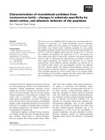
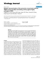
g
ure 1.1 Mechanism of allergy diseases. During the first encounter, the allergen will be
p
resented by the APCs to Th2 cells triggering release of cytokines like IL-4 and Il-13.
These cytokines stimulate antibody class switching and production of IgE antibodies. Pro-
inflammatory mediators will be released by mast cells once mast cells-
b
ound IgE
antibodies are cross-linked by the allergen, which ultimately results in acute allergic
reactions such as wheezing, asthma and sneezing.
7
America. On the other hand, storage mite B. tropicalis (family Echymyopodidae) has emerged
as a more important species in tropics and subtropical regions.
Dust mites reproduce sexually and the stages in its life cycle are the egg, a six-legged
larva, two eight legged nymphal stages (protonymph and tritonymph) and adult. Similar to
insects, adult mites have exoskeleton, jointed appendages and a blood-filled body cavity
(hemocoel) (Fernandez-Caldas 2002). However, instead of having three pairs of legs like
insects, mites have four. Dust mites are poikilothermic (unable to control body temperature),
the length of their life cycle is thus dependent on the temperature of the habitat. The growth in
population and egg-to-adult development of dust mites are controlled by both humidity and
temperature (Hart 1998). In laboratory, dust mite requires a high relative humidity (RH) from
75% to 80% to complete their life cycle, with an optimum temperature of approximately 25
°C to 30 °C (Fernandez-Caldas 2002). The life span of the adults is approximately 4-6 weeks,
during which time each female can produce 40-80 eggs.
The abundance of dust mites in household area is one of the main reasons why dust
mite is the major cause of asthma attacks in the world. The body and the feces of dust mite are
the major source of allergens. These “allergens” are the enzymes and other proteins from the
mites that react potently as antigenic molecule. For example, Der p 1, a group 1 allergen
isolated from D. pteronysinnus was shown to be strongly associated with the gut and faecal
pellets, based on its amino acid sequence; the group 4 allergens are amylase, common
functional enzymes found in most of the organisms (Colloff 2009). Based on the online
resource Allergome (www.allergome.org), more than 20 groups of proteins from dust mites
have been isolated and characterized, indicating the wide diversity of different proteins that
are involved in causing allergic reactions (Table 1.1). These allergens are grouped according
to their function, molecular weight and sequence identity, and numbered according to their
chronological characterization. As it can be seen, group 1 and group 2 are the two
predominant group of allergens from dust mites, with each of them accounting for more than
80% in IgE binding prevalence among patients sensitized to dust mites (Trombone, Tobias et
al. 2002).