Chemical and pharmacological studies of ardisia elliptica antiplatelet, anticoagulant activities and multivariate data analysis for drug discovery
Bạn đang xem bản rút gọn của tài liệu. Xem và tải ngay bản đầy đủ của tài liệu tại đây (1.56 MB, 213 trang )
CHEMICAL AND PHARMACOLOGICAL STUDIES
OF ARDISIA ELLIPTICA: ANTIPLATELET,
ANTICOAGULANT ACTIVITIES AND
MULTIVARIATE DATA ANALYSIS FOR DRUG
DISCOVERY
CHING JIANHONG
(B. Sc (Hons), NUS)
A THESIS SUBMITTED
FOR THE DEGREE OF DOCTOR OF PHILOSOPHY
DEPARTMENT OF PHARMACY
NATIONAL UNIVERSITY OF SINGAPORE
2011
ii
Acknowledgements
I would first of all, like to thank my supervisors, A/P Koh Hwee Ling
and A/P Tan Chay Hoon for the chance to work in their laboratories, and also
the guidance which they have given me, be it in academic, or life experiences.
I would like to thank Dr Yap Chun Wei for his numerous advices and
help on the metabolomics project. This part of the work forms a major part of
my thesis, without which, it would not be a success. I have also learnt many
important concepts of designing experiments from him.
Next, I would like to extend my greatest appreciation to Dr Lin Haishu,
who had very kindly helped me with the pharmacokinetic studies in this
project. Dr Lin had very patiently shared with me his expertise on PK.
Without his help, this project would not be as complete as I would hope it to
be.
Also, I would like to thank A/P Ho Chi Lui Paul for his kind comments
on my work on pharmacokinetics. Ms Kong Sing Teang had also been a great
help and company during weekends and late nights in the laboratory working
on the pharmacokinetic studies.
My appreciation goes to the final year undergraduate students whom I
have helped to guide. Their help and contribution to the project is essential to
the successful completion of this project. I would like to thank Ms Christina
Tan Juin Yu (Department of Pharmacy, graduated 2009) for her hard work in
helping with the sample collection and extraction, and the insights she has
given me on the anticancer effects of Ardisia elliptica. I would like to thank
Ms Soh Wei Li (Department of Pharmacy, graduated 2010), who had been a
great help with my work on metabolomics. I would like to thank Mr Lee Jun
iii
Feng (Department of Pharmacology, graduated 2010), for his help on the in
vivo work of the project, and especially his insightful advice and comments. I
am glad to say that I have learnt as much from them, as they have from me.
I appreciate the help rendered to me by my current and previous lab
mates, Dr Lau Aik Jiang, Dr Hou Peiling, Ms Agnes Chin, Dr Toh Ding Fung,
Mr Li Lin, Mr Patel Dhavalkumar Narendrabhai and Dr Sogand
Zareisedehizadeh. Also I must thank research assistants and lab technologists
in Departments of Pharmacy and Pharmacology, namely, Ms Yang Jun, Mr
Johannes Murti Jaya, Ms Ng Sek Eng, Mrs Khoo Yok Moi, Mdm Annie Hsu
and Mr Ang Seng Ban who had helped me at numerous occasions.
I would like to thank the Head of Department of Pharmacy, A/P Chan
Sui Yung for the chance to work in the department, and NUS for the research
scholarship.
Special thanks go to my friend of over 10 years, Mr Zhang Jiajie, who
had always been a great listening ear and source of moral support and
encouragement. Jiajie had also given me great advice on my future career
paths during my many discussions with him, which I find tremendously
useful.
Last but not least, I thank my family for their support, morally and
financially, which I am greatly indebted to. I thank my fiancée, Ms Ho Jia Pei,
who is a great source of comfort when times are down, and also her comments
on this thesis.
iv
List of publications and conference presentations
Publications
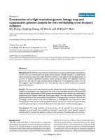
1. J. Ching, W.L. Soh, C.H. Tan, J.F. Lee, J.Y.C. Tan, J. Yang, C.W. Yap,
H.L. Koh. Identification of active compounds from medicinal plant
extracts using GC-MS and multivariate data analysis. Journal of
Separation Science 2012, 35: 53-59.
2. J. Ching*, H.S. Lin*, C.H. Tan, H.L. Koh. Quantification of α- and β-
amyrin in rat plasma by gas chromatography-mass spectrometry:
application to preclinical pharmacokinetic study. Journal of Mass
Spectrometry 2011, 46: 457-464. *Equal contribution
3. J. Ching, T.K. Chua, L.C. Chin, A.J. Lau, Y.K. Pang, J. Murti Jaya, C.H.
Tan, H.L Koh. β -Amyrin from Ardisia elliptica Thunb. is more potent
than aspirin in inhibiting collagen-induced platelet aggregation. Indian
Journal of Experimental Biology 2010, 48:275-279.
4. J. Ching, J.F. Lee, C.H. Tan, H.L. Koh. Antiplatelet activity of Ardisia
elliptica, and its isolated component, β-amyrin in rats (in preparation)
5. J. Ching, C.H. Tan, H.L. Koh. A study of antiplatelet and anticoagulant
activities in plants commonly found in Singapore. Annals Academy of
Medicine 2007, 36(11): S44.
6. Contributed to H.L. Koh, T.K. Chua, C.H. Tan. A guide to medicinal
plants: an illustrated, scientific and medicinal approach. Singapore: World
Scientific Pub, 2009, 312 pp.
Conference presentations
Oral presentations
1. W.L. Soh, J. Ching, C.H. Tan, C.W. Yap, H.L. Koh. Novel Method Using
Multivariate Data Analysis to Identify Antiplatelet Compounds from
Medicinal Plant Extract. 1st PharmSci@Indonesia 2011 Symposium,
Institute Technology of Bandung, Bandung, Indonesia, 11 June 2011 (Won
best presentation award)
2. J. Ching, C.H. Tan, H.L. Koh. Antiplatelet and anticoagulant effects of
Ardisia elliptica 5th PharmSci@Asia2010 (China) Symposium, Fudan
University, Shanghai, China, 27-28 May 2010 (Won presentation award)
3. J. Ching, D.F. Toh, C.H. Tan, H.L. Koh. Antiplatelet activities of Ardisia
elliptica and Swietenia macrophylla. 5
th
Congress of the Asian-Pacific
Society on Thrombosis and Haemostasis, Grand Copthorne Waterfront
Hotel, Singapore, 18-20 September 2008
v
4. J. Ching, D.F. Toh, C.H. Tan, H.L. Koh. Extracts of a local medicinal
plant Ardisia elliptica, inhibit collagen induced platelet aggregation. 3
rd
Scientific Meeting of Asian Society for Vascular Biology (Nominee,
Young Investigator Award Competition), National University of
Singapore, 4-5 August 2008
5. J. Ching. A study of antiplatelet and anticoagulant activities in plants
commonly found in Singapore. The Inaugural Singapore-Taiwan-Hong
Kong (CU) Meeting of Pharmacologists, National University of Singapore,
28-29 May 2007
6. J. Ching. Anticoagulant effects of extracts of Ardisia elliptica. 3
rd
American Association of Pharmaceutical Scientist-National University of
Singapore (AAPS-NUS, Student Symposium, 5 March 2007 (Won 2
nd
prize in podium competition)
Poster presentations
1. J. Ching, W.L. Soh, J.F. Lee, J.Y.C. Tan, J. Yang, C.H. Tan, C.W. Yap,
H.L. Koh. Novel method using multivariate data analysis to identify
antiplatelet compounds from medicinal plant extract. 10
th
Annual Oxford
International Conference on the Science of Botanicals, University of
Mississippi, 11-14 April 2011
2. J. Ching, W.L. Soh, J.F. Lee, J.Y.C. Tan, J. Yang, C.H. Tan, C.W. Yap,
H.L. Koh. Novel method using multivariate data analysis to identify
antiplatelet compounds from medicinal plant extract. 7
th
American
Association of Pharmaceutical Scientist-National University of Singapore
Student Chapter Scientific Symposium, National University of Singapore,
6 April 2011
3. J.F. Lee, J. Ching, H.L. Koh, C.H. Tan. Drug discovery from Ardisia
elliptica. Universitas 21 Undergraduate Research Conference 2010,
University of Melbourne, 1-7 July 2010
4. W.L. Soh, J. Ching, C.H. Tan, C.W. Yap, H.L. Koh. Investigations of
antiplatelet and anticoagulant compounds in Ardisia elliptica using
multivariate data analysis. Educating Pharmacists (Asia) Symposium
2010, National University of Singapore, 15-16 April 2010
5. W.L. Soh, J. Ching, C.H. Tan, C.W. Yap, H.L. Koh. Investigations of
antiplatelet and anticoagulant compounds in Ardisia elliptica using
multivariate data analysis. 6
th
American Association of Pharmaceutical
Scientist-National University of Singapore Student Chapter Scientific
Symposium, National University of Singapore, 7 April 2010 (Poster won
2
nd
Prize in Pharmaceutical Chemistry Category)
6. J.Y.C. Tan, D.F. Toh, J. Ching, S.Y. Neo, H.L. Koh. Effects of Ardisia
elliptica and Strobilanthes crispus on hepatocellular carcinoma cell
proliferation. NUS-AAPS, National University of Singapore, 1 April 2009
vi
7. L.C. Chin, J. Ching, HL Koh. Antiplatelet and anticoagulant effects of
Strobilanthes crispus. 5
th
Congress of the Asian-Pacific Society on
Thrombosis and Haemostasis, Grand Copthorne Waterfront Hotel,
Singapore, 18-20 September 2008
8. J. Ching, L.C. Chin, C.H. Tan, H.L. Koh. A study of antiplatelet and
anticoagulant activities in plants commonly found in Singapore. 3
rd
Medicinal Chemistry Symposium, National University of Singapore, 28
July 2008
9. J. Ching, L.C. Chin, C.H. Tan, H.L. Koh. A study of antiplatelet and
anticoagulant activities in plants commonly found in Singapore Medicinal
Chemistry Symposium, National University of Singapore, 23 January 2008
10. J. Ching, L.C. Chin, C.H. Tan, H.L. Koh. A study of antiplatelet and
anticoagulant activities in plants commonly found in Singapore National
Healthcare Group (NHG) Annual Scientific Congress 2007, Raffles City
Convention Centre, Singapore, 10-11 November 2007
vii
Table of contents
Acknowledgements ii
List of publications and conference presentations iv
Table of contents vii
Summary xi
List of tables xiii
List of figures xv
List of symbols and abbreviations xviii
CHAPTER 1 Introduction 1
1.1 Cardiovascular diseases and limitations of current treatments 1
1.1.1 Antiplatelet drugs 1
1.1.1.1 Cyclooxygenase inhibitors 2
1.1.1.2 ADP receptor antagonists 2
1.1.1.3 GP IIb/IIIa antagonists 4
1.1.1.4 Phosphodiesterase inhibitors 6
1.1.2 Anticoagulation drugs 6
1.2 Medicinal plants 9
1.2.1 Natural products in drug discovery 10
1.2.2 Antiplatelet and anticoagulant compounds from medicinal
plants 12
1.2.3 Ardisia elliptica 18
1.2.3.1 The genus Ardisia 18
1.2.3.2 Description of Ardisia elliptica 19
1.2.3.3 Traditional uses of Ardisia 20
1.2.3.4 Scientific findings of Ardisia elliptica 22
1.2.3.5 Chemical constituents of Ardisia elliptica 23
1.2.3.6 Biological activities of amyrins 24
1.3 Metabolomics 42
1.3.1 Metabolomics for quality control of medicinal plants 44
1.3.2 Metabolomics and analysis of pharmacological effects 45
1.3.3 Using metabolomics for drug discovery from medicinal plants 46
1.3.4 Techniques used in metabolomic studies 49
CHAPTER 2 Hypothesis and Objective 53
2.1 Hypothesis 53
2.2 Objectives 54
CHAPTER 3 Chemical analysis, antiplatelet and anticoagulation studies of A. elliptica
extract
56
3.1 Chemical analysis of A. elliptica extract 56
3.1.1 Introduction 56
3.1.2 Objectives 57
3.1.3 Materials and methods 58
3.1.3.1 Plant material 58
3.1.3.2 Reagents and standards 58
3.1.3.3 Extraction and preparation of plant extracts 58
3.1.3.4 Fractionation of A. elliptica 70% v/v methanol extract 59
3.1.3.5 Analysis of the 70% v/v methanol extract using HPLC 59
viii
3.1.3.6 Analysis of phytoconstituents in the 70% v/v methanol
extract using GC-MS 60
3.1.3.7 Isolation of β-amyrin using preparative and semi-
preparative HPLC 60
3.1.3.8 Sample preparation for amyrin quantification 61
3.1.3.9 GC-MS assay for amyrin quantification 61
3.1.3.10 Method validation for GC-MS assay 62
3.1.4 Results and discussion 64
3.1.4.1 Extraction and fractionation of A. elliptica 70% v/v
methanol extract 64
3.1.4.2 Analysis A. elliptica crude extract using HPLC 64
3.1.4.3 Identification of phytoconstituents in A. elliptica using
GC-MS 66
3.1.4.4 Isolation of β- amyrin from A. elliptica 68
3.1.4.5 GC-MS method for analysis of amyrins 71
3.1.4.6 GC-MS method validation 75
3.1.4.7 Quantification of α- and β-amyrins in the A. elliptica leaf
extract and the fresh leaves 77
3.2 Antiplatelet and anticoagulation studies of A. elliptica extract 78
3.2.1 Introduction 78
3.2.2 Objectives 79
3.2.3 Materials and methods 80
3.2.3.1 Plant material 80
3.2.3.2 Reagents and standards 80
3.2.3.3 Extraction and preparation of plant extracts 80
3.2.3.4 Fractionation of A. elliptica crude extract 80
3.2.3.5 Measurement of platelet aggregation 81
3.2.3.6 Plasma coagulation assays 82
3.2.3.7 Statistical analysis 83
3.2.4 Results and discussion 84
3.2.4.1 Antiplatelet effects of A. elliptica extracts and fractions 84
3.2.4.2 Antiplatelet effects of α- and β- amyrin 87
3.2.4.3 Anticoagulant effects of A. elliptica extracts and fractions 89
3.2.4.4 Anticoagulant effects of phytoconstituents found in A.
elliptica
93
3.3 Conclusion 93
CHAPTER 4 Multivariate data analysis for discovery of bioactive
components from A. elliptica 95
4.1 Introduction 95
4.2 Objectives 97
4.3 Methods and Materials 98
4.3.1 Plant material and chemicals 98
4.3.2 Extraction and preparation of plant extracts 98
4.3.3 Fractionation of A. elliptica extract 99
4.3.4 Derivatisation and development of GC-MS analysis of samples 99
4.3.5 GC-MS validation for MVDA 100
4.3.6 Measurement of platelet aggregation 101
4.3.7 Plasma coagulation assay 102
4.3.8 Preliminary data processing 102
4.3.9 Data processing 103
ix
4.3.9.1 Analysis using Mass Profiler Professional 103
4.3.9.2 Analysis using OPLS, PLS-DA, Chi-squared weighting
and InfoGain weighting 103
4.3.9.3 Analysis by correlating compounds with bioactivity 104
4.4 Results and discussion 105
4.4.1 Preliminary development of the MVDA method 105
4.4.1.1 PCA analysis of all extracts and fractions 107
4.4.1.2 Prediction of compounds with effects on platelet
aggregation 111
4.4.1.3 Prediction of compounds with effects on plasma
coagulation 115
4.4.2 Further development of the MVDA method 118
4.4.2.1 Validation of GC-MS method for MVDA study 118
4.4.2.2 GC-MS analysis of all extracts and fractions 119
4.4.2.3 PCA and PLS-DA analysis of the extracts and fractions 122
4.4.2.4 Antiplatelet activities of A. elliptica crude extract and its
fractions 124
4.4.2.5 Effects of A. elliptica crude extract and its fractions on
plasma coagulation 125
4.4.2.5.1 Effects of extracts and fractions on PT 126
4.4.2.5.2 Effects of extracts and fractions on aPTT 127
4.4.2.6 Prediction of potential antiplatelet compounds by MVDA 128
4.4.2.7 Prediction of anticoagulant compounds using MVDA 132
4.4.2.8 Confirmation of antiplatelet activity of β-amyrin 134
4.4.2.9 Advantage of using MVDA for natural product drug
discovery 135
4.5 Conclusion 136
CHAPTER 5 Antiplatelet, anticoagulation and pharmacokinetic studies of A
elliptica and its isolated bioactive component in rats 137
5.1 Ex vivo and in vivo antiplatelet and anticoagulant activities of A.
elliptica and β-amyrin in rats 137
5.1.1 Introduction 137
5.1.2 Objectives 138
5.1.3 Materials and Methods 139
5.1.3.1 Plant material and extraction 139
5.1.3.2 Chemical analysis of plant extract using HPLC and GC-
MS 139
5.1.3.3 Isolation of β-amyrin 139
5.1.3.4 Animals 140
5.1.3.5 In vivo tail-bleeding assay 140
5.1.3.6 Ex vivo platelet aggregation assays 138
5.1.3.7 Ex vivo
plasma coagulation assays 141
5.1.3.8 Statistical analysis 142
5.1.4 Results and Discussion 143
5.1.4.1 Isolation of β-amyrin 143
5.1.4.2 Tail bleeding assay 143
5.1.4.3 Ex vivo platelet aggregation assay 145
5.1.4.4 Ex vivo plasma coagulation assay 148
5.1.5 Conclusion 150
x
5.2 Pharmacokinetic study of A. elliptica and its bioactive components,
α-amyrin and β-amyrin in rats 151
5.2.1 Introduction 151
5.2.2 Objectives 152
5.2.3 Materials and methods 153
5.2.3.1 Reagents 153
5.2.3.2 Preparation of plant extract 153
5.2.3.3 GC-MS method development for detection of the amyrins
and internal standard methyltestosterone 153
5.2.3.4 Sample preparation 154
5.2.3.5 GC-MS assay validation for pharmacokinetic study 155
5.2.3.6 Pharmacokinetic study design 157
5.2.3.7 Pharmacokinetic analysis 158
5.2.3.8 Statistics 159
5.2.4 Results and discussion 160
5.2.4.1 GC-MS assay development and validation 160
5.2.4.2 Pharmacokinetic profiles of α- and β-amyrin 165
5.2.4.3 Application of pharmacokinetic study to antiplatelet and
anticoagulant activity of A. elliptica extract in rats 170
5.2.5 Conclusion 171
CHAPTER 6 Conclusion 172
References 178
xi
Summary
Medicinal plants have been important sources of novel therapeutics
since time immemorial. Current antiplatelet and anticoagulant drugs used to
treat cardiovascular diseases have numerous adverse effects. The objectives of
this study are to investigate the potential antiplatelet and anticoagulant effects
of a local medicinal plant, Ardisia elliptica Thunberg and to isolate and
identify the active compound(s) responsible for the actives.
Ardisia elliptica is a local medicinal plant used in Malay traditional
medicine for the treatment of pain in the region of the heart, parturition
complications, fever, diarrhoea and liver poisoning. We hypothesised that A.
elliptica possesses bioactive components that have antiplatelet and/or
anticoagulant properties.
A 70% v/v methanol extract was obtained from the leaves of the plant
and fractionated. HPLC and GC-MS were used for the analysis of the extract
and fractions. Platelet aggregation assay was performed on the extract and
fractions using a platelet aggregometer. Effects on plasma coagulation were
studied by measuring the prothrombin time and activated partial
thromboplastin time. The plant extract was found to have both antiplatelet and
anticoagulant activities. From the most active fraction, β-amyrin was
successfully isolated and purified by preparative and semi-preparative HPLC.
α –amyrin co-eluted with another compound and was not successfully
purified. The IC
50
values for inhibition of collagen-induced platelet
aggregation inhibition were 21.3 and 10.5 µM for α- and β-amyrin
respectively. These values indicated that α- and β-amyrin are three and six
times more active respectively than aspirin (IC
50
value= 62.7 µM). Hence, α-
xii
and β-amyrin are some of the active components in A. elliptica contributing to
its antiplatelet activity. However α- and β-amyrin did not exhibit anticoagulant
activity in the plasma coagulant assays, suggesting that other compounds are
responsible for the anticoagulant activity in extracts of A. elliptica.
As the conventional process of repeated fractionation is a tedious
process for the discovery of bioactive components, a platform method for drug
discovery from plant extracts using multivariate data analysis (MVDA) was
developed. The MVDA method independently predicted that α- and β-amyrin
were active components in the plant extract for antiplatelet activity. The
developed MVDA method is a more time-efficient and cost effective method
than the conventional bioassay guided fractionation method.
The 70% v/v methanol extract and β-amyrin were subsequently studied
in rats for their effects on tail bleeding, platelet aggregation and plasma
coagulation. The extract and β-amyrin administered to rats orally were shown
to prolong the tail bleeding times and inhibited platelet aggregation
significantly. However anticoagulant activity was not observed at these
dosages in vivo.
The pharmacokinetic profile of β-amyrin was then studied in rats. It
was found that β-amyrin had a very long terminal elimination half-life (t
1/2λz
=
10.2 ± 3.0 h) and slow clearance (Cl = 2.04 ± 0.24 ml min
−1
kg
−1
). The
absolute oral bioavailability of β-amyrin in the crude plant extract was found
to be generally low although slightly higher than that in the suspension of the
pure form (3.83% vs 0.86%).
In conclusion, the results presented in this thesis provide some
scientific evidence for the traditional uses of A. elliptica. Further work is
warranted to develop the lead compounds into useful therapeutics.
xiii
List of tables
Page
Table 1.1 Clinical trials conducted on the use of GPIIb/IIIa
antagonists.
5
Table 1.2 List of reports of active compounds from medicinal
plants with antiplatelet or anticoagulant activities
14
Table 1.3
List of biological activities studied scientifically for A.
elliptica.
22
Table 1.4
Phytochemical constituents obtained from different parts
of A. elliptica.
24
Table 1.5
Biological activities reported for α- and β-amyrin
mixture in alphabetical order.
37
Table 1.6
Biological activities reported for α-amyrin in alphabetical
order.
38
Table 1.7
Biological activities reported for β-amyrin in alphabetical
order.
40
Table 3.1
IC
50
values of A. elliptica extracts for inhibition of
collagen-induced platelet aggregation.
85
Table 3.2 IC
50
values of A. elliptica extract and bioactive
components for inhibition of collagen-induced platelet
aggregation.
87
Table 3.3 Percentage inhibition of platelet aggregation of amyrin
standards.
88
Table 4.1 List of putative compounds predicted with antiplatelet
and anticoagulation (prolong aPTT) activities.
113
Table 4.2 List of putative compounds with anticoagulation
(prolong PT) activity.
116
Table 4.3 Consensus list of potential antiplatelet compounds
(compounds identified as the top ten hits in at least three
of the four tests)
129
Table 4.4 Correlation list of potential antiplatelet compounds (top
ten compounds with the highest correlation coefficients).
130
Table 4.5 Consensus list of potential anticoagulant compounds 133
xiv
(compounds identified as the top ten hits in at least three
of the four tests).
Table 4.6 Correlation list of potential anticoagulant compounds
(top ten compounds with the highest correlation
coefficients).
133
Table 5.1 Tail-bleeding times after oral administration of test
samples (n denotes the actual number of rats being
analysed for each test sample). * p < 0.05; ** p < 0.01
compared with the control.
144
Table 5.2 Linearity, LOD and LOQ data of α-amyrin and β-amyrin
standard calibration curves.
162
Table 5.3
Absolute and analytical recovery of α-amyrin and β-
amyrin.
163
Table 5.4
Stability of α-amyrin and β-amyrin. 164
Table 5.5 Pharmacokinetic parameters of α-amyrin and β-amyrin. 167
xv
List of figures
Page
Figure 1.1 The coagulation cascade shown in conjunction with the
participation of the tissue factor pathway inhibitor (TFPI).
(PL, negatively charged phospholipids; TF, tissue factor;
HMWK, high molecular weight kininogen.)
7
Figure 1.2 Number of publication hits generated by Web of Science
using keywords “natural product*” from 1991 to 2010.
11
Figure 1.3 Photographs of A) trees B) flowers C) unripe fruits (Pink) D)
ripe fruits (dark purple) of A. elliptica.
19
Figure 1.4 Chemical structures of (A) α-amyrin (B) β-amyrin.
25
Figure 1.5
A general workflow of metabolomic study, adapted from
Okada et al., 2010.
51
Figure 3.1 HPLC chromatograms of (A) 70% v/v methanol extract, (B)
α- amyrin standard and (C) β- amyrin standard.
65
Figure 3.2 Gas chromatograms of (A) 70% methanol extract of A.
elliptica, (B) hexane fraction, (C) α-amyrin standard and (D)
β-amyrin standard.
67
Figure 3.3 Mass spectra of the standards (A) β- amyrin and (B) α-
amyrin.
68
Figure 3.4 HPLC chromatogram of 70% v/v methanol leaf extract from
preparative HPLC isolation.
69
Figure 3.5 HPLC chromatogram of 70% v/v methanol leaf extract from semi-
preparative HPLC isolation.
70
Figure 3.6 HPLC chromatograms of (A) isolated and purified β-amyrin and
(B) β-amyrin standard.
70
Figure 3.7 Gas chromatograms of (A) isolated and purified β-amyrin and (B)
β-amyrin standard.
71
Figure 3.8 Scanning mode mass spectra of (A) α-amyrin, (B) β-amyrin
and (C) methyltestosterone.
74
Figure 3.9 Gas chromatograms of (A) mixture of α-amyrin (peak 3; 2
ppm) and β-amyrin standards (peak 2; 2 ppm) and the
internal standard, methyltestosterone (peak 1; 1ppm) spiked
into HPLC grade methanol (B) A. elliptica 70% methanol
extract (100 ppm). Chromatograms are total ion
76
xvi
chromatograms of selective ion monitoring (SIM) of α- and
β-amyrin (m/z 203, 218, 428) and methyltestosterone (m/z
43, 124, 302).
Figure 3.10
Platelet aggregation inhibition by different A. elliptica
extracts and fractions derived from the 70% v/v methanol
extract at 0.2 mg ml
-1
. (n ≥ 3)
85
Figure 3.11 Plasma coagulation effects by different A. elliptica extracts
and fractions at 0.2 mg ml
-1
; bergenin, quercetin, syringic
acid at 0.1 mg ml
-1
; α- and β- amyrin at 0.01 mg ml
-1
.
(n ≥ 3.
* p <0.05; ** p < 0.01; *** p < 0.001)
92
Figure 4.1 Typical gas chromatograms of derivatised (A) 70% v/v
methanol extract, (B) hexane fraction, (C) chloroform
fraction, (D) butanol fraction, (E) water fraction of A.
elliptica 70% v/v methanol extract.
107
Figure 4.2 PCA analysis of chromatograms of the crude extracts and its
four fractions. The PCA plot shows good separation of the
crude 70% v/v methanol extract (●), the hexane fraction (●),
chloroform fraction (●), butanol fraction (●), water fraction
(●) and control (●) respectively. (n=6)
108
Figure 4.3 PCA analysis of chromatograms based on the extracts’
platelet aggregating activity. Yellow spots (●) represent
antiplatelet activity and red spots (●) represent and pro-
aggregating activity. Light blue spots (●) represent controls.
109
Figure 4.4 PCA analysis of chromatograms based on the extract’s
activity in affecting PT. Yellow (●) and red (●) spots
represent anticoagulation and procoagulation respectively.
Light blue spots (●) represent controls and extracts with no
effect on PT.
110
Figure 4.5 PCA analysis of chromatograms based on the extracts’
anticoagulant activity in prolonging aPTT. Yellow (●) and
red (●) spots represent strong (p <0.01 and p < 0.001) and
weak (p < 0.05) activity respectively. Light blue spots (●)
represent controls.
111
Figure 4.6 Typical gas chromatograms of (A) blank, (B) 70% v/v
methanol extract, (C) ethanol extract, (D) water extract, (E)
hexane fraction, (F) butanol fraction and (G) water fraction.
121
Figure 4.7 (A) PCA scatter plot (B) PLS-DA scatter plot of the
chromatograms showing distinct clustering of the different
extracts and fractions. ●—blank (MSTFA); ● 70% v/v
methanol extract; ● ethanol extract; ● water extract; ●
hexane fraction; ● butanol fraction; ● water fraction.
123
xvii
Figure 4.8 Percent inhibition of platelet aggregation by different A.
elliptica extracts (0.2 mg ml
-1
) and fractions (0.2 mg ml
-1
), β-
amyrin (10 µg ml
-1
) and aspirin (10 µg ml
-1
) compared to
control; n = 6 except for β-amyrin and aspirin where n = 3; *
p < 0.001
124
Figure 4.9 Effects of A. elliptica extracts (0.2 mg ml
-1
), fractions (0.2
mg ml
-1
) and heparin (1 µg ml
-1
and 5 µg ml
-1
) on PT
compared to control; n = 6 except heparin where n = 3; * p <
0.001
126
Figure 4.10 Effects of A. elliptica extracts (0.2 mg ml
-1
), fractions
derived from the 70% v/v methanol extract (0.2 mg ml
-1
) and
heparin (1 µg ml
-1
and 5 µg ml
-1
) on aPTT compared to
control; n = 6 except heparin where n = 3; * p < 0.001
128
Figure 5.1 Ex vivo comparison of percentage inhibition of collagen-
induced platelet aggregation after treatment with different
test samples in SD rats. Error bars represent standard
deviation and experiments on each animal were done in
triplicates. Doses of test samples indicated in brackets; n
denotes the actual number of rats being analysed for each
test sample. * p < 0.05 compared to aspirin
145
Figure 5.2 Ex vivo comparison of (A) PT and (B) APTT after treatment
with different test samples in SD rats. Error bars represent
standard deviation and experiments on each animal were
done in triplicates. Doses of test samples indicated in
brackets; n denotes the number of rats in each treatment
group.
149
Figure 5.3 GC-MS chromatograms of (A) a pre-dosing plasma sample
(B) a blank plasma sample spiked with 1 μg ml
-1
methyltestosterone (peak 1; 5.944 min) and 100 ng ml
-1
each
of β-amyrin (peak 2; 15.854 min) and α-amyrin (peak 3;
17.193 min) (C) methyltestosterone (peak 1; 5.937 min), β-
amyrin (peak 2; 15.836 min) and α-amyrin (peak 3; 17.170
min) in a plasma sample taken from a rat 5 h after being
dosed with 300 mg kg
-1
of the plant extract.
161
Figure 5.4 (A) Plasma concentration versus time profiles of amyrins in
rats after receiving: a single intravenous administration of 1
mg kg
-1
β-amyrin standard
(■) (n = 3); a single oral dose of
β-amyrin standard at 3 mg kg
-1
(
▲) (
n = 3); a single oral
dose of 300 mg kg
-1
plant extract equivalent of 3 mg kg
-1
of
β-amyrin (
▼) and 1.9 mg kg
-1
of α-amyrin (♦) (n = 4). (B)
Plasma concentration versus time profiles of amyrins for the
period 5 to 300 min. Data is presented as mean ± SD.
166
xviii
List of symbols and abbreviations
AA Arachidonic Acid
ADP Adenosine Diphosphate
APCI Atmospheric-Pressure Chemical Ionisation Mass
Spectrometry
APPI Atmospheric-Pressure Photoionisation
aPTT activated Partial Thromboplastin Time
ATP Adenosine Triphosphate
cAMP Cyclic Adenosine Monophosphate
AUC Area Under Curve
CMC Carboxymethylcellulose
COX Cyclooxygenase
DMSO Dimethylsulfoxide
ED
50
50% Effective Dose
EI Electron Impact
ESI Electrospray Ionisation
et al. et alii/et alia
FT-IR Fourier Transform Infrared Spectroscopy
g Gram
GC-MS Gas Chromatography-Mass Spectrometry
GP Glycoprotein
HIT Heparin Induced Thrombocytopenia
HMWK High Molecular Weight Kininogen
HPLC High Performance Liquid Chromatography
I.P. Intraperitoneal
IC
50
50% Inhibitory Concentration
IR Infrared
kg Kilogram
L Liter
LC-MS Liquid Chromatography-Mass Spectrometry
LOD Limit of Detection
LOQ Limit of Quantification
mg Milligram
MIC Minimum Inhibitory Concentration
min Minute
ml Milliliter
MOX Methoxyamine Hydrochloride
MRT Mean Residence Time
MSTFA N-Methyl-N-(trimethylsilyl)trifluoroacetamide
MVDA Multivariate Data Analysis
Ν. Α. Not Applicable
NaCl Sodium Chloride
Na
2
HPO
4
Sodium Hydrogen Phosphate
NCE New Chemical Entity
ng Nanogram
NMR Nuclear Magnetic Resonance
o
C
Degrees Celsius
OPLS Orthogonal Partial Least Squares
xix
PAF Platelet Activating Factor
PBS Phosphate Buffered Saline
PCA Principal Component Analysis
PKC Protein Kinase C
PL Phospholipids
PLS Partial Least-Squares
PLS-DA Partial Least Squares projection of latent structures-
Discriminant Analysis
P.O.
Per Os
PT Prothrombin Time
PXR Pregnane X Receptor
QCAR Quantitative Composition-Activity Relationship
RSD Relative Standard Deviation
s second
S.D. Sprague-Dawley
SIM Selective Ion Monitoring
TCM Traditional Chinese Medicine
TF Tissue Factor
TFPI Tissue Factor Pathway Inhibitor
TMS Trimethylsilyl
TNBS Trinitrobenzene Sulphonic Acid
TPA 12-O-tetradecanoylphorbol-13-acetate
TxA
2
Thromboxane A
2
UV Ultraviolet
vWF von Willebrand Factor
WE Water Extract
WF Water Fraction
WHO World Health Organisation
W/W Weight/Weight
Κ Kilo
KCl Potassium Chloride
KH
2
PO
4
Potassium Dihydrogen Phosphate
VKORC Vitamin K Epoxide Reductase Complex
V/V Volume/Volume
α Alpha
β Beta
μ Micro
1
CHAPTER 1
Introduction
1.1 Cardiovascular diseases and limitations of current treatments
Cardiovascular diseases such as coronary heart disease and stroke are the top
killer of people globally, and by 2030 almost 23.6 million people are projected to die
from cardiovascular diseases (WHO, 2010).
Patients of cardiovascular disease usually have myocardial infarction due to
coronary artery thrombosis. Myocardial infarction is generally caused by platelets
adhering onto the subendothelial matrix of the artery after it has been damaged by a
ruptured artherosclerotheic plague. The aggregation of platelet at the site induces the
formation of a prothrombotic surface which then induces a clot to form and
subsequently vascular blockage (Michelson, 2010). Patients with cardiovascular
diseases related to thromboembolism are usually treated with antiplatelets or
anticoagulants like aspirin and warfarin to decrease the risk of recurrences of heart
attack and stroke. Despite the efficacy of current drugs used in the treatment of such
diseases, drugs like aspirin and warfarin are associated with numerous adverse effects,
which will be elaborated later.
1.1.1 Antiplatelet drugs
Antiplatelet drugs used clinically are broadly classified into four classes:
cyclooxygenase (COX) inhibitors, adenosine diphosphate (ADP) receptor antagonists,
glycoprotein (GP) IIb/IIIa antagonists, and phosphodiesterase inhibitors (Michelson,
2010).
2
1.1.1.1 Cyclooxygenase inhibitors
There are two forms of COX: COX-1 and COX-2. COX-1 is constitutively
expressed in the endoplasmic reticular membrane of all cells, such as gastric, vascular
cell, kidney and platelets (Morita et al., 1995). It thus has varying roles such as
maintenance of renal blood flow, gastric mucosal protection and platelet activation,
though the generation of different prostaglandins (Smith, 1992). COX-2 exists in
microvascular endothelial cells, which generates prostaglandin I
2
(McAdam et al.,
1999) that has functions like decreasing platelet aggregation, vasodilation and
inhibition of gastric acid secretion (Michelson, 2007). Aspirin is an example of drugs
under the class of cyclooxygenase inhibitors. It works by inhibiting the catalytic
activity of cyclooxygenase-1 (COX-1), thereby preventing the conversion of
arachidonic acid into prostaglandin H
2
, and eventually thromboxane A
2
(TXA
2
) (Loll
et al., 1995). When TXA
2
is not generated, platelets are prevented from activation via
the thromboxane receptor. Because aspirin deactivates both COX-1 and COX-2,
gastric mucosal erosion is a common adverse effect in patients taking the drug.
Aspirin administration is associated with predisposition to Helicobacter pylori
infections (Patrono et al., 2001). In addition, aspirin administration is also associated
with Reye’s syndrome, making it difficult for usage in susceptible individuals
especially children and teenagers less than 18 years old (Glasgow, 2006).
1.1.1.2 ADP receptor antagonists
The second class of antiplatelet drugs are the ADP receptor antagonists. ADP
activates platelet aggregation by increasing the concentration of free cytoplasmic
calcium via the G
q
-coupled P2Y
1
receptor and the inhibition of the G
i
-coupled P2Y
12
receptor, which inhibits adenylyl cyclase. Both the receptors need to be activated
3
before platelets can aggregate (Michelson, 2007). When the P2Y
1
receptor is
activated, platelets undergo shape change and as well as a rapid reversible shape
change. When the P2Y
12
receptor is activated, platelets aggregate in a slow, sustained,
progressive fashion that is not preceded by shape change (Michelson, 2010).
Currently only P2Y
12
receptor antagonist are studied clinically. Examples include
ticlopidine, clopidogrel and prasugrel (Michelson, 2010). Ticlopidine is an
irreversible antagonist of the P2Y
12
receptor. It has adverse effects like bleeding,
gastrointestinal toxicity (heartburn, indigestion, nausea and vomiting), rash,
neutropaenia and rare cases of thrombotic thrombocytopaenic purpura (Michelson,
2007; Michelson, 2008). Because of its numerous adverse effects, ticlopidine has been
largely replaced by clopidogrel. Clopidogrel has a better adverse effect profile
compared to ticlopidine as it does not show gastrointestinal toxicity (Matetzky et al.,
2004; Sabatine et al., 2005; Michelson, 2007; Snoep et al., 2007; Michelson, 2010).
However clopidogrel has a slow onset of action and shows interindividual variability
where poor inhibition of platelet response was seen in some patients (Matetzky et al.,
2004; Sabatine et al., 2005; Snoep et al., 2007). Prasugrel is another antagonist of the
P2Y
12
receptor being introduced and it does not show the adverse effects exhibited by
both ticlopidine and clopidogrel (Michelson, 2010). Prasugrel is also more potent than
clopidogrel (Payne et al., 2007; Wiviott et al., 2007; Michelson et al., 2009), but
according to Wiviott et al. (2007), the TRITON-TIMI 38 (Trial to assess
Improvement in Therapeutic outcomes by optimizing platelet inhibition with
prasugrel–Thrombolysis In myocardial Infarction 38), a Phase III trial on patients
with acute coronary syndromes, patients with prasugrel has more haemorrhagic
adverse effects. There were more patients in the prasugrel group than clopidogrel
4
group experiencing major bleeding and the rate of life-threatening bleeding was also
higher.
1.1.1.3 GP IIb/IIIa antagonists
The third class of antiplatelet drugs are the GPIIb/IIIa antagonists. There are
three FDA approved GPIIb/IIIa antagonists, which includes abxicimab, eptifibatide
and tirofiban (Michelson, 2010). These drugs target the final pathway of platelet
aggregation, where fibrinogen, or under conditions of high shear stress, von
willibrand factor (VWF), binds to GPIIb/IIIa (Michelson, 2010). All the three drugs
require intravenous administration, and show adverse effects like bleeding and
thrombocytopaenia (Michelson, 2007; Michelson, 2010). Numerous clinical trials
have been conducted on the use of GPIIb/IIIa antagonists (Table 1.1).
These clinical trials show varying results. While many of the trials showed a
positive effect, there were also some trials which showed disappointing results. For
example in GUSTO-IV, there was no significant reduction in the number of acute
coronary syndromes in patients treated with either abciximab or placebo (Simoons et
al., 2001).
5
Table 1.1 Clinical trials conducted on the use of GPIIb/IIIa antagonists.
Clinical trial Reference
CAPTURE (C7E3 Anti-Platelet Therapy in Unstable
Refractory Angina)
Simoons et al., 1997
EPIC (Evaluation of c7E3 for Prevention of Ischemic
Complications)
Califf et al., 1994
EPILOG (Evaluation of PTCA to Improve Long- Term
Outcome with Abciximab GPIIb-IIIa Blockade)
Topol et al., 1997
EPISTENT (Evaluation of Platelet Inhibition in Stenting) Lincoff et al., 1999
ESPRIT (Enhanced Suppression of the Platelet IIb- IIIa
Receptor with Integrilin Therapy)
Tcheng et al., 2000
GUSTO-IV (Global Use of Strategies to Open Occluded
Coronary Arteries-IV)
Simoons et al., 2001
IMPACT II (Integrilin to Minimize Platelet Aggregation and
Coronary Thrombosis II)2
Tcheng et al., 1997
PRISM (Platelet Receptor Inhibition in Ischemic Syndrome
Management)
Bazzino et al., 1998
PRISM-Plus (Platelet Receptor Inhibition in Ischemic
Syndrome Management in Patients Limited by Unstable
Angina)
Bazzino et al., 1998
PURSUIT (Platelet IIb-IIIa in Unstable Angina: Receptor
Suppression Using Integrilin Therapy)
Harrington et al.,
1998
RESTORE (Randomized Efficacy Study of Tirofobanvfor
Outcomes and Restenosis)
Hanrath et al., 1997
TARGET (Do Tirofoban and ReoPro Give Similar Efficacy
Trial)
Topol et al., 2001
6
1.1.1.4 Phosphodiesterase inhibitors
The fourth class of drugs belong to the family of phosphodiesterase (PDE)
inhibitors. The majority of the PDEs found in platelets are PDE3 and PDE5, which
utilises mainly cyclic AMP (cAMP) and cyclic GMP (cGMP) as substrates
respectively (Hasalam et al., 1999). Phosphodiesterase inhibitors work by different
pathways, including the inhibition of cyclic nucleotide phosphodiesterase and
adenosine uptake blockage. This results in the increase in cAMP and cGMP levels in
the platelet, which inhibits signal transduction leading to platelet aggregation
(Michelson, 2007). Two examples of the phosphodiesterase inhibitors are
dipyridamole and cilostazol. Dipyridamole inhibits cGMP PDE5 in the platelets while
cilostazol is selective for cAMP PDE3. Dipyridamole was reported to cause headache,
dizziness, hypotension, flushing, gastrointestinal toxicity (nausea, vomiting, diarrhoea
and abdominal pain) and rash (Sacco et al., 2008; Michelson, 2010). Cilostazol was
reported to cause bleeding, headache, diarrhoea, palpitations, dizziness, rash and
pancytopaenia (Lee et al., 2007; Michelson, 2010). The adverse effects of cilostazol
led to approximately 15% of patients to discontinue use of the drug (Lee et al., 2007).
1.1.2 Anticoagulation drugs
The process of blood coagulation is complex. Briefly, there are three stages of
plasma coagulation: initiation, propagation and fibrin formation (Rang et al., 2003;
Weitz and Bates, 2005). The initiation step can occur by two pathways, the intrinsic
pathway and the extrinsic pathway (Figure 1.1). The coagulation cascade starts with
the formation of tissue factor (TF)/ factor VIIa (FVIIIa) complex at the site of tissue
injury via the extrinsic pathway. The intrinsic pathway is initiated by the interaction
of factor XII, high molecular weight kininogen (HMWK) and prekallikrein to form