Bone marrow derived mesenchymal stem cell (BM MSC) application in articular cartilage repair 10
Bạn đang xem bản rút gọn của tài liệu. Xem và tải ngay bản đầy đủ của tài liệu tại đây (7.24 MB, 59 trang )
107!
!
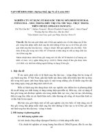
Figure 4-10. Migration quantification of the MSCs toward uninjured and
injured cartilage tissue.
Migration distance of the MSCs toward the complete media (CM) alone,
uninjured and injured tissues were showed in this graph (Error bars represent
standard errors of the means, one star: p-value < 0.05, two star: p-value <
0.01, and star: p-value < 0.001).
!
108!
4.4.6 Quantitative real-time reverse transcriptase-polymerase chain
reaction (RT-PCR)
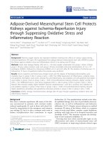
Custom-designed RT-PCR array was used to evaluate the differences in gene
expressions of uninjured and injured cartilage tissue. The factors were chosen
according to the literature review and the availability of the primers for the RT-
PCR (Table 4.1). RT-PCR results showed that the injured cartilage up-
regulated expressions of collagen type I A1 (COL1A1), chemokine C-X-C
motif 10 (CXCL10), transforming growth factor alpha (TGFA), insulin-like
growth factor 2 (IGF2), chemokine C-X-C motif 12 (CXCL12), angiopoietin 1
(ANGPT1), fibroblast growth factor 2 (FGF2), transforming growth factor beta-
3 (TGFβ3), bone morphogenetic protein 4 (BMP4), and vitronectin (VTN)
ligands. Figure 4.8 showed the relative increase of gene expressions of these
factors.
109!
!
Figure 4-11. Gene expression change of candidate ligands in injured
cartilage.
Gene expression level (sample number = 2) increases of the candidate
ligands, which could be involved in the MSCs migration.
110!
4.5 Discussion
To evaluate if the migration of MSCs toward the injured cartilage, which was
shown in previous chapter, is an active reaction to the injury site or a random
translocation of MSCs to that site, the behavior of MSCs were studied in a
microfluidic device. I developed a new microfluidic device to simulate the in
vivo interaction of MSCs with injured cartilage tissue in a three dimensional
(3D) environment. This simulation allows monitoring of the interactions
between MSCs and the tissue in cellular and molecular level. However, our
system still lacks many of the characteristics of an actual in vivo situation. For
example in living animals, the host cells such as immune cells (e.g.
macrophages, monocytes, T cells, etc.) have interactions with both injured
tissues and transplanted stem cells, which may affect their behaviors.
Moreover, in in vivo the blood circulation and joint motion may have some role
in stem cell migration, which I do not have in this in vitro model. Although,
these factors may make a difference between behavior of stem cells and
injured tissue in vivo and in vitro (microfluidic device), microfluidic device has
many advantages to the current methods and mimics the environment of real
tissue better than conventional methods such as Boyden chamber, scratch
migration assay, and under agarose migration system.
Boyden chamber, scratch test and under-agarose migration assay are the
most common migration assay systems. The Boyden chamber assay uses a
membrane to separate the upper chamber (cell chamber) from the bottom
chamber, which is too thin to make a gradient. In Boyden chamber assay only
the final results of the migration can be collected and this system allows
testing only one condition at a time. In the scratch migration test, cells are
111!
scratched and the migration of the cells is evaluated by monitoring how the
cells fill the gap.This system does not make a gradient of the chemotactic
factors, which is one of the effective components in the tissue regeneration.
Also with this system we cannot use any tissue as the origin of chemotactic
factors. Under agarose migration assay system is dependent on the concept
that solidified agarose does not attach to glass surfaces. To perform this
assay, a thick layer of warm agarose should be poured into a glass plate.
After solidification, three holes punched out; one hole for cell seeding, one for
the cytokine source and one as control. After gradient formation of the
cytokine the cell migration can be observed, however this migration can be in
any direction, and monitoring of the cells can be very difficult. Other potential
limitation of this system is the risk of cross-contamination of the cytokines
between the holes through the porous agarose.
By using microfluidic system, I could study the cell migration in a 3D
environment, which was more similar to in vivo situation and provided the
control of the gradient between channels. By having the specific channels for
the 3D scaffold parallel to the cell channel on both sides, I decreased the risk
of cross-contamination of the cytokines and also I could control the migration
of the cells in a certain direction (collagen channel), which made it easier to
monitor and image the cells migration. Moreover, the high quality imaging
capabilities of microfluidic system provided real-time monitoring of cells
simultaneously at two different conditions over time.
The results of this study showed that MSCs could be primed and migrated
toward the injured cartilage. The migration (distance) toward the injured tissue
112!
is longer than that of uninjured cartilage, suggesting injured tissue may secret
factors attracting the MSCs.
As I showed that the injured cartilage attracts the MSCs, and the engraftment
of the MSCs in the injured cartilage could be an active migration and homing,
I also evaluated the potential chemotactic candidates for this phenomenon.
There are many chemotactic factors named in the literature that are secreted
by different injured tissues such as skin wound, acute and chronic
inflammation in brain and etc. Previous studies (Table 4-2) have shown that
CXCL10 (247), TGFA (157), IGF2 (152), CXCL12 (248), FGF2 (148), TGFB3
(249), BMP4 (154) and ANGPT1 (158) are stimulatory factors for MSCs
migration. However, to our knowledge, there is not any study on the injured
cartilage. As the nature of the cartilage is different from the other tissues due
to lack of blood vessels and lymphatic drainage, in this study I evaluated the
factors, which were up-regulated by the chondrocytes after acute cartilage
injury. It is crucial to understand the chemotactic factors secreted by injured
cartilage to be able to use a sub-population of MSCs, which show stronger
response to such factors in the clinical setting to design more effective
treatment plan for patients.
RT-PCR results of injured cartilage tissues demonstrated that, chemotactic
factors such as CXCL10, TGFA, IGF2, CXCL12, ANGPT1, FGF2, TGFB3,
BMP4, and the extracellular matrix (ECM) proteins genes such as COL1A1,
and VTN were up-regulated after cartilage injury.
113!
Table 4-2 Other studies done on stem cell stimulatory chemotactic factors
Ligand
genes
Chemokine source
Assay method
Condition and outcome
Reference
COL1A1
Commercially
available
Modified Boyden
chamber
Collagen I induced significant motogenic activity for
both rabbit and human MSCs.
Thibault et al.
(161)
CXCL10
Recombinant human
chemokine
Agarose drop
migration assay
CXCL10 chemokine trigger hMSC migration and
promote hMSC proliferation.
Rice et al.
(247)
TGFA
Commercially
available
Boyden chamber /
Wound assay
The factors that induced the migration of rabbit and
human MSCs also enhanced their proliferation
Ozaki et al.
(157)
IGF2
Recombinant human
chemokine
Modified Boyden
chamber
IGF2 is a chemotactic factor for hMSCs and
stimulates migration of human mesenchymal
progenitor cells.
Fiedler et al.
(152)
CXCL12
Supernatant of
cultured human
pancreatic islets
Modified Boyden
chamber
Human pancreatic islets as an in vitro model released
CXCL12, which is able to attract BM MSCs in vitro.
Sordi et al.
(248)
ANGPT1
Commercially
available
Transwell dishes
Migration values of the TNFα-stimulated BM MSCs
were higher than un-stimulated cells.
Ponte et al.
(158)
FGF2
Commercially
available
Boyden chamber /
Wound Assay /
methyl cellulose
disc
Low concentrations of FGF2 leads to migration,
whereas higher concentrations resulted in repulsion
of the MSCs.
Schmidt et al.
(148)
TGFB3
Commercially
available
Modified Boyden
chamber
TGFB3 stimulates chemotaxis/chemokinesis of
multipotent C3H10T1/2 cells.
Makhijani et
al. (249)
BMP4
Commercially
available
Modified Boyden
chamber
Migration of primary human progenitor cells was
stimulated by rxBMP-4 in a dose-dependent manner
Fiedler et al.
(154)
VTN
Commercially
available
Modified Boyden
chamber
Vitronectin induced significant motogenic activity for
both rabbit and human MSCs.
Thibault et al.
(161)
114!
Our results agreed with those of Thibault et al. who demonstrated that ECM
proteins such as Col1 and VTN could induce significant migratory and
motogenic activity for MSCs (161). Then, these ECM proteins could be used
in the clinical setting for cartilage repair as a scaffold to carry the stem cells
and/or attract the endogenous or exogenous stem cells (endogenous from the
bone marrow and exogenous by multiple intra-articular injection of the
expanded autologous stem cells).
As I showed, in the previous chapter, that injection of stem cells is a promising
method for cartilage repair, in this chapter I confirmed that engraftment of the
MSCs in injured cartilage is an active migration and homing process and
injured cartilage encourage the migration of the MSCs toward the injury site. I
also showed that the cartilage injury up-regulate some specific chemotactic
factors, which can help to find and select a sub-population of MSCs which
show stronger response to such factors in cartilage repair. On one hand,
enhancement of the homing capacity of MSC can be achieved by modulating
their response to chemotactic factors; for example by finding and selecting
sub-population of MSCs which show stronger response to such factors
(because of higher expression of surface receptors which responsible for
those chemotactic signals) (250). On the other hand, modulation can be
applied in the site of injury for example with stimulating the target site to
attract more MSCs (with releasing more signals).
115!
Chapter 5 Autologous Bone Marrow
Derived Mesenchymal Stem Cell versus
Autologous Chondrocyte Implantation: An
Observational Cohort Study
1
1
The final, definitive version of this paper has been published in “The
American Journal of Sports Medicine”, 38(6): 1110-6, 2010 June by SAGE
Publications Ltd. SAGE Publications, Inc., All rights reserved. ©
116!
5.1 Abstract
Background: First generation ACI has limitations and introducing new
effective cell sources can improve cartilage repair.
Purpose: To compare the clinical outcomes of patients treated with first
generation autologous chondrocyte implantation (ACI) to patients treated with
autologous bone marrow derived mesenchymal stem cell (BM MSCs).
Study Design: Cohort Study, Level of Evidence, 3.
Methods: Seventy-two matched (lesion site and age) patients underwent
cartilage repair using chondrocytes (n=36) or BM MSCs (n=36). Clinical
outcomes were measured pre-operation and 3, 6, 9, 12, 18, and 24 months
post-operation using the International Cartilage Repair Society (ICRS)
Cartilage Injury Evaluation Package which included questions from the Short-
Form (SF-36) Health Survey, International Knee Documentation Committee
(IKDC) subjective knee evaluation form, Lysholm
24
knee scale, and Tegner
activity level scale.
Results: There was significant improvement in the patients’ quality of life
(physical and mental components of the SF-36 questionnaire included in the
ICRS package) after cartilage repair in both groups (ACI and BM MSCs).
However, there was no difference between the BM MSCs and the ACI group
in terms of clinical outcomes except for “Physical Role Functioning” with a
greater improvement over time in the BM MSCs group (P = 0.044 for
interaction effect). IKDC subjective knee evaluation (P = 0.861), Lysholm (P =
0.627), and Tegner (P = 0.200) scores did not have any significant difference
between groups over time. However, in general, men showed significantly
better improvements than women. Patients younger than 45 years scored
117!
significantly better than patients older than 45 years in the ACI group; but age
did not make a difference in outcomes in the BM MSCs group.
Conclusion: Using BM MSCs in cartilage repair is as effective as
chondrocytes for articular cartilage repair. In addition, it required one less
knee surgery, reduced costs, and minimized donor site morbidity.
Key Terms: chondrocyte; autologous chondrocyte implantation (ACI); bone
marrow derived mesenchymal stem cell
118!
5.2 Introduction
Full-thickness, focal cartilage defects causes knee symptoms such as pain,
popping and swelling (215); and it affects patient's quality of life and career.
Recent large arthroscopic studies indicated that the prevalence of cartilage
defects is between 11% to 63% (251-253). Treatment of articular cartilage
defects remains challenging (254-256), because cartilage tissue has a limited
capacity for repair (212, 213, 257). One of the most promising treatments for
cartilage defects is Autologous Chondrocyte Implantation (ACI) (29, 258-260),
which provides durable, hyaline-like cartilage (261, 262). ACI has some
limitations such as need for general anesthesia or at least regional anesthesia
to harvest the cartilage biopsy, a slow rate of chondrocyte proliferation,
difficulty in obtaining adequate number of chondrocytes for implantation, and
donor site morbidity. Some of these limitations could be solved by using other
techniques such as second or third generation ACI (263, 264), arthroscopic
second generation ACI, and microfracture (265, 266), or introducing new cell
sources like debrided waste chondrocytes, Bone Marrow derived
Mesenchymal Stem Cells (BM MSCs), or any combination of these cells (29,
214, 215, 267, 268).
Various authors have suggested the use of BM MSCs for cell-based cartilage
repair (51, 61, 214, 215, 269); Bone Marrow-derived Mesenchymal Stem
Cells (BM MSCs) have a better proliferation rate than chondrocytes and have
differentiation capacity to different tissues including chondrogenesis (270-
272). We also showed in the previous chapters that injured cartilage could
attract the BM MSCs to home and engraft in the cartilage defect and increase
the cartilage repair quality. However, as far as we know, cartilage repair by
119!
using BM MSCs has not been compared with other cell sources. Then in this
chapter, we compared the clinical outcomes of cartilage repair in patients
treated by autologous BM MSCs and chondrocytes.
As this was a clinical study, surgeries were done by my supervisor (A/prof
James Hui) and I assisted him in some of the surgeries. I designed the study
as historical cohort study and I used ACI treated patients’ data archive and
current data from the BM MSCs implanted patients. After I collected the data,
by consulting an independent biostatistician, I analyzed the data. Then I
interpreted the results and prepared the peer-reviewed manuscript.
120!
5.3 Methods
5.3.1 Participants
This non-randomized observational cohort study was designed to investigate
the effectiveness of Chondrocytes and BM MSCs as cell sources for repairing
full-thickness cartilage defects of the knee. The inclusion criteria were, at
least, one symptomatic chondral lesion diagnosed by clinical examination and
magnetic resonance imaging (MRI) on the femoral condyle, trochlea, or
patella and non-existent or correctable concomitant pathologies. The
exclusion criteria were patients with inflammatory arthritis, tri-compartmental
osteoarthritis, limited range of motion in particular fixed flexion deformity and
those who were 65 years of age or older. Cartilage repair was conducted with
informed consent of the patients.
Patients who fulfilled the inclusion and exclusion criteria were treated by our
senior author (JH Hui). Thirty-six consecutive patients underwent BM MSCs
and were matched by 36 cases of ACI performed earlier, in terms of lesion
sites and (10-year) age intervals.
The study protocol was approved by the National Healthcare Group Domain-
Specific Review Board (NHG DSRB reference number D/00/814) and the
University Hospital Ethic Committee. In addition, cells were processed at the
GMP cell processing facility at the National University Hospital of Singapore.
5.3.2 Cell Sources
5.3.2.1 Chondrocyte (ACI) preparation
The chondrocyte preparation method was adopted from Brittberg et al. (29) as
described here. A small amount of cartilage tissue (1cm x 0.5cm) was taken
121!
from non-weight bearing areas, which were deemed macroscopically healthy
by arthroscopy. The harvested tissue was transferred into a specimen
container filled with sterile saline (about 10ml) and processed within 60
minutes. The sample was washed twice with PBS (Gibco BRL, Grand Island,
NY, US) and then minced prior to being transferred aseptically into a tube with
5ml collagenase NB6 (Sigma, St Louis, Missouri, US) for overnight digestion
at 37°C in a water bath. Digested chondrocytes were washed with DMEM/F12
(Gibco BRL, Grand Island, NY, US) supplemented with 10% FBS (Gibco BRL,
Grand Island, NY, US) to stop the enzymatic reaction. These cells were then
cultured in T75cm
2
flasks with DMEM/F12 containing 10% FBS (Gibco) and
50µg/ml L-Ascorbic acid 2-phosphate sesquimagnesium salt hydrate (Sigma,
St Louis, Missouri, US) in a humidified atmosphere of 5% CO
2
, 37°C. Cells
were seeded at a cell density of 5,000 cells per square centimeter. Initial
medium change was done after 7 days, when adherent cells were recognized.
Subsequent medium change was done two to three times a week until the
preparation of cell sheets, which were formed in the presence of Ascorbic acid
(Passage 1). For each surgery, at least 4 cell sheets were prepared and
around two million cells /cm
2
were applied.
5.3.2.2 MSCs preparation
The detailed method is as follows; Under local anesthesia, 30 ml of bone
marrow (BM) was aspirated using a Jamshidi needle from the iliac crests of
each patient into heparinized syringes and transferred into sterile containers.
Seventy or eighty milliliters of each patient’s blood were collected as well. The
bone marrow aspirate was processed within 60 minutes. The heparinized
bone marrow aspirate was mixed with a one-fifth volume of 6% (w/v) dextran
122!
(molecular weight 100,000; Sigma, St Louis, Missouri, US) and left standing at
room temperature for 30 minutes to eliminate erythrocytes. The remaining
cells were washed twice with DMEM (Gibco BRL, Grand Island, NY, US).
These cells were cultured in T75cm
2
flasks with an initial culture medium
consisting of DMEM (Gibco) containing 10% FBS (Gibco BRL, Grand Island,
NY, US), 50µg/ml L-Ascorbic acid 2-phosphate sesquimagnesium salt hydrate
(Sigma, St Louis, Missouri, US) and 1% antibiotic-antimycotic (penicillin
100U/ml, streptomycin 0.1mg/ml, amphotericin B 0.25µg/ml) (Sigma, St Louis,
Missouri, US) in a humidified atmosphere of 5% CO
2
, at 37°C. The cells were
seeded at a density of 10,000 cells per square centimeter. Initial medium
change was done after 5 days when adherent cells were recognized.
Subsequently, culture media without antibiotics were used and changed two
to three times a week. Cell sheets were formed in the presence of Ascorbic
acid (Passage 1) and for each surgery, at least 4 cell sheets were prepared
and around two million cells /cm
2
(which is determined experimentally) were
applied. This MSC preparation method is a modified approach from Wakitani
et al. study (61). In this method we harvested the bone marrow and expanded
the cells the same as Wakitani’s approach however, we prepared the cell
sheets (by useing Ascorbic acid) instead of cell suspension.
Seventy milliliters of venous blood from each patient was transferred into two
50ml tubes for overnight incubation at 4 degree Celsius. After centrifuging the
tube with slow acceleration, the serum was carefully aspirated and transferred
to a new tube. Repeated centrifugation with slow acceleration for 3 minutes at
3000 rpm at ambient temperature was performed. The serum was aspirated
into a syringe and filtered with a sterile 0.2µm filter. The filtered serum was
123!
tested for sterility, anti-HIV and Hepatitis B antigen, and then stored at a
temperature of -20 degree Celsius.
Flow cytometry against CD90
+
, CD105
+
, CD14
-
and CD34
-
was used to
confirm that cultured cells were mesenchymal stem cells. Saline that was
used for transporting the cartilage biopsy to the laboratory, aspirated bone
marrow and culture media (without antibiotic) was tested for sterility and
Mycoplasma hominis contamination.
5.3.3 Operation techniques
Four to five weeks after harvesting cells, ACI surgery was done. For details on
ACI, refer to procedure described previously (29). In summary, approximately
10 to 15 million cells (with a viability rate of 96%) were returned for
implantation. The cell sheets were transported to the operating theater in a
sterile container within the patients’ own serum. The debrided chondral defect
(without damaging subchondral bone) was measured after arthrotomy.
Subsequently, periosteal patch harvesting from the proximal part of the tibia
or distal part of femur was done according to the measured size. Next, the
harvested periosteum was sutured precisely to the rim of the debrided
defect(s). The cultured chondrocytes or BM MSCs were implanted beneath
the patch and very fine stitches (micro suture 7-0) were used to hold the
periosteum to the defected site. To avoid cell leakage fibrin glue was used to
create a watertight seal.
5.3.4 Rehabilitation
To derive maximum benefit from the surgery, patients were advised to strictly
follow the rehabilitation protocol, which is one of the most important parts of
124!
recovery. The rehabilitation protocol began on the day of surgery and includes
passive range of motion and isometric muscle contractions. Patients were
able to begin active motion and partial weight bearing at 6 weeks, progressing
to full weight bearing exercises. The rehabilitation protocol varies according to
the location and size of the lesion, concomitant procedures, patient’s age and
previous activity level. There are four areas that rehabilitation focuses on:
walking/weight bearing, range of motion, strength, and cardiovascular
capacity.
5.3.5 Post operation evaluation
Patients were evaluated preoperatively (pre-operative assessment) as well as
at 3, 6, 9, 12, 18 and 24 months post-operatively. Assessments were
performed by our trained research staff using International Cartilage Repair
Society (ICRS) Cartilage Injury Evaluation Package which included questions
from the Short-Form (SF-36) (273) Health Survey, International Knee
Documentation Committee (IKDC) subjective knee evaluation form, Lysholm
knee scale (274), and Tegner activity level scale (275).
Second look arthroscopy was performed in 7 patients (4 in BM MSCs and 3 in
ACI group) 9 to 12 months after implantation. A biopsy of the repair tissue
was obtained in 2 cases (1 in each group). After fixation, paraffin sections
were stained with Alcian blue to evaluate aggrecan content and
immunohistochemistry staining was done to assess the collagen type I, II, X
content.
!
125!
5.3.6 Statistical analysis
Statistical analysis was performed by consulting an independent
biostatistician using STATA statistical software (Version 10). The MIXED
effect model (with random intercept) was used to evaluate the effect of cell-
type and time on the quality of life and other functional or pain outcomes of
patients, depending on gender. This method of analysis appropriately
accounts for the possible correlation between repeated measurements of an
individual. All statistical evaluations were made, based on an assumption of a
two-sided test at the conventional 5% level of significance.
126!
5.4 Results
Seventy-two patients who fulfilled the inclusion and exclusion criteria were
treated using ACI (n = 36) and BM MSCs (n = 36) between 2001-2005 and
2005-2007 respectively. All patients were followed up for 2 years. Table 5.1
shows the demographic characteristics of the patients. As anticipated,
patients in the two groups had similar age and gender distributions since they
were matched by age and gender. There were equal numbers of males and
females in the ACI group, with a mean age of 42.5 (SD 11.2) years.
Correspondingly, in the BM MSCs group, there were 20 men and 16 women
with a mean age of 44.0 (SD 11.4) years. However, the mean defect sizes of
ACI and BM MSCs group were 3.6 cm
2
(SD 2.84) and 4.6 cm
2
(SD 3.53)
respectively (P-value = 0.270). Concomitant procedures included patella
realignment (6 cases in ACI and 5 cases in BM MSCs group), high tibial
osteotomy (5 cases in BM MSCs group), partial meniscectomy (1 in each
group), and anterior cruciate ligament reconstruction (1 in each group).
127!
Table 5-1. Demographic characteristics of study patients
Characteristic
ACI
(n = 36)
ABMSCI
(n = 36)
Sex (%)
Male
18 (50)
20 (56)
Female
18 (50)
16 (44)
Age (%)
<45 years
19 (52.8)
16 (44.4)
>=45 years
17 (47.2)
20 (55.6)
Lesion Site (%)
Patellar
13 (36)
13 (36)
Trochlear
4 (11)
4 (11)
Femoral Condyle
12 (33)
12 (33)
Multiple lesions
7 (20)
7 (20)
Lesion Grade (%)
Grade 3
25 (70)
24 (67)
Grade 4
11 (30)
12 (33)
Mean defect size, cm
2
(SD)
3.6
*
(2.84)
4.6
*
(3.53)
Diagnosis (%)
Trauma
19 (53)
14 (39)
OA
15 (42)
20 (56)
Others
2 (5)
2(5)
*There was no significant difference between two groups (P-value = 0.270).
Note: Figures in parenthesis denotes percentages unless otherwise indicated.
5.4.1 ICRS package SF-36 components clinical outcomes
Table 5.2 shows the physical and mental components of the SF-36
questionnaire included in the ICRS package. Generally, there was a
significant improvement in these quality of life outcomes after ACI over time.
However, there were no differences between patients treated with BM MSCs
and Chondrocytes in terms of these clinical outcomes (p > 0.05) except for
“Physical Role Functioning” which suggested greater improvements in for
patients treated with BM MSCs as compared to chondrocytes, for both males
and females (p value = 0.044 for interaction effect).
128!
Table 5-2. Effects of cell type, time and gender on ICRS package SF-36
component outcomes.
Outcome/Parameter
Estimate
*
95% CI
p-value
Bodily Pain
Cell type
**
-2.16
-9.76 to 5.43
0.576
Time
***
0.42
-0.31 to 1.16
0.255
Sex
****
-5.65
-12.44 to 1.14
0.103
Cell Type x Time
0.53
0.05 to 1.01
0.030
Vitality
Cell type
-2.42
-8.86 to 4.03
0.463
Time
0.87
0.61 to 1.14
<0.001
Sex
2.76
-4.83 to 10.36
0.476
Social Functioning
Cell type
4.66
-6.37 to 15.68
0.408
Time
1.16
0.76 to 1.57
<0.001
Sex
8.88
-3.79 to 21.55
0.169
Role Functioning
(Emotional)
Cell type
-0.84
-6.16 to 4.49
0.758
Time
-0.28
-0.46 to -0.09
0.003
Sex
-0.83
-6.40 to 4.74
0.770
Mental Health
Cell type
-3.72
-7.90 to 0.46
0.081
Time
0.30
0.12 to 0.47
0.001
Sex
3.68
-1.01 to 8.36
0.124
Mental Health Summary
Cell type
-0.73
-3.20 to 1.75
0.565
Time
0.13
0.03 to 0.22
0.013
Sex
2.97
0.12 to 5.82
0.041
Physical Functioning
Cell type
0.85
-5.45 to 7.14
0.792
Time
1.12
0.88 to 1.35
<0.000
Sex
-6.86
-13.72 to -0.01
0.050
Physical Health Summary
Cell type
1.08
-1.37 to 3.53
0.387
Time
0.47
0.38 to 0.56
<0.001
Sex
-2.92
-5.70 to -0.14
0.039
Role Functioning
(Physical)
Cell type
13.22
-0.88 to 27.31
0.066
Time
3.76
2.24 to 5.27
<0.001
Sex
3.49
-10.46 to 17.44
0.624
Cell type x Time
-1.02
-2.02 to -0.03
0.044
* The estimate of the average level of each parameter.
** Evaluation of differences between patients treated with BM MSCs or chondrocytes for each
parameter.
*** Evaluation of the differences over time for each parameter.
**** Evaluation of the differences between male and female for each parameter.
129!
Within groups, there were significant differences in outcomes between
genders. In particular, males demonstrated greater improvements in scores
for “Physical functioning”, “Physical Health Summary”, “Physical Role
Functioning”, and “Mental Health Summary” (p < 0.05). On the other hand,
gender did not have any effect on the “Vitality”, “Social Functioning”,
Emotional Role Functioning”, “Mental Health”, and “Bodily Pain”.
However, there were no differences in physical and mental component scores
between the age groups (<45 years versus >= 45 years) within each cell type.
Table 5.3. Effect of cell type, time and gender on IKDC, Lysholm, and
Tegner outcomes.
Outcome/Parameter
Estimate *
95% CI
p-value
IKDC
Cell type
**
-0.46
-5.61 to 4.69
0.861
Time
***
1.08
0.90 to 1.25
<0.001
Sex
****
-6.03
-11.18 to -0.88
0.022
Lysholm
Cell type
-1.16
-5.83 to 3.52
0.627
Time
0.79
0.63 to 0.95
<0.001
Sex
-6.44
-11.17 t0 -1.71
0.008
Tegner
Cell type
0.25
-0.13 to 0.64
0.200
Time
0.06
0.05 to 0.07
<0.001
Sex
-0.55
-0.98 to -0.13
0.011
* The estimate of the average level of each parameter.
** Evaluation of differences between patients treated with BM MSCs or chondrocytes for each
parameter.
*** Evaluation of the differences over time for each parameter.
**** Evaluation of the differences between male and female for each parameter.
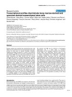
5.4.2 IKDC subjective knee evaluation outcomes
The postoperative IKDC scores (figure 5.1A) indicated a significant
improvement in performance over time throughout the follow-up period.
Patients treated with chondrocytes and BM MSCs did not differ significantly
130!
with regards to the improvements in the IKDC subjective knee evaluation.
However, men showed significantly better improvements than women (Table
5.3) (P value = 0.022). Moreover, there were no differences in IKDC scores
between patients younger than 45 years and those who were at least 45
years within ACI group (P value = 0.070) and within BM MSCs group (P value
= 0.671).
131!
Figure 5-1. IKDC, Tegner, and Lysholm activity level outcome.
!