Bone marrow derived mesenchymal stem cell (BM MSC) application in articular cartilage repair 1
Bạn đang xem bản rút gọn của tài liệu. Xem và tải ngay bản đầy đủ của tài liệu tại đây (5.16 MB, 70 trang )
BONE MARROW DERIVED MESENCHYMAL STEM
CELL (BM MSC) APPLICATION IN ARTICULAR
CARTILAGE REPAIR
HOSSEIN NEJADNIK
(M.D., Isfahan University of Medical Sciences, Isfahan, Iran)
A THESIS SUBMITTED
FOR THE DEGREE OF DOCTOR OF PHILOSOPHY
DEPARTMENT OF ORTHOPEDIC SURGERY
YONG LOO LIN SCHOOL OF MEDICINE
NATIONAL UNIVERSITY OF SINGAPORE
2013
ii!
ACKNOWLEDGEMENTS
It is my pleasure to thank all the kind people who made this thesis
possible with their great help and support. I would like to thank my supervisor,
Associate Professor James Hui, Department of Orthopedic Surgery, Yong Loo
Lin School of Medicine, National University of Singapore (NUS), for his critical
supervision and active support during my PhD study.
I am also deeply indebted to my co-supervisor Professor Shih-Chang
Wang, Head of Discipline of Medical Imaging at Sydney Medical School,
University of Sydney, for his constant support and encouragement.
My gratitude is also towards Professor Wong Hee Kit, the Head of
Orthopedic Surgery Department, for giving me this chance to pursue my PhD
degree and use the facilities in the department, and Professor Lee Eng Hin
and Professor James Goh Cho Hong, Head of National University of
Singapore Tissue Engineering Program (NUSTEP), for giving me the
opportunity to use the NUSTEP facilities.
I also would like to thank Professor Vincent Chong, Head of Radiology
Department, and Associate Professor Sudhakar Venkatesh for their help and
support.
I am most grateful to Professor Roger Kamm, lead investigator of
BioSystems and Micromechanics (BioSyM) at Singapore-MIT Alliance for
Research & Technology (SMART) and Cecil and Ida Green Distinguished
Professor of Biological and Mechanical Engineering at MIT University, for his
mentorship and support and Dr. Amirreza Aref, and Dr. Choong Kim for their
support in performing the bioengineering parts of my projects.
iii!
I also would like to extend my sincere gratitude to Professor Kishore
Bhakoo, Head of Translational Molecular Imaging Group at Singapore
Bioimaging Consortium (SBIC), who provided me a great support in the MR
imaging.
I owe my deepest gratitude to my colleagues and students in the
NUSTEP lab: Dr. Kenon Chua, Dr. Xiafei Ren, Dr. Zheng Yang, Dr. Sintje
Böhrensen, Afizah Binte Mohd Hassan, and Dr. Sari Panjang, who have
taught me a lot. They have been wonderful friends.
I am thankful to all the colleagues, students and staff members of
Department of Orthopedics Surgery, specially Ms Perumal Premalatha, and
Ms Sarojeni Shanmugam, Orthopaedic Surgery Department management
assistant officers, Yong Loo Lin School of Medicine for their timely help.
I would like to thank A*STAR and NUS for granting me graduate
student scholarship. This work was supported by grants from Singapore
Bioimaging Consercium (to A/P James Hui).
I wish to thank my great friends who made my PhD life a great memory
for me and I owe my loving thanks to my wife, Pooneh, and my parents and
I dedicate this thesis to them.
iv!
TABLE OF CONTENTS
ACKNOWLEDGEMENTS ii!
Summary x!
LIST OF TABLES AND FIGURES xiii!
LIST OF ABBREVIATIONS xvii!
LIST OF PUBLICATIONS xx!
Chapter 1 Background 1!
1.1 Basic Science of Articular Cartilage 2!
1.2 Cartilage injuries 4!
1.2.1 Focal cartilage defects 4!
1.2.2 Osteochondritis dissecans (OCD) 6!
1.3 Importance of cartilage repair 6!
1.4 Different methods of cartilage repair 6!
1.4.1 Palliative technique: Arthroscopic lavage and debridement 7!
1.4.2 Intrinsic repair enhancement: Microfracture 8!
1.4.3 Whole tissue transplantation: Osteochondral Autograft
Transplantation (OAT) 8!
1.4.5 Cell based cartilage repair 11!
1.4.5.1 Autologous chondrocyte implantation (ACI) 11!
1.4.5.2 Other Cell-based therapy methods 12!
1.4.5.3 Stem Cells in Articular Cartilage repair 13!
v!
1.5 Types of stem cells 14!
1.5.1 Embryonic stem cells (ESCs) 15!
1.5.2 Induced pluripotent stem (iPS) cells 15!
1.5.3 Adult stem cells 16!
1.5.4 Heterologous stem cells 17!
1.6 MSCs in cartilage repair 17!
1.7 Monitoring of Cell Therapy 18!
1.8 Histological Methods 19!
1.9 Imaging Modalities 19!
1.10 Contrast agents 22!
1.10.1 T1 Contrast Agents (Table 1-7) 23!
1.10.2 T2 Contrast Agents 23!
1.10.3 Iron Oxide Particles 24!
1.11 Cellular MRI 24!
1.11.1 Cellular Imaging with Iron Oxide Particles 25!
1.11.2 Mechanisms of Cellular Uptake 28!
1.12 Cell migration 30!
1.12.1 Stem cell migration mechanism 30!
1.12.2 MSC migration and homing 31!
1.12.3 Cell migration evaluation systems 32!
Chapter 2 Overall hypothesis and objectives 36!
Chapter 3 In vivo monitoring of the intra articular injected
SPIO-labeled stem cells for cartilage repair 39!
3.1 Abstract 40!
vi!
3.2 Introduction 43!
3.3 Methods 45!
3.3.1 Expansion of MSCs 45!
3.3.2 Flow cytometry assessment 45!
3.3.3 Labeling of the MSCs with SPIO 46!
3.3.4 Prussian blue staining 46!
3.3.5 Transmission electron microscopy (TEM) 47!
3.3.6 Atomic Absorption Spectroscopy (AAS) 47!
3.3.7 Viability and proliferation evaluation 48!
3.3.8 Differentiation of MSCs 48!
3.3.9 Histological evaluation 50!
3.3.10 Animal model 51!
3.3.11 Surgical procedure 51!
3.3.12 Preliminary MR imaging experiments 52!
3.3.13 MR imaging of live animals 53!
3.3.14 Postmortem analysis 55!
3.3.15 Statistical analysis 57!
3.4 Results 58!
3.4.1 Characterization of MSCs 58!
3.4.2 Prussian blue staining of SPIO-labeled MSCs 60!
3.4.3 Transmission Electron Microscopy (TEM) 60!
3.4.4 Iron content quantification in labeled-MSCs 61!
3.4.5 Viability and proliferation of labeled MSCs 62!
3.4.6 Differentiation potential of labeled MSC 64!
3.4.6.1 Adipogenic differentiation 64!
vii!
3.4.6.2 Osteogenic differentiation 65!
3.4.6.3 Chondrogenic differentiation 66!
3.4.7 MR imaging of animals 68!
3.4.7.1 Preliminary experiments 68!
3.4.7.2 MRI of the mini-pigs’ knee 70!
3.4.8 Postmortem analysis 73!
3.5 Discussion 76!
Chapter 4 Simulating Injured Articular Cartilage Environment
for Mesenchymal Stem Cell Migration Evaluation in A Three
Dimensional Microenvironment 78!
4.1 Abstract 79!
4.2 Introduction 82!
4.3 Methods 84!
4.3.1 Design of microfluidic device 87!
4.3.2 Computational modeling of concentration gradient 88!
4.3.3 Fabrication of microfluidic device 89!
4.3.4 MSC characterization and culture in microfluidic devices 90!
4.3.5 Microfluidic device migration validation 91!
4.3.6 Injured and uninjured sample preparation 91!
4.3.7 MSCs migration toward injured cartilage conditioned media 92!
4.3.8 Tissue placement and device assembly 93!
4.3.9 MSCs migration toward injured tissue 94!
4.3.10 Quantification of the MSCs migration 94!
viii!
4.3.11 Quantitative real-time reverse transcriptase-polymerase chain
reaction (RT-PCR) 95!
4.3.11 Statistical analysis 98!
4.4 Results 99!
4.4.1 Computational modeling of concentration gradient 99!
4.4.2 MSC characterization 100!
4.4.3 Microfluidic device migration validation 101!
4.4.4 MSCs migration stimulated by conditioned media 103!
4.4.5 MSCs migration toward injured tissue 105!
4.4.6 Quantitative real-time reverse transcriptase-polymerase chain
reaction (RT-PCR) 108!
4.5 Discussion 110!
Chapter 5 Autologous Bone Marrow Derived Mesenchymal
Stem Cell versus Autologous Chondrocyte Implantation: An
Observational Cohort Study
1
115!
5.1 Abstract 116!
5.2 Introduction 118!
5.3 Methods 120!
5.3.1 Participants 120!
5.3.2 Cell Sources 120!
5.3.2.1 Chondrocyte (ACI) preparation 120!
5.3.2.2 MSCs preparation 121!
5.3.3 Operation techniques 123!
5.3.4 Rehabilitation 123!
ix!
5.3.5 Post operation evaluation 124!
5.3.6 Statistical analysis 125!
5.4 Results 126!
5.4.1 ICRS package SF-36 components clinical outcomes 127!
5.4.2 IKDC subjective knee evaluation outcomes 129!
5.4.3 Tegner activity level outcomes 132!
5.4.4 Lysholm knee scale outcomes 132!
5.4.5 Second-look arthroscopy and histological outcomes 133!
5.5 Discussion 135!
Chapter 6 Conclusions and Future Direction 139!
Chapter 7 References 142!
x!
Summary
Articular cartilage repair is one the most challenging issues in orthopedic
surgery due to the avascular and aneural nature of the cartilage, which affects
its self-repair capability. Cell therapy shows a great potential for cartilage
defect repair. Brittberg et al. in 1994 used autologous chondrocytes as a cell
source to repair the injured knee cartilage, which remains a promising method
even to date. However, this method has some limitations such as inadequate
cell number, age dependent quality of the chondrocytes and donor site
morbidity. Recently bone marrow derived mesenchymal stem cells (MSCs)
become an alternative cell source for cartilage repair. Our group and others
had demonstrated the tri-lineage differentiation ability of MSCs to
chondrocytes, osteocytes and adipocytes. Wakitani et al. and our group
demonstrated that MSCs could be used as a new source for cartilage repair.
However, in order to develop this into an effective cell therapy treatment we
need to understand the behavior of cells, especially the proliferation,
differentiation, migration and engraftment of MSCs after implantation and in
vivo.
The use of MSCs for cartilage repair relies on their homing and engraftment to
the injured cartilage tissue. Although there is some theory that injured tissue
expresses ligands and chemotactic factors that encourage homing of MSCs,
these factors and their mechanism are not fully understood. In vitro modeling
of the in vivo simulation can be challenging. Microfluidic platforms allow the
study of cell migration in 3D environment, and at the same time, provide live
xi!
observation and time-lapse evaluation so that the dynamic interaction of
MSCs with injured tissue can be evaluated.
In summary, we showed that by labeling of the MSCs with superparamagnetic
iron oxide (SPIO) nanoparticles, we could monitor the fate of cells after
injection. The technique is useful tool to monitor the migration and localization
by MRI, a non-invasive and repeatable imaging method for in vivo evaluation.
In addition, we showed that labeled MSCs have tendency to move to the
injured cartilage site, engraft and increase the quality of the repaired cartilage
by production of hyaline-like cartilage. The MSCs also have tendency to home
in the other sources of the inflammation such as surgery site.
To have a better understanding of the migration behavior of the MSCs toward
injured cartilage, we developed a microfluidic device, and confirmed the
migration capacity of the MSCs in a three dimensional model system. An
increased migration of the MSCs was observed due to an increase in the
chemo-attractant factors secreted by the injured tissue. Cartilage injuries up-
regulated the expression of the chemotactic factors such as collagen type I A1
(COL1A1), chemokine C-X-C motif 10 (CXCL10), transforming growth factor
alpha (TGFA), insulin-like growth factor 2 (IGF2), chemokine C-X-C motif 12
(CXCL12), angiopoietin 1 (ANGPT1), fibroblast growth factor 2 (FGF2),
transforming growth factor beta-3 (TGFB3), bone morphogenetic protein 4
(BMP4), and vitronectin (VTN) ligands.
Furthermore, to assess the cartilage repair outcome by using MSCs, we
compared the clinical outcome of cartilage repair by using MSCs vs
chondrocytes, which is an FDA approved methods. In this part of our study,
we demonstrated that MSCs were as effective as chondrocytes for articular
xii!
cartilage repair. The use of MSCs offers advantages such as being less
invasiveness (bone marrow biopsy comparing with knee arthroscopy), having
little or no damage to normal articular cartilage, and requiring only regional
anesthesia. All of which leads to lower treatment costs for patient.
In conclusion, we demonstrated that MSCs are a promising cell source for
cartilage repair, and due to the migration capability of the cells and
chemotaxis factors secretion of the injured cartilage. Their use as an
injectable cell therapy treatment will increase in the coming years.
xiii!
LIST OF TABLES AND FIGURES
Tables
TABLE 1-1. COMPOSITION OF THE DIFFERENT LAYERS OF ARTICULAR
CARTILAGE (4). 4!
TABLE 1-2. DIFFERENT METHODS OF CARTILAGE REPAIR (12). 7!
TABLE 1-3. SCAFFOLDS USED IN VIVO STUDIES. (ADAPTED FROM (21))
10!
TABLE 1-4. DIFFERENT TYPES OF HUMAN ADULT STEM CELLS AND
THEIR DIFFERENTIATION CAPACITY (76). 16!
TABLE 1-5 THE SUMMARY OF IMAGING MODALITIES THAT CAN BE
USED TO EVALUATE AND MONITOR STEM CELLS (99). 21!
TABLE 1-6. COMPARISON OF IMAGING MODALITIES SCALE 22!
TABLE 1-7. CLASSIFICATION OF MRI CONTRAST AGENTS (ADAPTED
FROM (101)) 23!
TABLE 1-8. COMPARISON OF CELL TRANSPLANTATION AND HOMING
FOR CARTILAGE REGENERATION (ADAPTED FROM(141)). . 30!
TABLE 3-1. HISTOLOGICAL GRADING SCALE FOR CARTILAGE REPAIR.
56!
TABLE 4-1. LIST OF CANDIDATE LIGANDS USED FOR RT-PCR
ANALYSIS. 97!
TABLE 4-2 OTHER STUDIES DONE ON STEM CELL STIMULATORY
CHEMOTACTIC FACTORS 113!
TABLE 5-1. DEMOGRAPHIC CHARACTERISTICS OF STUDY PATIENTS
127!
TABLE 5-2. EFFECTS OF CELL TYPE, TIME AND GENDER ON ICRS
PACKAGE SF-36 COMPONENT OUTCOMES. 128!
Figures
FIGURE 1-1. LAYERS IN THE ARTICULAR CARTILAGE (2, 3). 3!
FIGURE 1-2. THE ICRS CARTILAGE INJURY CLASSIFICATION (8). 5!
xiv!
FIGURE 1-3. SCHEMATIC OF MICROFRACTURE TECHNIQUE OF
CARTILAGE REPAIR (18). 8!
FIGURE 1-4. SCHEMATIC OF OSTEOCHONDRAL AUTOGRAFT
TRANSPLANTATION (OAT) TECHNIQUE OF CARTILAGE REPAIR
(18). 9!
FIGURE 1-5. SCHEMATIC OF AUTOLOGOUS CHONDROCYTE
IMPLANTATION (ACI) TECHNIQUE OF CARTILAGE REPAIR (62). 12!
FIGURE 1-6. THE CARTILAGE REPAIR STRATEGY ALGORITHM BASED
ON THE LESION LOCATION AND CHARACTERISTICS. 13!
FIGURE 1-7. STEM CELL CLASSIFICATION AND DIFFERENTIATION
POTENTIAL (71). 14!
FIGURE 1-8. MECHANISMS OF CELLULAR UPTAKE OF NANO SIZED
PARTICLES. 28!
FIGURE 1-9. MECHANISMS OF PARTICLE UPTAKE BY ENDOCYTOSIS.
29!
FIGURE 1-10. CELL MIGRATION PROCESS. 31!
FIGURE 1-11. EXISTING MIGRATION ASSAY DEVICES. 32!
FIGURE 3-1. MINI-PIGS AS AN ANIMAL MODEL. 52!
FIGURE 3-2. MR IMAGING OF THE MINI-PIGS' KNEE JOINT. 54!
FIGURE 3-3. FLOW CYTOMETRY ANALYSIS OF THE STEM CELLS
SURFACE MARKERS. 59!
FIGURE 3-4. PRUSSIAN BLUE STAINING OF THE UNLABELED AND
LABELED MSCS. 60!
FIGURE 3-5. TRANSMISSION ELECTRON MICROSCOPY OF THE
LABELED MSCS. 61!
FIGURE 3-6. IRON CONTENT QUANTIFICATION. 62!
FIGURE 3-7. TRYPAN BLUE VIABILITY TEST. 63!
FIGURE 3-8. MTS ASSAY. 64!
FIGURE 3-9. OIL RED O STAINING. 65!
FIGURE 3-10. ALIZARIN RED STAINING. 65!
FIGURE 3-11. CHONDROGENIC DIFFERENTIATION POTENTIAL
EVALUATION OF THE MSCS. 67!
FIGURE 3-12. MR IMAGING OF THE PIG'S KNEE EXPLANT. 69!
xv!
FIGURE 3-13. MR SEQUENCE OPTIMIZATION. 70!
FIGURE 3-14. POST INJECTION MR IMAGES. 71!
FIGURE 3-15. MR IMAGING OF THE MINI-PIG'S FEMORAL CONDYLE. 72!
FIGURE 3-16. MRI OF THE DISTRIBUTION OF THE LABELED MSCS IN
THE MINI-PIG KNEE JOINT. 73!
FIGURE 3-17. POSTMORTEM HISTOLOGICAL EVALUATION OF THE
REPAIRED CHONDRAL DEFECTS 6 WEEKS AFTER STEM CELL
INJECTION. 74!
FIGURE 3-18. PRUSSIAN BLUE STAINING OF THE REPAIRED
CARTILAGE DEFECT. 74!
FIGURE 3-19. PRUSSIAN BLUE STAINING OF THE IRON LABELED MSCS
IN THE SURGICAL SCAR SITE. 75!
FIGURE 3-20. PRUSSIAN BLUE STAINING OF IRON LABELED MSCS IN
THE PARA-PATELLAR FAT. 75!
FIGURE 4-1 HISTORY OF MICROFLUIDIC DEVICE DESIGN. 86!
FIGURE 4-2 SCHEMATIC DESIGN AND DIMENSION OF MICROFLUIDIC
DEVICE. 88!
FIGURE 4-3 CARTILAGE TISSUE CONDITIONED MEDIA PREPARATION.
93!
FIGURE 4-4. THE GROWTH FACTORS DIFFUSION SIMULATION IN 3D
SCAFFOLD. 99!
FIGURE 4-5. FLOW CYTOMETRY ANALYSIS OF THE STEM CELLS
SURFACE MARKERS. 100!
FIGURE 4-6. MICROFLUIDIC DEVICE MIGRATION VALIDATION. 102!
FIGURE 4-7. MIGRATION EVALUATION OF THE MSCS TOWARD
UNINJURED AND INJURED CONDITIONED MEDIA. 104!
FIGURE 4-8. MIGRATION QUALIFICATION OF THE MSCS TOWARD
UNINJURED AND INJURED CONDITIONED MEDIA. 105!
FIGURE 4-9. MIGRATION EVALUATION OF THE MSCS TOWARD
UNINJURED AND INJURED CARTILAGE TISSUE. 106!
FIGURE 4-10. MIGRATION QUANTIFICATION OF THE MSCS TOWARD
UNINJURED AND INJURED CARTILAGE TISSUE. 107!
FIGURE 4-11. GENE EXPRESSION CHANGE OF CANDIDATE LIGANDS IN
INJURED CARTILAGE. 109!
xvi!
FIGURE 5-1. IKDC, TEGNER, AND LYSHOLM ACTIVITY LEVEL
OUTCOME. 131!
FIGURE 5-2. FEMORAL CONDYLE CARTILAGE DEFECT. 133!
FIGURE 5-3. HISTOLOGIC EVALUATION OF BIOPSY SPECIMENS.134!
xvii!
LIST OF ABBREVIATIONS
3D-GRE
3D Gradient Echo
AAS!
Atomic Absorption Spectroscopy !
ACI
Autologous chondrocyte implantation
ADSC
Adipose derived stem cells
AMNP
Anionic magnetic nanoparticle
BLI
Bioluminescence imaging
BM MSC
Bone marrow derived stem cells
CD
Cluster of differentiation
CEST
Chemical exchange saturation transfer
CPM
Continuous passive motion
ECM
Extracellular matrix
ESC
Embryonic stem cells
ETL
Echo train length
FA
Flip angle
FDA
Food and drug administration
FIESTA
Fast imaging employing steady state acquisition
FISH
Fluorescent in situ hybridization
FOV!
Field of view !
FSE PD FS
Proton density fast spin echo with fat saturation
G-CSF
Granulocyte colony stimulating factor
GA
Glutaraldehyde
GAG
Glycosaminoglycan
Gd-DTPA
Gadolinium-diethylenetriamine pentaacetic acid
GFP
Green fluorescence protein
xviii!
GRE
Gradient Echo
H&E
Haematoxylin and eosin
HLA
Human leukocyte antigen
ICM
Inner cell mass
ICRS!
International Cartilage Repair Society !
IGF-1
Insulin-like growth factor-1
IHC
Immunohistochemistry
IKDC
International Knee Documentation Committee
iPS cell
Induced pluripotent stem cell
IV
Intra-venous
IVM
Intra-vital microscopy
MACI
Matrix-associated autologous chondrocyte implantation
MACT
Matrix-associated autologous chondrocyte transplantation
MERGE
Multiple Echo Recombined Gradient Echo
MION
Monocrystalline iron oxide nanocompound
MPIO
Micron-size iron oxide particles
MRI
Magnetic Resonance Imaging
MTS
3-(4,5-dimethylthiazol-2-yl)-5-(3-carboxymethoxyphenyl)-2-
(4-sulfophenyl)-2H-tetrazolium)
OAT
Osteochondral Autograft Transplantation
OCD
Osteochondritis dissecans
OA
Osteoarthritis
PBS
Phosphate buffered saline
PET
Positron emission tomography
PG
Proteoglycans
SCF
Stem cell factor
xix!
SDF-1
Stromal derived factor-1
SE MT
Spin echo with magnetization transfer
SMSC
Synovium derived mesenchymal stem cells
SNR
Signal to noise ratio
SPECT
Single photon emission computed tomography
SPGR
Spoiled gradient recalled
SPIO
Superparamagnetic iron oxide nanoparticles
ST
Slice thickness
TE!
Echo time!
TEM
Transmission Electron Microscopy
TR
Repetition time
USPIO
Ultra small superparamagnetic iron oxide
VSOP
Very small superparamagnetic iron oxide particles
xx!
LIST OF PUBLICATIONS
Publication
- Nejadnik H, Hui JH, Feng Choong EP, Tai BC, Lee EH. Autologous
bone marrow-derived mesenchymal stem cells versus
autologous chondrocyte implantation: an observational cohort
study. Am J Sports Med. 2010 Jun; 38(6): 1110-6. Epub 2010 Apr 14.
PMID: 20392971
Publications in preparation
- Nejadnik H, Chua K, Boghensen S, Ren X, Yang Z, Hassan A, Hui J.
Live tracking of the intra articular injected mesenchymal stem
cell. 2013
- Nejadnik H, Aref AR, Kim C, Ren X, Yang Z, Kamm RD, Hui J.
Simulating Injured Articular Cartilage Environment for
Mesenchymal Stem Cell Migration Evaluation in A Three
Dimensional Microenvironment. 2013
Presentations
Oral
- Nejadnik H, Hui J. Is Stem Cell a Good Source for Autologous
Chondrocyte Implantation? A Comparative Study. ICRS 2009 - 8th
World Congress of the International Cartilage Repair Society, Miami,
USA, May 2009
xxi!
Poster
- Nejadnik H, Hui J. Iron oxide labeling does not affect the
chondrogenic differentiation capacity of mesenchymal stem
cells. ICRS 2010 - 9th World Congress of the International Cartilage
Repair Society, Barcelona, Spain September 2010.
- Nejadnik H, Aref A, Hui J, Kamm R. Simulating Injured Cartilage
Environment by Microfluidic Platform for Mesenchymal Stem Cell
Homing Evaluation. 6th World Congress on Biomechanics,
Singapore, August 2010.
Honors and Awards
- International Cartilage Repair Society Conference travel award (2010)
- National University of Singapore School of Medicine (SoM)
Scholarship award (2009 and 2010)
- International Cartilage Repair Society Conference travel award (2009)
- A* STAR International Graduate Scholarship award (IGS)(2007 and
2008)
1!
Chapter 1 Background
2!
1.1 Basic Science of Articular Cartilage
Articular cartilage is the hyaline cartilage, which covers the articular surfaces
of bones. The hyaline cartilage of the articular surface is an aneural,
avascular tissue with a relatively simple structure. 65-80% of the wet weight of
articular cartilage is made of water, 10-20% is collagen, while predominantly
type II Collagen and proteoglycans (PG) make up for another 10-20%. These
molecules are secreted by chondrocytes which themselves make up for 1-5%
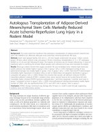
of the volume of articular. The ultra structure of articular cartilage varies
depending on the depth from the surface, which is shown in figure 1.1.
The superficial (tangential) zone is composed of flattened cells, a high
collagen content, which is packed tightly parallel to the surface and the lowest
concentration of proteoglycans. This zone provides tensile and shear
strength to the tissue. The transitional zone has a lower density of cells, which
are spheroid in shape, an abundant extracellular matrix (ECM), and collagen
fibers placed in a random pattern. The deep (radial) zone has the lowest cell
density and the highest proteoglycan content. In this zone collagen fibers are
oriented perpendicular to the articular surface to resist compressive loads.
Finally, the calcified zone is separated from the deep zone by the tidemark,
has few cells and the extracellular matrix is calcified (Table 1.1). In this zone,
production of type X collagen provides adhesive properties for tissue to
adhere to the underlying subchondral bone (1).
3!
!
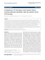
Figure 1-1. Layers in the articular cartilage (2, 3).
H&E staining of the articular cartilage (A), schematic of hyaline cartilage
collagen fiber arrangements (B).