Bone marrow derived mesenchymal stem cell (BM MSC) application in articular cartilage repair 3
Bạn đang xem bản rút gọn của tài liệu. Xem và tải ngay bản đầy đủ của tài liệu tại đây (6.72 MB, 7 trang )
63!
!
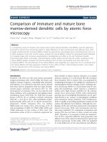
Figure 3-7. Trypan blue viability test.
Trypan blue viability test of the labeled and unlabeled MSCs showed a
significant decrease in high labeling concentration (100 and 125 µg/mL).
75
80
85
90
95
100
0 25 50 75 100 125
Mean cell viability (percent)
Labeling medium iron concentration (µg/ml)
!!*!
!*!
64!
!
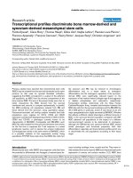
Figure 3-8. MTS assay.
MTS assay showed that labeling of the MSCs did not affect the proliferation
rate of the cells over time.
3.4.6 Differentiation potential of labeled MSC
MSCs are multipotent stem cells, which can differentiate to different lineage
(77, 78, 198, 199). To evaluate the effect of the ferucarbotran labeling on the
multipotent potential, we assessed the adipogenic (figure 3.9), osteogenic
(figure 3.10), and chondrogenic (figure 3.11) differentiation capacity of the
labeled cells.
3.4.6.1 Adipogenic differentiation
Qualitative evaluation of adipogenic differentiation after 21 days by Oil red O
staining demonstrated that unlabeled and labeled cells exhibited no difference
in developing fat vacuoles (Figure 3.9).
65!
!
Figure 3-9. Oil red O staining.
Oil red O staining of the MSCs after 21 days adipogenic differentiation
induction; Unlabeled (A), labeled with 25 µg/ml ferucarbotran (B), labeled with
50 µg/ml ferucarbotran (C), labeled with 75 µg/ml ferucarbotran (D), labeled
with 100 µg/ml ferucarbotran (E), labeled with 125 µg/ml ferucarbotran (F).
3.4.6.2 Osteogenic differentiation
Alizarin red staining showed that MSCs labeled with SPIO did not affect
osteogenic differentiation capacity of cells (figure 3.10).
!
Figure 3-10. Alizarin Red staining.
Alizarin Red staining of the MSCs was used to visualize the calcium
deposition of the cells after 21 days osteogenic differentiation induction;
Unlabeled (A), labeled with 25 µg/ml ferucarbotran (B), labeled with 50 µg/ml
ferucarbotran (C), labeled with 75 µg/ml ferucarbotran (D), labeled with 100
µg/ml ferucarbotran (E), labeled with 125 µg/ml ferucarbotran (F).
66!
3.4.6.3 Chondrogenic differentiation
Immunohistochemical staining after high density pellet culturing showed that
the distribution of collagen type II in extracellular matrix of cells was same
between unlabeled and 25 and 50 µg/mL SPIO labeling, however the matrix
production were decreased in labeling concentration of higher than 75 µg/mL
SPIO (Figure 3.11). Alcian blue staining also showed the inhibition of the
Aggrecan production in labeling concentration of the 75 µg/mL or higher.
Prussian blue was used to show the presence of iron particles.
Interestingly, SPIO labeled MSCs separated into distinct areas; the areas with
less Prussian blue stains (less iron content) showed more collagen type II and
aggrecan production. It appears that the inhibition seemed to be related to the
cellular iron content, increasing the labeling concentration could inhibit
chondrogenesis.
67!
!
Figure 3-11. Chondrogenic differentiation potential evaluation of the MSCs.
Upper panel shows the Alcian blue staining of unlabeled and labeled cells with different concentration of ferucarbotran
(25µg/mL, 50µg/mL, 75µg/mL, 100µg/mL, 125µg/mL). Middle panel shows the immunohistochemistry against collagen type
II in all groups and lower panel demonstrate the Prussian blue staining of the cell pellets to visualize the iron particles as
blue dots in the cells. (Scale bar is equal to 100 µm)
68!
3.4.7 MR imaging of animals
3.4.7.1 Preliminary experiments
To optimize the sequences that we needed to visualize the articular cartilage
of the pig knee, we performed a trial MRI to test different coils and sequences
on the pigs’ normal knee. Figure 3.12 showed chondral defect without / with
different fillings. Blank defect was the defect with no filling (air), which made a
signal loss (dark defect) in both FSE and GRE sequences. Defect with
scaffold only (1% agarose) filling showed an iso-intense signal to adjacent
cartilage in both FSE and GRE sequences. Defects filled with the agarose
mixed with different amount of Ferucarbotran, which can be a representative
of different concentrations of the labeled cells (the average amount of the iron
nanoparticles per cell was assumed as 10ng/cell; e.g. 1, 10, 1000 µg iron
nanoparticles were mixed with agarose as representative of 100, 1000,
100000 cells). We showed the efficacy of specifically imaging proton density
(short TE FSE) for anatomical data and the sensitivity of gradient echo
sequences (SPGR and 2D/3D-GRE) to signal loss due to iron nanoparticles to
reveal contrast between newly formed tissue and the incorporated iron
nanoparticles (Fe). The FIESTA sequence was also useful in the context of
proton density, however FIESTA images with Fe injected into the knee space
(to simulate unattached labeled cells) contained artifacts, which could
interfere with interpretation.
69!
!
Figure 3-12. MR imaging of the Pig's knee explant.
MR images of the pig’s knee explants with blank and scaffold only defect (A), 100 cells simulation (B), 1000 cells simulation (C),
and 100,000 cells simulation (D). Left panel of each image is 3D-FSE sequence and Right panel is 3D SPGR sequence.