Bone marrow derived mesenchymal stem cell (BM MSC) application in articular cartilage repair 5
Bạn đang xem bản rút gọn của tài liệu. Xem và tải ngay bản đầy đủ của tài liệu tại đây (4.3 MB, 1 trang )
74!
!
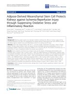
Figure 3-17. Postmortem histological evaluation of the repaired chondral
defects 6 weeks after stem cell injection.
H&E staining (A), IHC for collagen type I assessment (B), IHC for collagen
type II assessment (C), Masson’s trichrome for cartilage detection (D),
Toluidine blue staining for collagen type II detection (E), Safranin O staining
for cartilage matrix production evaluation (F). Arrowheads indicate the border
of repaired cartilage.
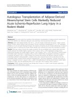
Serial sectioning and Prussian blue staining were performed to confirm the
presence of the labeled cells in the repaired cartilage. Results as shown in
Figure 3.18, 3.19 and 3.20 also confirmed the presence of the labeled MSCs
in the biopsies from the surgical scars and para-patellar fat tissues
respectively.
!
Figure 3-18. Prussian blue staining of the repaired cartilage defect.