The development of an in vivo humanized mouse model to investigate epstein barr virus infection and tumorigenesis
Bạn đang xem bản rút gọn của tài liệu. Xem và tải ngay bản đầy đủ của tài liệu tại đây (4.86 MB, 191 trang )
THE DEVELOPMENT OF AN IN VIVO HUMANIZED MOUSE MODEL
TO INVESTIGATE EPSTEIN-BARR VIRUS INFECTION AND
TUMORIGENESIS
MIN ZIN OO
(M.B., B.S.)
A THESIS SUBMITTED
FOR THE DEGREE OF DOCTOR OF PHILOSOPHY
IN COMPUTATION AND SYSTEMS BIOLOGY (CSB)
SINGAPORE-MIT ALLIANCE
NATIONAL UNIVERSITY OF SINGAPORE
2013
DECLARATION
I hereby declare that this thesis is my original work and it has been written by
me in its entirety. I have duly acknowledged all the sources of information
which have been used in the thesis.
This thesis has also not been submitted for any degree in any university
previously.
______________________________
MIN ZIN OO
25
th
March 2013
i
ACKNOWLEDGEMENT
I am highly indebted to my thesis advisors A/P Paul MacAry (Immunology
Program, NUS) and Prof Jianzhu Chen (SMART ID-IRG; Department of
Biology, MIT) for their advice, funding support, and guidance throughout the
course of the project from its conception until its end. I would also like to
thank my mentor Dr Maroun Khoury (Universidad de los Andes, Faculty of
Medicine, Santiago, Chile) for his wonderful guidance during the initial stages
of my project during his tenure at SMART-ID IRG as a research scientist.
The success of this research project and thesis would not be possible
without the help and support of each and every one of my colleagues and lab
mates in both the PAM and SMART labs. I would like to express special
thanks to Ms Zhenying Song who taught me the basic research skills; Dr
Adrian Sim who has consistently been a good friend, a comrade, and an
academic counsel; Mr. Wei Jian Tan who has never turned down my call for
help during the hectic period of the final stages of my project; Ms Fatimah Bte
Mustafa and Ms Chien Tei Too for their administrative support and relentless
efforts to make everybody happy during the troughs of our mood; Ms Siew
Chin Loh for her friendly technical support with flow cytometry; Ms Lan
Hiong Wong for taking excellent care of our laboratory mice; and Ms Hooi
Linn Loo for her technical and administrative assistance.
ii
I would like to express my gratitude to Singapore-MIT Alliance and
SMART ID-IRG for funding support; Emeritus Prof Chan So Ha and Ms
Nalini Srinivasan for providing us with the B95-8 strain of the Epstein-Barr
virus and quantitative PCR viremia assessment system; A/P Jerry Chan (Duke-
NUS) and his research team who kindly provided us with human fetal livers;
A/P Chng Wee Joo for his time for discussion and expert comments and
opinion on my project; and lastly Ms Carol Cheng, Ms Juliana Chai, Ms Hong
Yanling and Ms Chua Lay Peng for their administrative support throughout
my PhD candidature at SMA.
iii
TABLE OF CONTENTS
Acknowledgement i
Table of Contents iii
Summary vii
List of Tables ix
List of Figures x
List of Abbreviations xii
Chapter 1 : INTRODUCTION 2
1.1 Epstein-Barr virus life cycle 2
1.2 EBV and the immune system 5
1.2.1 CD8
+
cytotoxic T cell (CTL) responses 5
1.2.2 CD4
+
helper T cell responses 6
1.2.3 EBV and NK cells 8
1.2.4 EBV and the human cytokine system 8
1.2.5 Anti-EBV antibody responses 11
1.2.6 Immune evasion mechanisms of EBV 12
1.3 Latent EBV infection 15
1.3.1 EBV-encoded RNAs (EBERs) 18
1.3.2 Latency I or ‘EBNA1 only’ program 18
1.3.3 Latency II or the ‘Default’ program 19
1.3.4 Latency III or the ‘Growth’ program 20
1.3.5 Other latency programs 21
1.3.6 Regulation of EBV latency 22
1.3.7 The persistence of EBV 23
1.4 EBV-associated malignancies and the role of EBV in tumorigenesis .27
1.4.1 EBV and Burkitt’s lymphoma 27
1.4.2 EBV and Hodgkin’s lymphoma 29
1.4.3 EBV and Immunodeficiency-related lymphoproliferative
disease 30
1.5 EBV-associated Post-transplant Lymphoproliferative disease (PTLD)
32
iv
1.5.1 Post-transplant lymphoproliferative disease 32
1.5.2 The role of lytic EBV infection 33
1.5.3 The role of cytokines 34
1.5.4 The role of anti-EBV antibodies 35
1.5.5 The role of T cell immunity 36
1.6 Humanized mouse models of Epstein-Barr virus infection 38
1.6.1 Early mouse models of EBV-associated lymphoproliferative
disease 39
1.6.2 Humanized mouse models of EBV infection and
lymphoproliferative disease 40
1.6.3 Differences in the humanized mouse strains in previous studies
43
1.6.4 Differences in virus strains used in previous humanized mouse
studies 44
1.6.5 Controversy of the published models 45
1.6.6 Current concepts about the role of human B cells in EBV-
associated lymphoproliferative diseases based on animal
models 46
CHAPTER 2 : MATERIALS AND METHODS 51
2.1 Isolation of hemopoietic stem and progenitor cells (HSCs) from human
fetal liver 51
2.2 Establishment of the humanized mouse 52
2.3 Epstein-Barr virus culture, virus titer assay and in vivo infection 52
2.4 Flow cytometric immunophenotyping 53
2.5 Histopathology 56
2.6 Viremia assay 57
2.7 Multiplex cytokine assay 60
2.8 Treatment of EBV-infected mice with Rituximab 61
2.9 Secondary adoptive transfer tumor model 61
2.10 Statistical analysis 62
CHAPTER 3 : RESULTS AND INTERPRETATION 64
3.1 Establishment of the humanized mouse model 64
3.1.1 Experimental scheme for development of a humanized mouse
model of EBV infection 64
v
3.1.2 Isolation of hemopoietic progenitor cells from the human fetal
liver 65
3.1.3 In vivo infection of the humanized mouse model with Epstein-
Barr Virus 68
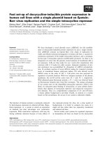
3.2 Characterization of the humanized mouse model of EBV infection 71
3.2.1 Demonstration of virus replication in the plasma 72
3.2.2 Evidences of human cell responses in the peripheral blood 76
3.2.3 Peripheral blood activation marker profile 81
3.2.4 Gross pathological examination of fatal systemic
lymphoproliferative lesions in EBV-infected humanized mice.
85
3.2.5 Histopathological study of the systemic lesions 87
3.2.6 Significant T cell expansion with higher CD8:CD4 ratios in the
spleens of infected mice 90
3.2.7 Activation marker profile in the spleens of infected mice 92
3.2.8 Marked expansion of CCR5
+
Th1 cells in infected mice with
poor memory T cell development 95
3.2.9 Human cytokine response in infected humanized mice 99
3.2.10 Distribution of human B cell subsets in the spleen 105
3.2.11 Clonality of splenic B cells 107
3.3 Demonstration of a pivotal role of human B cells in the pathogenesis of
disease in EBV-infected mice 111
3.3.1 Experimental scheme of Rituximab treatment of EBV-infected
mice 112
3.3.2 Complete clearance of viremia after B cell depletion 115
3.3.3 Immune correlates of rituximab-treated mice 117
3.3.4 Survival benefit of rituximab treated mice – Evidence that B
cells are the major driving force in the pathogenesis of the
disease 119
3.4 Experimental proof of the malignant nature of human B cells in EBV-
infected mice 122
3.4.1 Experimental scheme of secondary adoptive transfer
experiments 123
3.4.2 Life span of secondary mice 124
3.4.3 Appearance of systemic fatal tumors in secondary mice which
received unfractionated splenocytes 126
vi
3.4.4 Activated human cells in the spleens of secondary mice which
received unfractionated splenocytes 128
3.4.5 The B cell population alone is both necessary and sufficient for
tumorigenesis 131
3.4.6 Tissue lesions in secondary B cell tumor mice 134
3.4.7 Histopathological examination of secondary tumors 136
3.4.8 Demonstration of latency III pattern of EBV gene expression in
secondary tumors 139
3.4.9 Demonstration of cellular proteins in secondary tumors 140
CHAPTER 4 : DISCUSSION 144
4.1 Disease phenotype of the EBV infection in our humanized mouse
model 145
4.2 Human immune responses in our mouse model 146
4.3 Proof of tumorigenicity of human B cells in EBV-infected mice 152
4.4 Future directions 155
REFERENCES 158
vii
SUMMARY
Epstein-Barr virus (EBV) is a human-specific B lymphotrophic
gammaherpesvirus which causes life-long latent infection. Although latent
infection is mainly asymptomatic and thus inconsequential in the majority of
cases, EBV latency is also consistently associated with many aggressive
hematological and solid tissue malignancies of diverse tissue origins in
different populations throughout the world. The ubiquitous presence of the
virus in more than 90% of the adult human population and the benign nature
of latent infection make the pathogenic role of EBV questionable and thus
many critics claim that the virus is simply an innocent bystander in most of the
associated malignancies and does not play a direct causal role in
tumorigenesis. Clinical evidences from post-transplant lymphoproliferative
diseases have implied a direct causative role of EBV in pathogenesis.
However, requirement for additional factors including signaling components
and cytokine support from T lymphocytes were also identified as essential in
the rapidly fatal spectrum of lymphoproliferative disease. In this study, we
describe a humanized mouse model that incorporates the clinical, virological
and immunological features of latent EBV infection. We employ a secondary
adoptive transfer lymphoproliferative disease model to prove the tumorigenic
potential of EBV-infected B lymphocytes in severely immunocompromised
mice. These data strongly suggest that B lymphocytes in infected hosts are
both necessary and sufficient for the development of fatal systemic
lymphoproliferative disease. Treatment of EBV-infected humanized mice
viii
with the B lymphocyte depleting antibody Rituximab further highlighted the
pivotal role of B lymphocytes in the pathogenesis of the disease. Taken
together, these data provide the first unequivocal evidence for a causal link
between Epstein-Barr virus infection of human B cells with
lymphoproliferative disease and tumorigenesis.
ix
LIST OF TABLES
Table 1. Latent EBV gene expression programs. 17
Table 2. EBV latency gene expression programs in physiological B cell
maturation stages and disease associations. 17
Table 3. Summary of published humanized mouse models of EBV
infection and salient observations. 42
Table 4. Fluorochrome-conjugated anti-human antibodies used for flow
cytometry analysis. 55
Table 5. Antibodies used for histopathological examination 56
Table 6. Quantitative representation of reconstitution and lymphoid
lineage of an average batch of humanized mice. 69
x
LIST OF FIGURES
Figure 1. Epstein-Barr virus (EBV) life cycle. . 4
Figure 2. Immunodominance pattern of selected lytic and latent EBV
antigens in healthy virus carriers. 7
Figure 3. The structure of the circular plasmid-like genome of the
prototypic B95-8 strain of EBV in latently infected cells 16
Figure 4. EBV viremia qPCR standard curves. 59
Figure 5. Experimental scheme of the development of a humanized
mouse model of EBV infection. . 64
Figure 6. Assessment of purity of CD34 immunomagnetic bead isolation
of hemopoietic progenitor cells from a human fetal liver. 67
Figure 7. Assessment of the reconstitution of humanized mice 10 weeks
after intracardiac injection of CD34
+
hemopoietic progenitor
cells. 69
Figure 8. Monitoring infected mice by measurement of body weight and
weekly plasma viremia by qPCR. 72
Figure 9. Human immune cell response against EBV infection. 77
Figure 10. Activation marker profile of human immune cell response
against EBV infection. . 81
Figure 11. Autopsy of the humanized EBV-infected mice. 85
Figure 12. Histopathological examination of infected mice tissues. 87
Figure 13. Flow cytometric analysis of host immune response against
EBV infection – splenic lymphocyte lineages. 90
Figure 14. Expression of activation markers on splenic lymphocytes. 92
Figure 15. Flow cytometric analysis of splenic T cell and memory cell
subsets. 95
Figure 16. Analysis of the human cytokine response against EBV infection
102
Figure 17. Flow cytometric analysis of splenic B cell lineages. . 105
xi
Figure 18. Clonality assay by surface expression of immunoglobulin light
chain measured by flow cytometry. 108
Figure 19. Experimental scheme of rituximab treatment and depletion of B
cells after treatment. 113
Figure 20. Weekly plasma viremia of rituximab treated and untreated
mice. 115
Figure 21. Comparison of distribution of peripheral blood immune cells in
rituximab treated and untreated mice. 117
Figure 22. Kaplan-Meier survival analysis of rituximab treated mice 119
Figure 23. Experimental scheme of secondary splenocyte transfer
experiments. 123
Figure 24. Survival of secondary mice injected with unsorted total
splenocytes of infected and uninfected humanized mice of the
same age. 124
Figure 25. Fatal systemic lesions in secondary mice and expansion of
human cells in secondary spleens. 126
Figure 26. Flow cytometry of the splenocytes of the secondary mice 128
Figure 27. Experimental scheme of secondary splenocyte transfer
experiment using sorted human B cells. 131
Figure 28. Survival of secondary mice injected with sorted splenocytes
from infected and uninfected humanized mice of the same age.
132
Figure 29. Kidney lesions in secondary mice 134
Figure 30. Histopathological examination of lesions in secondary mice.
136
Figure 31. Immunohistochemistry staining of secondary tumors to
determine EBV latency. . 139
Figure 32. Immunofluorescent staining of secondary tumor tissues 140
xii
LIST OF ABBREVIATIONS
BLCL B Lymphoblastoid Cell Line
BLT Bone marrow, Liver and Thymus-transplanted humanized mice
CB Cord Blood
CD Cluster of Differentiation
CTL Cytotoxic T Lymphocyte
EBERs EBV-encoded RNAs
EBNA EBV Nuclear Antigen
EBV Epstein-Barr Virus
FL Fetal Liver
GM-CSF Granulocyte-Monocyte Colony Stimulating Factor
IL Interleukin
LMP Latent Membrane Protein
LPD Lymphoproliferative Disease
NOD Non-obese Diabetic mouse strain
NOG NOD/SCID/ IL2Rγc
truncated
NRG Rag
-/-
IL2Rγc
-/-
NSG NOD-scid IL2Rγc
null
mouse strain
NOD.Cg-Prkdc
scid
Il2rg
tm1Wjl
/SzJ
PBS Phosphate Buffered Saline
PBMC Peripheral Blood Mononuclear Cells
PTLD Post-transplant Lymphoproliferative Disease
SCID Severe Combined Immunodeficiency syndrome
TAP-1 Transport-Associated Protein-1
Treg Regulatory T cells
CHAPTER 1 : INTRODUCTION
Humanized mouse model of EBV infection Introduction
2
CHAPTER 1 : INTRODUCTION
1.1 EPSTEIN-BARR VIRUS LIFE CYCLE
Epstein-Barr Virus (EBV) is a ubiquitous human-specific virus found in over
90% of the adult population worldwide according to seroepidemiology
1
. It is
a large double-stranded DNA virus (172 kbp)
2
and belongs to the gamma-
herpesvirus subfamily of the human herpesvirus super family, which is
characterized by life-long latent infection and periodic reactivation. The virus
primarily infects B cells but viral genomes have also been found in T cells and
natural killer (NK) cells in chronic active EBV infection. The virus is also
postulated to infect oropharyngeal epithelial cells.
Primary childhood infection is usually asymptomatic but the primary
infection in adolescents is manifested by non-life-threatening self-limiting
infectious mononucleosis (IM) or glandular fever accompanied by flu-like
syndrome and systemic lymphadenopathy
3
. The mode of transmission is by
inoculation of infected saliva. EBV infects B lymphocytes by engaging its
envelope protein gp350/220 with CD21 (complement receptor 2)
4
. The
invasion of the virus into non-B cells, oropharyngeal epithelial cells and
tissues like smooth muscle, gastric epithelium and other cell types that do not
express CD21 suggests the involvement of additional as yet undefined host
receptors for this virus. According to current model of EBV life cycle, the
virus invades the oropharyngeal epithelium and infects naïve B cells in the
Humanized mouse model of EBV infection Introduction
3
subepithelial lymphoid tissues. EBV drives massive proliferation of infected
B cells and expresses lytic as well as some latent proteins that elicit a strong
cytotoxic T lymphocyte (CTL) response, which destroys the infected B cells.
During acute infection and reactivation, EBV undergoes a lytic replicative
cycle in which overwhelming production of virus progeny results in host cell
rupture and cytopathology. The virus, after some time, adopts the latent life
cycle, associated with a limited transcriptional activity, evading immune
recognition by the host CTLs, and enabling life-long persistence in the
memory B cell compartment with periodic reactivation
5
. A comprehensive
overview of the life cycle of EBV is illustrated below in Figure 1.
Humanized mouse model of EBV infection Introduction
4
Figure 1. Epstein-Barr virus (EBV) life cycle. Epstein-Barr virus
(EBV) is transmitted by contact with the infected saliva. The current concept
holds that EBV invades the nasopharyngeal mucosa and infects circulating B
lymphocytes in the subepithelial lymphoid tissues. Infection of
nasopharyngeal epithelium is controversial. EBV causes massive proliferation
of infected B lymphocytes which prime cytotoxic T lymphocytes (CTLs)
important for the immune clearance of EBV-transformed B cells. EBV drives
maturation of the infected B cells into memory B cells which move into the
peripheral circulation. Differentiation of memory B cells into plasma cells on
antigen exposure triggers the lytic EBV life cycle, resulting in the release of
virus particles which infect B lymphocytes in the vicinity and shed in the
saliva to infect new susceptible hosts. Physiological homeostasis of the
memory B cell compartment maintains the life-long persistence of the virus.
Humanized mouse model of EBV infection Introduction
5
1.2 EBV AND THE IMMUNE SYSTEM
Cytotoxic T cell immunity is the mainstay of the human immune defense
against EBV infection and virus-induced B cell proliferation. In
immunocompromised patients, such as those on post-transplant
immunosuppressive drug regimes, those receiving cancer chemotherapy, or
patients with AIDS, suppression of T cell function results in an outgrowth of
EBV-transformed B lymphocytes, which give rise to lymphoproliferative
disorders that have high mortality rates
6,7
. This problem is particularly
marked in transplant patients receiving immunosuppressive drug regimes. In
many cases, the lymphoproliferative disorder is controlled by reducing
immunosuppression which results in an increased risk of graft rejection. This
reflects the important role of CTLs in controlling EBV-induced
lymphoproliferation.
1.2.1 CD8
+
cytotoxic T cell (CTL) responses
CD8
+
T cells are massively expanded in acute symptomatic EBV infection
(infectious mononucleosis) and the expanded cells exert functional, EBV-
specific, oligoclonal CD8 responses which strongly skew towards immediate
early and certain early lytic proteins
8
. Up to 40% of the total CD8
+
T cell
population is EBV-specific. CTL responses against latent proteins are
relatively smaller and tend to focus strongly on epitopes derived from the
EBNA3A, B, C family of proteins and the frequency of specific cells comprise
Humanized mouse model of EBV infection Introduction
6
up to 5% of total CD8
+
T cells
9
. In the late stages of infection and in healthy
carriers, the frequency of CD8
+
T cells specific to latent epitopes selectively
increases, implying that CTLs are the most important cells responsible for the
long term control of the virus
10
. There is an age-related expansion of EBV-
specific CTLs which comprise up to 14% of total CD8
+
T cell population in
healthy carriers over 60 years of age
11
.
1.2.2 CD4
+
helper T cell responses
The CD4 response in acute infection, is more widely spread across immediate
early, early and late antigens and the frequencies are much lower compared to
the CD8 response
12
. The CD4 memory response against lytic antigens has
been documented but not comprehensively characterized. Latent antigen-
specific CD4
+
T cell response is relatively spread across EBNAs - more
towards EBNA1 and EBNA2 in contrast to EBNA3-skewed CD8 response
13,14
. LMP1 is a subdominant CD4 epitope but this latent protein and derived
peptides stimulate IL10 production in vitro
15
, suggesting that LMP1 may
induce Treg-like cells
16
. The frequency of EBV-specific CD4
+
T cells is
much lower than that of CD8.
A diagrammatic representation of the immunodominance pattern of
CD8 and CD4 epitopes in healthy virus carriers is given below in Figure 2
(adapted from Hislop, et al.
17
).
Figure 2. Immunodominance pattern of selected lytic and latent EBV antigens in healthy virus carriers. Blue boxes
represent lytic (IE – immediate early; E – early; L – late; n.t. – not tested) and latent EBV proteins, epitopes derived from which
are recognized by CD4
+
or CD8
+
T cells. Solid arrows denote relative immunodominance of defined epitopes and the height of
the arrows reflects relative abundance of the response against respective epitopes. Dotted arrows show documented epitopes
whose relative immunodominance is not yet determined. During the lytic replicative cycle of EBV, the majority of the CD8
+
CTL responses target immediate early lytic and some early lytic proteins. CD4
+
T cell responses, although poorly studied, appear
to be more widespread across lytic antigens. In latent EBV infection, the EBNA3 group of nuclear antigens are the most
immunodominant in terms of CD8
+
CTL response, followed by LMP2 and EBNAs. The CD4+ T cell response is more widely
distributed across latent antigens and is skewed towards EBNA1, EBNA2 and EBNA3C with some contribution from EBN3A,
EBNA3B and LMPs.
7
Humanized mouse model of EBV infection Introduction
Humanized mouse model of EBV infection Introduction
8
1.2.3 EBV and NK cells
Expansion of activated NK cells in the blood of acute infectious
mononucleosis patients was reported and the cell number was inversely
correlated with virus load
18
. In vitro experiments also showed that NK cells
from human tonsil can be primed by co-resident dendritic cells to produce
IFN-γ which delays the outgrowth of EBV-transformed B cells
19
. However,
clinical evidence in T cell depleted stem cell transplant patients did not
support a major role for NK cells in the control of EBV-transformed B cell
outgrowth in vivo - lymphoproliferative disease is most common in the first
three to six months post-transplant, by which time NK cell numbers have
usually recovered
20
. Several EBV gene products have inhibitory effects on
MHC class I expression on the infected cell’s surface and this may render
infected cells susceptible to NK-mediated recognition and lysis. However,
EBV microRNA (miR-BART2) has recently been shown to reduce expression
of the NK activating ligand MIC-B, thereby nullifying the susceptibility of the
infected cells to NK-mediated lysis
21
.
1.2.4 EBV and the human cytokine system
Both acute lytic EBV infection and latent persistence are closely linked with
cytokines and chemokines which shape the immune response in the
microenvironment of the infected cells. Furthermore, EBV genome encodes
Humanized mouse model of EBV infection Introduction
9
an IL10 homolog and several proteins which regulate the expression and
biological functions of cytokines
22
.
Cytokines play a major role in the pathogenesis of acute infectious
mononucleosis in which patient sera were shown to be rich in IL1α, IL2, IL6,
and IFN-γ
23
. Release of cytokines and chemokines results from massive
expansion of activated CTL clones which is a characteristic pathological
feature of acute IM. EBV-infected cells in the tonsil of acute IM patients
express lymphotoxin, TNF-α, IL6, IFN-γ
24
, IL18, monokine induced by IFN-γ
and IFN-γ inducible protein 10 (IP10)
25
while EBV-negative interfollicular
cells expressed IL1α, IL1β and IL8.
Among other cytokines and chemokines, IL6 is closely linked in EBV
infection and immunodeficiency-associated lymphoproliferative disease.
Cross-linking of CD21 by viral envelope glycoprotein gp.350/220 induces
expression of IL6
26
, which activates NF-κB in lymphocytes
27
and regulates B
cell growth and differentiation during primary EBV infection. IL6 also acts as
an autocrine growth factor for EBV-immortalized B cells
28
. IL6 promotes the
growth of B cells in lymphoproliferative disease and anti-IL6 antibodies
decrease the incidence of the tumor
29
, highlighting the role of IL6 in disease
progression.
EBNA1-specific helper T cells were shown to preferentially secrete a
Th2-type cytokine IL5 in response to antigenic stimuli
30
. However, another
group reported that the cytokine secretion of EBNA1-specific helper T cells
Humanized mouse model of EBV infection Introduction
10
was polarized towards a Th1 pattern IFN-γ secretion
31
. Secretion of IL8 and
macrophage inhibitory protein 1α (MIP-1α) was triggered by EBV binding of
monocytes and the secretory response was enhanced by GM-CSF
32
. GM-CSF
was also known to facilitate the spontaneous outgrowth of EBV-infected B
cells
33
. GM-CSF orchestrates the host immune response towards virus
eradication but at the same time, facilitates more infection events as the
immune cells are recruited to the site of infection.
Th1 cytokines IFN-γ and TNF-α were reported to be expressed in the
lymph nodes of infectious mononucleosis patients but not in those of
lymphoproliferative disease
25
. IL18, which induces IFN-γ expression, acts
together with IL12 and IP10 caused the regression of EBV-positive Burkitt’s
lymphoma
34
.
The cytokine profile in post-transplant lymphoproliferative disease was
reported to be skewed towards a Th2 pattern with IL2
-
IFN-γ
-
IL4
+
IL10
+
profile by using semi-quantitative RT-PCR, suggesting that PTLD was
developed in a Th2 cytokine milieu
35
. Th2 polarization in PTLD may reduce
the host CTL response which requires Th1 cytokines, and favor the growth of
tumor cells in autocrine fashion. Furthermore, Paul, et al.
36
showed EBV-
transformed B lymphoblasts secrete Th2 cytokine IL5. The same group also
reported that GM-CSF modulated the spontaneous outgrowth of B
lymphoblastoid cell clones after in vitro infection
33
.
Humanized mouse model of EBV infection Introduction
11
EBV encodes several cytokine homologs and regulatory factors to
manipulate the host cytokine response to abrogate the anti-viral immune
response. EBV-encoded viral analog of IL10, EBV-induced gene 3 (EBI3)
and BARF1 proteins regulate the secretion and biological actions of several
human cytokines and thus important for evasion of immune response
(elaborated in Section 1.2.6 – Immune evasion mechanisms). EBNA2, which
is a transcriptional transactivator, regulates the expression of lymphotoxin,
TNF-α and G-CSF in EBV-infected B cells
37
. LMP1 can induce the secretion
of IL6, IL8, IL10 and IFN regulatory factor-7 (IRF7) to favor the survival of
host cells
38-40
.
1.2.5 Anti-EBV antibody responses
The function of the anti-EBV humoral immune response is to limit the
infectious virus particles and therefore, to control the spread of the virus in the
late stages of infection
41
. Important antibody targets include the early antigen
complex, which comprises several immediate early and early EBV proteins
including the lytic cycle transactivator BZLF1, and the late structural viral
capsid antigen complex early in infectious mononucleosis. In addition, the
membrane antigen complex, including gp350, induces neutralizing antibodies
in acute IM
42
. Thus by the onset of clinical symptoms, high titers of anti-viral
capsid antigen IgM and IgG as well as IgG against early antigen and
membrane antigen complexes can be detected. IgA against viral capsid