The role of b cells in the pathogenesis of atherosclerosis 2
Bạn đang xem bản rút gọn của tài liệu. Xem và tải ngay bản đầy đủ của tài liệu tại đây (9.26 MB, 38 trang )
3.3.2 Plasmablasts in extrafollicular responses were IgM+
Our data, thus far, indicated that extrafollicular responses in apoE-/mice may be responsible for the generation of total IgM+ and also oxLDLspecific IgM+ plasma cells. Thus, we performed immunofluorescence staining
to evaluate whether IgM+ plasmablasts were generated from extrafollicular
responses. Our result showed that IgM+ plasmablasts were indeed colocalizing
with CD11chi DCs at the bridging channel of the follicles (Figure 21A).
Extending our findings, we observed these IgM+ plasmablasts that colocalized
with CD11chi DCs were proliferating as they incorporated thymidine analog,
EdU in a 12hr pulsed chase experiment (Figure 21B).
Because of the lack of tools to evaluate if these IgM+ plasmablasts
were oxLDL-specific, we immunized young WT mice with oxLDL via
intravenous route to evaluate if immunization with oxLDL could elicit splenic
extrafollicular responses. Activated
B
cells
migration
to
the
extrafollicular
sites
could
be
detected
from
day
3.5
and
maximally
at
day
4.5
after
immunization
as
described
(Gatto
et
al.,
2009).
Therefore,
these mice were
sacrificed on Day 4 after immunization to evaluate extrafollicular responses.
However, the use of common adjuvant such as CFA alone could induce the
increased titer of MDA-LDL specific antibodies (Khallou-Laschet et al., 2006).
Therefore, the use of adjuvants was excluded in our immunization studies. Our
results showed that although the CD138+ plasmablasts colocalized with
CD11chi DCs were not as numerous as in apoE-/- mice (data not shown), we
were still able to observe an increased extent of extrafollicular responses in
oxLDL-immunized mice compared to PBS-immunized controls (Figure 21C).
We also evaluated if the immunization with oxLDL could elicit GC reactions
104
in these mice on Day 14 by flow cytometry. However, we did not detect an
increase in percentage and number of GC B cells in these oxLDL-immunized
mice compared to PBS-immunized controls (Figure 21D & 21E).
105
106
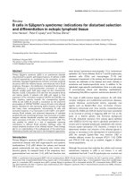
Figure 21. IgM+ plasmablasts were generated in extrafollicular responses
in the spleen of apoE-/- mice.
(A) Representative image of IgM+ plasmablasts colocalizing with CD11c+
DCs at bridging channel of follicle in spleen of apoE-/-. Outer laying region
(dotted lines) denotes B cell follicle regions (n=6). Data were representative of
two independent experiments. (B) Representative image of proliferating IgM+
plasmablasts in EdU pulse chase (n=3). White arrows denote proliferating
IgM+EdU+ plasmablasts colocalizing with CD11c+ DCs at bridging channel of
follicle. (C) Quantification of extrafollicular responses sites per area (mm2)
analyzed in spleen of i.v. oxLDL immunized WT mice at Day 4 (n=4). Data
were pooled from two independent experiments. (D-E) Flow cytometry
analysis of GC B cells in terms of (D) percentage and (E) number in the
spleen of i.v. oxLDL immunized WT mice (n=5) at Day 14. ***, P < 0.001.
107
3.3.3 Summary
Collectively, we provide direct evidence that IgM+ plasmablasts were
generated through the extrafollicular response pathway in the spleen of apoE-/mice. The increased humoral IgM responses in spleen of apoE-/- mice were not
due to defective antibodies class-switching from the GC reactions. Also, we
showed that extrafollicular responses, but not GC reactions, were elicited in
WT mice when we immunized WT mice with oxLDL.
108
3.4 Evaluation of molecular cues to direct extrafollicular responses in the
spleen of apoE-/- mice
Activated B cells that migrate to the bridging channel in extrafollicular
responses requires the expression of chemokine receptor, EBI2 (Gatto et al.,
2009). Until recently, the natural ligand for EBI2 was identified to be 7α, 25OHC (Hannedouche et al., 2011; Liu et al., 2011). The formation of 7α, 25OHC is by the stepwise actions of two enzymes, CH25H and CYP7B1
(Figure 2). This oxysterol could be further metabolized into 4-cholesten-7α,
25-ol-3-one by HSD3B7 (Figure 2). The deficiency of any of the three
enzymes is associated with decreased antigen-specific plasma cell numbers
(Hannedouche et al., 2011; Yi et al., 2012). Therefore, we investigated if the
robust splenic extrafollicular responses seen in apoE-/- mice could be
attributed to increased EBI2 expression and/or increased bioavailability of 7α,
25-OHC.
3.4.1 Increased ch25h mRNA expression in the spleen of apoE-/- mice
We examined the mRNA expression of ebi2, ch25h, cyp7b1 and
hsd3b7 relative to the house keeping gene, hprt1 in spleen from WT and apoE/-
mice. Our analysis revealed no change in ebi2 mRNA expression in spleen
of apoE-/- mice compared to WT mice (Figure 22A). However, when we
examined the mRNA expression level of the enzymes critical for 7α, 25-OHC
synthesis, we found statistical significant increased ch25h mRNA expression
but not cyp7b1 and hsd3b7 mRNA expression (Figure 22B, 22C & 22D).
Thus, our data suggests that mRNA expression of ebi2 may not account for
the robust extrafollicular responses in the spleen of apoE-/- mice. Furthermore,
109
the increased ch25h mRNA expression observed may translate into higher
bioavailability of 7α, 25-OHC in spleen of apoE-/- mice since mRNA
expression level of hsd3b7 was not elevated to indicate higher efficiency to
metabolize 7α, 25-OHC into 4-cholesten-7α, 25-ol-3-one.
3.4.2 Increased oxysterol in the spleen of apoE-/- mice
To confirm our hypothesis, we collaborated with Dr. Andreas Sailor
from Novartis (Basel, Switzerland) to measure the amount of oxysterol in the
spleen of apoE-/- mice compared to WT mice using high performance liquid
chromatography mass spectrometry (HPLC-MS). Indeed, our data analysis
showed higher amount of 25-OHC and 7α, 25-OHC oxysterol in the spleen of
apoE-/- mice compared to WT mice (Figure 22E & 22F). Therefore, our data
indicates the possibility that the robust splenic extrafollicular responses in
apoE-/- mice may be supported, at least in part, by increased bioavailability of
7α, 25-OHC.
110
Figure 22. Elevated 7α, 25-OHC oxysterol in the spleen of apoE-/- mice.
(A-D) Quantitative mRNA transcript expression of (A) EBI2, (B) CH25H, (C)
CYP7B1 and (D) HSD3B7, relative to HPRT1 (mean ± SEM, n = 8). Data
were pooled from two independent experiments. (E-F) Quantitative oxysterol
measurement of (E) 25-OHC and (F) 7α, 25-OHC (n=5) by LC-MS using D67α, 25-OHC as reference. *, P < 0.05
111
3.4.3 Summary
Our data suggests the robust extrafollicular responses in the spleen of
apoE-/- mice may be due to the increased amount of EBI2 ligand, 7α, 25-OHC.
This was facilitated by the increased ch25h, but not hsd3b7, mRNA
expression observed in the spleen of apoE-/- mice. Our result also suggests
changes in EBI2 expression unlikely contribute to the robust extrafollicular
responses although it remains possible that B cell subpopulations may display
increased ebi2 mRNA expression.
112
3.5 Antibody production of B1a cells in apoE-/- mice
The peritoneal cavity (PEC) is highly enriched for B1 cells and also
exists in the spleen but constitute a minor B cell population (Baumgarth, 2011).
B1 cell population could be divided into two sub-populations; CD19+CD5+
B1a and CD19+CD5- B1b cells. Since the amount of oxLDL-specific IgM
autoantibodies in circulation were elevated in apoE-/- mice and B1a cells are
implicated in the production of oxidation epitope-specific antibodies in
atherosclerosis (Chou et al., 2009), we examined if B1a cell population
increased in apoE-/- mice. These B1a cells had also been described to retain
CD5+ marker before losing expression 5 days after LPS stimulation (Yang et
al., 2007). Therefore, it allows a window of opportunity to investigate B1a
cells differentiating into IgM+CD138+ ASCs (Yang et al., 2007).
3.5.1 B1a cells were not expanded in PEC of apoE-/- mice
Our flow cytometry analysis demonstrated that there were no
differences in relative percentage and number in B1a cell population in PEC
of apoE-/- mice compared to WT mice (Figure 23A & 23B). Furthermore,
when we examined extracellular IgM+CD138+ B1a cell population, we also
could not detect any difference in relative percentage and number when
compared to WT mice (Figure 23C & 23D). Therefore, our data do not
support the hypothesis that an increased in B1a cell population accompanied
by differentiation into IgM+ ASCs in PEC of apoE-/- mice may account for
increased titer of oxLDL-specific IgM autoantibodies.
113
3.5.2 Increased splenic B1a cells differentiation into IgM+ plasmablasts
B1 cells also exists in the spleen but constituted only 1-2% of CD19+ B
cell population (Baumgarth, 2011). Therefore, it remained possible that B1a
cells increased in the spleen of apoE-/- mice and differentiated into IgM+
plasma cells in apoE-/- mice.
Our preliminary analysis of CD19+CD5+ B1a cell population showed
that apoE-/- mice had higher relative percentage of B1a in the spleen (Figure
24A). However, we did not detect an increase in relative cell number of B1a
population in the spleen of apoE-/- mice (Figure 24B). Therefore, these sets of
observations suggest increased frequency of splenic B1a cell population was
due to changes in frequency of other lymphocyte sub-populations instead of
indications that there was B1a cell population expansion in apoE-/- mice.
Next, we examined if there were more B1a cells differentiating into
IgM+ ASCs in apoE-/- mice. Preliminarily, we observed a non-statistical
significant increase in relative percentage but a statistical significant increase
in relative number of extracellular IgM+CD138+ B1a cells in apoE-/- mice
(Figure 24C & 24D). On the contrary, when we examined for intracellular
IgM+CD138+ B1a cells, we found significant decrease in relative percentage
and number in the spleen of apoE-/- mice (Figure 24E & 24F). Two
possibilities could explain the decreased IgM+CD138+ B1a plasma cells; 1) the
plasma cells died in situ or, 2) the plasmablasts migrated to the bone marrow
for long-term maintenance. With more CD11chi DCs colocalizing with IgM+
plasmablasts in extrafollicular responses to aid in the survival and successful
differentiation of plasmablasts in apoE-/- mice, it is unlikely to explain that
114
these IgM+CD138+CD19+CD5+ died rapidly to account for the decreased
population. We favoured the latter possibility that these IgM+ plasmablasts
migrate to the bone marrow for long-term maintenance that we will explore in
the subsequent sections.
115
Figure 23. No difference in B1a cell population in peritoneal cavity of
apoE-/- mice.
(A-B) Comparative flow cytometry analysis of relative (A) percentage and (B)
number of CD19+CD5+ B1a cells (mean ± SEM, n = 9). Data were pooled
from three independent experiments. (C-D) Comparative flow cytometry
analysis of relative percentage and number of extracellular
IgM+CD138+CD19+CD5+ B1a cells (mean ± SEM, n = 6). Data were pooled
from two independent experiments.
116
Figure 24. Increased population of B1a cells differentiating into IgM+
plasmablasts in the spleen of apoE-/- mice.
(A-B) Comparative flow cytometry analysis of relative (A) percentage and (B)
number of CD19+CD5+ B1a cells in the spleen (mean ± SEM, n=9). Data were
pooled from three independent experiments. (C-D) Comparative flow
cytometry analysis of relative (C) percentage and (D) number of extracellular
IgM+CD138+CD19+CD5+ B1a cells in the spleen (mean ± SEM, n=6). Data
were pooled from two independent experiments. (E-F) Comparative flow
cytometry analysis of relative (E) percentage and (F) number of intracellular
IgM+CD138+CD19+CD5+ B1a cells in the spleen (mean ± SEM, n=9). Data
were pooled from three independent experiments. * P < 0.05; ** P < 0.01
117
3.5.3 Summary
Collectively, our data suggests that PEC B1a cells were not affected in
the apoE-/- mice. In addition, our preliminary data suggests that although B1a
cells did not expand in the spleen of apoE-/- mice, we did detect increased
differentiation of B1a cells into IgM+CD138+ plasmablasts and their
subsequent disappearance/ decrease in relative percentage and number as
intracellular IgM+CD138+ B1a plasma cells in the spleen.
118
3.6 Evaluation of the impact of splenic CD138+ ASCs on atherosclerosis
Elegant studies by Caliguri et al, demonstrated that the adoptive
transfer of splenic B cells, but not T cells, from old apoE-/- mice donor into
young apoE-/- mice recipient led to decreased lesion size in the aorta (Caligiuri
et al., 2002). They also showed that this protective effect was not observed
when adoptive transfer of splenic B cells were from young apoE-/- mice
(Caligiuri et al., 2002). This prompted us to investigate if the protective effect
of B cells could be due to ASCs existing in the old apoE-/- mice.
3.6.1 Adoptive transfer of splenic CD138+ ASCs into apoE-/- mice
To investigate this, we carried out a pilot experiment in which sorted
CD138+ splenic ASCs from apoE-/- or WT mice were adoptively transferred
via i.v. route into 10 weeks old apoE-/- recipient mice. Due to cell number
limitation ( < 1.0 x 10⁶ cells) after sorting from six spleens from donor mice,
only one recipient mouse per group was used. Recipient mice were sacrificed
at 28 weeks old and aorta analysis for lesion was carried out. We performed
Oil Red-O staining to visualize lipid distribution throughout the aortic tree of
the mice. As reported, aortic arches of the aorta are lesion prone sites and most
affected in disease severity (Figure 25A). Our observation of the whole mount
Oil Red-O stained aortas suggested that less amount of lipids were
accumulated in the aortic arch region of apoE-/- ASCs recipient mouse (Figure
25A). To confirm, we sectioned the aortic arch and performed
immunofluorescence staining with α-actin and DAPI to visualize smooth
muscle cells and necrotic core respectively, to aid visualization of lesion area
within the lumen of aortic arch (Figure 25B). At the same time, we performed
119
quantitative analysis of the lesion size but we did not detect any differences
between recipient mouse receiving apoE-/- ASCs and recipient mouse
receiving WT ASCs or apoE-/- control mice (Figure 25C).
120
121
Figure 25. No differences in lesion size of apoE-/- mice after adoptive
transfer of ASCs.
(A) Representative whole mount images of aorta from apoE-/- mice (n=2), WT
mice (n=3), apoE-/- mice recipient for apoE-/- ASCs (n=1) and apoE-/- mice
recipient for WT ASCs (n=1). (B) Representative immunofluorescence image
of aorta staining to reveal lesion size. Scale bar represents 200µm. (C)
Quantification of lesion size in aorta of apoE-/- mice (n=2), WT mice (n=3),
apoE-/- mice recipient for apoE-/- ASCs (n=1) and apoE-/- mice recipient for
WT ASCs (n=1) (mean ± SEM).
122
3.6.2 Summary
Although we observed less amount of Oil Red-O staining on the aortic
arch of recipient mouse for apoE-/- ASCs, we did not detect decreased
atherosclerotic lesion size. Therefore, together with small sample size in each
group, we could not conclude if apoE-/- ASCs were atheroprotective.
123
3.7 Evaluation of ASCs in bone marrow of apoE-/- mice
Our data thus far, indicate that robust extrafollicular responses
occurring in the spleen of apoE-/- mice were associated with elevated
circulating IgM antibodies in plasma. Critically, oxLDL-specific IgM ASCs
were generated in the spleen, but not the LN compartment. As B1a cell
population is implicated in atherosclerosis in the production of modified lipid
antibodies, our examination of intracellular IgM+CD138+ B1a plasma cells
showed a decreased population in the spleen. Although high affinity IgG
isotype plasma cells from GC reactions could form long-lived population in
the bone marrow, we reasoned that the decrease in intracellular IgM+CD138+
B1a plasma cells population in the spleen may be explained by their
subsequent migration into the bone marrow of apoE-/- mice. To support our
hypothesis, recent reports showed that IgM+ plasma cells could be detected in
the bone marrow after immunization (Bortnick et al., 2012; Foote et al., 2012;
Racine et al., 2011).
3.7.1 Accumulation of IgM+ long-lived plasma cells in the bone marrow of
apoE-/- mice
Because B cells also express CD138 in almost all stages of B cell
development in the bone marrow of adult mice (Tung et al., 2006), we
included the non-proliferative state of plasma cells as an additional parameter
when examining plasma cells in the bone marrow. To begin our investigation
into ASCs in the bone marrow of apoE-/- mice, we maintained apoE-/- mice
with thymidine analog, BrdU in drinking water for one month. The
administration of BrdU on apoE-/- mice facilitates the detection of non-
124
proliferating plasma cells in the bone marrow. Indeed, our flow cytometry
analysis on BrdU-B220-CD138+ plasma cells in the bone marrow compartment
revealed an increased percentage and number of the population in 32 weeks
old apoE-/- mice (Figure 26A & 26B).
Next, we examined if accumulated plasma cells in the bone marrow of
apoE-/- mice were of IgM isotype in our 12hrs Edu-pulsed chase assay. Our
flow cytometry analysis of intracellular IgM+CD138+EdU-B220- plasma cells
showed that IgM+ plasma cells were increased in relative percentage and
number in bone marrow of apoE-/- mice (Figure 26C & 26D). However, our
ELISpot analysis revealed that there were no changes in total IgM ASCs in
bone marrow of apoE-/- mice (Figure 26E). This suggests that while
frequency of IgM+ ASCs in bone marrow of apoE-/- mice was similar to WT
mice, the changes occurred in the IgM+ plasma cells compartment. More
importantly, in our ELISpot analysis, we were able to detect statistically
significant increased frequency of oxLDL-specific IgM ASCs in the bone
marrow of apoE-/- mice (Figure 26F).
125
Figure 26. Increased IgM+ long-lived plasma cells in bone marrow of
apoE-/- mice.
(A-B) Comparative flow cytometry analysis of (A) percentage and (B) number
of BrdU-B220-CD138+ long-lived plasma cells in bone marrow of 32 weeks
old apoE-/- mice maintained on BrdU in drinking water for 1 month (mean ±
SEM, n=2). (C-D) Comparative flow cytometry analysis of relative (C)
percentage and (D) number of EdU-B220-IgM+CD138+ plasma cells in bone
marrow of apoE-/- mice in 12 hrs EdU pulsed-chase assay (mean ± SEM,
n=11-13). Data were pooled from three independent experiments. (E-F)
Comparative ELISpot quantification on frequency of (E) total IgM and (F)
anti-oxLDL IgM ASCs in the bone marrow (mean ± SEM, n=8-12). Data were
pooled from three independent experiments. * P < 0.05; ** P < 0.01
126
3.7.2 Summary
Collectively, our data established an accumulation of plasma cells in
the bone marrow of apoE-/- mice. Because we maintained the mice on BrdU in
drinking water for one month, the result is also indicative that these plasma
cells accumulated in the bone marrow of apoE-/- mice were long-lived.
Subsequent analysis revealed that the accumulated plasma cells in the bone
marrow of apoE-/- mice were of IgM isotype, accompanied by observation of
increased oxLDL-specific IgM ASCs.
127
3.8 Evaluation of IgM+ plasmablasts migration from the spleen to bone
marrow of apoE-/- mice
The increased population of IgM+ plasma cells and oxLDL-specific
ASCs in the bone marrow of apoE-/- mice suggests migration of these
populations from the spleen. To address the possibility of migration of ASCs
from the spleen into the bone marrow compartment for long-term maintenance,
we used two different approaches 1) performing splenectomy and 2)
disrupting humoral responses through CD11c depletion.
3.8.1 Bone marrow was generating humoral responses in the absence of
spleen in apoE-/- mice
Since our findings established the spleen as a major site in generating
humoral responses via extrafollicular response pathway in apoE-/- mice, we
performed an intervention experiment where apoE-/- mice before 10 weeks of
age were splenectomized (Sx) and thereafter, evaluated for the humoral
responses in these mice when they were at least 24 weeks old.
Our evaluation of antibodies responses in the plasma showed that the
total IgM in circulation in Sx apoE-/- mice were similar to that of shamoperated apoE-/- mice (Figure 27A). In addition, the titer of oxLDL-specific
IgM autoantibodies was also similar, if not non-statistical significantly higher
than that of sham-operated apoE-/- mice (Figure 27B) in agreement with
previous report that increased MDA-LDL IgM autoantibodies in Sx apoE-/mice was associated with increased atherosclerotic lesion size in the aorta
compared to sham-operated mice (Caligiuri et al., 2002). This implied that the
humoral responses in Sx apoE-/- mice were not affected by the lack of spleen.
128