Discovery and mechanism of action study of anti viral compounds for dengue virus
Bạn đang xem bản rút gọn của tài liệu. Xem và tải ngay bản đầy đủ của tài liệu tại đây (2.83 MB, 175 trang )
1
DISCOVERY AND MECHANISM OF ACTION STUDY OF
ANTI-VIRAL COMPOUNDS FOR DENGUE VIRUS
POH MEE KIAN
B.Sc. (Hons.), Uni. East London
A THESIS SUBMITTED
FOR THE DEGREE OF DOCTOR OF PHILOSOPHY
BIOCHEMISTRY DEPARTMENT
YONG LOO LIN SCHOOL OF MEDICINE
NATIONAL UNIVERSITY OF SINGAPORE
2010
I
ACKNOWLEDGEMENTS
First and foremost, I would like to express my gratitude to both my supervisors Dr
Markus WENK from NUS and Dr Feng GU from NITD for their unfailing support
and guidance during the past four years. I appreciated the freedom they gave me, as a
PhD student, to explore my topics of interest in dengue research and assistance they
gave me when needed. I would like to offer my sincere gratitude to Dr Pei-yong SHI
for his endless effort and interest in the students and post-docs' scientific
development, despite his busy schedule as the head of dengue unit in NITD. My
acknowledgement is also extended to Dr Mary NG and Dr Yuru DENG, for their
critical views on my work and concern on my progress during my studies. I am also
very grateful to Dr Jolanda Smit and Dr Jan Wilschut, my previous mentors from
University of Groningen, for their understanding and the kindness they had given me
when I decided to return to Singapore to pursue my PhD.
My four years went past smoothly with the help of nice colleagues that I am fortunate
to meet and work with in NUS and NITD. I am thankful to Joyce, Lissya and Huimin,
our ever-helpful lab managers in Markus' lab. They have helped me a lot with
handling the administrative paperwork involved during my studies. I am also thankful
to our seniors (Guanghou, Weifun, Anne and Aaron) who were there to organize and
chair the monthly lab meetings and for “glue-ing” the team spirit of the 40-members
in Lipidprofiles. A special mention to the students, Xueli, Robin, Kai Leng, Gek-
Huey, Hong-san, Gladys, Lukas, Madhu, Lynette, Husna, Jin Yan and other recent
fellow students. Many thanks for the sweet treats and "ears" for listening during the
stressful times
II
In NITD, the dengue unit is indeed a very united team, I felt spoiled, having the
opportunity to work with talented principal investigators (Yen, Siew-pheng, Gu Feng,
Qing-yin, Christian, Wouter, Mark), who are ever willing to share their knowledge
and latest findings. I am especially thankful to Christophe and Paul, my immediate
neighbors in lab, for their enthusiasm and helpfulness whenever I am challenged with
technical problems beyond "gel-science". Their sense of humor that kept me going
through the endless pipetting and long incubation hours will surely be missed. I am
also very grateful to Liu Wei, Hao-ying, Andy, Cheah-chen, Chin-chin and Boping,
the "pillars" of dengue unit, for ensuring everyone is doing good basic science and
keeping level 6 a tidy and safe environment to work in. I would also like to express
my gratitude to the students and post-docs in NITD (Paula, David, Hong-ping, Zhou
Gan, Kayan, Sam, Thai-leong, Dai-hai, Indira, Swee-hoe, Wai-yee, Qin-ming, Xue-
ping and Edna). I am very grateful for their willingness to share with me their
reagents and protocols.
Thank you once again Paul, Husna and Rebecca, for your time and effort spent in
proof reading my thesis. I am so thankful that I met a very good friend and colleague,
Jeanette Wu. A big hug goes out to my parents and sisters for their support and
concern. Of course, not forgetting, a big kiss to my fiancé, Bryan, for his
understanding, patience and unfailing support of my dreams. Lastly, I would like to
dedicate this thesis to my mum.
III
TABLE OF CONTENTS
ACKNOWLEDGEMENTS I
TABLE OF CONTENTS III
SUMMARY VIII
LIST OF TABLES X
LIST OF FIGURES XI
LIST OF ABBREVIATIONS XIII
LIST OF PUBLICATIONS XV
1. INTRODUCTION 1
1.1. HISTORY OF DENGUE INFECTIONS 1
1.2. BIOLOGY OF DENGUE VIRUS 2
1.2.1. Taxonomy of dengue virus 2
1.2.2. Structure and genetic organization of dengue virus 4
1.2.3. Viral infection cycle 5
1.2.4. Viral proteins 7
1.3. PATHOGENESIS OF DENGUE INFECTION 10
1.3.1. The course of dengue infection 10
1.3.2. Cross reactive T cells and dysregulation cytokine production 11
1.3.3. Antibody-dependent enchancement of dengue virus infection 12
1.3.4. Genotype and viral factors involvement in pathogenesis of DHF 13
1.4. DRUG DISCOVERY OF DENGUE VIRUS 14
1.4.1. Vector control 14
1.4.2. Vaccine development 15
1.4.3. Targeting the E protein to inhibit viral fusion 17
1.4.4. Targeting viral enzymes involved in viral replication 24
IV
1.4.4.1. NS2A-NS3 protease 24
1.4.4.2. NS3 helicase 26
1.4.4.3. NS5 methyltransferase 28
1.4.4.4. NS5 polymerase 30
1.4.5. Host lipids as targets for anti-viral compounds 32
1.4.5.1. Host cholesterol metabolism 32
1.4.5.2. Host fatty acids metabolism 34
1.4.5.3. Host ceramides 36
1.5. SCOPE AND OUTLINE OF THIS THESIS 39
2. METHODS AND MATERIALS 41
2.1. CELL-BASED VIRAL FUSION ASSAY 41
2.2. CELL-BASED FLAVIVIRUS IMMUNODETECTION (CFI) ASSAY 41
2.3. CELL CULTURES 42
2.4. CYTOTOXICITY DETERMINATION 42
2.5. DRUG SYNERGY STUDY USING MACSYNERGY II 42
2.6. EXPRESSION AND PURIFICATION OF DENV NS5 PROTEIN AND
MUTANTS 43
2.7. HPLC/APCI/MS ANALYSIS OF CHOLESTEROL AND ZYMOSTEROL 44
2.8. INDIRECT IMMUNO-FLUORESCENCE MICROSCOPY 45
2.8.1. Immuno-fluorescence microscopy for DENV envelope in C6/36 cells 45
2.8.2. Immuno-fluorescence microscopy for viral trafficking and co-labeling of DENV
envelope protein with endosomes 46
2.8.3. Cholesterol staining using FILIPIN III 46
2.9. IN- VITRO FLUORESCENCE POLYMERASE ASSAY 47
2.10. ISOLATION OF LIPID RAFTS 48
2.11. LIPID EXTRACTION 48
2.12. LIPOSOME-BASED VIRAL FUSION ASSAY 49
2.13. PLAQUE ASSAY FOR VIRAL TITER DETERMINATION 50
2.14. PURIFICATION OF DENGUE VIRUS 50
2.14.1. Dengue virus purification using potassium tartrate. 50
2.14.2. Dengue virus purification using Optiprep. 51
V
2.15. QUANTITATIVE REAL-TIME RT-PCR 52
2.16. RAISING AND SEQUENCING RESISTANT VIRUSES 53
2.17. REPLICON ASSAY FOR VIRAL REPLICATION STUDY 55
2.18. TRANSMISSION ELECTRON MICROSCOPY 56
3. DISCOVERY OF SMALL MOLECULE FUSION INHIBITOR OF
DENGUE VIRUS 57
3.1. INTRODUCTION 57
3.2. RESULTS 60
3.2.1. In-silico virtual screening to build a focused library of dengue envelope protein
binding compounds 60
3.2.2. Development of a medium throughput cell-based fusion assay to screen a
focused compound library 63
3.2.3. Compound NITD448 inhibits E protein-mediated membrane fusion in liposome-
based fusion assay 68
3.2.4. Anti-viral activity of compound NITD448 73
3.3. DISCUSSION 75
4. A STUDY OF THE MODE OF ACTION OF NITD770, A SMALL
MOLECULE INHIBITOR OF DENGUE VIRUS 79
4.1. INTRODUCTION 79
4.2. RESULTS 81
4.2.1. NITD770 shows specific anti-viral activity across several flaviviruses 81
4.2.2. The lack of inhibition of NITD770 in MVD enzymatic assay and host cholesterol
biosynthesis pathway 82
4.2.3. Validating host lipids as potential host target of NITD770 85
4.2.4. Raising resistant mutant viruses against NITD770 88
4.2.5. Isolation and sequencing of individual isolate of viruses resistant to NITD770
found conserved mutations within the NS5 polymerase, near to the surface of the
RNA entry tunnel 90
4.2.6. Studying the effect of NITD770 and its resistant mutations on the polymerase
activity of NS5 protein 93
4.3. DISCUSSION 95
VI
5. U18666A, A CHOLESTEROL TRANSPORT INHIBITOR AND ITS
EFFECTS ON DENGUE VIRAL ENTRY AND REPLICATION 99
5.1. INTRODUCTION 99
5.2. RESULTS 101
5.2.1. Anti-viral activity of U18666A, a cholesterol transport inhibitor and its effect
during viral infection 101
5.2.2. The importance of cholesterol in viral trafficking in cells 103
5.2.3. The importance of cholesterol in the replication of dengue viruses 107
5.2.4. Modulation of host cholesterol and zymosterol levels by U18666A. 107
5.2.5. U18666A has no effect on the association of viral proteins with lipid rafts and
the formation of viral induced membranous structure 110
5.2.6. Effect of various intermediate sterol inhibitors on dengue replication 113
5.2.7. C75, a fatty acid synthase inhibitor, has an additive anti-viral effect when used in
combination with U18666A 115
5.3. DISCUSSION 118
6. SUMMARIZING CONCLUSION AND FUTURE OUTLOOK 120
6.1. TARGETING VIRAL FUSION VIA THE OG POCKET NEAR TO THE
HINGE REGION OF DENV E PROTEIN 120
6.2. NITD770, AN ANTI-VIRAL SMALL MOLECULE WITH UNKNOWN
MECHANISM OF ACTION 122
6.3. THE DEPENDENCE OF HOST CHOLESTEROL BY DENGUE VIRUS AND
TARGETING HOST LIPID METABOLISM AS AN ANTI-VIRAL
STRATEGY 124
6.4. TARGETING VIRAL AND HOST FACTORS IN ANTI-DENGUE DRUG
DISCOVERY 128
LIST OF REFERENCES 130
ANNEXES 155
ANNEX 1: CLONING OF DENGUE 4 NS5 MUTANT PROTEINS 155
ANNEX 2: QUENCHING OF INTRINSIC FLUORESCENCE OF NS5 BY
NITD770 157
VII
ANNEX 3: IN-VIVO EFFICACY OF NITD770 IN MOUSE VIREMIA MODEL
158
ANNEX 4: CHIKUNGUNIA VIRUS CPE ASSAY 159
VIII
SUMMARY
Dengue fever is a mosquito-borne disease that is prevalent in tropical and
subtropical regions of the world. In some severe cases, this disease leads to dengue
hemorrhagic fever (DHF) or dengue shock syndrome (DSS), which may lead to loss
of life. The WHO estimates more than fifty million cases of dengue fever occurring
every year, hence there is a need for drug-discovery and vaccine development for
dengue fever. The aim of this thesis is to identify and characterize three antiviral
compounds, NITD448, NITD770 and U18666A, as novel anti-dengue compounds.
In the first study, a rational approach was used to create a library of small
molecules. These compounds were structurally predicted to bind to the dengue
envelope protein. A medium throughput assay measuring cell-cell fusion activity was
developed to screen this library and this screening led to the identification of a novel
small molecule compound (NITD448) which was validated to block dengue fusion
and infection.
In the second study, a small molecule mevalonate pyrophosphate
decarboxylase (MVD) inhibitor (NITD770) was tested for anti-viral activity in
DENV. It exhibited a good anti-viral activity with a therapeutic window of more than
100. Its anti-viral activity was also found to be specific against flaviviruses. However,
subsequent studies confirmed that MVD was not the target of NITD770 and hence,
there was a need to determine its mode of mechanism. During the studies to determine
the mode of mechanism of NITD770, host lipid rafts (as suggested by chemogenomic
profiling data) and cholesterol were confirmed not targeted by this compound. Gene
sequencing of resistant viruses raised against the compound revealed that resistant
mutations were within the NS5 RNA-dependent RNA polymerase (RdRp) coding
region. When these mutations were introduced into wild type RdRp, an increased in
IX
polymerase activity was observed but these mutations did not rescue the suppression
effect of NITD770, implying that these were compensatory mutations.
In the final study, the importance of host cholesterol to dengue infection was
investigated using an amphiphile, U18666A.When two main sources of cholesterol in
the host cell, i.e., extracellular cholesterol intake and cholesterol biosynthesis, were
inhibited by U18666A, dengue infection was suppressed. Subsequent studies further
showed that when extracellular cholesterol transport into host cell was arrested by
U18666A, it resulted in inefficient trafficking of dengue viruses. Immuno-
flourescence studies revealed that these viruses were trapped in the host late
endosomes, which were heavily loaded with the accumulated cholesterol, and unable
to undergo fusion. This resulted in reduced infection. U18666A was also shown in
this study to have a suppression effect on viral replication and further studies
suggested that it could be caused by the reduction of host de-novo biosynthesis of
cholesterol by this compound.
X
LIST OF TABLES
Table 1-1: Summary of small molecule fusion inhibitors of DENV 22
Table 2-1: Primers used for the amplification of dengue viral genome 53
Table 2-2: Seeding density and cell culture conditions for replicon cell lines 55
Table 4-1: Anti-viral activity profile of NITD770 in several assays 81
Table 4-2: Sequencing results of the genome of the NITD770 resistant viruses 91
Table 5-1: Effect of sterol inhibitors on DENV and HCV replicon cell lines 114
XI
LIST OF FIGURES
Figure 1-1: Whole genome phylogenetic tree of family Flaviviridae 3
Figure 1-2: Dengue virus genome 4
Figure 1-3: Infection cycle of dengue virus in host cell 6
Figure 1-4: Overall architecture of class II fusion protein 19
Figure 1-5: A pictorial representation of DENV envelope monomer highlighting the
OG site 21
Figure 1-6: Dengue RNA 5′ cap formation 29
Figure 1-7: A cartoon depicting the mechanistic effects of ceramides in mediating cell
bending and fusion 38
Figure 2-1: A cartoon representation of the gradient set up for (A) Potassium-tartrate
medium and (B) Optiprep medium 51
Figure 3-1: A diagram depicting the outline of the screening program to look for small
molecules, which are able to inhibit dengue virus fusion 59
Figure 3-2: A schematic representation of the virtual screening process used to
assemble a focused library of small molecules for the primary fusion assay 62
Figure 3-3: Low pH induced fusion of dengue infected C6/36 cells mediated by viral
E-protein on the cell surface 64
Figure 3-4: Characterization and optimization of the primary cell-cell fusion assay. 66
Figure 3-5: Compound structure and inhibition of fusion in primary assay 67
Figure 3-6: A cartoon representation of the liposome based viral fusion 69
Figure 3-7: Purification of DENV using two different mediums for density gradient 70
Figure 3-8: SDS-PAGE analysis to check for the purity of the purified viruses 71
Figure 3-9: Inhibition of fusion in secondary assay 72
Figure 3-10: Antiviral activity of compound NITD448 74
Figure 3-11: Putative binding mode of NITD448 78
Figure 4-1: The disruption of host cholesterol biosynthesis using various inhibitors . 80
Figure 4-2: Mevalonate diphospho decarboxylase (MVD) enzymatic assay 83
Figure 4-3: Determination of total cholesterol and zymosterol level in cells using GC-
MS 84
Figure 4-4: Looking at the integrity of lipid rafts in cells upon treatment with
NITD770 87
XII
Figure 4-5: Raising resistant viruses against NITD770 89
Figure 4-6: Location of the conserved mutations in the NITD770-resistant viruses 92
Figure 4-7: Determination of polymerase activity of NS5 and its resistant mutants 94
Figure 5-1: Antiviral effect of U18666A on dengue viruses 102
Figure 5-2: Characterization of the effect of U18666A on the viral binding 103
Figure 5-3: The effects of U18666A on viral trafficking 105
Figure 5-4: Association of trapped viruses in Lamp-1 labeled compartments 106
Figure 5-5: Inhibition of viral replication by U18666A 108
Figure 5-6: Quantification of cholesterol and zymosterol level 109
Figure 5-7: Association of viral proteins with lipid rafts 111
Figure 5-8: Ultra-structural study of viral induced membranous structures 112
Figure 5-9: Inhibition of viral replication by C75, a fatty acid synthase inhibitor 116
Figure 5-10: A detailed calculation of combined dose effect of U18666A and C75 in
inhibition of dengue replication 117
XIII
LIST OF ABBREVIATIONS
OG
n-octyl-b-D-glucosidase
C75
4-Methylene-2-octyl-5-oxotetrahydrofuran-3-carboxylic acid
CC
50
50% cytotoxic concentration
CFI
Cell-based Flavivirus Inhibition
CTL
Control
DENV
Dengue Virus
DF
Dengue Fever
DHF
Dengue Hemorrhagic Fever
DRM
Detergent Resistant Membrane
DSS
Dengue Shock Syndrome
E
Envelope
EC
50
Half maximal effective concentration
ER
Endoplasmic Reticulum
FASN
Fatty acid synthase
GC MS
Gas Chromatography Mass Spectrometry
HCV
Hepatitis C Virus
HIV
Human Immunodeficiency Virus
IC
50
Half maximal inhibitory concentration
IFN
Interferon
ITC
Isothermal Titration Calorimetry
MBCD
Methyl-beta-cyclodextrin
MOI
Multiplicity of Infection
MVD
Mevalonate pyrophosphate decarboxylase
NGC
New Guinea C laboratory strain of dengue virus type 2
XIV
NS
Non Structural
PCR
Polymerase Chain Reaction
PFU
Plaque Forming Unit
PI
Propidium Iodide
RdRp
RNA dependent RNA polymerase
RFU
Relative Fluorescence Unit
SAR
Structure Activity Relation
TEM
Transmission Electron Microscopy
U18666A
3-β-[2-(diethylamino)ethoxy]androst-5-en-17-one
WHO
World Health Organization
WNV
West Nile Virus
XV
LIST OF PUBLICATIONS
First Author Publications:
Poh MK, Yip A, Zhang S, Priestle JP, Ma NL, Smit JM, Wilschut J, Shi PY, Wenk
MR, Schul W (2009) A small molecule fusion inhibitor of dengue virus Antiviral
Res. 84(3):260-6.
Poh MK, Shui GH, Shi PY, Wenk MR, Gu F (2011) U18666A, an intra-cellular
cholesterol transport inhibitor, inhibits dengue virus entry and replication. Manuscript
submitted to Antiviral Research Journal in May 2011.
Collaborative Publications:
Wang QY, Patel SJ, Vangrevelinghe E, Xu HY, Rao R, Jaber D, Schul W, Gu F,
Heudi O, Ma NL, Poh MK, Phong WY, Keller TH, Jacoby E, Vasudevan SG (A
small molecule dengue virus entry inhibitor. Antimicrob Agents Chemother.53
(5):1823-31.
POSTER PRESENTATION
3rd ASIAN Regional Dengue Research Network Meeting
Grand Hotel, Taipei, Taiwan. 22-24th August 2007
Poh MK, Yip A, Zhang S, Priestle JP, Ma NL, Smit JM, Wilschut J, Shi PY, Wenk
MR, Schul W. A screening program to look for dengue virus fusion inhibitors.
Gordon Research Conference 2009 (Virus & Cells)
IL Ciocco, Italy. 7-12th June 2009.
Poh MK, Shui GH, Shi PY, Wenk MR, Gu F. A study of the role of cholesterol in
dengue infection.
12th Western Pacific Congress on Chemotherapy & Infectious Diseases
Shangri la, Singapore. 2-5th December 2010.
Poh MK, Shui GH, Shi PY, Wenk MR, Gu F. The role of cholesterol in dengue viral
entry and replication.
1
1. INTRODUCTION
1.1. HISTORY OF DENGUE INFECTIONS
Dengue fever (DF) is a mosquito-borne viral disease that affects humans. The
disease is caused by a virus known as dengue virus (DENV). DENV was first
successfully isolated from human patients in Hawaii (DENV-1) and New Guinea
(DENV-2) in 1944, and subsequently in the Philippines (DENV-3 & DENV-4).
Dengue fever can be caused by four distinct but related serotypes of dengue virus
(DENV-1 to 4). It made its deadly presence known to the medical field in the 1950s
when a severe form of dengue fever, Dengue Hemorrhagic Fever (DHF), surfaced
during epidemics in the Philippines and Thailand. This disease is also known as
“break-bone” fever, perhaps owing to the symptoms observed in DF patients, which
include intense headaches and body aches.
Dengue fever is classified as an emerging disease, with initially less than ten
countries reporting to have DHF (prior to 1970), to more than hundred countries
displaying cases of DHF. The World Health Organization (WHO) currently estimates
more than fifty million cases of dengue fever every year. The escalating number of
cases is a worrying issue as there is no cure to date. Tropical and sub-tropical climates
are environments where dengue thrives and with the increasing global travelling, a
spread in the disease is thought to be inevitable (Gubler 2002). Reports of epidemics
in several countries are occurring more frequently in this century; often with more
severity than ever displayed before. This is of particular concern to countries with
limited resources in medical care, as patients require constant and careful monitoring.
2
1.2. BIOLOGY OF DENGUE VIRUS
1.2.1. Taxonomy of dengue virus
Dengue virus (DENV) belongs to the family of Flaviviridae that consists of
three genera, flavivirus (e.g. dengue virus, West Nile virus, and yellow fever virus),
hepacivirus (hepatitis C virus) and pestivirus (e.g. bovine viral diarrhea virus), as
shown in figure 1. The detailed taxonomy and classification can be found at the
International Committee on Taxonomy of Viruses website at
This family of viruses is mostly
arthropod-borne, with a complex transmission cycling between the vectors (mostly
mosquitoes and ticks) and host vertebrates. There are two known transmission cycles
for DENV: (i) human hosts and Aedes mosquitoes (mainly, Aedes Aegypti) and (ii) a
sylvatic cycle involving non-human primates and Aedes mosquitoes (Gubler 1988;
Gubler and Trent 1993).
3
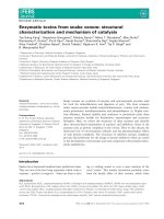
Figure 1-1: Whole genome phylogenetic tree of family Flaviviridae. This tree is
reconstructed using maximum parsimony. Color coding for arcs is as follows: Red
(Aedes borne Flaviviruses), Purple (Culex borne Flaviviruses), Blue (Tick borne
Flaviviruses), Orange (No known vector Flaviviruses), Green (Pestiviruses), Cyan
(Hepaciviruses) and Black (unassigned members of family Flaviviridae).
Reprinted by permission from BioMed Central: [BMC Bioinformatics] (Kulkarni-
Kale U, Bhosle SG, Manjari GS, Joshi M, Bansode S, and Kolaskar AS. 2006.
Curation of viral genomes: challenges, applications and the way forward. BMC
Bioinformatics 7 Suppl 5:S12.)
4
1.2.2. Structure and genetic organization of dengue virus
Previous ultra-structural studies showed that dengue virus is a 50 nm
icosahedral entity with a surprisingly smooth surface. DENV particle is made up of a
RNA core that is encapsidated by nucleo-capsid, which is wrapped by a lipid bilayer
membrane, followed by an organized outer protein shell (Zhang et al. 2003).
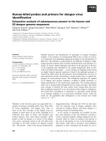
The dengue genome (as shown in figure 2) is composed of a positive single stranded
RNA of approximately 11 kb in size. It is organized into three structural viral proteins
(capsid (C), pre-membrane (prM) and envelope (E)), and seven non-structural
proteins essential for viral replication (NS1, NS2A, NS2B, NS3, NS4A, NS4B, NS5)
(Lindenbach et al. 2007).
Figure 1-2: Dengue virus genome: It is a polyprotein composed of three structural
proteins (highlighted in green) and seven non-structural proteins (highlighted in blue).
Reprinted by permission from Macmillan Publishers Ltd: [Nature Reviews
Microbiology] (Whitehead S, Blaney J, Durbin A, and Murphy B. 2007. Prospects for
a dengue virus vaccine. Nat Rev Microbiol 5(7):518-528), copyright (2007).
5
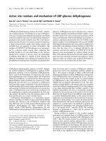
1.2.3. Viral infection cycle
The infection cycle of DENV (as depicted in Figure 3) begins with the binding
of DENV onto the host cell surface with a receptor, probably a low affinity but
abundance receptor, such as DC-SIGN (Tassaneetrithep et al. 2003). The virus is then
endocytosed into the cell via an unknown high affinity specific receptor (Lozach et al.
2005). When encountering a change in pH within the acidic environment of the late
endosome, protonation of the histidine residues of the viral envelope occurs (Mueller
et al, 2008). This triggers the E protein to undergo major conformational changes
which leads to the fusion of the viral membrane with host endosomal membrane. The
fusion event releases the nucleocapsid, containing the genetic material, into the
cytoplasm. Positive strand viral RNA is translated into a single polyprotein that is
further processed into structural and non structural viral proteins by viral and host
cellular proteases. Viral replication is initiated on the intercellular membranes near to
the host endoplasmic reticulum (ER). Newly synthesized viral proteins are assembled,
in the ER, into immature non-infectious virions. These immature virions are
subsequently transported to the trans-Golgi apparatus for processing; resulting in the
release of the mature infectious virions to the extracellular environment via the host
secretory pathway.
6
Figure 1-3: Infection cycle of dengue virus in host cell.
Reprinted from Host Cell. Cell Host & Microbe, 5(4), Fernandez-Garcia M-D,
Mazzon M, Jacobs M, and Amara, A., Pathogenesis of Flavivirus Infections: Using
and Abusing the Host Cell, p318-328, Copyright (2009), with permission from
Elsevier.
Viral entry
(Endocytosis)
Low-pH viral fusion
Viral uncoating &
genome release
Genome release
Translation of vRNA
Processing of polyprotein
Replication of ssRNA in
viral replication complex
Viral assembly
Viral maturation
Viral exit
(Exocytosis)
7
1.2.4. Viral proteins
Viral RNA is packaged inside the DENV capsid to forms the RNA core,
known as nucleocapsid. This RNA core protects the viral genome before its delivery
into the host cell cytoplasm for initiation of viral replication. After viral fusion, the
viral capsid localizes to both cytoplasm and nuclease of the host cell. The reason for
nuclear localization is still poorly understood. There were studies done suggesting the
possible roles of capsid in (i) virus-induced apoptosis, (ii) viral assembly (Khromykh
and Westaway 1996), (iii) viral morphogenesis (Samsa et al. 2009) and (iv) acting as
an antagonist of an excocyst protein (hSec3p), which is a repressor of viral replication
(Bhuvanakantham et al. 2009).
The membrane protein of DENV is initially presented as a pre-membrane
form (prM). It acts as a shield covering the fusion peptides of DENV envelope
protein, preventing it from premature fusion with the cellular membrane during the
synthesis of new viral particles. During the final step of viral assembly (known as
maturation), it is cleaved by a host protease, furin, with the “pr” peptide remains
associated with the virion in the environment of the TGN. This association keeps the
virus in a non-infectious state inside the cell. Upon exiting the cell, “pr” peptide
dissociates from the virus, making the virion infectious. This primes the mature virion
for fusion upon entry into acidic compartments of host cells.
DENV is an enveloped virus which contains a lipid bilayer that has 180 copies
of E protein and M protein embedded in it. The E proteins are arranged in an
icosahedral scaffold of 90 dimers (Kuhn et al. 2002). The fusion protein of DENV E
protein is classified as a class II fusion protein, similar to those belonging to
alphaviruses and flaviviruses. The E protein has three domains, domain I being the
central domain, flanked by a dimerization domain (domain II) on one end and
8
immunoglobulin-like domain (domain III) on the other end (Mukhopadhyay et al.
2005). The discovery of a hydrophobic pocket, occupied by a small detergent
molecule, n-octyl-β-D-glucoside (βOG), near to the hinge region of the E protein,
highlighted an attractive region for anti-viral targeting (details are to be further
discussed in later section, see chapter 1.3.3). Other important roles of E proteins
include receptor-mediated binding, neutralization and viral assembly (Chin et al.
2007; Crill and Roehrig 2001; Hiramatsu et al. 1996; Stiasny et al. 2006)
The non-structural protein, NS1, exists in both soluble and insoluble forms
(Winkler et al. 1989). During the replication event of DENV, NS1 is anchored to the
intracellular membrane of the endoplasmic reticulum, mainly as homodimers (Falgout
and Markoff 1995; Mackenzie et al. 1996), and is implicated to participate in viral
replication. It is also found to be associated with the cell surface via a GPI-anchor and
is capable of triggering signal transduction (Jacobs et al. 2000). The soluble form of
NS1 circulates in the extra-cellular compartment in the form of a hexamer (Flamand
et al. 1999). Soluble NS1 levels were found to be elevated in the blood serum of
dengue fever patients during the acute phase of the disease (Young et al. 2000). It is
believed that these circulating soluble NS1 hexamers could contribute to the
pathogenesis of the disease by reacting with the host complement system, causing
activation and the onset of host immune responses, leading to a vascular leakage
(Avirutnan et al. 2006).
NS2B-NS3 complex of DENV is an excellent example of efficient fusion of
various functional proteins into one protein which allows sequential processes to take
place in close association. NS3 has the viral protease at its N-terminal end that cleaves
the viral polyprotein together with host proteases. At the C-terminal end of NS3 is the
viral helicase with a RNA-stimulated nucleoside triphosphatase to provide the energy
9
to unwind viral RNA replication intermediates during amplification of viral RNA.
NS2B is a co-factor of NS3, acting as a putative anchor for NS3 protease to the host
membrane in order to allow efficient proteolytic activity. Other non-enzymatic
functions of flaviviral NS3 have been suggested, such as substrate recognition
(capped viral RNA) (Luo et al. 2008; Patkar and Kuhn 2008) and the recruitment of
fatty acid synthase to the site of replication for fatty acid biosynthesis, which is
necessary for viral replication (Heaton et al. 2010).
NS5 is another non-structural protein of DENV with multi-enzymatic
properties. At the N-terminus of NS5 is the viral methyltransferase, which is involved
in the methylation of the N7 and 2‟-O positions of viral RNA cap. RNA capping
stabilizes viral mRNA for efficient translation (Furuichi and Shatkin 2000; Wengler
1993). The RNA-dependent RNA polymerase (RdRp) domain is found in the C-
terminal region of NS5 and is responsible for the amplification of viral RNA
(Ackermann and Padmanabhan 2001; Tan et al. 1996; Yap et al. 2007). NS5 also has
a non-enzymatic function in modulating the host responses by binding to STAT2,
resulting in the suppression of interferon signalling involved in anti-viral response in
host (Ashour et al. 2009; Mazzon et al. 2009).
NS2A, NS4A and NS4B are non-structural proteins that are generally believed
to possess non-enzymatic functions. All have been shown to antagonize IFN
signalling with NS4B having the most potent anti-IFN activity. The function of NS4A
is still relatively elusive, although that it is known to be localized to the replication
site, and is shown to induce the formation of membranous structures similar to those
observed in infected cells (Miller et al. 2007). NS4B has been implicated in enhancing
the overall helicase activity of NS3 by causing the dissociation of NS3 from single-
stranded RNA (Umareddy et al. 2006).