A dosimetric skin study on postmastectomy breast cancer patients undergoing radiation therapy
Bạn đang xem bản rút gọn của tài liệu. Xem và tải ngay bản đầy đủ của tài liệu tại đây (2.68 MB, 148 trang )
A DOSIMETRIC SKIN STUDY ON POSTMASTECTOMY
BREAST CANCER PATIENTS UNDERGOING
RADIATION THERAPY
SHARON WONG MEI MEI
THE NATIONAL UNIVERSITY OF SINGAPORE
2011
A DOSIMETRIC SKIN STUDY ON POSTMASTECTOMY
BREAST CANCER PATIENTS UNDERGOING
RADIATION THERAPY
SHARON WONG MEI MEI
(MSc (Biomedical Science), BSc(Medical Radiation Science)
A THESIS SUBMITTED
FOR THE DEGREE OF DOCTOR OF PHILOSOPHY
DEPARTMENT OF MEDICINE
FACULTY OF MEDICINE
THE NATIONAL UNIVERSITY OF SINGAPORE
2011
DECLARATION
I hereby declare that the thesis is my original work and it has been written by me in
its entirety. I have duly acknowledged all the sources of information which have been
used in the thesis.
This thesis has also not been submitted for any degree in any university previously.
Sharon Wong Mei Mei
25 April 2012
Page | II
ACKNOWLEDGEMENTS
I would like to express my sincere thanks to my supervisor Professor Jiade Jay Lu, for
without his support, his wide range of resources, his splendid vision and keen mind;
this work would not have been possible. Professor Phan Toan Thang, my cosupervisor for supervising me in my translational research work. For helping me get
through the well-guarded door into the department of Surgery and patiently guiding
me in the planning and execution of experiments.
My sincere gratitude to the staff at the Department of Radiation Oncology (NUCIS),
Department of Medicine (NUH), Department of Surgery (NUH) and the numerous
lecturers at Faculty of Medicine (NUS) who provided me with their continuous and
encouraging support through the years of my study. Of special mention are my
collaborators and mentors who provided valuable advice and critique, Prof Bay Boon
Huat, Prof Ho Khek Yu, Dr Elaine Lim Hsuen, Dr Lee Khai Mun, Dr Michael Back,
Dr Fong Kum Weng and Dr Susan Loong. Special thanks to my beloved friends and
colleagues especially Alvin Chua, Han Hwan Chour, Ong Chee Tian, Bala
Rajaratnam, Dr Sathiyamoorthy Selvarajan and Amarjit Sardul.
And most importantly, I dedicate this thesis to my husband Royston, my two
daughters Nicole and Kylie and my son Cayden who sacrificed their days and nights
without me at home as I complete this course of study. I share with them this thesis
as an expression of my deepest love and happiness for their endless support.
Page | III
TABLE OF CONTENTS
Declaration
II
Acknowledgements
III
Table of Contents
III
Publications derived from this thesis
X
Summary
XI
List of Tables
XIV
List of Figures
XVI
Chapter 1
1.1.
Chapter 2
Introduction
Aims and objectives of this thesis
1
9
Overview of Breast Cancer
11
11
2.1.2. Lymphatic Drainage
11
2.1.3. Local Involvements
12
2.1.4. Recurrence in the skin
2.2.
10
2.1.1. Anatomy of the breast
2.1.
Background
12
Role of Radiation Therapy
13
2.2.1. Postmastectomy Radiation Therapy
14
2.2.2
15
Use of postmastectomy radiation therapy with
systemic therapy
2.2.3. Radiation Therapy Techniques after mastectomy
17
Page | IV
2.2.4. Definition of Clinical target volume
2.2.5
2.3.
18
20
Complications of Radiation Therapy
20
2.3.1. Acute skin reactions
21
2.3.2. Chronic skin reactions
22
2.3.3. Dose Distribution in skin
23
Radiation Physics on Production of X-rays
24
2.4.1. Characteristic Radiation
25
2.4.2. Bremsstrahlung
26
2.4.3. Dose computation using 3D Monte Carlo Radiotherapy
2.4.
Response of the skin to radiation dose
27
Treatment Planning System Algorithms
Chapter 3
A dosimetric study on the use of radiation therapy
28
treatment planning system to predict for surface doses
in postmastectomy radiation therapy patients
3.1.
Introduction
29
3.2.
Materials and method
31
3.2.1. Construction of mastectomy phantom
31
3.2.2. Thermoluminescence dosimeter placements and invivo
33
dosimetry
3.2.3. Thermoluminescence dosimeters (TLD)
36
3.2.4. Treatment Planning System
36
3.2.5. Statistical analysis
37
Page | V
3.3.
Results
37
3.3.1. Surface doses between absorbed doses (TLD) and
calculated doses (TPS)
38
3.3.2. Entrance dose at the build-up region between absorbed
41
Doses (TLD) and calculated doses (TPS)
3.4.
Discussion
44
3.5.
Conclusion and future direction
48
3.6.
References
50
An ultrasonographic evaluation of skin thickness in
54
Chapter 4
breast cancer patients after postmastectomy radiation therapy
4.1.
Introduction
55
4.2.
Methods and Materials
58
4.2.1. Patients selection
58
4.2.2. Postmastectomy breast radiation therapy
59
4.2.3. Ultrasound measurements
60
4.2.4. Statistical analysis
64
Results
65
4.3.
4.3.1. Skin thickness on the mastectomy side with radiation in
comparison to the non-irradiated breast
4.3.2
65
Correlations of acute skin scoring (RTOG) and
66
FibroticThickness
4.4.
Discussion
67
Page | VI
4.4.1. Correlations of Acute skin scoring (RTOG) and fibrotic skin
thickness
4.5.
71
76
4.5.2. Authors’ contributions
76
4.5.3. Acknowledgement
76
4.5.4. Patient’s consent
Chapter 5
75
4.5.1. Competing interest
4.6.
Conclusion
76
References
77
Epidermal keratinocytes death and expression of marker proteins
of apoptosis in human skin after ionizing radiation exposure
81
5.1.
Introduction
82
5.2.
Materials and Methods
83
5.2.1. Biopsies and Irradiation
83
5.2.2. Measurement of Radiation Induced Apoptotic
85
Keratinocytes
5.2.2.1.
Hematoxylin and Eosin (H&E) staining
5.2.3.
TUNEL assay
85
85
5.2.3. Immunohistochemistry (IHC) Analysis of Apoptotic
Biomarkers
85
5.2.3.1.
Histological Procedures
85
5.2.3.2.
Histolopathological Evaluation
87
5.2.5. Western Blot Analysis of Apoptotic Biomarkers
88
Page | VII
5.2.5.1.
Protein extraction from tissue culture cells
88
5.2.5.2.
Determination of protein concentrations
89
5.2.5.3.
SDS-polyacrylamide gel electrophoresis
90
5.2.5.4.
Protein electroblotting
91
5.2.5.5.
Blocking, antibody incubation, washing and
stripping
92
5.2.6. Computerized gel densitometry
5.2.7. Statistical Analysis
5.3.
93
93
Results
94
5.3.1. Radiation induced apoptotic keratinocyte cell death
is dose and fraction size dependent
94
5.3.1.1
Increased Apoptotic Keratinocyte cell count 94
5.3.1.2.
Morphological changes of radiation induced
apoptotic keratinocytes
95
5.3.2. Expression of apoptosis related protein markers with
increasing radiation dose
5.3.2.1.
97
Accumulation of PCNA, p21 and p53
Proteins with increasing dose
97
5.3.3. Morphological changes of PCNA, p21 and p53 with
increasing dose
101
5.3.4. Accumulation of, PCNA, p21and p53 proteins with
increasing fraction size
102
Page | VIII
5.3.5. Western Blot Analysis revealed elevated levels of PCNA
and p21 proteins in irradiated keratinocytes and this is
dose and fraction size dependent
107
5.4.
Discussion
109
5.5.
Conclusion
116
5.6.
References
118
Bibliography
122
Appendices
130
Page | IX
PUBLICATIONS DERIVED FROM THIS THESIS
1.
S. Wong, M. Back, W.P.Tan, K.M. Lee, S. Baggarley, J.J. Lu.
Can Radiation Therapy Treatment Planning System accurately predict for
Surface doses in Postmastectomy Radiation Therapy Patients?
Medical Dosimetry 2011;5762
2.
S. Wong, A. Kaur, M. Back, K.M. Lee, S. Baggarley, J.J. Lu.
An Ultrasonographic evaluation of skin thickness in breast cancer patients
after undergoing postmastectomy radiation therapy.
Radiation Oncology 2011; 6:9
3.
S. Wong, H.H. Chor, M. Sathiya, C.T. Ong, T.T. Phan, J.J. Lu. Human
epidermal keratinocytes death and expression of protein markers of apoptosis
after ionizing radiation exposure.
Submitted to Radiation Oncology
Page | X
SUMMARY
Postmastectomy radiation therapy (PMRT) has been proven to decrease locoregional
recurrence and increase survival for women with large tumors and/or node-positive
disease. However, the clinical benefit of radiotherapy in the treatment of breast cancer
must be balanced against the documented risk for early and late toxicity. Adverse
effects after breast irradiation have been reported in a range of organs with the skin
being the most commonly affected during breast cancer radiation. The focus of this
thesis is to evaluate the skin reactions of postmastectomy breast cancer patients
undergoing radiation therapy treatments and its dosimetric effects. The proteomic
response of human skin cells after exposure to PMRT regimens and the expression of
apoptotic biomarkers that reflect cell death or biology using multiplexed
immunoassays have also been studied in depth.
Accurate assessments of skin doses in PMRT are important to ensure sufficient dose
to the surface target volume without excessive skin reaction. In our first study, we
assessed the accuracy of surface dose calculation by a clinically used 3D treatment
planning system (TPS) and those measured by Thermoluminescence dosimeters
(TLDs) in a customized chest wall phantom. Dose accuracy of up to 2.21% was
found. The deviations from the calculated absorbed doses were overall larger when
wedges and bolus were used. These findings suggest that 3D TPS is a useful and
accurate tool to assess the accuracy of surface dose and that radiation treatment
Page | XI
accuracy can be accurately predicted for tangential treatment of the chest wall after
mastectomy.
Skin reaction is the most common side effects during breast cancer irradiation.
Unfortunately, current clinical assessment of radiation induced skin changes is
generally limited to clinician-based rating scales, which are usually not sufficient for
quantitative and objective evaluations. In our study, we determined the usefulness of
ultrasonography in the assessment of post radiotherapy skin changes in PMRT breast
cancer patients. Our results demonstrated statistical significant difference between
the skin thickness of irradiated chestwall and the contral lateral non-irradiated breast
and a predisposition to severe chronic reactions was found in patients with RTOG
scoring of grade 1 and grade 2.
Knowledge of the pathophysiology of the irradiated skin is important to understand
the tolerance and cosmetic response of the human skin to radiation. Unfortunately the
cellular radiation response of the skin to different radiation therapy treatment regimen
has never been studied. The practice of radiotherapy would also greatly benefit from
the discovery of biomarkers that correlate with symptoms and side effects pertaining
to tissues within the irradiated volume.
We investigated the radiation induced
apoptotic cell death and apoptotic proteins expression in human skin after exposure to
PMRT regimens. There is strong evidence of cellular damaged and accumulation of
apoptotic proteins caused by ionizing radiation and these are radiation dosage and
fraction size dependent.
Page | XII
In summary, results derived from this thesis have demonstrated that radiation induced
skin reaction in PMRT breast cancer patients can be accurately predicted using
image-based technology and multiplexed immunoassays. Taken together, it is
conceivable that in the near future these measures will be used to monitor therapeutic
response and predict local control and toxicity to Radiation Therapy.
Page | XIII
LIST OF TABLES
Table No.
Table 1.1
Description
Ten most frequent cancers in Singapore females (%) between 20052009. Singapore Cancer Registry Interim Report, Trends in Cancer
Incidence in Singapore 2005-2009
Table 1.2
Ten Most Frequent Cancer Deaths in Singapore Females,
2005-
2009. Singapore Cancer Registry Report No. 7
Table 3.1
The mean surface dose measurements of all 7 positions as measured
in the customised mastectomy breast phantom
Table 3.2
The mean entrance dose (buildup region) measurements of all 7
positions as measured in the customised mastectomy breast phantom
Table 4.1
Relevant Equipment Settings for the ‘Breast Detail’ preset
Table 4.2
Reduced mean skin thickness on the Right mastectomy side with
radiation in comparison to the Left non-irradiated breast
Table 4.3
Reduced mean skin thickness on the Left mastectomy side with
radiation in comparison to the Right non-irradiated breast
Table 4.4
Skin thickness of points marked on the medial and lateral side
Table 4.5
RTOG Scoring Criteria for Acute Radiation Skin Reactions
Table 4.6
Reduced mean skin thickness in patients with grade 2 acute skin
reactions
Page | XIV
LIST OF TABLES
Table No.
Description
Table 5.1
Experimental design
Table 5.2
List of antibodies used in immunohistochemistry procedures
Table 5.3
The scale for staining intensity and pattern scoring
Table 5.4
Composition of stacking and separation gels for electrophoresis
Table 5.5
Composition of self-prepared reagents for western blotting
Table 5.6
List of antibodies used in Western Blot Analysis
Table 5.7
Percent of radiation induced apoptotic cells from H&E and TUNEL
staining
Table 5.8
Correlations between different radiation fractionation and TUNEL
staining
Table 5.9
Apoptotic protein expression in different radiation dosage starting
from 2 – 50Gy
Table 5.10
Correlations between different radiation fractionation and PCNA
expression
Table 5.11
Correlations between different radiation fractionation and p21
expression
Page | XV
LIST OF FIGURES
Figure No.
Figure 1.1
Description
Ten most frequent cancers in females in Singapore between
2002-
2006. Singapore Cancer Registry Interim Report, Trends in Cancer
Incidence in Singapore 2005-2009
Figure 1.2
Breast cancers in Singapore: incidence and age-standardized
incidence rate 1968 – 2002. Singapore Cancer Registry Report No. 7
Figure 2.1
Treatment marks on chest wall
Figure 2.2
A tangential breast setup showing (a) medial and (b) lateral tangent
fields
Figure 2.3
Treatment marks drawn on patient
Figure 2.4
Examples of central-axis depth-dose curves for (A) photon beams
and (B) electron beams of various energies used in external beam
radiation therapy
Figure 2.5
The interaction of an incoming electron with an inner orbital electron
Figure 2.6
The emission of characteristic X-ray
Figure 3.1
Chest wall thickness and lung measured using CT transversal slice
Figure 3.2
A customised chestwall phantom using wax and cord material
Figure 3.3
Display of TLD positions on the mastectomy phantom
Figure 3.4
Schematic representation of the TLD positions
Page | XVI
LIST OF FIGURES
Figure No.
Figure 3.5
Description
Schematic representation of the TLD positions on a phantom with 1cm
Bolus
Figure 3.6
TLD vs TPS surface dose (buildup) with 1cm bolus
Figure 3.7
TLD vs TPS surface dose (buildup) with wedge
Figure 3.8
TLD vs TPS surface dose (buildup) with 1cm bolus and wedge
Figure 3.9
TLD vs TPS entrance dose (dmax) with 1cm bolus
Figure 3.10
TLD vs TPS entrance dose (dmax) with wedge
Figure 3.11
TLD vs TPS entrance (dose (dmax) with 1cm bolus and wedge
Figure 4.1
Treatment marks on the patient
Figure 4.2
Representation of points on the chest wall and contra-lateral breast
Figure 4.3
Points marked on the chest wall and contra-lateral breast prior to
ultrasound
Figure 4.4
Scar showing shadowing
Figure 4.5
Transducer resting on the thick layer of gel on the skin
Figure 4.6
Echogenic border between the skin and the subcutaneous tissue
Page | XVII
LIST OF FIGURES
Figure No.
Description
Figure 4.7
Erythema and dry desquamation seen in Grade 1
Figure 5.1
Transfer stack
Figure 5.2
Morphological evaluation
Figure 5.3
Morphological changes in cell after irradiation
Figure 5.4
HE slide showing a detached epidermis layer due to radiation
Figure 5.5
The boxplot for PCNA immunoreactitivity in different radiation
dosage starting from 2Gy to 50Gy
Figure 5.6
The boxplot for p21 immunoreactitivity in different radiation dosage
starting from 2Gy to 50Gy
Figure 5.7
The boxplot for p53 immunoreactitivity in different radiation dosage
starting from 2Gy to 50Gy
Figure 5.8
Dose series of Irradiated skin stained with monoclonal anti-PCNA
Figure 5.9
Dose series of Irradiated skin stained with monoclonal anti-p21
Figure 5.10
Dose series of Irradiated skin stained with monoclonal anti-p53
Figure 5.11
Boxplot for PCNA at 10Gy delivered to human skin at different
fractionation
Figure 5.12
Boxplot for PCNA at 30Gy delivered to human skin at different
fractionation
Page | XVIII
LIST OF FIGURES
Figure No.
Figure 5.13
Description
Boxplot for PCNA at 50Gy delivered to human skin at different
fractionation
Figure 5.14
Boxplot for P21 at 10Gy delivered to human skin at different
fractionation
Figure 5.15
Boxplot for P21 at 30Gy delivered to human skin at different
fractionation
Figure 5.16
Boxplot for P21 at 50Gy delivered to human skin at different
fractionation
Figure 5.17
Little or no immunoreactive band specific for p53 was observed at
dose above 10Gy
Figure 5.18
Western blot analysis of p21 protein with increasing radiation dosage
Figure 5.19
Western blot analysis of PCNA protein with increasing radiation
dosage
Figure 5.20
Western blot analysis of P21 reveals a higher level of protein with
increasing fraction size at 10Gy
Figure 5.21
Western blot analysis of PCNA reveals a higher level of protein with
increasing fraction size at 10Gy
Page | XIX
LIST OF FIGURES
Figure No.
Figure 5.22
Description
Western blot analysis of P21 reveals a higher level of protein with
increasing fraction size at 50Gy
Figure 5.23
Western blot analysis of PCNA reveals a higher level of protein with
increasing fraction size at 50Gy
Figure 5.24
Radiation Induced Injury
Page | XX
Chapter One - Introduction
CHAPTER ONE
INTRODUCTION
Page | 1
Chapter One - Introduction
INTRODUCTION
Breast cancer is one of the most common cancers amongst women. Its incidence in
Singapore has risen significantly over the last two decades [Chia et al 2002] and is
expected to continue to rise sharply through the years. Published results by Chia et
al. 2002, demonstrated that the age standardized incidence rates in Singapore
increased from 46.1 to 53.1 cases per 100,000 persons per year from 1998 – 2002. A
separate report taken from the Singapore Cancer Registry showed a similar increasing
trend for breast cancer in Singapore females between 2005 and 2009 (Table 1.1 &
Figure 1.1). Singapore has one of the highest age-adjusted breast cancer incidences
in Asia with increasing incidence in women in their 50’s [J Tey 2008].
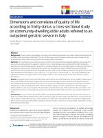
Table 1.1 Ten most frequent cancers in Singapore females (%) between 2005-2009.
Singapore Cancer Registry Interim Report, Trends in Cancer Incidence in Singapore
2005-2009.
Page | 2
Chapter One - Introduction
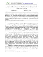
Figure 1.1 Ten most frequent cancers in Singapore females (%) between 2005-2009.
Singapore Cancer Registry Interim Report, Trends in Cancer Incidence in Singapore
2005-2009.
Figure 1.2 Breast cancer in Singapore: incidence and age-standardized incidence rate
1968 – 2007. Singapore Cancer Registry Report No. 7
Page | 3
Chapter One - Introduction
Breast cancer is also the leading cause of death in Singapore women (Table 1.2 Singapore Cancer Registry Interim Report, Trends in Cancer Incidence in Singapore
2005-2009). Published results by Jara-Lazaro et al 2010, demonstrated that about
1,100 new cases are diagnosed annually and approximately 270 women die in
Singapore each year from breast cancer, translating to breast cancer diagnoses in
about three women daily, with approximately three cancer deaths every four days.
Table 1.2 Ten Most Frequent Cancer Deaths in Singapore Females, 2005-2009.
Singapore Cancer Registry Report No. 7
Radiotherapy has played an important role in the treatment of breast cancer. It is
routinely employed in breast conservation therapy. Its role as adjuvant therapy in
selected patients undergoing mastectomy for stages I and II disease is evolving, and it
has become an essential component of the combined modality approach for stage III
disease.
Page | 4