Claudin 1 is the direct target of RUNX3 in gastric epithelial cells
Bạn đang xem bản rút gọn của tài liệu. Xem và tải ngay bản đầy đủ của tài liệu tại đây (1.96 MB, 146 trang )
CLAUDIN-1 IS THE DIRECT TARGET OF RUNX3 IN
GASTRIC EPITHELIAL CELLS
CHANG TI LING
NATIONAL UNIVERSITY OF SINGAPORE
2009
CLAUDIN-1 IS THE DIRECT TARGET OF RUNX3 IN
GASTRIC EPITHELIAL CELLS
CHANG TI LING
(M.Sc., National University of Malaysia)
A THESIS SUBMITTED
FOR THE DEGREE OF DOCTOR OF PHILOSOPHY
DEPARTMENT OF MEDICINE
NATIONAL UNIVERSITY OF SINGAPORE
2009
i
ACKNOWLEDGEMENTS
My gratitude goes to both my supervisors, Prof. Yoshiaki Ito and A/P Kosei Ito for
their patient guidance and discussions throughout my PhD study. I thank them for
critically reviewing my work during our progress report sessions, as well as their
useful advice for improvements.
I would also like to specially thank my ex-supervisor, A/P Evelyn Koay, the Director
of Molecular Diagnosis Centre, NUH for her constant support and encouragement.
Without her nurturing and many opportunities given, I will not be where I am today.
My gratitude also goes to the Singapore Millennium Foundation for providing me the
SMF scholarship during the first three years of my PhD project and the sponsored trip
to the Gordon Research Conferences, as well as to the NUS for my final year of
scholarship. I also thank the Oncology Research Institute common grant for
supporting my PhD work throughout.
My sincere appreciation also goes to all my fellow colleagues and friends at ORI
(Tomoko, Tada-san, TK, Angela, KumChew, PeiYi, FenYi, BeeKeow, Michelle,
Kathryn, Dr. LihWen, Dr. Vidya, Baidah, Diyanah, Erna, Judy, Mei Xian and
ShenKiat) and IMCB RUNX group (Cecilia, Dominic, Anthony, Dr. Osato,
YungKiang, Eunice, Ida, Yano and Ken-ichi) for their kind assistance and
constructive advice along my PhD journey. I thank them for their friendship. Many
thanks also to the ORI administrative team, Selena Gan, Deborah, Ivy, Alexis and
Siew Hong for their kind help.
Last but not least, I would like to specially thank my beloved husband, Andy Yip for
his loving care, trust, sacrifice and constant moral support and encouragement to
make my PhD journey a possible one. I thank God for our baby Jasper who brought
much joy and hope towards the end of my PhD project. Endless gratitude also goes to
ii
my beloved parents, family members, cell members (Doris, BoonLay & FeeYoon,
Selena & Willie, PohChoo, Anna & Charles, SiorPeng & ChekLeong, Winnie &
ShenKong) and friends (Hazel, Eileen, HueyFen, HongLing, Maisy, Tony and
JennHui) for their patient support, understanding, constant encouragement and
prayers.
To God be the Glory! He is my pillar of support in times of troubles.
Chang Ti Ling
January 2009
iii
TABLE OF CONTENTS
Page
ACKNOWLEDGEMENTS i
TABLE OF CONTENTS iii
SUMMARY vii
LIST OF TABLES ix
LIST OF FIGURES x
LIST OF ABBREVATIONS xii
CHAPTER 1 OBJECTIVE 1
CHAPTER 2 INTRODUCTION 2
2.1 Gastric Cancer 2
2.1.1 Genetics of Gastric Cancer 3
2.2 RUNX Protein Family 5
2.2.1 Nomenclature of RUNX 7
2.2.2 Evolutionary Conservation of RUNX 9
2.3 Role of RUNX Protein Family 9
2.3.1 RUNX1 9
2.3.2 RUNX2 11
2.3.3 RUNX3 11
2.4. RUNX and TGF-β Tumor Suppressor Pathway 12
2.5 RUNX3 and Gastric Cancer 15
2.5.1 Tumor Suppressive Mechanism of RUNX3 in 19
Gastric Cancer
2.6 Tight junction (TJ) Protein Family 21
2.7 Claudin Superfamily 23
2.7.1 Emergence of Claudin Superfamily 23
2.7.2 Evolution of Claudin Genes Family 24
iv
2.7.3 Claudin Protein Structure and Functions 25
2.8 Claudin and Cancer 29
2.8.1 Claudin and Gastric Cancer 30
2.9 Crosstalk of TJ Components with Signaling Pathways 31
2.10 TJ, AJ and Mechanism in Tumor Metastasis 33
CHAPTER 3 MATERIALS AND METHODS
3.1 MATERIALS 39
3.1.1 Primers 39
3.1.2 Oligonucleotide probes for EMSA 42
3.1.3 Commercial kit 43
3.1.4 Antibodies 44
3.1.5 General Buffer Preparation 45
3.2 METHODS
3.2.1 Establishment of Gastric Epithelial Cell Lines 49
3.2.2 Cell Lines and Cell Culture 51
3.2.2.1 Treatment of Cells by TGF-β1 52
3.2.3 Semiquantitative RT-PCR, Quantitative RT-PCR 52
3.2.4 Protein Isolation 53
3.2.5 SDS-PAGE and Western Blot Analysis 54
3.2.6 Promoter Assay 55
3.2.6.1 Cloning of hclaudin-1 Promoter 55
3.2.6.2 Site-Directed Mutagenesis 56
3.2.6.3 Dual-Luciferase Reporter Assay 58
3.2.7 Generation of Stable Cell Line 60
3.2.7.1 Plasmids and Stable Cell Line 60
3.2.7.2 Stable Transfection and Stable Cloning 61
v
3.2.8 Xenografts in Nude Mice 62
3.2.9 Collection and Processing of Mouse and Human 64
Tissue Samples
3.2.9.1 Fixing, Processing and Embedding of Mouse 64
Stomach
3.2.9.2 Human Gastric Cancer Specimens 64
3.2.10 Microscopy Technique 65
3.2.10.1 Immunocytochemistry (IF) 65
3.2.10.2 Immunohistochemistry (IHC)
66
3.2.11 Electrophoresis Mobility Shift Assay (EMSA) 67
3.2.12 Chromation Immunoprecipitation (ChIP) 69
CHAPTER 4 RESULTS AND DISCUSSIONS
4.1 Results
4.1.1 Expression of TJ Proteins in Mouse 71
Gastric Epithelial Cells
4.1.2 TJ Proteins and TGF-β Pathway 73
4.1.3 RUNX3 and claudin-1 in TGF- β Pathway 74
4.1.4 claudin-1 Expression in Mouse Gastric Epithelial Cells 77
4.1.5 claudin-1 Promoter Assay 79
4.1.6 Electrophoresis Mobility Shift Assay (EMSA) 84
4.1.7 Chromatin Immunoprecipitation (ChIP) 86
4.1.8 Nude Mice Assay 88
4.1.8.1 Restoration of claudin-1 Expression and Its Tumor 88
Suppressive Effect
4.1.8.2 Reduced claudin-1Expression Enhances 92
vi
Tumorigenicity
4.1.9 Claudin-1 and RUNX3 Expression in Human Gastric 92
Cancer Samples
4.2 Discussions 99
CHAPTER 5 CONCLUSIONS AND FUTURE PERSPECTIVE
104
REFERENCES 106
APPENDICES
1 Genomic Structure of Runx3, Structure of The Targeting 125
Vector For Homologous Recombination, and The Gene
Structure of The Targeted Locus
2 Full Length Sequence of hclaudin-1 Promoter (1530 bp) 126
3 pGL3 Vector for Cloning of Promoter 127
4 pRLSV40 Vector 128
5 Human RUNX3 cDNA 129
6 Full Length Sequence of mclaudin-1 Open Reading Frame (636 bp) 130
7 Full Length Sequence of hclaudin-1 Open Reading Frame (636 bp) 131
vii
SUMMARY
The genes controlling cell-cell contact and cellular polarity are known to be heavily
involved in cancer progression. Tumorigenic mouse GIF cells isolated from
Runx3
-/-
gastric epithelium attached weakly to each other and did not form glandular structures
on collagen gels as previously reported, suggesting that cellular polarity could not be
established in the
Runx3
-/-
cells. In a search for
RUNX
3 target genes functioning in
gastric carcinogenesis,
claudin-1
, a gene from the tight junction protein family which
functions in cell-cell contact and cellular polarity was found to be expressed at high
level in
Runx3
+/+
mouse gastric epithelial cells, but at very low level in
Runx3
-/-
ones.
In human gastric cancer cell line, SNU16, RUNX3 is expressed in the
cytoplasm in an inactive form and, upon treatment of cells by TGF-β, RUNX3
translocates into the nucleus and functions as a tumor suppressor. In SNU16, claudin-
1 is expressed after the treatment of cells with TGF-β. The TGF-β-dependent
expression of claudin-1, however, was not observed in
RUNX3
-knocked-down
SNU16 cells. Furthermore,
hclaudin-1
promoter activity was dose-dependently up-
regulated by expression of
RUNX3
. Chromatin immunoprecipitation assay showed
that RUNX3 is bound to the cognate RUNX3 binding site in the promoter region of
hclaudin-1
.
SNU16 cells express claudin-1 and knock-down of claudin-1 expression
enhanced tumorigenicity in nude mice. Furthermore, the tumorigenicity of
Runx3
-/-
GIF clones stably expressing claudin-1 was significantly less than parental cell lines.
viii
Altogether, these results showed that claudin-1 has a tumor suppressor activity in
gastric epithelial cells. Consistent with these observations, expression of claudin-1
and RUNX3 expression were found to be correlated in the human gastric cancer
specimens.
For the first time, claudin-1 was identified as a novel downstream target of
RUNX3 in the TGF-β pathway. Strong evidence showed that RUNX3 transcriptionally
regulates the expression of claudin-1. Since claudin-1 exhibits tumor suppressive
activity, a part of tumor suppressor activity of RUNX3 is likely to be mediated by
claudin-1.
ix
LIST OF TABLES
Table Page
2.1 The mammalian RUNX genes synonyms and their locus 8
2.2 Claudin expression in human cancer 29
4.1 RUNX3 vs claudin-1 expression in 52 gastric cancers 97
4.2 RUNX3 vs claudin-1 expression in differentiated and 98
diffuse type of gastric cancers
x
LIST OF FIGURES
Figures Page
2.1 Crystal structure of the Runt domain heterodimerized 7
with PEBP2/CBF bound to DNA
2.2 The TGF-β tumor suppressor pathway 14
2.3 Characteristic of mouse gastric epithelial cell lines in vitro 18
2.4 Claudin-based tight junctions in simple epithelia 27
2.5 Model of claudin-based TJ and its alteration during 34
2.6 Schematic representation of the proposed TGF-β1 36
signaling mechanism that promotes EMT
2.7 Putative NCAM-associated signaling changes during EMT 38
cancer progression
3.1 Schematic diagram showing the generation of Runx3+/+p53-/- 50
and Runx3-/-p53-/- cell lines
3.2 Overview of QuikChange XL site-directed mutagenesis 57
procedure
3.3 Format of the Dual-Luciferase Reporter Assay 59
3.4 Nude mice assay in Runx3-/- GIF and SNU16 cell lines 63
4.1 Western blot analysis of tight junction (TJ) and adhesion 72
junction (AJ) proteins in Runx3-/- and Runx3+/+
mouse gastric epithelial GIF cell lines
4.2 TGF-β treatment and expression of tight junction 75
and adhesion junction proteins
4.3 Regulation of claudin-1 by TGF-β 76
4.4 Quantitative RT-PCR analysis of claudin-1 expression 77
upon induction by RUNX3 in SNU16 cell line
4.5 Immunodetection of claudin-1 in mouse gastric epithelial 78
xi
cells and tissue samples
4.6 hclaudin-1 promoter 81
4.7 hclaudin-1 reporter assay in SNU16 cell line 82
4.8 hclaudin-1 reporter assay in AGS cell line 83
4.9 Electrophoretic Mobility Shift Assay (EMSA) 85
4.10 Chromatin Immunoprecipitation (ChIP) assay 87
4.11 Nude mice assay in Runx3-/- GIF5 cell line 89
4.12 Nude mice assay in Runx3-/- GIF14 cell line 90
4.13 Nude mice assay in the SNU16 human gastric cancer cells 91
4.14 Expression pattern of claudin-1 and RUNX3 in normal 94
human gastric sample
4.15 RUNX3 and claudin-1 expression pattern in differentiated 95
and diffuse type of human gastric cancers
4.16 Staining pattern of claudin-1 in the RUNX3 positive 96
and negative cases
xii
LIST OF ABBREVATIONS
AJ adheren junction
APS ammonium persulfate
AS anti-sense
bp base pair
BS binding site
CBF core-binding factor
cDNA complementary DNA
ChIP chromatin immunoprecipitation
DAPI 4',6-Diamidino-2-phenylindole dihydrochloride
DMEM Dulbecco’s modified eagle’s medium
dNTP deoxynucleotide triphosphate
EMSA electrophoresis mobility shift assay
FBS fetal bovine serum
G gauge
GAPDH glyceraldehyde-3-phosphate dehydrogenase
GI gastro-intestinal
GIF gastric Ito-Fukamachi cells
kb kilo base
kDa kilo Dalton
LDS lithium dodecyl sulfate
mAb monoclonal antibody
min minute
mRNA messenger RNA
ORF open reading frame
PBS phosphate buffered saline
PBST phosphate buffered saline with tween-20
PCR polymerase chain reaction
PEBP polyomavirus enhancer-binding protein
pmol pico mole
PVDF polyvinylidene fluoride
xiii
RT room temperature
S sense
SDM site-directed mutagenesis
SDS sodium dodecyl sulfate
TBE Tris, Boric Acid, EDTA
TJ tight junction
TEMED N,N,N,N-Tetramethyl-Ethylenediamine
Tgf-β transforming growth factor-beta
V volt
wt wild type
ZO zonula occluden
1
CHAPTER 1 OBJECTIVE
The aim of the study is to understand the full potential of RUNX3 as a tumor
suppressor. Since RUNX3 is a transcription factor, the identification of the target
genes that are induced or suppressed by RUNX3 should provide important insights
into the molecular mechanism behind its tumor suppressor activity. Since RUNX3 is a
nuclear effecter of TGF-β pathway and TGF-β pathway is considered to be a tumor
suppressor pathway, it is important to identify TGF-β dependent, RUNX3 mediated
target genes.
Based on a previous observation that the loss of RUNX3 affects cell-cell
contact and polarity, I would like to examine whether RUNX3 regulates genes involve
in this function and its possible link to gastric carcinogenesis. Previously, it was
observed that claudin-1, a member of the tight junction protein family exhibited a
similar knockout phenotype as those of Runx3
-/-
. As RUNX3 functions as a tumor
suppressor under the TGF-β pathway, it would be feasible to observe if tight junction
genes are regulated under this pathway in the gastric epithelial cells, which is an area
yet to be examined. Findings of possible connections between RUNX3 and tight
junction proteins in the gastric system may enable the development of useful
molecular markers for diagnostic purposes.
2
CHAPTER 2 INTRODUCTION
2.1 Gastric Cancer
With the steady decline in the incidence of gastric cancer worldwide, it has become
the fourth most common cancer, after cancers of the lung, breast, and colorectal. This
is in comparison to its ranking as the second most common cancer worldwide in the
past (1). However, gastric cancer remains a major public health problem as it remains
the second most common type of fatal cancer worldwide (1, 2). There has been little
improvement in survival as gastric cancer is too often diagnosed at an advanced stage,
despite the extensive diagnostic and therapeutic investigations of gastric cancer.
Although gastric cancer frequencies have been clearly linked to environmental
factors, such as Helicobacter pylori infection and diet, the genetic basis for gastric
cancer development is still largely unclear. It is evident that transformation of a
normal epithelial cell to a malignant cell is a result from the accumulation of several
gene abnormalities which involves multiple steps. Correa postulated a model
involving histomorphological changes that leads to gastric cancer (3). In this model,
development of chronic gastritis, atrophy, intestinal metaplasia and eventually
dysplasia results in gastric cancer. However, this model is still open to debate as it
remains unclear whether these changes follow each other step by step, or whether
some histomorphological changes directly precede gastric cancer development (3, 4).
3
2.1.1 Genetics of Gastric Cancer
Over the years, various genetic and epigenetic alterations have been associated with
the development and progression of gastric cancer. These include microsatellite
instability (MSI), reactivation of telomerase, inactivation of tumor suppressor genes
and activation of oncogenes.
Due to DNA mismatch repair deficiency, replication errors in simple
repetitive microsatellite sequences may occur which is defined as MSI. MSI can be
classified as high-frequency (MSI-H), low-frequency (MSI-L), or stable (MSS). MSI
has been recognized as one of the earliest changes in carcinogenesis, resulting in
genomic instability. It was discovered that gastric cancer cases with MSI-H often
show hypermethylation of CpG islands in the promoter region of the hMlh1 gene
which is associated with decreased hMlh1 protein expression. This indicates that
epigenetic inactivation of hMlh1 due to promoter methylation could be the underlying
cause of MSI (5, 6). A subset of gastric cancers including gastric tumors was found to
harbor MSI (7, 8). Various genes involved in the regulation of cell-cycle progression
and apoptotic signaling that have been found to be specifically altered in gastric
cancer displaying MSI include BAX, hMSH3, hMSH6, E2F-4, TGF-β receptor II, and
insulin-like growth factor receptor II (7). In view of this, MSI has been suggested as a
genetic marker for the development of multiple gastric cancers (9).
4
Tumor suppressor genes and oncogenes play a general role in regulating the
developmental and differentiation processes. Deregulation of these genes enables the
development of cancer. Inactivation of tumor suppressor genes is frequently
discovered in gastric carcinogenesis (10). It is known that at least two independent
‘hits’ are required to fully inactivate a tumor suppressor. The attention in cancer
research has typically been on two of the mechanisms that inactivate tumor
suppressors, namely loss of heterozygosity (LOH) or homozygous deletion. LOH in
chromosomal loci such as 1p, 2q, 4p, 5q, 6p, 7q, 11q, 12q, 14q, 17p, 18q and 21q
have been discovered in differentiated type of gastric cancer (11-14). However, it
remained unclear which are the genes that are specifically involved in gastric
carcinogenesis. It was then found that DNA methylation is also a powerful
mechanism that suppresses gene transcription, which represents an alternative
mechanism of tumor suppressor inactivation in cancer. Genes that are inactivated by
DNA methylation include RB, the von Hippel-Lindau gene (VHL), CDKN2A
(p16
INK4A
), CDKN2B (p15
INK4B
), E-cadherin (E-cad), hMLH1, APC, RASSF1 and
caspase-8 (15-17).
The group of activated oncogenes consists primarily of various growth factors
and growth factor receptors. c-met, a proto-oncogene which encodes a tyrosine kinase
receptor for the hepatocyte growth factor, was overexpressed in 50% of diffuse and
intestinal-type of gastric cancers. Tumors overexpressing c-met also display increased
invasiveness and are poorly differentiated (18). The overexpression of c-erB2 (HER-
2/neu) gene, a proto-oncogene and a transmembrane tyrosine kinase receptor was also
5
found to associate with approximately one-fourth of all gastrointestinal tract
malignancies (19), and has been implicated as a potential marker for the prognosis in
gastric cancers (20). Oncogenes, such as cyclin E and and c-myc were also discovered
to be amplified and overexpressed in gastric carcinoma (21, 22).
Though numerous genetic abnormalities associated with gastric cancer have
been described, they were either associated with a limited number of cases or were
still poorly understood. The molecular mechanisms underlying the pathogenesis of
gastric cancer have only been characterized recently with the discovery of RUNX3
and its role in gastric carcinogenesis. The reason why RUNX3 was not identified as a
tumor suppressor earlier could be because this gene is inactivated mainly by
epigenetic modification and genetic alteration of both alleles is very rare. RUNX3
appears to be a new addition to the list of genes that are inactivated by DNA
methylation. Since its discovery as a strong candidate tumor suppressor in gastric
carcinogenesis (23), the underlying mechanisms of RUNX3 regulation and its
downstream target genes in gastric carcinogenesis became the subject of active
investigations.
2.2 RUNX Protein Family
RUNX genes encode the α subunits called the polyomavirus enhancer-binding protein
2 (PEBP2)α/core binding factor (CBF)α of the Runt domain transcription factors. The
α subunit heterodimerizes with the β subunits (PEBP2β/CBFβ) to form the
6
heterodimeric transcription factor, initially discovered as the PEBP2, or the CBF,
which interacts with the enhancer core of Moloney murine leukemia virus. RUNX
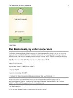
proteins alone are unstable, as they are subjected to ubiquitination followed by
proteolytic degradation by proteasome enzymes (24). Heterodimerization with the β
subunit prevents ubiquitination, thus stabilizing RUNX proteins (Figure 2.1).
However, RUNX heterodimers are relatively weakly acting transcriptional
regulators. Associations with transcriptional co-activators, such as MYB, ETS, and
p300/CBP, or co-repressors such as TLE1 and mSin3A however can induce the
potency of its transcriptional function (25). Due to the low expression level of RUNX
proteins, subcellular localization of RUNX proteins has been studied largely using
exogenously expressed RUNX proteins in fibroblasts and leukemic cells.
Immunocytochemistry shows that RUNX proteins are localized in the nucleus,
whereas exogenously expressed PEBP2β/CBFβ is in the cytoplasm.
To date, three RUNX genes, namely RUNX1, RUNX2 and RUNX3 have been
identified in mammals, whereby all three genes contain a conserved region, termed
the Runt domain (26). By comparing between the mouse PEBP2αA1 (Runx2), mouse
PEBP2αB1 (Runx1), and human PEBP2αC1 (RUNX3) as well as Drosophila Runt
proteins, PEBP2αA1 and PEBP2αC1 was found to be 93.8% identical in homology,
whereas PEBP2αB1 and PEBP2αC1 was 93.0% identical in homology. Besides, all
three Runt domain proteins also have a C-terminal end containing a unique five
amino-acid sequence, identified as VWRPY, which is 100% conserved from
7
Drosophila to human (27). This C-terminal part of the RUNX molecule plays a role
in transcription regulation (28, 29).
Figure 2.1: Crystal structure of the Runt domain heterodimerized with the 134
amino-acid region of PEBP2/CBF bound to DNA. [Ref. 23]
2.2.1 Nomenclature of RUNX
The runt-domain transcription factors RUNX1, RUNX2 and RUNX3 have previously
been assigned various designations by different laboratories. The designation acute
myelogenous leukemia (AML) factors (ie. AML1, AML2 and AML3) was generated
8
based on the genetic studies of leukemia-related chromosomal translocations. Core-
binding factor alpha (CBFA) was initially characterized as sequence specific DNA-
binding proteins that interact with the enhancers of retroviruses. PEBP2 was named
after the murine cDNAs polyoma enhancer-binding proteins. Other aliases, such as
nuclear matrix protein 2 (NMP2), osteoblast-specific complex (OBSC) and
osteoblast-specific factor 2 (OSF2) were also generated. In November 1999, the
Nomenclature Committee of the Human Genome Organization (HUGO) adopted the
use of the term ‘RUNX’ to refer to the genes encoding the runt-related proteins, also
an abbreviation for the term ‘runt-related protein’. The mammalian RUNX proteins
and their synonyms as well as their locus are as listed in Table 2.1. The order of the
numbers was given according to the order in which the knock-outs for each of the
mouse Runx genes were published (Runx1/Aml1 in 1996 (30, 31), Runx2/Cbfa1 in
1997 (32, 33) and Runx3/Pebp2αC in 2002 (23).
Table 2.1: The mammalian RUNX genes synonyms and their locus
(Adapted from Oncogene 2004; 23:4209-10)
RUNX1 CBFA2 AML1 PEBP2alphaB 21q22
RUNX2 CBFA1 AML3 PEBP2alphaA 6p21
RUNX3 CBFA3 AML2 PEBP2alphaC 1p36
9
2.2.2 Evolutionary Conservation of RUNX
Besides the three RUNX genes in mammals, four genes have been reported in D.
melanogaster (34), (35), (36), one in sea urchin (37), one in Xenopus (38), four in
zebra fish (39), (40), (41), and one in C. elegens (42). RUNX genes also appear to be
conserved throughout in metazoa, the most primitive organism described so far, with
the findings of RUNX homologs in basal metazoans such as starlet sea anemone
(Nematostella vectensis) (43) and sponge (Oscarella carmela) (44). This shows that
RUNX genes were highly conserved throughout the evolution, and very likely to play
an important role in the early metazoan development and evolution. Like their
counterparts in D. melanogaster and C. elegans, mammalian RUNX family
transcription factors also play important roles in cell fate determination during
development. As reported for Runx1 (30), Runx2 (32, 33) and Runx3 (23), genetic
ablation of all these genes have profound effects on development processes.
2.3 Role of RUNX Protein Family
2.3.1 RUNX1
RUNX1 is known to play a critical role in hematopoietic development and is
genetically altered in leukemia. RUNX1 is the most frequent target of chromosomal
translocations associated with human leukemia, with the TEL-AML t(12;21) fusion
10
accounting for 20% of acute lymphoblastic leukemia (ALL) cases and the AML-ETO
t(8;21) fusion accounting for 12% of acute myeloid leukemias (AML) (45).
Heterozygous loss-of-function mutations which cause haploinsufficiency of RUNX1
are associated with familial platelet disorder with predisposition to acute myeloid
leukemia (FPD-AML) (46). This supported the hypothetical function of RUNX1 as
tumor suppressor for AML. Besides, sporadic heterozygous mutations and point
mutations of RUNX1 are also leukemogenic (47, 48). Its association with several
autoimmune diseases, namely, systemic lupus erythematosus, rheumatoid arthritis
and psoriasis has also been reported (49), (50).
In mouse model, genetic ablation of Runx1 results in embryonic lethality and
a complete lack of fetal liver hematopoiesis (30), (31). Runx1 is also essential for the
generation of hematopoietic stem cells (HSCs) (51, 52). Runx1-deficient hematogenic
endothelial cells are incapable of producing hematopoietic stem cells (HSCs),
suppo rting its role in the initiation of the hematopoietic system. However, Runx1 is
not necessary for the maintenance of HSCs in the adult stage or expansion of
HSC/progenitor cells (HSC/Ps). Instead, lack of Runx1 induces myeloproliferative
disease and T-cell lymphoma. It was thus suggested that Runx1 plays a role as a
global regulator of hematopoiesis, from initiation to terminal differentiation.