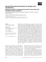
The generation of native human monoclonal antibodies with neutralising activity for dengue virus 2
Bạn đang xem bản rút gọn của tài liệu. Xem và tải ngay bản đầy đủ của tài liệu tại đây (204.6 KB, 17 trang )
i
Acknowledgements
First and foremost I offer my sincerest gratitude to my supervisor, Dr. Paul A.
MacAry, who has supported me throughout my thesis with his patience and
knowledge whilst allowing me the room to work in my own way. I attribute the
level of my Doctor of Philosophy degree to his encouragement and effort and
without him this thesis, too, would not have been completed or written. One
simply could not wish for a better or friendlier supervisor.
Next, I will like to thank my co-supervisor, Professor Ng Mah Lee, Mary for
offering so much advice and insight throughout my work on Dengue. She,
together with Mdm Boon had smoothen the path of my PhD journey by providing
me with the necessary materials and skills that are crucial for building a strong
foundation at the beginning.
In my daily work I have been blessed with a friendly and cheerful group of fellow
colleagues who will not hesitate to lend a helping hand. Teo En Wei, especially,
has been a great learner, friend and confidante and I attribute part of this thesis to
her. Life at work will not be as enjoyable and interesting without her around. The
continuation of all that we had set up together and the prospect of future
discovery, I now consider them to be in her capable hands. Too Chien Tei has
provided me with her technical expertise in animal handling and antibody
production and purification. Without her, I will not have the materials vital for the
platform of my project.
In the various laboratories I have been aided for many years in various
experiments, I got to make many friends along the way who provided valuable
insight and expertise. Dr. Brendon J. Hanson and Ms. Angeline Lim Pei Chiew
from DSO National Laboratories, Dr. Wouter Schul and Mr. Andy Yip from
Norvartis Institute of Tropical Diseases, Nalini Srinivasan and Emeritus Professor
Chan Soh Ha from World Health Organisation Singapore, Professor Mike D.
Kemeny from National University of Singapore and Wang Jin from National
University Hospital all contributed to the completion of this project.
To the rest of the friends in Paul A. MacAry (PAM) laboratory, thank all of you,
for the good laughs that never fail to pull me out of the brink of insanity. I thank
my family for letting me be, especially Adrian Lee Kok Hee in supporting me in
whichever way he can. I thank my mom, Lee Geck Keng, for all the sacrifices she
had made. I also thank a special someone, Alex Liau Whatt Meng, for the moral
support and encouragement. Most importantly, I thank God for the strength to
meet the demands of a Doctor of Philosophy degree.
ii
Table of contents
Acknowledgments i
Table of contents ii
Summary viii
List of Tables x
List of Figures xi
Abbreviations xv
Chapter 1 – Introduction 1
1.1 Dengue virus 1
1.1.1 Classification 1
1.1.2 Epidemiology 3
1.1.3 Structure of dengue virions 5
1.1.4 Organization of the Flavivirus genome 6
1.1.5 Replication strategy 8
1.1.5.1 Receptor interaction 8
1.1.5.2 Viral entry and the E protein 11
1.1.5.3 Translation of the DV genome 14
1.1.5.4 Virus assembly and propagation 17
1.1.6 Phylogeny of Dengue Virus 19
1.1.7 Pathogenesis of Dengue Virus 22
1.1.8 Immune response to Dengue Virus 26
1.1.8.1 Innate immunity to DV 26
1.1.8.2 Adaptive immunity to DV 28
1.1.9 Antibody dependent enhancement 35
iii
1.2 Epstein-Barr Virus 39
1.2.1 Classification 39
1.2.2 Epidemiology 39
1.2.3 Structure of EBV virions and organization of the virus genome 40
1.2.4 EBV patterns of latency 42
1.2.5 Replication strategy 44
1.2.6 EBV latent gene products in B cell immortalization 47
1.2.6.1 EBNAs in B cell immortalization 47
1.2.6.2 LMPs in B cell immortalization 49
1.2.7 Pathogenesis of EBV 50
1.3 Somatic hypermutation during EBV-driven B cell growth 54
1.4 Dengue human monoclonal antibody therapeutics and vaccine 55
Chapter 2 – Material and methods 60
2.1 Cell lines 60
2.2 EBV virus production 60
2.3 Dengue virus production 60
2.4 Cryopreservation of cells 61
2.5 Hybridoma cultures and antibody purification 61
2.6 Ascites and antibody purification 62
2.7 Primary cell culture 62
2.8 Source of primary CD22
+
B cells 63
2.9 RNA extraction and RT-PCR for serotyping of patients 63
2.10 Cloning of B cells from Dengue virus-infected patients 64
2.10.1 Preparation of feeder layer
2.10.2 Serial dilution and immortalization of patients’ memory B cell 64
2.11 Plaque reduction neutralization assay 65
iv
2.12 Titration of virus stocks 66
2.13 Cytopathic effect assay 66
2.14 Captured ELISA for B cell screening 66
2.15 Captured ELISA for Antibody/Fab binding 67
2.16 RNA extraction and cDNA amplification for antibody genes 67
2.17 PCR amplification 71
2.18 Generation of single chain fragment variable of antibody 72
2.18.1 SOE PCR 72
2.18.2 Single chain fragment variable cloning into pCANTAB vector 75
2.19 Production of recombinant neutralizing dengue antibody 77
2.19.1 Amplification of heavy and light chain sequences 77
2.19.2 Purification of heavy and light chain PCR products 79
2.19.3 Gel extraction 80
2.19.4 Gel electrophoresis 81
2.19.5 Restriction enzyme digestion of purified PCR products 82
2.19.6 Preparation of IgG1, IgG3 and IgG4 framework vector 82
2.19.7 Ligation of insert and vector 84
2.19.8 Bacterial transformation of chemically competent cells 85
2.19.9 Miniprep purification of DNA from bacteria 85
2.19.10 Restriction enzyme screening of Miniprep DNA 86
2.19.11 Maxiprep purification of DNA from bacteria 86
2.19.12 Establishing Freestyle
TM
293-F cells 87
2.19.13 Transfection of Freestyle
TM
293-F cells 88
2.19.14 Quantification of purified recombinant IgG antibodies 89
2.19.15 SDS PAGE analysis of purified IgG antibody 90
2.20 Western blotting analysis 91
2.21 Flow Cytometry 91
v
2.22 Co-immunoprecipitation and radioactive immunoblotting 91
2.23 Immunocytochemistry 92
2.24 Mouse infection and blood sampling 92
Chapter 3 – Establishment and optimization of EBV immortalized
human B cell lines 94
3.1 Optimization of transformation of B cells with EBV 94
3.2 Optimization of cloning of EBV immortalized B cells 95
3.3 Markers of immortalized B cells 97
3.4 Heat-killed DV neither enhanced B cell growth nor increase
DV-specific neutralizing clones. 99
3.5 Maintenance of immortalized B cells 100
3.6 Optimization of ELISA for screening of B cell supernatants 101
Chapter 4 – Screening of neutralizing clones derived from dengue
Patients 103
4.1 Recruitment of Dengue patients from NUH cohort 103
4.2 Screening of EBV immortalized B cell clones for dengue specificity 104
4.3 Purification of antibodies form B cell supernatants 110
4.4 Determination of lowest antibody concentration for
complete neutralization and serotype specificity 112
4.5 Total number of clones identified 115
vi
Chapter 5 – Reactivation of somatic hypermutation in EBV-infected
B cells 116
5.1 Loss of neutralizing activity of supernatant from identified
clones over time 116
5.2 Sequences of IgG genes extracted from EBV-infected B
cells suggest reactivation of SHM 117
5.3 Upregulation of AID in EBV infected B cells 119
Chapter 6 – Production of neutralizing recombinant human
monoclonal antibody 121
6.1 Generation of single chain variable fragment 121
6.2 Generation of recombinant human IgG (whole antibody) 123
Chapter 7 – Characterization of neutralizing 14C10
recombinant mAb 130
7.1 Sequence analysis of neutralizing antibody 130
7.2 Serotype specificity of generated fully human recombinant
14C10 antibody 131
7.3 14C10 mAb neutralizes both mammalian and insect derived DV 134
7.4 14C10 mAb as a reagent for immunocytochemistry 136
7.5 Mapping of 14C10 mAb binding to DV E protein 137
7.6 Binding activity and affinity determination on different
genotypes of DV1 141
7.6.1Comparison of binding affinity of 14C10 mAb with hu4G2 141
7.6.2 Determination of K
aff
values for both 14C10 and hu4G2 144
vii
7.7 Neutralization efficiency on different genotypes of DV1 147
7.7.1 Comparison of neutralizing efficiency of 14C10 mAb with
commercial 4G2 antibody 147
7.7.2 Comparison of neutralizing efficiency of 14C10 mAb within
Genotypes 150
7.8 Mechanism of 14C10 mAb 153
7.9 ADE profiles of 14C10 mAb 155
7.9.1 Development of ADE among the DV serotype 155
7.9.2 Development of ADE among the antibody subclasses 157
7.10 Protective capacity of 14C10 mAb in DV-infected mice 159
Chapter 8 – Discussion 162
Chapter 9 – References 178
Chapter 10 – Appendix 196
viii
Summary
Dengue is the most significant mosquito-borne viral disease affecting humans. At
present close to 2.5 billion people living in more than 100 dengue endemic
countries in the tropical/sub-tropical belt are considered to be at risk of dengue
infection. Dengue diseases affect 50 million people yearly, with frequent and
recurrent epidemics (Stephenson 2005). The 1990’s saw a return of dengue
diseases in Singapore despite stringent mosquito controls, peaking with the largest
ever outbreak in 2005. Over 80% of the reported cases were young adults. In
addition to the mortality and morbidity associated with infection, dengue also
imposes a considerable burden on the finances and infrastructure of the health-
care systems in Singapore and developing countries. Hence, alternatives to
dengue vaccines, such as passive antibody therapies and/or antivirals are needed
urgently to help control dengue-associated diseases in the immediate term. These
proposed therapeutics have the potential to help large numbers of infected
individuals in Singapore and elsewhere until such time that safe vaccines become
available and can achieve a decent coverage in target populations in endemic
countries in approximately 10-20 years. As part of the project, we have isolated a
first ever fully human antibody with potential clinical utility using a clone of an
immortalized human B memory lymphocyte capable of producing a human
antibody with remarkable neutralizing activity to Dengue Virus Type 1in vitro
and in vivo. We have isolated the genes that form the template for this antibody
and successfully generated a Fab (for X-ray crystallography) and various human
IgG subclasses (IgG1, IgG2, IgG3 and IgG4) of recombinant monoclonal
ix
antibody. Our study provided greater insights to the ADE hypothesis and
demonstrated that this antibody can be a good prophylactic and therapeutic
candidate in the treatment of dengue.
x
List of tables
Table 1.1 Flavivirus classification. 2
Table 1.2 Pattern of EBV latency and gene expression. 42
Table 1.3 Five transcription programs with listed genes
and functions. 46
Table 1.4 Disease associated with EBV. 51
Table 2.1 Nucleotide sequences and positions of upstream consensus. 64
Table 2.2 Human immunoglobulin gene PCR primers for heavy chain .68
Table 2.3 Human immunoglobulin gene PCR primers for Kappa
light chain. 69
Table 2.4 Human immunoglobulin gene PCR primers for Lamda
light chain. 70
Table 2.5 Primers for cDNA synthesis of human immunoglobulin
genes. 72
Table 2.6 Primers for attachment of (Gly
4
Ser)
3
linker onto heavy
chain. 73
Table 2.7 Primers for attachment of (Gly
4
Ser)
3
linker onto Kappa
light chain. 75
Table 2.8 Primers for attachment of (Gly
4
Ser)
3
linker onto Lamda
light chain. 75
Table 4.1 Number of clones isolated per batch and respective target
serotypes. 115
Table 6.1 The presence of heavy and light chains in clones. 123
Table 6.2 Heavy and light chain sequences of 12 candidate
recombinant antibodies from clone 14C10. 127
xi
List of Figures
Figure 1.1 Global prevalence of DF and DHF as shown by WHO. 4
Figure 1.2 Ribbon drawing of E protein. 6
Figure 1.3 Schematic representation of the polyprotein processing
for flaviviruses. 8
Figure 1.4 Proposed rearrangement of E dimer in Flaviviruses upon
exposure to low pH. 13
Figure 1.5 Schematic diagram of E glycoprotein in neutral and the
proposed acidic pH conformation. 13
Figure 1.6 Life cycle of dengue virus. 18
Figure 1.7 Course of dengue infection and timing of diagnosis 24
Figure 1.8 Type I Interferon transduction pathway and putative
inhibition by flavivirus. 28
Figure 1.9 Immunopathogenesis of dengue virus infection. 33
Figure 1.10 Location and transcription of the EBV virus genes on the
doubled stranded viral genome. 41
Figure 2.1 Human IgG1, IgG3 and IgG4 framework vector. 83
Figure 3.1 EBV immortalized B cell numbers versus time (days)
in culture with addition of CpG and EBV. 95
Figure 3.2 EBV immortalized B cell numbers versus time (days)
in culture with addition of IL-2 and IL-4. 96
Figure 3.3 Markers on EBV immortalized B cells. 98
Figure 3.4 EBV immortalized B cell numbers versus time (days)
in culture with addition of heat-killed dengue virus. 99
xii
Figure 3.5 Microscopy picture of EBV-immortalized B cells 101
Figure 3.6 Graph of the optimization of ELISA for all four dengue
serotypes. 102
Figure 4.1 Graph of number of patients versus dengue serotypes 104
Figure 4.2 Pie-chart of the distribution of neutralizing and
non-neutralizing clones. 105
Figure 4.3 PRNT of screening of B cell clones. 106
Figure 4.4 Graph of CPE assay with ranking order of fluorescence
intensity. 108
Figure 4.5a PRNT screening of B cell clones identified by CPE assay. 109
Figure 4.5b Graph of percentage neutralization versus B cell clones. 110
Figure 4.6 SDS PAGE of antibody in supernatant of Clone 14C10. 111
Figure 4.7 PRNT of 14C10 antibody at increasing concentrations. 112
Figure 4.8 Graph showing serotype specificity of 14C10 antibody. 113
Figure 4.9 PRNT of 14C10 antibody at different concentrations to
show serotype specificity. 114
Figure 5.1 Graph showing decreasing neutralizing activity of
neutralizing clones over time. 116
Figure 5.2 Mutations in DNA sequence of the IgG heavy chain of
Clone 20G6. 118
Figure 5.3 Upregulation of AID in EBV-infected cells. 119
Figure 5.4 Densitometry graph of AID with β – actin as internal
control. 120
Figure 6.1a PRNT of increasing concentrations of 17D11 scFv. 121
Figure 6.1b PRNT of increasing concentrations of 20G6 scFv. 122
xiii
Figure 6.2 Gel photo of cloned and amplified DNA sequence of 124
heavy and light chains of Clone 14C10
Figure 6.3 A schematic diagram of the in-house p-CMV human IgG
expression vector. 124
Figure 6.4 Gel photo of human IgG expression vector after digestion 125
Figure 6.5a ELISA data on the 12 recombinant antibodies mAb
derived from 14C10 against DV1. 128
Figure 6.5b ELISA plate demonstrating mAb 8 from 14C10 binding
to DV. 129
Figure 7.1 Sequence of DV neutralizing recombinant 14C10 mAb. 130
Figure 7.2 Serotype specificity of 14C10 mAb. 131
Figure 7.3 Serotype specificity of 14C10 mAb at different
concentrations. 132
Figure 7.4 PRNT of 14C10 mAb at increasing concentrations against
4 dengue serotypes. 133
Figure 7.5 Graph representative of Figure 7.4 showing PRNT
90
and PRNT
50
. 134
Figure 7.6 Neutralizing activity against insect-derived and
mammalian-derived DV. 135
Figure 7.7 Immunocytochemistry of 14C10 mAb detecting DV in
DV-infected BHK 136
Figure 7.8 Immunoprecipitation of 14C10 mAb to E protein of DV1. 137
Figure 7.9 Western blot of 14C10 mAb binds to E protein of DV1. 138
Figure 7.10 Western blot and native gel of 14C10 mAb against DIII
of E protein of DV. 139
xiv
Figure 7.11 Graph of binding affinities of 14C10 Fab, 14C10 mAb
and Hu 4G2. 140
Figure 7.12 PRNT of 14C10 Fab at increasing concentrations. 140
Figure 7.13 Comparison of binding affinities of 14C10 to 4G2 in
various genotypes of DV1. 141
Figure 7.14a K
aff
graph of 14C10 mAb. 145
Figure 7.14b K
aff
graph of 4G2 antibody. 146
Figure 7.15a Graph of percentage neutralization versus 14C10 mAb
concentration against seven different genotypes of DV1. 148
Figure 7.15b Graph of percentage neutralization versus 4G2 antibody
concentration against seven different genotypes of DV1. 149
Figure 7.16 Sequence alignment of various genotypes of DV1. 151
Figure 7.17 Neutralization efficiency of 14C10 mAb against
individual genotypes of DV. 152
2
Figure 7.18 PRNT to elucidate mechanism of DV. 154
Figure 7.19 ADE profiles of 14C10 mAb and humanized 4G2 on
all 4 DV serotypes. 156
Figure 7.20 ADE profile of 14C10 mAb subclasses on DV1. 158
Figure 7.21 Effect of prophylactic and therapeutic treatment in mice. 160
Figure 7.22 Lowest effective therapeutic dose for treatment in mice. 161
Figure 8.1 Probable epitopes that antibodies bind to neutralize DV. 166
xv
Abbreviations
ADE Antibody Dependent Enhancement
AID Activation-Induced cytidine Deaminase
BL Burkitt’s Lymphoma
BLCL B lymphocyte cell line
bp base pair
C Core protein
CDR Complementarity Determining Region
CLEC-5 C-type Lectin domain family 5, member A
CRD Carbohydrate Recognition Domain
CPE Cytopathic Effect
DC Dendritic Cell
DC-SIGN DC-Specific ICAM-3-Grabbing Nonintegrin
DF Dengue Fever
DHF Dengue Hemorrhagic Fever
DV Dengue Virus
DSS Dengue Shock Syndrome
E Envelope protein
EBER Epstein-Barr virus Encoded RNAs
EBNA EBV Nuclear Antigens
EBV Epstein-Barr Virus
ER Endoplasmic Reticulum
Fab Fragment, Antigen Binding
xvi
g gram
HLA Human Leukocyte Antigens
HRP Horse Radish Peroxidase
IFN Interferon
IL Interleukin
IM Infectious Mononucleosis
JEV Japanese Encephalitis Virus
kD kilo Daltons
Lat Latency
LCL Lymphoblastoid Cell Lines
LMP Latent Membrane Proteins
M Membrane protein
mAb Monoclonal Antibody
MHC Major Histocompatibility Complex
min minute
ml mililitre
NC Nucleocapsid core
NPC Nasopharyngeal Carcinoma
NS Non-structural protein
OD Optical Density
PBMC Peripheral Blood Mononuclear Cell
PBS Phosphate Buffered Saline
PCR Polymerase Chain Reaction
RNA RiboNucleic Acid
prM Pre-membrane Protein
xvii
PRNT Plaque Reduction Neutralizing Test
scFv Single Chain Variable Fragment
SHM Somatic HyperMutation
TNF Tumor Necrosis Factor
UTR Untranslated Region
WHO World Health Organization
WNV West Nile Virus
YFV Yellow Fever Virus
µg microgram
µl microliter